ABSTRACT. Non-O157 Shiga toxin-producing Escherichia coli (STEC) are emerging
|
|
|
- Annis Dorsey
- 6 years ago
- Views:
Transcription
1 ABSTRACT Title of Document: NON-O157 SHIGA TOXIN-PRODUCING ESCHERICHIA COLI: PRESENCE IN FOOD, PATHOGENICITY ISLAND AND MOLECULAR EVOLUTION Wenting Ju, Doctor of Philosophy, 2013 Directed By: Professor, Jianghong Meng Department of Nutrition and Food Science Non-O157 Shiga toxin-producing Escherichia coli (STEC) are emerging foodborne pathogens that can cause life-threatening hemolytic uremic syndrome (HUS) as well as foodborne outbreaks worldwide. Compared with O157, non-o157 STEC are not well studied. The objective of this study is to determine the presence of non-o157 STEC in food, identity virulence markers to distinguish highly pathogenic strains and determine its phylogenetic relationship. Ground beef (259) and pork (231) samples were collected weekly for one year period ( ) to determine the prevalence of non-o157 in retail meat in the Washington, D.C., area. Colony hybridization was used to identify potential non- O157 STEC after PCR screening for the stx gene from the enrichment broth. Non- O157 isolates were characterized phenotypically and genotypically by serogrouping, virulence genes, pulsed-field gel electrophoresis (PFGE), cytotoxicity assay and
2 antimicrobial susceptibility assay. The results demonstrated that both ground beef and pork were contaminated with heterogeneous non-o157 STEC at similar levels (5%) and a subset of isolates were potential human pathogens based on the virulence genes and serogrouping information. In addition, this study demonstrated that antimicrobial resistance was common in STEC isolates from retail meat. Additionally, the distribution of pathogenicity islands (OI-122, OI-57, OI- 43/48 and high pathogenicity island) was investigated in 98 STEC strains classified into 5 seropathotypes. PCR-RFLP was used to determine eae and stx subtypes, and 14 PCR assays were used to amplify virulence marker genes of PAIs. In addition, phylogenetic dendrograms were constructed for pagc and iha. The prevalence of OI- 122 and OI-57 was significantly higher in seropathotypes associated with severe diseases and outbreaks than in other seropathotypes (P<0.0001). Most virulence genes located on OI-122, OI-43/48 and OI-57 were found more often in seropathotypes associated with severe disease and outbreak than in other seropathotypes (P<0.0001). Interestingly, OI-122, OI-57 and OI-43/48 were found highly associated with eae-positive STEC strains, while the presence of HPI mostly occurred independently of eae presence. Last, the phylogenetic relationship of non-o157 STEC was determined based on whole genome wide study of 33 STEC and 10 other pathogenic E. coli. Dendrogram of PFGE, MLST and whole genome level single nucleotide polymorphisms indicated that O26:H11 and O111:H11 were closely related and may have a common ancestor.
3 NON-O157 SHIGA TOXIN-PRODUCING ESCHERICHIA COLI: PRESENCE IN FOOD, PATHOGENICITY ISLAND AND MOLECULAR EVOLUTION By Wenting Ju Dissertation submitted to the Faculty of the Graduate School of the University of Maryland, College Park, in partial fulfillment of the requirements for the degree of Doctor of Philosophy 2013 Advisory Committee: Professor Jianghong Meng, Chair Associate Professor Kevin McIver Assistant Professor Qin Wang Dr. Mickey Parish Dr. Jie Zheng
4 Copyright by Wenting Ju 2013
5 Acknowledgements First of all, I would like to acknowledge my advisor Dr. Jianghong Meng for his guidance, patience, support, and encouragement in my study and life for the last four and half years. Thanks for guiding me to the world of food safety. I cannot imagine I could be where I am today without his guidance and help. I also want to express my deepest thanks to Dr. Kevin McIver, Dr. Qin Wang, Dr. Mickey Parish, and Dr. Jie Zheng for serving on my committee and helping me through numerous discussions and suggestions for my study and dissertation. I would like to give my special thanks to Dr. Shaohua Zhao from FDA-CVM for her revisions on my manuscripts and encouragement in my life. I also want to thank Ms. Sherry Ayes for her help of antimicrobial susceptibility study. In addition, I want to thank Dr. Marc Allard and Dr. Eric Brown from CFSAN, FDA for their support for the whole genome sequencing study. I am grateful to all the help and discussions from my previous and current labmates: Dr. Xiaodong Xia, Dr. Lydia Rump, Dr. Likou Zou, Dr. Xin Wang, Dr. Fei Wang, Dr. Mohamed Najjar, Guojie Cao, Magaly Toro, Yu Hu, Jinling Shen, Yi Li, and Qianru Yang. Last but most important, I want to thank my family in China for their love and support for my study abroad. Thanks to my father for his decision to support my education during the hard times of our family. Thanks to my mother for her love and encouragement. Thanks to my brother for taking care of my parents when I could not be around with them. ii
6 Table of Contents Acknowledgements... ii List of Tables... iv List of Figures... v List of Abbreviations... vi CHAPTER I: LITERATURE REVIEW... 1 Shiga toxin-producing E. coli: an overview... 1 Serotyping and seropathotypes... 2 Non-O157 STEC: underrated pathogens... 4 Outbreak caused by non-o157 STEC... 5 Virulence factors... 6 Reservoirs and transmission Contamination in meat Isolation and detection challenge Project overview References CHAPTER II: NON-O157 SHIGA TOXIN-PRODUCING ESCHERICHIA COLI IN RETAIL GROUND BEEF AND PORK IN THEWASHINGTON D.C. AREA Abstract Introduction Materials and Methods Results Discussion References CHAPTER III: DISTRIBUTION OF PATHOGENICITY ISLANDS OI-122, OI- 43/48, OI-57 AND HIGH PATHOGENICITY ISLAND (HPI) IN SHIGA TOXIN- PRODUCING ESCHERICHIA COLI Abstract Introduction Materials and Methods Results Discussion References CHAPTER IV: PHYLOGENETIC ANALYSIS OF NON-O157 SHIGA TOXIN- PRODUCING ESCHERICHIA COLI BY WHOLE GENOME SEQUENCING Abstract Introduction Materials and Methods Results Discussion References CHAPTER V: SUMMARY AND FUTURE STUDY MASTER REFERENCES iii
7 List of Tables TABLE II-1. Oligonucleotide primers based on wzy for PCR serogrouping TABLE II-2. Antimicrobial resistance profiles of non-o157 STEC recovered from retail ground beef and pork TABLE II1-1. Distribution of eae subtypes, and virulence genes of OI-122, OI-43/48, OI-57 and high pathogenicity island (HPI) by serotypes TABLE III-2. Distribution of pathogenicity islands (PAIs) in seropathotypes associated outbreak (SPT A and B) and severe disease (SPT A, B and C) TABLE III-3. Distribution of virulence genes located at pathogenicity islands (PAIs) in seropathotypes associated outbreak (SPT A and B) and severe disease (SPT A, B and C) TABLE III-4. Characterization and presence of PAIs associated virulence genes among O157:H7 and O157:NM TABLE IV-1. Serotypes, pathotypes, toxin genotypes, sources and genome sizes of Escherichia coli strains used in this study TABLE IV-2. Pairwise distance matrix analysis of six selected groups iv
8 List of Figures FIGII-1. Dendrogram of PFGE profiles with XbaI of 32 STEC isolates from ground beef and pork FIG II-2. Vero cell cytotoxicity of 32 STEC isolates from ground beef and pork FIG III-1. Distribution of OI-122, OI-43/48, OI-57 and high pathogenicity island (HPI) in STEC strains (n=98) of different seropathotype FIG III-2. Distribution of virulence genes located on OI-122, OI-43/48, OI-57 and high pathogenicity island (HPI) between eae-positive (n=54) and eae-negative STEC (n=44) strains FIG III-3. Phylogenetic tree based on iha sequences from 67 E. coli and Shigella strains FIG III-4. Phylogenetic tree based on pagc sequences from 35 pathogenic E. coli strains Figure IV-1. Dendrogram of PFGE profiles of 15 O26, O103 and O111 STEC isolates Figure IV-2. Dendrogram of MLST analyses using aspc, clpx, fadd, icda, lysp, mdh, and uida Figure IV-3. Parsimony phylogenetic tree of 43 E. coli from diverse pathotypes based on genome wide single nucleotide polymorphisms (SNPs) with 10,000 iterations. 111 v
9 List of Abbreviations AidA ATCC BioNJ CDC CVM DAEC DEC EAEC Efa EHEC EIA EIEC EMEM EPEC ETEC FDA Gb3 Gb4 GUD HC HlyA HPI Adhesin involved in diffuse adherence American Type Culture Collection Bio Neighbor Joining Centers for Disease Control and Prevention Center for Veterinary Medicine Diffuse-adherent E. coli Diarrheagenic E. coli Enteroaggregative E. coli Enterohemorrhagic E. coli factor for adherence Enterohemorrhagic E. coli Enzyme immunoassay Enteroinvasive E. coli Eagle s Minimum Essential Medium Enteropathogenic E. coli Enterotoxigenic E. coli Food and Drug Administration Globotriaosylceramide Gloctetraosylceramide Glucuronidase Hemorrhagic colitis Hemolysin A High pathogenicity island vi
10 HUS Iha LEE LifA LPF ML MLST MLVA MRA Nle NMEC PAI PagC PCR PFGE RAJ RTX Saa SNPs SPT STEC Stx TTSS Hemolytic uremic syndrome Iron-regulated gene A Locus of enterocyte effacement Lymphostatin Long polar fimbriae Maximum likelihood Multi-locus sequence typing Multiple locus variable-number tandem repeat analysis Molecular risk assessment Non-LEE effector Neonatal meningitis E. coli Pathogenicity island phop-activated gene C Polymerase chain reaction Pulsed-field gel electrophoresis Recto-anal conjunction Repeats in toxin STEC autoagglutinating adhesion Single nucleotide polymorphisms Seropathotype Shiga toxin-producing E. coli Shiga toxin Type III secretion system vii
11 TNT UPEC UPGMA VT VTEC WGS Tree analysis using New Technology Uropathogenic E. coli Unweighted pair group means with arithmetic averages Vero toxin Verocytotoxin-producing E. coli Whole genome sequencing viii
12 CHAPTER I: LITERATURE REVIEW E. coli are Gram negative, short rod, and facultative anaerobic bacteria (1). Optimal growth of E. coli could be reached at 37, but it also can grow well at 42 (1). The main niches of E. coli are the intestinal tract of humans and warm-blooded animals, where they appear almost ubiquitously. Most E. coli are commensal bacteria and do not cause any disease except to immunocompromised people. However, during evolution, certain subsets of E. coli acquire a combination of virulence genes and become human pathogenic strains. Until now, six main pathotypes of diarrheagenic E. coli have been identified based on their virulence genes, pathogenic mechanisms and clinical symptoms. The six pathotypes associated with disease are enteropathogenic E. coli (EPEC), enterotoxigenic E. coli (ETEC), Shiga toxinproducing E. coli (STEC), enteroinvasive E. coli (EIEC), enteroaggregative E. coli (EAEC) and diffuse-adherent E. coli (DAEC). Recently, an emerging pathotype called Shiga toxin-producing EAEC has been identified that harbors a combination of virulence factors from STEC and EAEC. Of all pathogenic E. coli, STEC are the most devastating and a major public health concern for its association with large foodborne outbreaks and life-threatening hemolytic uremic syndrome (HUS) (2). Shiga toxin-producing E. coli: an overview Shiga toxin-producing E. coli (STEC) are E. coli producing one or more Shiga toxin (s). STEC can also be called Verocytotoxin-producing E. coli (VTEC) for its toxigenic effect to Vero cells (3). Generally speaking, STEC is primarily used in the United States, whereas VTEC is mostly used in Europe (2). STEC can cause a variety 1
13 of diseases such as watery diarrhea, bloody diarrhea, hemorrhagic colitis (HC), and HUS. Most of the time, STEC infections are self-limiting; however 5-10% of diarrhea infection develop into a potential life-threatening HUS ( The STEC causing HC and HUS are also called enterohemorrhagic E. coli (EHEC) (4). All EHEC are pathogenic, whereas not all STEC are pathogenic to human beings. E. coli O157:H7 are the most wellknown serotype of STEC because of its strong association with outbreaks and HUS. However, non-o157 STEC can also cause human infection and some serotypes, such as O26, are considered as virulent as O157. In total, more than 470 serotypes of STEC have been isolated from humans; yet, less than 10 groups are responsible for the majority of cases (1). For example, in the United States, six of the most common serogroups (O157, O26, O111, O121, O45 and O145) made up 82% of non-o157 human isolates in 2009 (5). Serotyping and seropathotypes Serotyping is one of the most extensive methods used to characterize E. coli. Serotyping is based on the immunologic reactivity of two surface structures: cell wall polysaccharide (LPS) (O antigen) and flagella (H antigen). O antigen determines serogroup of a strain, and H antigen identifies its serotype. In total, there are 174 O antigens (numbered from 1 to 181, with 31, 47, 67, 72, 93, 94, and 122 deleted) and 53 H antigens that have been identified with various O and H combinations (1). Karmali classified all STEC into five seropathotypes based on a specific serotype s association with diarrhea, HUS, and outbreak (6). Seropathotype A (SPT A) consists of O157:H7 and O157:NM, which are considered to be most virulent 2
14 STEC and they are commonly associated with foodborne outbreak and severe disease. Seropathotype B (SPT B) consists of O26:H11, O103:H2, O111:H8/NM, O121:H19, and O145:NM. Strains in SPT B can cause severe disease and outbreaks similar to SPT A, but occur at lower frequency. Seropathotype C (SPT C) is composed of serotypes that are infrequently implicated in sporadic HUS and rarely associated with outbreaks such as O91:H21, O113:H21, O45:H2. Seropathotype D (SPT D) is composed of numerous serotypes that have been implicated in sporadic cases of diarrhea, but never in HC and HUS. Seropathotype E (SPT E) is composed of the many STEC serotypes that are mainly isolated from animal and food products and have not been implicated in human disease (1). Although serotyping is often regarded as the starting point of STEC characterization, there are many disadvantages of this technique. First of all, the serotyping process is tedious and lab intensive. Second, cross-reaction also happens between different serotypes and gives confusing results. Third, it is common that O and H antigens cannot be serotyped because they are not within the international scheme. Last, only a small number of labs worldwide are authorized for E. coli serotyping, which makes it not accessible for some researchers. Although serotyping can be used to reveal the diversity of STEC, it is often not effective enough to distinguish STEC strains. Thus, subtyping methods beyond the level of serotyping are needed for epidemiological study, outbreak investigation and early detection of geographical distribution of foodborne pathogens. A variety of subtyping methods such as pulsed-field gel electrophoresis (PFGE), multi-locus 3
15 sequence typing (MLST), multiple locus variable-number tandem repeat analysis (MLVA) and phage typing, have been developed to differentiate STEC. Non-O157 STEC: underrated pathogens It is well known that O157 causes most of the STEC HUS infections and foodborne outbreaks worldwide. However, more and more data show that non-o157 STEC are as important as O157 and illnesses caused by non-o157 are rising worldwide (7). In the United States, the active surveillance of non-o157 began in Since then, the number of cases caused by non-o157 is increasing sharply. According to recent surveillance by the Center for Disease Control and Prevention (CDC), non-o157 can cause almost twice as much infection as O157:H7. Specifically, O157:H7 and O157:NM cause about 63,153 cases of diseases annually and non-o57 STEC can cause 112,752 cases each year (8). In the Netherlands, survey data has shown that 80% of STEC infections are caused by non-o157 (9). Similarly, 74% of STEC infections are non-o157 in Denmark (10). However, the real number of diseases caused by non-o157 STEC may still be underestimated. Currently, there are only a few public health clinical labs routinely looking for them (11). In addition, the isolation of non-o157 is time consuming and lab intensive, and there is no biochemical maker that can distinguish non-o157 from commensal E. coli, which makes isolation of non-o157 a big challenge. An enhanced surveillance targeting STEC by comprehensive laboratory analysis that reported that non-o157 STEC increased from < 1 to 11 cases/year/100,000 populations, supported that non-o157 is still underreported. 4
16 Outbreak caused by non-o157 STEC The first recorded outbreak caused by non-o157 STEC occurred in Japan in 1984, and O145:H- was determined to be the causing serotype; however, the source of the outbreak was not identified (12). After this, outbreaks of non-o157 have been reported worldwide, including in the United States, Europe, Australia, and Japan. There have been more than 80 outbreaks worldwide associated with non-o157 since 1984 (13). The real number of outbreaks caused by non-o157 is likely more, because it is very hard to isolate and characterize non-o157 STEC from food and clinical samples. In the United States, there were at least 24 outbreaks associated with non- O157 from 1990 to 2011 (5, 14). The first outbreak caused by non-o157 was reported in 1990 in Ohio. O111:NM was the serotype that caused a family outbreak, and the source of the outbreak was unknown (15). The largest non-o157 STEC outbreak in the United States was also caused by O111:NM; it occurred in Oklahoma in The outbreak led to 341 cases of illness: 26 patients developed HUS and 1 death occurred (16). Although the outbreak was traced back to a restaurant, an extensive effort has been made to identify the source, but no particular source has ever been identified (16). Although some of the sources are not determined, the main transmission vehicles of non-o157 STEC associated outbreaks are food, water and person-toperson transmission. The food vehicles included milk, salad bars, beef, beef sausages, apple cider, lettuce, ice cream and sprouts (5). The serogroups associated were O111 (10), O121 (5), O26 (3), O45 (2), O104 (2), O103 (1) and O145 (1) (5). 5
17 Virulence factors Shiga toxin Shiga toxin is the primary virulence factor of STEC. Shiga toxin can be divided into two groups: Stx1 and Stx2. The Stx1 group is highly homogenous and consists of Stx1a, Stx1c and Stx1d (17, 18), whereas the Stx2 group contains several variants including Stx2a, Stx2b, Stx2c, Stx2d, Stx2dact, Stx2e, Stx2f and Stx2g (19-24). Although Stx1 and Stx2 share a common receptor and have the same intracellular mechanism of action, they only share 56% identity at the amino acid level (25). The Stx2 variants share about 84%-99% similarity to Stx2a (25). Shiga toxin types and an association with the clinical symptoms of STEC infection have been demonstrated (26). Stx2a, Stx2c and Stx2dact were demonstrated with high pathogenic STEC and the ability to cause HUS, whereas Stx1a, Stx1c, Stx2e and other Stx subtypes occurred mainly in patients with milder diarrhea, asymptomatic carriers or in animals (26). Stxs are encoded by temperate lambdoid phages inserted at various positions within the STEC chromosomes (7). Phages are highly mobile genomic elements, and play an important role for horizontal gene transfer and diversification of bacteria genomes. Stxs are highly expressed when the lytic cycles of phage is induced (25). The production of Stx is regulated by several factors such as promoter activity, amplification of the copy number and toxin release (27). Once assembled, the final toxin secretion is achieved by lysis of the host cells. Shiga toxins are AB 5 toxins. The A subunit (32 kda) of the toxin possesses enzymatic activity that can cleave one adenine from the 28S rrna, thus preventing 6
18 protein synthesis of the host cells, and leads to apoptosis. B pentamer (7.7 kda per unit) binds to the receptor molecular globotriaosylceramide (Gb3) or gloctetraosylceramide (Gb4)(stx2e) located at the cell membrane (28). The A subunit and B subunit are linked through a disulfide bond. The toxin molecule is taken up into the cell through receptor-mediated endocytosis. Shiga toxin can be destroyed if the toxin carrying vesicle fuses with the lysosome. In susceptible cells, the toxin can be transported to the Golgi apparatus and endoplasmic reticulum, leading to cell apoptosis. In humans, STEC mainly colonizes the large intestine. Although the detailed mechanism is unknown, Shiga toxin can be transferred to the blood stream and disseminated to other organs that express Gb3. Binding sites in human tissue include kidney tubules, intestinal lymphoid aggregates, sinusoidal liver cells, alveolar macrophages, and peripheral blood leukocytes (29). Renal glomerular endothelium cells can express high levels of Gb3 in humans, which is the main reason that STEC infection can lead to kidney failure. Intimin Intimin, which is a 95 kda outer membrane protein encoded by the eae gene, is associated with intimate attachment of STEC to intestinal epithelial cells (13). Two functional groups have been identified in Intimin: the highly conserved N terminus that inserts into the bacteria outer membrane and the C terminus that binds to translocated intimin receptor (Tir), integrin and nucleolin. Based on sequence and antigenic variation, at least 17 intimin types (α1, α2, β1, β2, γ1, γ2/, δ/κ, ε, ζ, η, η2, ι, λ, μ, ν, ξ, o) have been identified. The main difference between various intimin subtypes are sequences located at the C-terminus (30). STEC strains producing stx2 7
19 and eae genes are most associated with severe human illness. However, many STEC strains without eae have also been isolated from patients with severe disease. Hemolysin Hly is one member of the pore forming repeats in toxin (RTX) family toxins and that lyse erythrocytes, which can provide iron for bacteria from hemoglobin (2). Hly is encoded by four open reading frames (hlycadb) and secreted into the extracellular space using a type I secretion system. Besides erythrocytes, Hly can cause membrane damage to a wide variety of other cell types and also may induce proinflammatory cytokine production (1). Hly presents in both eae-positive and eaenegative STEC. However, a Finish study showed that hlya was positive in 92% of eae-positive strains, but only 35% in eae-negative strains (31). Pathogenicity islands in STEC Increasing evidence has shown that the main difference between pathogenic strains and non-pathogenic strains of the same or closely related species are virulence genes encoded on large, horizontally acquired "gene cassettes" referred to as pathogenicity islands (PAIs) (6). PAIs are genome regions that contain large blocks of virulence genes (32). There are several features shared by all pathogenicity islands. The size of PAIs is usually larger than 10 kb. The G+C content and codon usage of PAIs are usually different from the core genome. A t-rna gene is often present in one side of PAIs. In addition, PAIs are often flanked by direct repeat sequences and its genome commonly contains mobile genetic elements, such as integrases, transposases and insertion sequences (32). PAIs can carry a variety of 8
20 virulence genes such as adhesion genes, type III secretion system (TTSS), invasion genes, enterotoxin, or siderphores. In STEC, the locus of enterocyte effacement (LEE) is required for formation of attaching and effacing lesions and is the most characterized PAI (33). In EDL 933 O157:H7, LEE is 43 kb and carries five functional polycistronic gene clusters (LEE1- LEE5) consisting of 41 open reading frames. The five LEE functional modules include a type three secretion system; Intimin and its receptor; the secreted effectors EspF, EspG, EspZ, EspH, and CesT; the secreted translocators (EspA, EspD and EspB) that are required to inject effectors into host; and regulators such as Ler (LEEencoded regulator), GrlA (global regulator of LEE) and GrlR (global regulator of LEE-repressor) (32). Besides LEE, OI-O122 is another PAI that is well described (6, 34). In O157:H7, OI-122 is a 23 kb island. Several virulence genes have been identified in this island. Efa1 (EHEC factor for adherence) is an adhesin originally described in some EHEC strains (35). The efa1 gene is almost identical to lifa, an EPEC gene encoding lymphostatin (LifA), which can inhibit the proliferation of mitogenactivated lymphocytes and the synthesis of proinflammatory cytokines (36). In addition, Efa1/LifA contributes to EPEC adherence to epithelial cells and is critical for intestinal colonization by Citrobacter rodentium, which is an AE lesion-producing bacterial pathogen of mice (37). PAI OI-122 also harbors genes that are very similar to pagc of Salmonella enterica serovar Typhimurium, sen of Shigella flexneri, and two C. rodentium non-lee encoded effector genes (nleb and nlee). PagC is required for bacterial survival within macrophages and is immunogenic to humans, while sen 9
21 encodes an S. flexneri enterotoxin (6). NleB is linked to colonization and disease in mice (38). NleE induces polymorphonuclear (PMN) transepithelial migration and is involved in the blockage of NF- κb activation (39, 40). OI-43 and OI-48 are duplicate genomic islands found in the EDL933 genome (41). OI-43/48 genes can be divided into three function groups. The first group is a gene cluster of seven genes (uredabcefg) related to urea resistance. The inactive aproprotein (Ure ABC) is activated by the addition of two Ni 2+ ions to each of the three active sites in the enzyme. Accessory genes, Ure D, UreE, UreF and UreG, are all needed for the function of urease (42). The second group is a gene cluster for tellurite resistance. Ter r in EHEC strain EDL933 is encoded by terzabcdef (43). The third group includes putative adhesion genes iha (iron-regulated gene A) and aida-1 (Adhesin involved in diffuse adherence) (42). OI-57 is another genomic island associated with virulence (44, 45). Three non-lee secreted effectors (NleG2-3, NleG6-2 and NleG 5-2) are located in this island. Although the biological function of those three genes is not clear yet, similar proteins as NleG are related that act to compromise the immune response of the host. The high pathogenicity island (HPI), which encodes for a siderophore (yersiniabactin) mediating iron-uptake system, was first detected in Yersinia pestis (46). HPI is required for full virulence expression in Yersinia. Interestingly, an orthologous and highly conserved HPI is widely distributed among different species and genera of the family Enterobacteriaceae. FyuA is an outer membrane protein acting as a receptor for ferric-yersiniabactin uptake and for the bacteriocin pesticin (47). Irp2 is involved in the process of yersiniabactin synthesis. 10
22 Reservoirs and transmission STEC strains have been isolated from a variety of hosts such as pigs, sheep, goats, cats, deer, hogs, dogs, flies and birds (5). Ruminants, especially cattle, are recognized as the major reservoirs for STEC O157 and apparently also for the reservoirs of non-o157 STEC. In the United States, cattle are considered the major reservoirs for STEC. However, in Australia, sheep are the major carriers of STEC (1). There are more than 435 serotypes of STEC that have been isolated from cattle (1, 48). A German study found a high association with the population of cattle and the number of non-o157 infection cases (49). A large number of the serotypes isolated from animals has also been isolated from patients. Cattle transit STEC to humans mainly through fecal shedding. Some cattle are super shedders. Although they account for only a small portion of the whole cattle community, super shedders are responsible for over 95% of STEC shed (50). Intimate attachment of STEC in the recto-anal conjunction (RAJ) contributes to the high concentration of STEC in the feces and prolonged shedding (51). STEC from cattle manure can be transferred to humans through contaminated meat, milk, and dairy products. Drinking water or lakes may also be contaminated by fecal materials. In addition, fruits and vegetables can be contaminated by irrigation water or directly by fertilized manure. Furthermore, non-o157 can be transmitted to humans by contact with animals or infected people within families, daycare centers or hospitals. 11
23 Contamination in meat Non-O157 STEC has been isolated from a variety of foods such as raw milk, beef, minced meat, pork, cheese, sausage, ice cream and lettuce. For meat products, Sekala et al. reported a prevalence of 5.45% in beef in Canada (52). In France, Pradel reported that 3.9% of beef samples were contaminated with STEC (33). However, STEC was found in 12% of ground beef in Spain (53), and in 16% of ground beef in Australia (54). For pork products, very few studies are available for the contamination rates. Samapour et al. reported that 9 of 51 pork samples were positive for stx probe, but only one isolate was recovered (55). In Austria, Mayrhofer et al. reported 1.7% pork samples were positive for STEC in (56). In a recent study in Korea, only 2.0% of pork samples were positive for STEC (57). Isolation and detection challenge Sorbitol-MacConkey (SMAC) is the most widely used media to isolate O157 and is based on the fact that most of O157:H7 cannot ferment sorbitol. SMAC is an effective media to differentiate O157:H7 and inhibit the growth of Gram-positive bacteria by crystal violet and bile salt. However, it is almost impossible to differentiate non-o157 in SMAC because all non-o157 colonies will be pink to mauve on this media. Currently, isolation of non-o157 STEC from food faces many challenges. Food samples usually contain a variety of high background flora and it is common that the target cells, non-o157 STEC cells, are present in low number (58). Food processing often injures the bacteria cells or transfers them into viable but nonculturable (VBNC) state. Other factors such as the complex food particles 12
24 involved and possible nonhomogeneous distribution of non-o157 STEC make recovery of them from food samples even harder (58). Enrichment of the food samples is often required to overcome these challenges. Enrichment can recover the injured and VBNC cells, dilute the effect of food matrix inhibitors, and increase the number of target cells by millions of times (58). Besides the common challenges facing isolation of all targets cells, there are some unique challenges for isolation of non-o157 STEC. STEC comprises over 400 serotypes and they differ greatly in their pathogenic potential and physical characteristics (11). There is no single biochemical characteristic that can distinguish non-o157 STEC from generic E. coli. The most common media used for enriching non-o157 STEC are Trypticase Soy Broth (TSB), Buffered Peptone Water (BPW), and E. coli broth (EC). In order to inhibit the growth of Gram positive or other background flora, many researchers add bile salts and novobiocin to the base media (59). However, research has demonstrated that novobiocin can inhibit the growth of non-o157 and it is not recommended to add it to the media. Other antibiotics such as cefixime, vancomycin and selective agents (tellurite) are also used to inhibit background flora; however, some of non-o157 are sensitive to one or more of these agents and adding antibiotics to the media may affect recovery rate for non-o157 STEC from food (59). In addition to the enrichment media, the incubation temperature is very critical in recovering STEC from food samples. Several studies have found that enrichment at 42 o C is more effective for isolating non-o157 STEC than at 37 o C from food products such as ground beef, cheese, apple juice and radish sprouts (60-63). Baylis found that incubation temperature alone (37 o C versuss 42 o C) is not a significant 13
25 factor contributing to the growth of 20 STEC strains in pure culture (59). However, enrichment at 42 o C can effectively inhibit the growth of background flora (59). Recently, an acid treatment procedure was reported that can effectively reduce the background flora in fecal samples and enhance the recovery of non-o157 STEC (64). The newly released USDA non-o157 isolation protocol uses 42 o C incubation temperature and exposure to acid treatment to increase the STEC recovery rate (65). PCR or real-time PCR (singular or multiplex) targeting stx genes post enrichment acts as an effective, rapid and very specific screening tool. However, the drawback of this method is that it may cause false positives because some other bacteria species such as Shigella dysenteriae also contains one or more stx genes. Not only other bacteria, but also free phage containing stx phage can cause a false positive. Enzyme immunoassay (EIA) targeting Stx is another method to detect the existence of non-o157 STEC. The advantage of this method is that it targets all strains of STEC and it is often used as golden standard to detect STEC infection. However, false positives can also be observed when other bacteria or free phage are present. Due to the importance of O26, O45, O91, O103, O111, O113, O121, and O145 STEC, numerous methods targeting genes or antigens of lipopolysaccharide have been developed. These methods include PCR, real-time PCR, Luminex array, immunomagnetic separation (IMS), loop-mediated isothermal amplification and so on. Although these methods can rapidly detect specific E. coli serotypes. However, the E. coli serogroups detected by these methods are not necessarily STEC because those serogroups can be non pathogenic or other pathogenic E. coli. 14
26 Project overview As emerging pathogens, non-o157 can cause both life-threatening diseases and huge foodborne outbreaks. Some non-o157 STEC are considered as virulent as O157 STEC. The public health concern of non-o157 is continuously increasing. Most of the STEC research focuses on O157; non-o157 STEC are not well characterized as foodborne pathogens and there is an urgent need to differentiate pathogenic and nonpathogenic STEC. The objective of this study was to explore non-o157 STEC of its presence in food, pathogenicity islands, and molecular evolution. Three specific objectives were as follows: 1) To identify the presence and characterization of non-o157 STEC from retail meats. Meat, especially ground meat, is easily contaminated by STEC during processing. Thus, meat can serve as transmission vehicles for STEC. Although a lot of studies have been done on O157 in meat products, there is a paucity of data about non-o157 STEC contamination in meat in the United States. Thus, the first objective of this study is to determine the prevalence of non-o157 STEC from retail meat and explore its potential as a human pathogen. 2) To determine distribution of pathogenicity islands and its associated virulence genes in non-o157 STEC. Although more than 470 non-o157 serotypes have been isolated from humans, not all non-o157 STEC are created equal. The scientific base for this is not well understood. Pathogenicity island (PAI), which is normally highly prevalent in high pathogenic strains, but almost absent in non-pathogenic strains of the same or closely related species, may contribute to the virulence difference and be used as a marker to distinguish non-o157 STEC, causing severe disease with nonpathogenic or low pathogenic strains. Thus, it is important to determine the distribution of PAIs in non-o157 STEC. 15
27 3) To determine the phylogenetic relationship of non-o157 STEC O157:H7 is proposed to be evolved from EPEC O55:H7. However, the evolutional relationship of non-o157 STEC has not been well unveiled. Among all non-o157 STEC, serogroups O26, O111 and O103 caused most of the known outbreaks and severe diseases. Identifying its phylogenetic evolution can provide us with a model to study the evolution of non-o157 STEC and to help understand the pathogenesis evolution of non-o157 STEC. Determination of non-o157 phylogenetic relationship can also help to set bases for new markers. In the following chapters, three studies are presented for each objective. 16
28 References 1. Konowalchuk J, Speirs JI, Stavric S Vero response to a cytotoxin of Escherichia coli. Infect Immun 18: Bolton DJ Verocytotoxigenic (Shiga toxin-producing) Escherichia coli : virulence factors and pathogenicity in the farm to fork paradigm. Foodborne Pathog Dis 8: Xia X, Meng J, McDermott PF, Ayers S, Blickenstaff K, Tran TT, Abbott J, Zheng J, Zhao S Presence and characterization of shiga toxinproducing Escherichia coli and other potentially diarrheagenic E. coli strains in retail meats. Appl Environ Microbiol 76: Gyles CL Shiga toxin-producing Escherichia coli : an overview. J Anim Sci 85:E Mathusa EC, Chen Y, Enache E, Hontz L Non-O157 Shiga toxinproducing Escherichia coli in foods. J Food Prot 73: Karmali MA, Mascarenhas M, Shen S, Ziebell K, Johnson S, Reid-Smith R, Isaac-Renton J, Clark C, Rahn K, Kaper JB Association of genomic O island 122 of Escherichia coli EDL 933 with verocytotoxinproducing Escherichia coli seropathotypes that are linked to epidemic and/or serious disease. J Clin Microbiol 41: Nataro JP, Kaper JB Diarrheagenic Escherichia coli. Clinical microbiology reviews 11:
29 8. Scallan E, Hoekstra RM, Angulo FJ, Tauxe RV, Widdowson MA, Roy SL, Jones JL, Griffin PM Foodborne illness acquired in the United States--major pathogens. Emerg Infect Dis 17: Van Duynhoven YT, Friesema IH, Schuurman T, Roovers A, van Zwet AA, Sabbe LJ, van der Zwaluw WK, Notermans DW, Mulder B, van Hannen EJ, Heilmann FG, Buiting A, Jansen R, Kooistra-Smid AM Prevalence, characterisation and clinical profiles of Shiga toxinproducing Escherichia coli in The Netherlands. Clinical microbiology and infection : the official publication of the European Society of Clinical Microbiology and Infectious Diseases 14: Nielsen EM, Scheutz F, Torpdahl M Continuous surveillance of Shiga toxin-producing Escherichia coli infections by pulsed-field gel electrophoresis shows that most infections are sporadic. Foodborne pathogens and disease 3: Bettelheim KA The non-o157 Shiga-toxigenic (Verocytotoxigenic) Escherichia coli ; under-rated pathogens. Crit. Rev. Microbio. 33: Johnson RP, Clarke RC, Wilson JB, Read SC, Rahn K, Renwick SA, Sandhu KA, Alves D, Karmali MA, Lior H, McEwen SA, Spika JS, Gyles CL Growing concerns and recent outbreaks involving non-o157:h7 serotypes of Verotoxigenic Escherichia coli. J Food Prot 59: Kaspar C, Doyle M, Archer J White paper on non-o157 Shiga toxinproducing E. coli from meat and non-meat sources. Food Research Institute, UW-Madison. 18
30 14. Scheutz F, Moller Nielsen E, Frimodt-Moller J, Boisen N, Morabito S, Tozzoli R, Nataro J, Caprioli A Characteristics of the enteroaggregative Shiga toxin/verotoxin-producing Escherichia coli O104:H4 strain causing the outbreak of haemolytic uraemic syndrome in Germany, May to June Euro Surveill Banatvala N, Debeukelaer MM, Griffin PM, Barrett TJ, Greene KD, Green JH, Wells JG Shiga-like toxin-producing Escherichia coli O111 and associated hemolytic-uremic syndrome: a family outbreak. Pediatr Infect Dis J 15: Piercefield EW, Bradley KK, Coffman RL, Mallonee SM Hemolytic uremic syndrome after an Escherichia coli O111 outbreak. Arch Intern Med 170: O'Brien AD, LaVeck GD Purification and characterization of a Shigella dysenteriae 1-like toxin produced by Escherichia coli. Infect Immun 40: Johannes L, Romer W Shiga toxins--from cell biology to biomedical applications. Nature reviews. Microbiology 8: Tyler JS, Mills MJ, Friedman DI The operator and early promoter region of the Shiga toxin type 2-encoding bacteriophage 933W and control of toxin expression. Journal of bacteriology 186: Burk C, Dietrich R, Acar G, Moravek M, Bulte M, Martlbauer E Identification and characterization of a new variant of Shiga toxin 1 in Escherichia coli ONT:H19 of bovine origin. J Clin Microbiol41:
31 21. Zhang W, Bielaszewska M, Kuczius T, Karch H Identification, characterization, and distribution of a Shiga toxin 1 gene variant (stx1c) in Escherichia coli strains isolated from humans. J Clin Microbiol40: Schmitt CK, McKee ML, O'Brien AD Two copies of Shiga-like toxin II-related genes common in enterohemorrhagic Escherichia coli strains are responsible for the antigenic heterogeneity of the O157:H- strain E Infect Immun 59: Pierard D, Muyldermans G, Moriau L, Stevens D, Lauwers S Identification of new verocytotoxin type 2 variant B-subunit genes in human and animal Escherichia coli isolates. J Clin Microbiol36: Melton-Celsa A, Darnell S, O'Brien A Activation of Shiga-like toxins by mouse and human intestinal mucus correlates with virulence of enterohemorrhagic Escherichia coli O91:H21 isolates in orally infected, streptomycin-treated mice. Infect Immun 64: Weinstein DL, Jackson MP, Samuel JE, Holmes RK, O'Brien AD Cloning and sequencing of a Shiga-like toxin type II variant from Escherichia coli strain responsible for edema disease of swine. J. Bacteriol. 170: Schmidt H, Scheef J, Morabito S, Caprioli A, Wieler LH, Karch H A new Shiga toxin 2 variant (Stx2f) from Escherichia coli isolated from pigeons. Appl Environ Microbiol66:
32 27. Leung PHM, Peiris JSM, Ng WWS, Robins-Browne RM, Bettelheim KA, Yam WC A newly discovered verotoxin variant, VT2g, produced by bovine verocytotoxigenic Escherichia coli. Appl Environ Microbiol69: Eklund M, Scheutz F, Siitonen A Clinical isolates of non-o157 Shiga toxin-producing Escherichia coli : serotypes, virulence characteristics, and molecular profiles of strains of the same serotype. J Clin Microbiol 39: Gyles CL Shiga toxin-producing Escherichia coli : an overview. Journal of animal science 85:E Winter KR, Stoffregen WC, Dean-Nystrom EA Shiga toxin binding to isolated porcine tissues and peripheral blood leukocytes. Infect Immun 72: Garrido P, Blanco M, Moreno-Paz M, Briones C, Dahbi G, Blanco J, Parro V STEC-EPEC oligonucleotide microarray: a new tool for typing genetic variants of the LEE pathogenicity island of human and animal Shiga toxin-producing Escherichia coli (STEC) and enteropathogenic E. coli (EPEC) strains. Clin Chem 52: Eklund M, Scheutz F, Siitonen A Clinical isolates of non-o157 Shiga toxin-producing Escherichia coli : serotypes, virulence characteristics, and molecular profiles of strains of the same serotype. J Clin Microbiol 39:
33 33. Gal-Mor O, Finlay BB Pathogenicity islands: a molecular toolbox for bacterial virulence. Cell Microbiol 8: Pradel N, Livrelli V, De Champs C, Palcoux J-B, Reynaud A, Scheutz F, Sirot J, Joly B, Forestier C Prevalence and characterization of Shiga toxin-producing Escherichia coli isolated from cattle, food, and children during a one-year prospective study in France. J Clin Microbiol38: Wickham ME, Lupp C, Mascarenhas M, Vazquez A, Coombes BK, Brown NF, Coburn BA, Deng W, Puente JL, Karmali MA, Finlay BB Bacterial genetic determinants of non-o157 STEC outbreaks and hemolytic-uremic syndrome after infection. J Infect Dis 194: Nicholls L, Grant TH, Robins-Browne RM Identification of a novel genetic locus that is required for in vitro adhesion of a clinical isolate of enterohaemorrhagic Escherichia coli to epithelial cells. Mol Microbiol 35: Klapproth JM, Scaletsky IC, McNamara BP, Lai LC, Malstrom C, James SP, Donnenberg MS A large toxin from pathogenic Escherichia coli strains that inhibits lymphocyte activation. Infect Immun 68: Klapproth JMA, Sasaki M, Sherman M, Babbin B, Donnenberg MS, Fernandes PJ, Scaletsky ICA, Kalman D, Nusrat A, Williams IR Citrobacter rodentium lifa/efa1 is essential for colonic colonization and crypt cell hyperplasia in vivo (vol 73, pg 1441, 2005). Infect Immun 73: Kelly M, Hart E, Mundy R, Marches O, Wiles S, Badea L, Luck S, Tauschek M, Frankel G, Robins-Browne RM, Hartland EL
34 Essential role of the type III secretion system effector NleB in colonization of mice by Citrobacter rodentium. Infect Immun 74: Nadler C, Baruch K, Kobi S, Mills E, Haviv G, Farago M, Alkalay I, Bartfeld S, Meyer TF, Ben-Neriah Y, Rosenshine I The type III secretion effector NleE inhibits NF-kappaB activation. PLoS Pathog 6:e Zurawski DV, Mumy KL, Badea L, Prentice JA, Hartland EL, McCormick BA, Maurelli AT The NleE/OspZ family of effector proteins is required for polymorphonuclear transepithelial migration, a characteristic shared by enteropathogenic Escherichia coli and Shigella flexneri infections. Infect Immun 76: Perna NT, Plunkett G, 3rd, Burland V, Mau B, Glasner JD, Rose DJ, Mayhew GF, Evans PS, Gregor J, Kirkpatrick HA, Posfai G, Hackett J, Klink S, Boutin A, Shao Y, Miller L, Grotbeck EJ, Davis NW, Lim A, Dimalanta ET, Potamousis KD, Apodaca J, Anantharaman TS, Lin J, Yen G, Schwartz DC, Welch RA, Blattner FR Genome sequence of enterohaemorrhagic Escherichia coli O157:H7. Nature 409: Yin X, Wheatcroft R, Chambers JR, Liu B, Zhu J, Gyles CL Contributions of O island 48 to adherence of enterohemorrhagic Escherichia coli O157:H7 to epithelial cells in vitro and in ligated pig ileal loops. Appl Environ Microbiol 75: Taylor DE, Rooker M, Keelan M, Ng LK, Martin I, Perna NT, Burland NT, Blattner FR Genomic variability of O islands encoding tellurite 23
35 resistance in enterohemorrhagic Escherichia coli O157:H7 isolates. J Bacteriol 184: Coombes BK, Wickham ME, Mascarenhas M, Gruenheid S, Finlay BB, Karmali MA Molecular analysis as an aid to assess the public health risk of non-o157 Shiga toxin-producing Escherichia coli strains. Appl Environ Microbiol 74: Imamovic L, Tozzoli R, Michelacci V, Minelli F, Marziano ML, Caprioli A, Morabito S OI-57, a genomic island of Escherichia coli O157, is present in other seropathotypes of Shiga toxin-producing E. coli associated with severe human disease. Infect Immun 78: Schubert S, Darlu P, Clermont O, Wieser A, Magistro G, Hoffmann C, Weinert K, Tenaillon O, Matic I, Denamur E Role of intraspecies recombination in the spread of pathogenicity islands within the Escherichia coli species. PLoS Pathog 5:e Benedek O, Schubert S Mobility of the Yersinia High-Pathogenicity Island (HPI): transfer mechanisms of pathogenicity islands (PAIS) revisited (a review). Acta Microbiol Immunol Hung 54: Blanco M, Blanco JE, Mora A, Dahbi G, Alonso MP, Gonzalez EA, Bernardez MI, Blanco J Serotypes, virulence genes, and intimin types of Shiga toxin (verotoxin)-producing Escherichia coli isolates from cattle in Spain and identification of a new intimin variant gene (eae-xi). J Clin Microbiol 42:
36 50. Frank C, Kapfhammer S, Werber D, Stark K, Held L Cattle density and Shiga toxin-producing Escherichia coli infection in Germany: increased risk for most but not all serogroups. Vector Borne Zoonotic Dis 8: Chase-Topping ME, McKendrick IJ, Pearce MC, MacDonald P, Matthews L, Halliday J, Allison L, Fenlon D, Low JC, Gunn G, Woolhouse ME Risk factors for the presence of high-level shedders of Escherichia coli O157 on Scottish farms. J Clin Microbiol 45: Cobbold RN, Hancock DD, Rice DH, Berg J, Stilborn R, Hovde CJ, Besser TE Rectoanal junction colonization of feedlot cattle by Escherichia coli O157:H7 and its association with supershedders and excretion dynamics. Appl Environ Microbiol 73: Sekla L, Milley D, Stackiw W, Sisler J, Drew J, Sargent D Verotoxin-producing Escherichia coli in ground beef--manitoba. Can Dis Wkly Rep 16: Mora A, Blanco M, Blanco J, Dahbi G, Lopez C, Justel P, Alonso M, Echeita A, Bernardez M, Gonzalez E, Blanco J Serotypes, virulence genes and intimin types of Shiga toxin (verocytotoxin)-producing Escherichia coli isolates from minced beef in Lugo (Spain) from 1995 through BMC Microbiology 7: Barlow RS, Gobius KS, Desmarchelier PM Shiga toxin-producing Escherichia coli in ground beef and lamb cuts: results of a one-year study. Int J Food Microbiol 111:
37 56. Samadpour M, Ongerth JE, Liston J, Tran N, Nguyen D, Whittam TS, Wilson RA, Tarr PI Occurrence of Shiga-like toxin-producing Escherichia coli in retail fresh seafood, beef, lamb, pork, and poultry from grocery stores in Seattle, Washington. Appl Environ Microbiol 60: Mayrhofer S, Paulsen P, Smulders FJ, Hilbert F Antimicrobial resistance profile of five major food-borne pathogens isolated from beef, pork and poultry. Int J Food Microbiol 97: Lee GY, Jang HI, Hwang IG, Rhee MS Prevalence and classification of pathogenic Escherichia coli isolated from fresh beef, poultry, and pork in Korea. Int J Food Microbiol 134: Ge B, Meng J Advanced technologies for pathogen and toxin detection in foods: current applications and future directions. JALA 14: Baylis CL Growth of pure cultures of Verocytotoxin-producing Escherichia coli in a range of enrichment media. J Appl Microbiol 105: Hara-Kudo Y, Konuma H, Nakagawa H, Kumagai S Escherichia coli O26 detection from foods using an enrichment procedure and an immunomagnetic separation method. Lett Appl Microbiol 30: Catarame TM, O'Hanlon KA, Duffy G, Sheridan JJ, Blair IS, McDowell DA Optimization of enrichment and plating procedures for the recovery of Escherichia coli O111 and O26 from minced beef. J Appl Microbiol 95:
38 63. Drysdale M, MacRae M, Strachan NJ, Reid TM, Ogden ID The detection of non-o157 E. coli in food by immunomagnetic separation. J Appl Microbiol 97: Gill A, Martinez-Perez A, McIlwham S, Blais B Development of a method for the detection of verotoxin-producing Escherichia coli in food. J Food Prot 75: Hu J, Green D, Swoveland J, Grant M, Boyle DS Preliminary evaluation of a procedure for improved detection of Shiga toxin-producing Escherichia coli in fecal specimens. Diagn Microbiol Infect Dis 65: USDA Microbiology Laboratory Guidebook 5B.02: Detection and Isolation of non-o157 Shiga Toxin-Producing Escherichia coli (STEC) from Meat Products
39 CHAPTER II: NON-O157 SHIGA TOXIN-PRODUCING ESCHERICHIA COLI IN RETAIL GROUND BEEF AND PORK IN THEWASHINGTON D.C. AREA Abstract The prevalence and characteristics of non-o157 Shiga toxin-producing Escherichia coli (STEC) in retail ground meat from the Washington D.C. area were investigated in this study. STEC from 480 ground beef and pork samples were identified using PCR screening followed by colony hybridization. The STEC isolates were serogrouped and examined for the presence of virulence genes (stx1, stx2, eae and hlya), and antimicrobial susceptibility. PFGE was used to identify the clonal relationships of STEC isolates, and PCR-RFLP was employed to determine stx subtypes. In addition, the cytotoxicity of STEC isolates was determined using a Vero cell assay. STEC were identified in 12 (5.2%) of 231 ground pork and 13 (5.2%) of 249 ground beef samples. Among 32 STEC isolates recovered from the 25 samples, 12 (37.5%) carried stx2dact and 7 (21.9%) carried hlya, but none carried eae. Nine isolates were identified as O91, and 17 (53.1%) isolates were resistant to two or more antimicrobials. Verotoxicity was detected in 26 (81.3%) of the STEC isolates. Thus, the retail ground meat was contaminated with a heterogeneous population of non- O157 STEC, some of which were potential human pathogens. 28
40 Introduction Shiga toxin-producing Escherichia coli (STEC) are important foodborne pathogens worldwide (1). Most STEC infections cause self-limiting diarrhea, but some can progress to life-threatening diseases such as hemolytic uremic syndrome (HUS). To date, more than 470 STEC serotypes have been reported to be associated with human illness (2). E. coli O157: H7 is the predominant serotype associated with outbreaks and sporadic cases of STEC infections in the United States (3). However, a growing number of non-o157 serotypes have also been linked to human illnesses, including HUS (4). In the United States, non-o157 STEC cause an estimated 112,752 cases of illness each year, whereas E. coli O157:H7 causes 63,153 cases per year (5). Depending on geographic location, a variety of non-o157 serotypes were isolated from patients with STEC infections (6, 7). As only a limited number of laboratories test for non-o157 STEC, the public health risk associated with non-o157 STEC is likely underestimated. Shiga toxin-producing E. coli are defined by their production of one or more Shiga toxins (Stx). The Stx family consists of two groups: Stx1 and Stx2. Stx1 is highly homogenous and consists of Stx1a, Stx1c and Stx1d (8, 9), whereas, Stx2 contains several variants including Stx2a, Stx2c, Stx2d, Stx2dact, Stx2e, Stx2f and Stx2g (10). Certain Shiga toxin subtypes are highly associated with clinical syndromes (11). STEC strains carrying certain stx2 genes were frequently associated with severe diseases such as hemorrhagic colitis (HC) and HUS (12). Additionally, Intimin, an outer membrane protein encoded by eae that resides in the locus of enterocyte effacement (LEE), is highly associated with STEC infections (13). 29
41 However, STEC strains lacking eae have also been isolated from patients with severe disease (14). Additional adhesion factors, such as Saa (STEC autoagglutinating adhesion), Iha (IrgA homologues adhesion), and LPF (long polar fimbriae), may contribute to the adhesion process of eae-negative STEC (15). Shiga toxin-producing E. coli are transmitted to humans mainly through consumption of contaminated food and water (16). Ground meat presents a greater risk than intact muscle because it can be contaminated with STEC during processing, and the pathogens present inside the ground product are more likely to survive during cooking (17). However, limited information is available about non-o157 STEC contamination in retail ground meat in the United States. The objectives of this study were to determine the prevalence of non-o157 STEC in ground meat in the Washington D.C. area, and to characterize STEC isolates to determine their virulence potential. 30
42 Materials and Methods Sampling, culture enrichment and PCR assay. From March 2009 to March 2010, 480 samples (249 ground beef and 231ground pork) were collected weekly from three grocery chain stores in the Washington D.C. area, USA. The USDA-FSIS enrichment method was used in this study with modification (18). Briefly, a 25 g portion of each sample was placed in a plastic filter bag with 225 ml of modified tryptic soy broth (mtsb; 30 g TSB base, 1.5 g bile salts No. 3 and 1.5 g dipotassium phosphate per liter of distilled water) (Becton Dickinson, Sparks, MD). After homogenizing in a stomacher (Seward, Bohemia, NY), each sample was incubated for 15 to 22 h at 42 C in a water bath with shaking at 100 rpm. One milliliter of the broth culture was taken for DNA extraction using the InstaGene DNA extraction kit (Bio-Rad, Hercules, CA) according to the manufacturer s instructions. A PCR assay described by Lin et al was used to identify stx-positive samples (19). It amplified stx1a, stx1c, stx1d, stx2a, stx2c, stx2d, stx2dactive, stx2e, stx2f (19-22). The PCR was performed in a 25 µl reaction mixture containing 3µl of DNA template, 2.5 µl of 10 x PCR buffer, 2 µl of 25 mmol l -1 MgCl 2, 2 µl 1.25 mmol l -1 dntp mix, µl of 5 U µl -1 AmpliTaq Gold DNA polymerase mix (Applied Biosystems, Branchburg, NJ) and 0.2 µl of 50 pmol µl -1 of each primer. Thermal cycle condition was used as previously described (19). E. coli O157:H7 EDL933 and E. coli K-12 were used as positive and negative controls, respectively, in all PCR assays. Colony hybridization. A colony hybridization procedure targeting stx genes was used to identify suspect STEC as previous described (6). A DNA probe targeting 31
43 both stx1 and stx2 was prepared by labeling stx-pcr amplicons from E. coli EDL 933 using the PCR DIG probe synthesis kit (Roche Applied Science, Indianapolis, IN) according to the manufacturer s instructions. In order to isolate STEC, up to six hybridization positive colonies were randomly picked and grown at 37 C overnight on LB agar (Becton Dickinson, Sparks, MD). Isolates were determined as E. coli using the Vitek 2.0 system (biomerieux, Marcyl Etoile, France). Molecular characterization of virulence genes. Two multiplex PCRs were used to determine the presence of stx1, stx2, eaea and hlya in STEC isolates (15). PCRs were performed in a 25 µl reaction system with 0.2 µl of 50 pmol µl -1 of each primer. stx subtypes were determined using PCR-RFLP as described previously (23) with the following control strains: EDL933 (stx1a and stx2a), E32511 (stx2c), EH250 (stx2d), S1191 (stx2e), B2F1 (stx2dactive) and N15018 (stx1c). stx2dact was confirmed by PCR according to our previously reported method (25). Pulsed-field gel electrophoresis (PFGE). PFGE was performed following the updated protocol for non-o157 from PulseNet (24). Salmonella Braenderup H9812 was used as control for PFGE. PFGE gel pictures were analyzed with Bionumerics Software (Applied Maths, Austin, TX) using dice coefficients and unweighted pair group method with a 1.5% band position tolerance. 32
44 Vero cell cytotoxicity assay. Shiga toxin production of STEC isolates was evaluated using a Vero cell cytotoxicity assay as previously described (15, 25). Briefly, Vero cells were grown in Eagle's Minimum Essential Medium (EMEM) (ATCC, Manassas, VA) supplemented with 10% fecal calf serum (Phenix Research Product, Candler, NC) under 5% CO 2 at 37 C. STEC isolates were inoculated in LB broth (Becton Dickinson, Sparks, MD) and incubated at 37 C overnight with shaking at 100 rpm. After adjusting the cell concentration to 10 9 CFU/ml with LB broth, 2 ml of the culture was centrifuged at 10,000 rpm for 10 min, and the supernatant was filtered through a 0.45μm pore-size membrane filter (Fisher HealthCare, Houston, TX). The filtrate was serially diluted (1:5) and 100μl of each dilution was added to each well that was preseeded with Vera cells. After incubation under 5% CO 2 at 37 C for 48 h, 200 µl of 2% formalin in M phosphate-buffered saline (ph 7.2) was added to fix Vero cells. After 1 h, the fixed cells were stained with 0.13% crystal violet in 5% ethanol for 30 min. The color density of each well was measured using a Elx800 microplate reader (Bio-Tek Instruments, Winooski, VT) at 600 nm. EDL933 and E. coli k-12 were used as positive and negative controls, respectively. All assays were conducted in triplicate and repeated independently three times. Molecular serogrouping. Serogroups including O8, O26, O28, O45, O91, O103, O111, O121, O145, and O157 were screened using PCR assays with primers based on the wzy gene (Table II-1). Each PCR was performed in a 25 µl reaction mixture containing 2 µl of DNA template, 2.5 µl of 10 x PCR buffer, 2 µl of 25 mmol l -1 MgCl 2, 2 µl of 1.25 mmol l -1 dntp mix, µl of 5 U µl -1 AmpliTaq Gold 33
45 DNA polymerase mix (Applied Biosystems) and 0.2 µl of 25 pmol µl -1 of each primer. An annealing temperature of 55 C was used for all PCRs. The following strains were used as positive controls: EDL933 (O157:H7), UMD141 (O26:H11), UMD144 (O45:H2), TW7990 (O103:NM), UMD168 (O111:NM), SJ19 (O121:H19), UMD248 (O8:H4), P1334 (O91:H21), UMD327 (O28:NM), and SJ23 (O145:NM). E.coli K12 was used as negative control for all PCR assays. Antimicrobial susceptibility. The antimicrobial susceptibilities of the STEC isolates were determined using a broth microdilition method (Sensititre system; Trek Diagnostic systems, Westlake, OH) and interpreted according to Clinical and Laboratory Standards Institute (26). The following antimicrobials were used: tetracycline, streptomycin, sulfisoxazole, nalidixic acid, kanamycin, amikacin, amoxicillin/clavulanic acid, ampicillin, cefoxitin, ceftiofur, ceftriaxone, chloramphenicol, ciprofloxacin, gentamicin, and trimethoprim/sulfamethoxazole. Quality control organisms were E. coli ATCC 25922, Staphylococcus aureus ATCC 29213, Pseudomonas aeruginosa ATCC 27853, and Enterococcus faecalis ATCC Data analysis. Chi square or Fisher's exact tests and T-test were used to analysis the data using SAS9.2 (SAS Institute, Cary, NC). A p-value of < 0.05 was considered statistically significant for comparisons. 34
46 Results Prevalence of STEC in ground beef and pork. Among the 249 ground beef and 231 ground pork samples, 21 (8.5%) beef and 31 (13.4%) pork samples were positive for stx gene(s) by the PCR screening. Positive broth samples were further analyzed for STEC using colony hybridization. Overall, STEC were isolated from 13 (5.2%) of the beef and 12 (5.2%) of the pork samples. There was no significant difference in the prevalence of stx genes and STEC between pork and beef samples. Multiple STEC isolates were recovered from several samples resulting in a total of 32 isolates (16 from beef and 16 from pork). Molecular characteristic of STEC isolates. PCR assays revealed that 10 (31.3%) of the 32 STEC isolates contained stx1, whereas 22 (68.7%) carried stx2 but none harbored both stx1 and stx2 (Figure II-1). stx1 was significantly more prevalent in STEC isolates from pork (50.0%) than in isolates from beef (12.5%) (P< 0.05); and stx2 was significantly more prevalent in STEC isolates from beef (87.5%) than in isolates from pork (47.1%) (P< 0.05). Of 10 STEC isolates carrying stx1, 9 were positive for stx1a and 1 for stx1d. Among 22 stx2-carrying STEC isolates, 8 were positive for stx2a, 3 for stx2a and stx2dact, 9 for stx2dact, and 2 for stx2e. None of the STEC isolates contained the eae gene, but seven isolates (21.9%) harbored hlya. There was no significant difference in the carriage of hlya between STEC recovered from ground beef (31.3%) and STEC from ground pork (17.6%). Of the seven hlyapositive isolates, six carried stx2, only one carried stx1. Nine isolates were identified 35
47 as serogroup O91, including eight from pork and one from beef (Figure II-1). The others did not belong to any of the other serogroups examined. Pulsed-field gel electrophoresis (PFGE). A total of 26 distinct PFGE profiles were identified among the 32 STEC isolates, indicating heterogeneous STEC were present in the retail meat (Figure II-1). There was more than one PFGE pattern among STEC isolated from six samples (2, 158,197, 347, 348 and 380), suggesting that these samples were contaminated with multiple STEC clones. Additionally, identical PFGE profiles were found among STEC recovered from pork and beef samples (195-1, 196-6, and 199-4; and 419-1). Identical PFGE patterns were also obtained with different samples from the same source (347-2 and 348-1) (Figure II-1). Vero cell cytotoxicity. Among the 32 isolates, 26 demonstrated toxicity to Vero cells, whereas 6 (3 from beef and 3 from pork) had no detectable cytotoxicity effect when compared with the negative control (Figure II-2). Three isolates (126-1, and ) displayed high cytotoxicity similar to that of the positive control strain E. coli O157:H7 EDL 933. All O91 isolates except showed high cytotoxic effect to Vero cells. No significant difference in cytotoxicity between beef and pork STEC isolates was observed. Antimicrobial susceptibility. Eighteen of the 32 STEC isolates (56.3%) were resistant to one or more antimicrobials. Seventeen (53.1%) isolates were resistant to 36
48 tetracycline, 14 (43.8%) to streptomycin, 13 (40.6%) to sulfisoxazole, 4 (12.5%) to nalidixic acid and 3 (9.4%) to kanamycin (Table II-2). Significantly more pork isolates (75.0%) than beef isolates (37.5%) were resistant to one or more antimicrobials (P< 0.05). Among the pork isolates, 12 (75.0%) were resistant to tetracycline, 8 (50.0%) to sulfisoxazole, 10 (62.5%) to streptomycin, 2 (12.5%) to nalidixic acid and 3 (18.8%) to kanamycin. Of the beef STEC isolates, 5 (31.2%) were resistant to tetracycline, 5 (31.2%) to sulfisoxazole, 3 (18.8%) to streptomycin, and 2 (12.5%) to nalidixic acid. Resistance to streptomycin and tetracycline was significantly more common in pork than in beef isolates (P< 0.05). All isolates were susceptible to amikacin, amoxicillin/clavulanic acid, ampicillin, cefoxitin, ceftiofur, ceftriaxone, chloramphenicol, ciprofloxacin, gentamicin, and trimethoprim/sulfamethoxazole. 37
49 Discussion Isolation of non-o157 STEC from meat products containing a large number of other bacteria is still a challenge, since non-o157 STEC strains show great genetic and biochemical diversity, and there is no unique phenotypic marker that can differentiate them from other E. coli. There was no standard method to detect non- O157 STEC in meat product at the time the study was conducted. An enrichment protocol used originally for detecting O157:H7 in meat products by USDA-FSIS was adapted to detect STEC in ground beef and pork (18). As we used the same meat samples for testing other pathogens, 25 g instead of 325 g of sample was taken for the culture enrichment and isolation of STEC. In order to support the growth of all STEC, novobiocin was not added as studies showed that nonvobiocin inhibits the growth of some non-o157 STEC (27, 28). Instead, bile salt No. 3 was added to inhibit Gram-positive bacteria, and a higher temperature (42 C) was used to control the growth of other Enterobacteriaceae (29). Shaking at 100 rpm was also employed to enhance the growth of non-o157 STEC, especially those that might be injured during meat processing. However, a study showed that shaking could inhibit the detection of O157:H7 (30). The above variations may have contributed to a possible lower recovery rate of non-o157 STEC in our study. Colony hybridization was employed to increase isolation rates of STEC as it allows the simultaneously screening of colonies. Overall, 21 (8.5%) beef and 31 (13.4%) pork samples were positive for stx gene, and STEC isolates were recovered from 13 (5.2%) and 12 (5.2%) beef and pork samples, respectively. Failure to isolate STEC from stx-positive samples is a common problem. There are several 38
50 possible reasons like low numbers of STEC in the meat sample, high levels of background bacteria, the presence of stx carrying phages, the loss of stx during subculture of the bacteria, or the presence of other bacteria carrying stx (1, 31). Contamination of STEC in ground beef has been studied by researchers around the world and it has been shown that the prevalence of non-o157 STEC ranges from 2.4% to 30% (32). A recent study of non-o157 STEC in the United States showed that 7.3% ground beef samples were positive for non-o157 STEC (33). Sekala et al. reported a prevalence of 5.5% STEC from 165 ground beef samples in Canada, in which 3.03% (5/165) were non-o157 STEC (34). In France, 3.9% of 411 beef samples were contaminated by non-o157 STEC (35). A much higher prevalence of non-o157 STEC was reported in ground beef samples in France (11%) (36) and Australia (16%) (17). The present study had a positive rate of 5.2% after testing 249 ground beef samples, and was in agreement with some of these reports. However, it is very difficult to compare different studies as geographical location, sampling, isolation and detection methods can affect the prevalence data significantly. Compared to beef, fewer reports on the contamination of STEC in pork are available. In the present study, 12 (5.2%) pork samples were positive for non-o157 STEC, which was similar to the prevalence of non-o157 STEC in ground beef. We also identified 8 out of 16 STEC isolates from pork were O91, which has been associated with diarrhea and HUS (37). Read et al reported that 3.8% (9/235) ground pork samples were positive for non-o157 STEC in Canada (38), whereas only one STEC was isolated from 35 pork samples in New Zealand (39). In Austria, 1.7% of 39
51 120 pork samples were positive of STEC (40). More recently, 2.0% of 201 pork samples were found positive for STEC in Korea (41). These studies indicated that pork may also be a vehicle for transmission of STEC to humans. Shiga toxin subtypes have been highly associated with clinical syndromes. Stx2a and Stx2dact are linked with high virulence and ability to cause severe disease. In this study, 9 out of 32 STEC isolates from retail meat harbored stx2dact, 3 with both stx2a and stx2dact. Although these STEC did not carry eae, they could still cause severe disease and outbreaks (42, 43). Only limited data on the presence of stx2dact-positive STEC in food are available. We previously reported that 5 of 16 STEC recovered from retail ground beef harbored stx2dact (15), and that 7 out of 153 STEC from food, cattle and human carried stx2dact, of which 2 were from food (25). In addition, Gobius et al found that 4 of 63 STEC isolates from ground beef and 1 of 103 from lamb meat contained stx2dact (44). These findings indicated that stx2dactpositive STEC are not uncommon in meat products. While most STEC in the present study showed Vero cytotoxicity, six isolates were not cytotoxic to Vero cells. One possible explanation could be that the expression of stx was at very low level in those isolates. Previous studies found that some STEC strains produced low levels of Stx that could not be detected using tissue culture (45). It is also possible that although some STEC carry intact stx, it is not expressed (45). Lack of stx expression has been reported in stx2g STEC (23, 46, 47). Additionally, it is possible that those stx genes were not phage born or were from defective phages (45). 40
52 STEC particularly E. coli O157:H7 often carry other virulence genes in addition to stx (s), including eae and hlya. Although eae is considered essential for causing attaching and effacing lesions in human intestinal epithelial cells, it may not be necessary for STEC pathogenicity. STEC lacking eae have been reported to cause illnesses including life threatening HUS and foodborne outbreaks (48, 49). None of the 32 STEC isolates in the present study carried eae, but nine belonged to O91, a STEC serogroup associated with clinical cases (37, 50). O91 is one of the most common serogroups isolated from adult patients (37). Additionally, the O91 STEC isolates except demonstrated high cytotoxicity for Vero cells, indicating the potential as human pathogens. While most E. coli O157:H7 are susceptible to antimicrobials, many non- O157 STEC isolated from humans and animals have shown resistance to multiple antimicrobials (51). More than half of the STEC isolates in the current study were resistant to two or more antimicrobials. Tetracycline, sulfisoxazole and streptomycin were the most common antimicrobials to which STEC were resistance to, similar to the findings of other studies (52-54). A greater resistance level was found in STEC from ground pork (12/16) than from ground beef (6/16). This is likely due to differences in animal husbandry practices in the raising of hogs and cattle for meat. This study showed that ground beef and pork can be contaminated with heterogeneous STEC, many of which were resistant to commonly used antimicrobials. Some STEC belonged to serogroup associated with human illness and showed significant Vero cell cytotoxicity, suggesting their potential to cause illness in 41
53 humans. Education of consumers and food handlers on STEC in food are needed to reduce the risk of STEC infections in humans. 42
54 References 1. Islam MA, Mondol AS, de Boer E, Beumer RR, Zwietering MH, Talukder KA, Heuvelink AE Prevalence and genetic characterization of Shiga toxin-producing Escherichia coli isolates from slaughtered animals in Bangladesh. Appl Environ Microbiol74: Blanco M, Blanco JE, Mora A, Dahbi G, Alonso MP, Gonzalez EA, Bernardez MI, Blanco J Serotypes, virulence genes, and intimin types of Shiga toxin (verotoxin)-producing Escherichia coli isolates from cattle in Spain and identification of a new intimin variant gene (eae-xi). J Clin Microbiol 42: Manning SD, Motiwala AS, Springman AC, Qi W, Lacher DW, Ouellette LM, Mladonicky JM, Somsel P, Rudrik JT, Dietrich SE, Zhang W, Swaminathan B, Alland D, Whittam TS Variation in virulence among clades of Escherichia coli O157:H7 associated with disease outbreaks. Proc Natl Acad Sci U S A 105: Bettelheim KA The non-o157 Shiga-toxigenic (Verocytotoxigenic) Escherichia coli ; under-rated pathogens. Crit. Rev. Microbio. 33: Scallan E, Hoekstra RM, Angulo FJ, Tauxe RV, Widdowson MA, Roy SL, Jones JL, Griffin PM Foodborne illness acquired in the United States--major pathogens. Emerg Infect Dis 17: Stephan R, Schumacher S, Corti S, Krause G, Danuser J, Beutin L Prevalence and characteristics of Shiga toxin-producing Escherichia coli in Swiss raw milk cheeses collected at producer level. J Dairy Sci 91:
55 7. Brooks JT, Sowers EG, Wells JG, Greene KD, Griffin PM, Hoekstra RM, Strockbine NA Non-O157 Shiga toxin-producing Escherichia coli infections in the United States, Journal of Infectious Diseases 192: Burk C, Dietrich R, Acar G, Moravek M, Bulte M, Martlbauer E Identification and characterization of a new variant of Shiga toxin 1 in Escherichia coli ONT:H19 of bovine origin. J Clin Microbiol41: Zhang W, Bielaszewska M, Kuczius T, Karch H Identification, characterization, and distribution of a Shiga toxin 1 gene variant (stx1c) in Escherichia coli strains isolated from humans. J Clin Microbiol40: Feng PC, Jinneman K, Scheutz F, Monday SR Specificity of PCR and serological assays in the detection of Escherichia coli Shiga toxin subtypes. Appl Environ Microbiol 77: Jelacic JK, Damrow T, Chen GS, Jelacic S, Bielaszewska M, Ciol M, Carvalho HM, Melton-Celsa AR, O'Brien AD, Tarr PI Shiga toxinproducing Escherichia coli in Montana: bacterial genotypes and clinical profiles. J Infect Dis 188: Miliwebsky E, Deza N, Chinen I, Martinez Espinosa E, Gomez D, Pedroni E, Caprile L, Bashckier A, Manfredi E, Leotta G, Rivas M Prolonged fecal shedding of Shiga toxin-producing Escherichia coli among children attending day-care centers in Argentina. Rev Argent Microbiol 39:
56 13. Karmali MA, Gannon V, Sargeant JM Verocytotoxin-producing Escherichia coli (VTEC). Vet Microbiol 140: Bonnet R, Souweine B, Gauthier G, Rich C, Livrelli V, Sirot J, Joly B, Forestier C Non-O157:H7 Stx2-Producing Escherichia coli strains associated with sporadic cases of hemolytic-uremic syndrome in adults. J Clin Microbiol36: Xia X, Meng J, McDermott PF, Ayers S, Blickenstaff K, Tran TT, Abbott J, Zheng J, Zhao S Presence and characterization of shiga toxinproducing Escherichia coli and other potentially diarrheagenic E. coli strains in retail meats. Appl Environ Microbiol 76: Erickson MC, Doyle MP Food as a vehicle for transmission of Shiga toxin-producing Escherichia coli. J Food Protect 70: Barlow RS, Gobius KS, Desmarchelier PM Shiga toxin-producing Escherichia coli in ground beef and lamb cuts: results of a one-year study. Int J Food Microbiol 111: USDA-FSIS Detection, Isolation and Identification of Escherichia coli O157:H7 from meat products, Microbiology Laboratory Guildbook, vol. MLG Lin Z, Kurazono H, Yamasaki S, Takeda Y Detection of various variant verotoxin genes in Escherichia coli by polymerase chain reaction. Microbiol Immunol 37:
57 20. Bastian SN, Carle I, Grimont F Comparison of 14 PCR systems for the detection and subtyping of stx genes in Shiga-toxin-producing Escherichia coli. Res Microbiol 149: Kruger A, Lucchesi PM, Parma AE Verotoxins in bovine and meat verotoxin-producing Escherichia coli isolates: type, number of variants, and relationship to cytotoxicity. Appl Environ Microbiol 77: Schmidt H, Scheef J, Morabito S, Caprioli A, Wieler LH, Karch H A new Shiga toxin 2 variant (Stx2f) from Escherichia coli isolated from pigeons. Appl Environ Microbiol66: Beutin L, Miko A, Krause G, Pries K, Haby S, Steege K, Albrecht N Identification of human-pathogenic strains of Shiga toxin-producing Escherichia coli from food by a combination of serotyping and molecular typing of Shiga toxin genes. Appl Environ Microbiol 73: PulseNet 2009, posting date. %202_5%204_PNetStand_Ecoli_with_Sflexneri.pdf. [Online.] 25. Zheng J, Cui S, Teel LD, Zhao S, Singh R, O'Brien AD, Meng J Identification and characterization of Shiga toxin type 2 variants in Escherichia coli isolates from animals, food, and humans. Appl Environ Microbiol 74: CLSI Performance standards for antimicrobial susceptibility testing; fifteenth informational supplement (M100-S20). Clinical and laboratory Standards Institute, Wayne, PA. 46
58 27. Kanki M, Seto K, Harada T, Yonogi S, Kumeda Y Comparison of four enrichment broths for the detection of non-o157 Shiga-toxin-producing Escherichia coli O91, O103, O111, O119, O121, O145 and O165 from pure culture and food samples. Lett Appl Microbiol 53: Vimont A, Delignette-Muller ML, Vernozy-Rozand C Supplementation of enrichment broths by novobiocin for detecting Shiga toxin-producing Escherichia coli from food: a controversial use. Lett Appl Microbiol 44: Hussein HS, Bollinger LM Influence of selective media on successful detection of Shiga toxin-producing Escherichia coli in food, fecal, and environmental samples. Foodborne Pathog Dis 5: Blais BW, Booth RA, Phillippe LM, Yamazaki H Effect of temperature and agitation on enrichment of Escherichia coli O157:H7 in ground beef using modified EC broth with novobiocin. Int J Food Microbiol 36: Imamovic L, Jofre J, Schmidt H, Serra-Moreno R, Muniesa M Phage-mediated Shiga toxin 2 gene transfer in food and water. Appl Environ Microbiol 75: Hussein HS Prevalence and pathogenicity of Shiga toxin-producing Escherichia coli in beef cattle and their products. J Anim Sci 85:E Bosilevac JM, Koohmaraie M Prevalence and characterization of non-o157 Shiga toxin producing Escherichia coli isolated from commercial 47
59 ground beef in the United States. Appl. Environ. Microbiol.:AEM Sekla L, Milley D, Stackiw W, Sisler J, Drew J, Sargent D Verotoxin-producing Escherichia coli in ground beef--manitoba. Can Dis Wkly Rep 16: Pradel N, Livrelli V, De Champs C, Palcoux J-B, Reynaud A, Scheutz F, Sirot J, Joly B, Forestier C Prevalence and characterization of Shiga toxin-producing Escherichia coli isolated from cattle, food, and children during a one-year prospective study in France. J Clin Microbiol38: Mora A, Blanco M, Blanco J, Dahbi G, Lopez C, Justel P, Alonso M, Echeita A, Bernardez M, Gonzalez E, Blanco J Serotypes, virulence genes and intimin types of Shiga toxin (verocytotoxin)-producing Escherichia coli isolates from minced beef in Lugo (Spain) from 1995 through BMC Microbiology 7: Bielaszewska M, Stoewe F, Fruth A, Zhang W, Prager R, Brockmeyer J, Mellmann A, Karch H, Friedrich AW Shiga toxin, cytolethal distending toxin, and hemolysin repertoires in clinical Escherichia coli O91 isolates. J Clin Microbiol 47: Read SC, Gyles CL, Clarke RC, Lior H, McEwen S Prevalence of verocytotoxigenic Escherichia coli in ground beef, pork, and chicken in southwestern Ontario. Epidemiol Infect 105: Brooks HJ, Mollison BD, Bettelheim KA, Matejka K, Paterson KA, Ward VK Occurrence and virulence factors of non-o157 Shiga toxin- 48
60 producing Escherichia coli in retail meat in Dunedin, New Zealand. Lett Appl Microbiol 32: Mayrhofer S, Paulsen P, Smulders FJ, Hilbert F Antimicrobial resistance profile of five major food-borne pathogens isolated from beef, pork and poultry. Int J Food Microbiol 97: Lee GY, Jang HI, Hwang IG, Rhee MS Prevalence and classification of pathogenic Escherichia coli isolated from fresh beef, poultry, and pork in Korea. Int J Food Microbiol 134: Bielaszewska M, Friedrich AW, Aldick T, Schürk-Bulgrin R, Karch H Shiga toxin activatable by intestinal mucus in Escherichia coli isolated from humans: predictor for a severe clinical outcome. Clin Infect Dis 43: Gobius KS, Higgs GM, Desmarchelier PM Presence of activatable Shiga toxin genotype (stx(2d)) in Shiga toxigenic Escherichia coli from livestock sources. J Clin Microbiol 41: Zhang W, Bielaszewska M, Friedrich AW, Kuczius T, Karch H Transcriptional analysis of genes encoding Shiga toxin 2 and its variants in Escherichia coli. Appl Environ Microb 71: Miko A, Pries K, Haby S, Steege K, Albrecht N, Krause G, Beutin L Assessment of Shiga toxin-producing Escherichia coli isolates from wildlife meat as potential pathogens for humans. Appl Environ Microbiol 75:
61 46. Prager RP, R., Fruth A, Busch U, Tietze E Comparative analysis of virulence genes, genetic diversity, and phylogeny of Shiga toxin 2g and heatstable enterotoxin STIa encoding Escherichia coli isolates from humans, animals, and environmental sources. Int J Med Microbiol 301: Paton AW, Woodrow MC, Doyle RM, Lanser JA, Paton JC Molecular characterization of a Shiga toxigenic Escherichia coli O113:H21 strain lacking eae responsible for a cluster of cases of hemolytic-uremic syndrome. J Clin Microbiol 37: Werber D, Beutin L, Pichner R, Stark K, Fruth A Shiga toxinproducing Escherichia coli serogroups in food and patients, Germany. Emerg Infect Dis 14: Perelle S, Dilasser F, Grout J, Fach P Detection by 5'-nuclease PCR of Shiga-toxin producing Escherichia coli O26, O55, O91, O103, O111, O113, O145 and O157:H7, associated with the world's most frequent clinical cases. Mol Cell Probes 18: Khan A, Das SC, Ramamurthy T, Sikdar A, Khanam J, Yamasaki S, Takeda Y, Nair GB Antibiotic resistance, virulence gene, and molecular profiles of Shiga toxin-producing Escherichia coli isolates from diverse sources in Calcutta, India. J Clin Microbiol 40: Singh R, Schroeder CM, Meng J, White DG, McDermott PF, Wagner DD, Yang H, Simjee S, Debroy C, Walker RD, Zhao S Identification of antimicrobial resistance and class 1 integrons in Shiga toxin-producing 50
62 Escherichia coli recovered from humans and food animals. J Antimicrob Chemother 56: Mora A, Blanco JE, Blanco M, Alonso MP, Dhabi G, Echeita A, Gonzalez EA, Bernandez MI, Blanco J Antimicrobial resistance of Shiga toxin (verotoxin)-producing Escherichia coli O157 : H7 and non-o157 strains isolated from humans, cattle, sheep and food in Spain. Research in Microbiology 156: Cergole-Novella MC, Nishimura LS, Irino K, Vaz TM, de Castro AF, Leomil L, Guth BE Stx genotypes and antimicrobial resistance profiles of Shiga toxin-producing Escherichia coli strains isolated from human infections, cattle and foods in Brazil. FEMS Microbiol Lett 259:
63 TABLE II-1. Oligonucleotide primers based on wzy for PCR serogrouping. Serogroup Primer Size Accession Number O8 5'-ACACCACAAACGATAATCAATGCC-3' 272 5'-GACCGATTAACAGCCAACACAGGT-3' O26 5'-TAATTGGCTTGCTGGGTTTATTCC-3' 422 5'-AGAATGAACCTTAATGCCATCAGC-3' O28 5'-GTCCAAAACGCAAGACGGTTC-3' 387 5'-CCATACGCACGAGTGAATGTCC-3' O45 5'-GGCTCATCATTTGGTGCTTTGTG-3' 404 5'-ATAAGGATTTTCAGCGCCCCTG-3' O91 5'-CTGGAATGCTTGATGAACCTGGG-3' 28 5'-AAGCCCCGACTCACTGTCAGAAAT-3' 4 O103 5'-TTATACAAATGGCGTGGATTGGAG-3' 385 5'-TGCAGACACATGAAAAGTTGATGC-3' O111 5'-TAGGGGGCAGATTTTATATTCCGT-3' 379 5'-AACCAATGCTCCTATCACACCAAT-3' O121 5'-AGTGGGGAAGGGCGTTACTTATC-3' 366 5'-CAATGAGTGCAGGCAAAATGGAG-3' O145 5'-CCTGTCTGTTGCTTCAGCCCTTT-3' 392 5'-CTGTGCGCGAACCACTGCTAAT-3' O157 5'-TCGTTCTGAATTGGTGTTGCTCA-3' 278 AF AF DQ AY AY AY AF AY AY AF '-CTGGTGTCGGAAAGAAATCGTTC-3' 52
64 TABLE II-2. Antimicrobial resistance profiles of non-o157 STEC recovered from retail ground beef and pork. Antimicrobial resistance pattern a No. (%) Isolates from beef No. (%) isolates from pork NAL-STR-FIS-TET 1 (6.25%) 2 (12.50%) KAN-STR-FIS-TET 0 (0.00%) 1 (6.25%) NAL-FIS-TET 1 (6.25%) 0 (0.00%) KAN-FIS-TET 0 (0.00%) 1 (6.25%) KAN-STR-TET 0 (0.00%) 1 (6.25%) STR-FIS-TET 1 (6.25%) 3 (18.75%) STR-TET 1 (6.25%) 2 (12.50%) FIS-TET 2 (12.50%) 1 (6.25%) FIS 0 (0.00%) 1 (6.25%) Total 6 (37.50%) 12 (75.00%) a KAN, kanamycin; NAL, nalidixic acid; STR, streptomycin; FIS, sulfisoxazole; TET, tetracycline. 53
65 FIGII-1. Dendrogram of PFGE profiles with XbaI of 32 STEC isolates from ground beef and pork. Similarity of the PFGE profiles was based on the Dice algorithm with 1.5% tolerance. (UN: unidentified by PCR for E. coli serogroups O8, O26, O28, O45, O91, O103, O111, O121, O145, and O157) 54
66 Dice (Opt:1.50%) (Tol 1.5%-1.5%) (H>0.0% S>0.0%) [0.0%-100.0%] PFGE-Xbal I Isolates No. Source Serogroup* stx subtype hlya Ground beef UN stx2dact Ground beef UN stx2dact Ground pork UN stx2dact Ground pork UN stx2dact Ground pork UN stx2e Ground beef UN stx2a Ground beef UN stx2a, stx2dact Ground beef UN stx2a, stx2dact Ground beef UN stx2dact Ground beef UN stx2a Ground beef UN stx2dact Ground beef UN stx2a, stx2dact Ground beef UN stx2a Ground beef O91 stx1a Ground pork O91 stx1a Ground pork O91 stx1a Ground beef UN stx1d Ground pork O91 stx1a Ground pork O91 stx2a Ground pork O91 stx1a Ground pork O91 stx1a Ground pork UN stx1a Ground pork O91 stx1a Ground pork O91 stx1a Ground beef UN stx2a Ground beef UN stx2dact Ground pork UN stx2b Ground beef UN stx2a Ground beef UN stx2dact Ground pork UN stx2dact Ground pork UN stx2a - 55
67 FIG II-2. Vero cell cytotoxicity of 32 STEC isolates from ground beef and pork. (CD50 = toxin dilution needed to cause 50% of cell detachment compared with untreated cell. CD50 value for each strain was an average of three independent assays. Values on the Y axis indicate the log of the reciprocal of CD50 values. E. coli O157:H7 EDL933 and E. coli K12 were used as positive and negative controls, respectively) 56
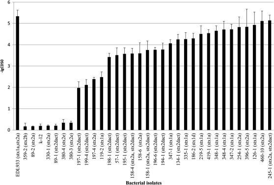
68 57
69 CHAPTER III: DISTRIBUTION OF PATHOGENICITY ISLANDS OI-122, OI-43/48, OI-57 AND HIGH PATHOGENICITY ISLAND (HPI) IN SHIGA TOXIN- PRODUCING ESCHERICHIA COLI Abstract Pathogenicity islands (PAIs) play an important role in STEC pathogenicity. The distribution of PAIs OI-122, OI-43/48, OI-57 and high pathogenicity island (HPI) were determined among 98 STEC strains assigned to seropathotypes (SPT) A to E. PCR and PCR-RFLP assays were used to identify 14 virulence genes that belonged to the four PAIs, and to subtype eae and stx genes, respectively. Phylogenetic trees were constructed based on the sequences of pagc among 34 STEC strains and iha among 67 diverse pathogenic E. coli, respectively. Statistical analysis demonstrated that the prevalence of OI-122 (55.82%) and OI-57 (82.35%) was significantly greater in seropathotypes (SPT A, B and C) that are frequently associated with severe disease than in other seropathotypes. terc (62.5%) and urec (62.5%) in OI-43/48 were also significantly more prevalent in SPT A, B and C than in SPT D and E. Additionally, OI-122, OI-57, OI-43/48 and their associated virulence genes (except iha) were found to be primarily associated with eae-positive STEC, whereas HPI occurred independently of the eae presence. The strong association of OI-122, OI-43/48 and OI-57 with eae-positive STEC suggests in part different pathogenic mechanisms exist between eae-positive and eae-negative STEC. Virulence genes in PAIs that are associated with severe diseases can be used as potential markers to aid in identifying highly virulent STEC. 58
70 Introduction Shiga toxin-producing Escherichia coli can cause human illnesses ranging from self-limiting diarrhea to life-threatening diseases such as hemolytic uremic syndrome (HUS), a leading cause of kidney failure in children (1). E. coli O157:H7 is the single serotype that causes most STEC outbreaks and HUS cases. Like O157, non-o157 STEC can also cause severe diseases and foodborne outbreaks (1). There are more than 470 non-o157 STEC serotypes have been associated with human illness (2) and the public health concerns of non-o157 STEC is increasing (1). Estimations indicate that non-o157 STEC cause 112,752 illness each year in the United States, almost doubling the numbers of O157:H7 illnesses (63,153) (3). While some non-o157 STEC have been associated with disease symptoms indistinguishable from O157:H7, not all STEC can cause HUS and outbreaks, and some STEC serotypes have never been reported related to any human illness (4). The scientific basis for this difference, however, is poorly understood. Increasing evidence shows that differences in virulence between pathogenic and nonpathogenic bacterial strains can be attributed in part to virulence genes located in pathogenicity islands (PAIs) (5). PAIs usually contain blocks of virulence genes and are greater than 10 Kb (6). Several PAIs have been identified and characterized in STEC. A chromosomal pathogenicity island termed the locus of enterocyte effacement (LEE) was identified in E. coli O157:H7 strain EDL933, which encodes a type III secretion system (TTSS) as well as virulence genes (eae and tir) associated with the intimate attachment of bacteria to intestinal epithelial cells (4). LEE appears to confer enhanced virulence, since LEE-positive STEC are much more 59
71 commonly associated with HUS and outbreaks than LEE-negative STEC (5). However, some LEE-positive STEC serotypes have never been associated with disease, and some LEE-negative STEC can cause HUS and outbreaks, indicating that virulence factors other than those in LEE may contribute to pathogenesis of STEC (5). Pathogenicity island OI-122 is also well characterized in O157:H7 (5, 7). OI- 122 is a 23 kb PAI consisting of three modules (5, 8, 9). Z4321 is located in module one and encodes a protein sharing 46% similarity with the phop-activated gene C (pagc) of Salmonella Typhimurium (5, 9). Z4326, Z4328, and Z4329 are located in module two. Z4326 (sen) encodes a protein that shares 38.2% similarity to Shigella flexneri enterotoxin 2 (5), whereas Z4328 and Z4329 encode proteins that have 89% and 86% similarity to non-lee encoded effectors NleB and NleE, respectively (9). The enterohemorrhagic E. coli factor for adherence (Efa), which is involved in epithelial cell adhesion and proliferating the inhibition of bovine peripheral blood lymphocytes, is located in module three (10). OI-43 and OI-48 are duplicate genomic islands found in EDL933 (8). OI- 43/48 genes are divided into three functional groups: a seven-gene cluster uredabcefg that encodes urease and accessory proteins hydrolyzing urea to ammonia and carbon dioxide; telluride resistance genes terzabcdef (11); and two putative adhesion genes, iha (iron-regulated gene A) and aida-1 (autotransporter adhesin involved in diffuse adherence) (12). 60
72 In EDL933, OI-57 contains non-lee encoded effector genes nleg2-3, nleg6-2, and nleg 5-2 (13, 14). NleG proteins are E3 ubiquitin ligases analogous to RING finger and U-box enzymes in eukaryotes. Although the exact functions of NleG2-3, NleG6-2, and NleG 5-2 are still unclear, similar proteins have been identified as effectors that suppress immune response from the host (15). High pathogenicity island (HPI) was first detected in Yersinia pestis and other highly virulent Yersinia species, and encodes a siderophore (yersiniabactin) mediated iron-uptake system (16). HPI is required for full virulence expression in Yersinia (16), and contains two main virulence genes, fyua and irp2. FyuA is an outer membrane protein acting as a receptor for ferric-yersiniabactin uptake and for bacteriocin pesticin, while Irp2 is involved in yersiniabactin synthesis (17). An orthologous and highly conserved HPI is widely distributed among different species and genera of the family Enterobacteriaceae (16). Few studies have investigated PAIs other than LEE in STEC to date. Since PAIs are normally absent in non-pathogenic strains of the same or closely related species, they may serve as useful markers to distinguish highly virulent from less virulent or harmless strains (5, 6). In addition, PAIs can be used to identify new and emerging pathogenic bacteria. In this study, we reported the distribution of OI-122, OI-43/48, OI-57, and HPI and their virulence genes in STEC, and evaluated the association of the PAIs and individual virulence genes with STEC seropathotypes linked to severe diseases and outbreaks. In addition, the association of the four PAIs with LEE was determined. 61
73 Materials and Methods Bacterial strains. A total of 98 STEC strains from humans, animals and food were used in this study (TABLE III-1). Strains were classified into seropathotypes A to E according to the criteria described by Karmali et al. (5). The assignment of seropathotypes was based on published references (5, 14, 18) and a large online database on non-o157 STEC ( stx and eae subtyping. stx and eae subtypes were determined using PCR- RFLP analysis (19, 20), and stx2dact was confirmed by PCR as previously described (21). Genomic DNA was extracted using boiling method as previously described (22, 23). STEC strains S1191 (stx2e), EDL933 (stx1a and stx2a), E32511 (stx2c), EH250 (stx2b), B2F1 (stx2dact) and N15018 (stx1c) were used as positive controls for the stx subtyping; STEC strains (gamma 1), EDL933 (gamma 1), TW06584 (kappa), E (alpha), TW07920 (epsilon), RDEC-1 (beta), TW10366 (rho), TW03501 (iota), TW07892 (eta), and TW01387 (gamma 2/theta) were used as positive controls for the eae subtyping. E. coli K12 was used as a negative control strain for both stx and eae subtyping. Presence of OI-122, OI-43/48, O-57 and HPI. PCR assays were used to determine the presence of 14 virulence genes in STEC OI-122, OI-43/48, O-57 and HPI as described (5, 11, 13, 24, 25). The presence of a PAI was determined by several marker genes located in different regions of the island, including pagc, sen, nleb, efa-1, and efa-2 for OI-122; terc, urec, iha and aida-1 for OI-43/48; nle2-3, nleg6-2 and nleg5-2 for OI-57; and irp2 and fyua for HPI. PCR was performed in a 25 µl reaction mixture, containing 2 µl of DNA template, 2.5 µl of 10x PCR buffer, 2 62
74 µl of 25 mmol l -1 MgCl 2, 2 µl of 1.25 mmol l -1 dntp mix, µl of 5 U µl -1 AmpliTaq Gold DNA polymerase mix (Applied Biosystems, Branchburg, NJ) and 0.2 µl of 50 pmol µl -1 of each primer. E. coli O157:H7 EDL 933 was used as a positive control for the virulence genes of OI-122, OI-43/48 and OI-57, and E. coli O26:H11 SJ-13 for virulence genes of HPI. E. coli K12 was used as a negative control for all PCR assays. Phylogenetic and sequences analysis. iha and pagc were the only two genes that were highly prevalent in both eae-positive and eae-negative STEC. To determine the evolutionary relationship between the two groups of STEC, iha and pagc were selected for phylogenetic analysis studies. iha sequences from 67 E. coli and Shigella were obtained from the GenBank. A multiple sequence alignment of iha was performed using ClustalW in MEGA 5.05, and a maximum likelihood phylogenetic tree was generated using the General Time Reversible model (26). A bootstrapping of 2,000 replicates was used to estimate the confidence of the branching patterns of the phylogenetic tree using iha of E. coli SMS-3-5 as the phylogenetic tree s root. Additionally, PCR was used to amplify pagc of OI-122 from 12 selected STEC strains representing different serotypes as described by Konczy et al (9). PCR products were sequenced by GeneWiz (Germantown, MD). Twenty two pagc sequences representing different STEC serotypes and one Citrobacter were downloaded from the GenBank. The pagc sequences were cropped to 446 bp prior alignment. Phylogenetic analysis was performed using ClustalW within MEGA 5.05 (26). A phylogenetic tree based on pagc sequences was constructed using maximum 63
75 likelihood methods by MEGA 5.05 with bootstrapping of 2,000 replicates using pagc of Citrobacter rodentium IC168 as the tree s root. Statistical analysis. Chi square or Fisher's exact test was used for data analysis using SAS9.2 (SAS Institute, Cary, N.C.). A P-value of < 0.01 was considered statistically significant. 64
76 Results Distribution of OI-122, OI-43/48, OI-57 and HPI in STEC. The 98 STEC strains were classified into seropathotypes A to E (TABLE III-1). Overall, the prevalence of OI-122 and OI-57 decreased progressively from seropathotype A (SPT A) to seropathotype E (SPT E) (FIG III-1). The prevalence of OI-122 and OI-57 was significantly higher in seropathotypes associated with severe diseases (SPT A, B, and C) and outbreaks (SPT A and B) than in other seropathotypes (P<0.0001) (TABLE III-2). Although the prevalence of OI-43/48 was greater in seropathotypes associated with HUS (SPT A, B and C) and outbreaks (SPT A and B) than in other seropathotypes, the difference was not statistically significant (P= and 0.02, respectively). HPI was not found in SPT A (O157), but was almost evenly distributed from SPT B to SPT E (FIG III-1). sen, nleb, efa-1, efa-2, terc, urec, nleg2-1, nleg5-2 and nleg6-2 were significantly more prevalent in SPT A and B than in SPT C, D and E (TABLE III-3). pagc, sen, nleb, efa-1, efa-2, terc, urec, nleg2-1, nleg5-2 and nleg6-2 were statistically more prevalent in SPT A, B and C than in SPT D and E (TABLE III-3). Although aida-1 was more prevalent in SPT A and B than in SPT C, D and E, the difference was not statistically significant (P=0.27). iha, fyua, and irp2 were less prevalent in SPT A, B and C than in SPT D and E, and the differences were not statistically significant neither. Distribution of OI-122, OI-43/48, OI-57 and HPI in EHEC O157. PAIs showed three patterns of distribution in EHEC O157 (TABLE III-4). As to β- 65
77 glucuronidase (GUD)-negative O157:H7, four strains all contained marker genes for OI-122, OI-57, and OI-43/48. In GUD-positive O157: H7, none of the five strains carried efa-1 and efa-2 (located at the third module of OI-122) or aida-1(located at the end of OI-43/48). Sorbitol fermenting O157:NM strains contained all virulence genes of OI-122 and OI-57 but were negative for all the OI-43/48 virulence marker genes, indicating the absence of OI-43/48 in O157:NM. Additionally, none of the O157:H7 and O157:NM strains were positive of HPI virulence genes. Association of OI-122, OI-43/48, OI-57 and HPI with eae. We compared the distribution of virulence genes of OI-122, OI-43/48, OI-57, and HPI between eaepositive and eae-negative STEC. All virulence genes of OI-122 and OI-57 (pagc, sen, nleb, efa-1, efa-2, nleg2-3, nleg5-2 and nleg6-2) were highly prevalent in eaepositive strains (FIG III-2). However, these genes, with the exception of pagc, were less prevalent in eae-negative STEC (FIG III-2). Although 38.6% of eae-negative STEC strains were positive for pagc, its prevalence was significantly higher (64.8%) in eae-positive STEC (P=0.005). There was no apparent physical or functional relationship identified between OI-43/48 and LEE, but three OI-43/48 virulence genes (urec, terc and aida-1) were mainly associated with the presence of eae (P<0.0001). On the other hand, iha was more prevalent in eae-negative than in eaepositive STEC strains (P=0.007). As for HPI, there were no significant differences in the distribution of fyua or irp2 between eae-positive and eae-negative STEC (P=0.36). 66
78 Phylogenetic analysis of iha from diverse pathogenic E. coli. A phylogenetic tree based on iha separated eae-positive and eae-negative STEC strains into two distinct clades (FIG III-3). In clade I, two subgroups, Ia and Ib, shared at least 98.0% sequence similarity. eae-positive EHEC serotypes highly associated with outbreaks and severe diseases were located in clade I (O157:H7, O26:H11, O103:H2, O111:NM, O145:H28). Those sequences shared at least 99% similarity and clustered together with iha from other pathogenic E. coli including enteropathogenic E. coli (EPEC), enteroinvasive E. coli (EIEC), eneteroaggregative E. coli (EAEC), enterotoxigenic E. coli (ETEC), uropathogenic E. coli (UPEC), neonatal meningitis E. coli (NMEC), and Shiga toxin-producing EAEC O104:H4 (from a German outbreak in 2011). iha sequences from the O26:H11, O111:H11, and O111:NM strains formed subgroup Ib and shared at least 98.0% sequence similarity with subgroup Ia. Interestingly, strains DEC10A (O26:H11), DEC10C (O26:H11), (O26:H11) and DEC8C (O111:NM) carried two iha that clustered separately in Ia and Ib. All 15 iha sequences from eae-negative STEC clustered together to form clade II. Multiple sequence alignments demonstrated that iha from eae-negative STEC shared only % sequence similarity with iha from clade I. iha from subgroups IIa and IIb shared only % sequence similarity. As in some eaepositive strains, eae-negative STEC strains CL-3 (O113:H21), (O91:H21) and B2F1 (O91:H21) also carried two iha genes that clustered separately in subgroups IIa and IIb. 67
79 Phylogenetic and sequence analysis of pagc. The pagc phylogenetic tree showed four clades (FIG III-4). The eae-positive E. coli STEC formed a single clade with EPEC and ETEC, whereas the eae-negative STEC strains formed two clades, along with one strain that clustered with a Citrobacter rodentium strain. We identified 15 single nucleotide polymorphisms (SNPs) and one indel among the 35 pagc sequences. Sequence analysis revealed that an insertion of adenine at nucleotide 388 in two O103:H25, two O45:H2, one O103:H2 and one O103:H6 strains led to a frame shift mutation, and that a premature stop codon truncated the protein at the third loop resulting in the loss of the fourth and last loops. 68
80 Discussion In the present study, PAIs OI-122 and OI-57 of STEC were found highly associated with seropathotypes that can cause severe disease and outbreaks, as previously demonstrated (7, 13, 14). Several OI-122 virulence factors play important roles in bacterial pathogenesis. For example, PagC can promote the survival of Salmonella within macrophages (5, 9). Efa is an adhesion protein originally described in some EHEC strains (27). The efa-1 gene is almost identical to lifa, an EPEC gene encoding lymphostatin (LifA) (28), which inhibits the proliferation of mitogen-activated lymphocytes and the synthesis of proinflammatory cytokines (28). Efa1/LifA also contributes to EPEC adherence to epithelial cells and is critical for the intestinal colonization by C. rodentium (29). NleB was required for full colonization and colonic hyperplasia in mice and a mutation of nleb abolished lethality of C. rodentium in C3H/HeJ mice (7, 30). Whereas OI-122 is highly related to colonization and suppression of the host immune system, the function of OI-57 is largely unknown. Wu et al. (15) determined that NleG like proteins including NleG2-3, NleG6-2 and NleG5-1 were E3 ubiquitin ligases, analogous to RING finger and U-box enzymes in eukaryotes. Although the targets of the OI-57 Nle effectors are unknown, several similar effectors are primarily involved in suppressing host immune response by degrading immune-related host proteins (15). Thus, it is possible that OI-57, similar to OI-122, would be also related to suppression of host immune system. In addition to the virulence genes in OI-122 and OI-57, urec and terc located on OI-43/48 were also highly associated with seropathotypes related to severe disease 69
81 and outbreaks. Urease has been confirmed as an important virulence factor in several bacterial species, such as Helicobacter pylori, Yersinia enterocolitica, Proteus mirabilis, Brucella species, and Klebsiella pneumonia (31). Mutation of urec has led to a reduced adherence of EHEC O157:H7 in ligated pig intestine (12). A recent study by Steyert et al (32) revealed that strains with non-functional urease were two times unlikely to survive passage through the stomach and had a reduced ability to colonize the mouse intestinal tract compared with urease-positive strains. These data demonstrate that urease can help STEC strains survive in the stomach and enhance its competitiveness in colonization in calf and human intestinal tracts. The role of tellurite resistance genes (terzabcdef) in STEC is still not well understood. Yin et al (12) showed that mutation of the ter cluster in O157:H7 led to fewer adherence to epithelial cells and smaller bacterial clusters compared with wild-type strains. Therefore, ter genes might encode an adhesin or a gene product that promotes the function of adhesion(s). In addition, tellurite salts are strong oxidative agents, and it is possible that ter genes might offer a selective advantage in the host environment and aid STEC in general stress response (12). Interestingly, urec has been more frequently found in eae-positive STEC (113/132) than in eae-negative strains (4/70) although no physical linkage of urec and eae has been identified (33). The prevalence of urec in eae-positive STEC (45/55) was significantly higher than in eae-negative STEC (2/44) (P< ). Similarly, terc was also more prevalent in eae-positive STEC (45/55) than eaenegative strains (5/55) (P< ). Even though OI-43/48 and LEE are physically 70
82 distant, our observations indicated that there might be a functional relationship between LEE and OI-43/48. The arrangement of OI-122 genes was found to be serotype dependent and all O157:H7 strains have a complete OI-122 (5, 9). However, we found that two patterns of OI-122 existed in O157:H7. An incomplete OI-122 lack of third module was identified in all GUD-positive O157:H7 strains. Additionally, aida-1 of OI-43/48 was absent in GUD-positive O157:H7. Most OI-122, OI-43/48 and OI-57 virulence genes (pagc, sen, nleb, efa-1, efa-2, terc, urec, iha, aida-1, nleg2-3, nleg6-2, and nleg5-2) were highly prevalent in eae-positive STEC. However, they were largely absent in eae-negative STEC with the exception of pagc and iha. Phylogenetic analysis revealed that iha genes from eae-positive STEC had high similarity (99.6%), whereas they had lower sequence similarity ( %) with iha genes from eae-negative STEC, indicating that iha from eae-positive and eae-negative STEC may have evolved independently or have different origins. Such a difference also existed in pagc between eae-positive and eae-negative STEC. Schemidt et al (34) reported that iha was carried by a 33,014 bp PAI in STEC serotype O91:H- strains (eae-negative). In addition, iha was found in po113 plasmid of STEC serotype O113:H21 (eae-negative) (35). Moreover, Shen et al (36, 37) reported that pagc was identified within a mosaic PAI from STECO113:H21strain CL-3 (eae-negative). Thus, the higher prevalence of iha and pagc in the eae-negative STEC strains, as compared with other virulence marker genes in this study, is likely due to the presence of the same or similar PAIs and/ or plasmids as previously described. The similar prevalence rate of iha genes in the 71
83 seropathotypes highly associated with severe diseases and other seropathotypes indicates that iha is not related with severe clinical outcomes, but the significantly higher prevalence of pagc in the seropathotypes associated with severe diseases indicates that this gene has some association with severe clinical outcome whether a strain carries the gene in OI-122 or in some other PAIs. The distribution of PAI virulence genes and the phylogenetic analysis of iha and pagc support the hypothesis that OI-122, OI-43/48 and OI-57 are primarily associated with eae-positive strains in STEC. However, some eae-negative STEC serotypes, for example, O113:H21 and O91:H21, are also associated with life threatening diseases such as HUS (5). Virulence factors such as subtilase cytotoxin AB5 (subab5) and Saa (STEC autoagglutinating adhesion) are more commonly associated with eae-negative STEC. Moreover, it has been shown that some LEEnegative STEC, especially O113:H21, can invade tissue culture cells (38).Whole genome comparison between nine eae-negative and five eae-positive STEC strains revealed that eae-negative strains did not carry any LEE or other phage encoded non- LEE effectors (39). These observations indicate that some differences in pathogenesis mechanisms may exist between eae-positive and eae-negative STEC. Additional studies, especially genomics and proteomics, are needed to unveil the difference in the pathogenicity mechanisms between eae-negative and eae-positive STEC. The strong association of OI-122, OI-57 and OI-43 with eae-positive STEC offers an important basis for STEC molecular risk assessment (MRA). The MRA, which uses 14 non-lee encoded virulence factors to distinguish high risk from low 72
84 risk non-o157 STEC, was proposed by Coombes et al in 2008 (13). Other researchers adopted this concept and applied it to their own studies (40-43). However, the current work demonstrated that some of non-lee encoded effectors (nleb, nle2-3, nleg5-2 and nleg6-2) were primarily associated with eae-positive STEC strains. In addition, Mundy et al (44) reported that nlea was present in 37 out of 43 (86%) eaepositive STEC, but absent in 50 eae-negative STEC clinical strains. Konczy et al (9) reported that nleb and nlee of OI-122 were highly correlated with LEE. Moreover, comparative genomics analysis demonstrated that all known phage encoded non-lee effector genes were absent in eae-negative STEC (39). Based on the MRA framework, which uses non-lee effector genes as sole markers, all eae-negative virulence STEC, including HUS associated O113:H21, O91:H21 and O104:H21, would be grouped as harmless STEC; other serotypes, for example, O103:H11 and O119:H25, which have not been reported as associated with severe disease or outbreaks but carry similar non-lee encoded virulence effectors as O157 EHEC, would be considered as outbreak- and severe disease-associated serotypes. Therefore, additional markers or methods of assessment, especially for eae-negative STEC, are needed to accurately distinguish highly pathogenic STEC from low virulent or harmless STEC. In summary, O-122 and OI-57, and their virulence genes were highly associated with seropathotypes that cause severe diseases and outbreaks. In addition, urec and terc located at OI-43/48 were also identified as markers related to high risk seropathotypes. Virulence genes in PAIs that are associated with severe diseases can be used as markers to identify potentially highly virulent STEC. Furthermore, this 73
85 study demonstrated that OI-122, OI-43/48, and OI-57 are highly associated with eaepositive STEC, which offers an important basis for STEC MRA. 74
86 References 1. Bettelheim KA The non-o157 Shiga-toxigenic (Verocytotoxigenic) Escherichia coli; under-rated pathogens. Crit. Rev. Microbio. 33: Blanco M, Blanco JE, Mora A, Dahbi G, Alonso MP, Gonzalez EA, Bernardez MI, Blanco J Serotypes, virulence genes, and intimin types of Shiga toxin (verotoxin)-producing Escherichia coli isolates from cattle in Spain and identification of a new intimin variant gene (eae-xi). J Clin Microbiol 42: Scallan E, Hoekstra RM, Angulo FJ, Tauxe RV, Widdowson MA, Roy SL, Jones JL, Griffin PM Foodborne illness acquired in the United States--major pathogens. Emerg Infect Dis 17: Coombes BK, Gilmour MW, Goodman CD The evolution of virulence in non-o157 shiga toxin-producing Escherichia coli. Front Microbiol 2: Karmali MA, Mascarenhas M, Shen S, Ziebell K, Johnson S, Reid-Smith R, Isaac-Renton J, Clark C, Rahn K, Kaper JB Association of genomic O island 122 of Escherichia coli EDL 933 with verocytotoxinproducing Escherichia coli seropathotypes that are linked to epidemic and/or serious disease. J Clin Microbiol 41: Gal-Mor O, Finlay BB Pathogenicity islands: a molecular toolbox for bacterial virulence. Cell Microbiol 8:
87 7. Wickham ME, Lupp C, Mascarenhas M, Vazquez A, Coombes BK, Brown NF, Coburn BA, Deng W, Puente JL, Karmali MA, Finlay BB Bacterial genetic determinants of non-o157 STEC outbreaks and hemolytic-uremic syndrome after infection. J Infect Dis 194: Perna NT, Plunkett G, 3rd, Burland V, Mau B, Glasner JD, Rose DJ, Mayhew GF, Evans PS, Gregor J, Kirkpatrick HA, Posfai G, Hackett J, Klink S, Boutin A, Shao Y, Miller L, Grotbeck EJ, Davis NW, Lim A, Dimalanta ET, Potamousis KD, Apodaca J, Anantharaman TS, Lin J, Yen G, Schwartz DC, Welch RA, Blattner FR Genome sequence of enterohaemorrhagic Escherichia coli O157:H7. Nature 409: Konczy P, Ziebell K, Mascarenhas M, Choi A, Michaud C, Kropinski AM, Whittam TS, Wickham M, Finlay B, Karmali MA Genomic O island 122, locus for enterocyte effacement, and the evolution of virulent verocytotoxin-producing Escherichia coli. J Bacteriol 190: Abu-Median AB, van Diemen PM, Dziva F, Vlisidou I, Wallis TS, Stevens MP Functional analysis of lymphostatin homologues in enterohaemorrhagic Escherichia coli. FEMS Microbiol Lett 258: Taylor DE, Rooker M, Keelan M, Ng LK, Martin I, Perna NT, Burland NT, Blattner FR Genomic variability of O islands encoding tellurite resistance in enterohemorrhagic Escherichia coli O157:H7 isolates. J Bacteriol 184: Yin X, Wheatcroft R, Chambers JR, Liu B, Zhu J, Gyles CL Contributions of O island 48 to adherence of enterohemorrhagic Escherichia 76
88 coli O157:H7 to epithelial cells in vitro and in ligated pig ileal loops. Appl Environ Microbiol 75: Coombes BK, Wickham ME, Mascarenhas M, Gruenheid S, Finlay BB, Karmali MA Molecular analysis as an aid to assess the public health risk of non-o157 Shiga toxin-producing Escherichia coli strains. Appl Environ Microbiol 74: Imamovic L, Tozzoli R, Michelacci V, Minelli F, Marziano ML, Caprioli A, Morabito S OI-57, a genomic island of Escherichia coli O157, is present in other seropathotypes of Shiga toxin-producing E. coli associated with severe human disease. Infect Immun 78: Wu B, Skarina T, Yee A, Jobin MC, Dileo R, Semesi A, Fares C, Lemak A, Coombes BK, Arrowsmith CH, Singer AU, Savchenko A NleG Type 3 effectors from enterohaemorrhagic Escherichia coli are U-Box E3 ubiquitin ligases. PLoS Pathog 6:e Schubert S, Darlu P, Clermont O, Wieser A, Magistro G, Hoffmann C, Weinert K, Tenaillon O, Matic I, Denamur E Role of intraspecies recombination in the spread of pathogenicity islands within the Escherichia coli species. PLoS Pathog 5:e Benedek O, Schubert S Mobility of the Yersinia High-Pathogenicity Island (HPI): transfer mechanisms of pathogenicity islands (PAIS) revisited (a review). Acta Microbiol Immunol Hung 54: Toma C, Martínez Espinosa E, Song T, Miliwebsky E, Chinen I, Iyoda S, Iwanaga M, Rivas M Distribution of putative adhesins in different 77
89 seropathotypes of Shiga toxin-producing Escherichia coli. J Clin Microbiol 42: Beutin L, Miko A, Krause G, Pries K, Haby S, Steege K, Albrecht N Identification of human-pathogenic strains of Shiga toxin-producing Escherichia coli from food by a combination of serotyping and molecular typing of Shiga toxin genes. Appl Environ Microbiol 73: Tramuta C, Robino P, Oswald E, Nebbia P Identification of intimin alleles in pathogenic Escherichia coli by PCR-restriction fragment length polymorphism analysis. Vet Res Commun 32: Zheng J, Cui S, Teel LD, Zhao S, Singh R, O'Brien AD, Meng J Identification and characterization of Shiga toxin type 2 variants in Escherichia coli isolates from animals, food, and humans. Appl Environ Microbiol 74: Xia X, Meng J, McDermott PF, Ayers S, Blickenstaff K, Tran TT, Abbott J, Zheng J, Zhao S Presence and characterization of shiga toxinproducing Escherichia coli and other potentially diarrheagenic E. coli strains in retail meats. Appl Environ Microbiol 76: Ju W, Shen J, Li Y, Toro MA, Zhao S, Ayers S, Najjar MB, Meng J Non-O157 Shiga toxin-producing Escherichia coli in retail ground beef and pork in the Washington D.C. area. Food Microbiol 32: Nakano M, Iida T, Ohnishi M, Kurokawa K, Takahashi A, Tsukamoto T, Yasunaga T, Hayashi T, Honda T Association of the urease gene 78
90 with enterohemorrhagic Escherichia coli strains irrespective of their serogroups. J Clin Microbiol 39: Karch H, Schubert S, Zhang D, Zhang W, Schmidt H, Olschläger T, Hacker J A genomic island, termed high-pathogenicity island, is present in certain non-o157 Shiga toxin-producing Escherichia coli clonal lineages. Infect Immun 67: Tamura K, Peterson D, Peterson N, Stecher G, Nei M, Kumar S MEGA5: molecular evolutionary genetics analysis using maximum likelihood, evolutionary distance, and maximum parsimony methods. Mol Biol Evol 28: Nicholls L, Grant TH, Robins-Browne RM Identification of a novel genetic locus that is required for in vitro adhesion of a clinical isolate of enterohaemorrhagic Escherichia coli to epithelial cells. Mol Microbiol 35: Klapproth JM, Scaletsky IC, McNamara BP, Lai LC, Malstrom C, James SP, Donnenberg MS A large toxin from pathogenic Escherichia coli strains that inhibits lymphocyte activation. Infect Immun 68: Klapproth JMA, Sasaki M, Sherman M, Babbin B, Donnenberg MS, Fernandes PJ, Scaletsky ICA, Kalman D, Nusrat A, Williams IR Citrobacter rodentium lifa/efa1 is essential for colonic colonization and crypt cell hyperplasia in vivo (vol 73, pg 1441, 2005). Infection and Immunity 73:
91 30. Kelly M, Hart E, Mundy R, Marches O, Wiles S, Badea L, Luck S, Tauschek M, Frankel G, Robins-Browne RM, Hartland EL Essential role of the type III secretion system effector NleB in colonization of mice by Citrobacter rodentium. Infect Immun 74: Steyert SR, Rasko DA, Kaper JB Functional and phylogenetic analysis of ured in Shiga toxin-producing Escherichia coli. J Bacteriol 193: Steyert SR, Kaper JB Contribution of urease to colonization by Shiga toxin-producing Escherichia coli. Infect Immun 80: Friedrich AW, Lukas R, Mellmann A, Kock R, Zhang W, Mathys W, Bielaszewska M, Karch H Urease genes in non-o157 Shiga toxinproducing Escherichia coli: mostly silent but valuable markers for pathogenicity. Clin Microbiol Infect 12: Schmidt H, Zhang WL, Hemmrich U, Jelacic S, Brunder W, Tarr PI, Dobrindt U, Hacker J, Karch H Identification and characterization of a novel genomic island integrated at selc in locus of enterocyte effacementnegative, Shiga toxin-producing Escherichia coli. Infect Immun 69: Newton HJ, Sloan J, Bulach DM, Seemann T, Allison CC, Tauschek M, Robins-Browne RM, Paton JC, Whittam TS, Paton AW, Hartland EL Shiga toxin-producing Escherichia coli strains negative for locus of enterocyte effacement. Emerg Infect Dis 15:
92 36. Shen S, Mascarenhas M, Rahn K, Kaper JB, Karmali MA Evidence for a hybrid genomic island in verocytotoxin-producing Escherichia coli CL3 (serotype O113:H21) containing segments of EDL933 (serotype O157:H7) O islands 122 and 48. Infect Immun 72: Girardeau JP, Bertin Y, Martin C Genomic analysis of the PAI ICL3 locus in pathogenic LEE-negative Shiga toxin-producing Escherichia coli and Citrobacter rodentium. Microbiology 155: Luck SN, Badea L, Bennett-Wood V, Robins-Browne R, Hartland EL Contribution of FliC to epithelial cell invasion by enterohemorrhagic Escherichia coli O113:H21. Infect Immun 74: Steyert SR, Sahl JW, Fraser CM, Teel LD, Scheutz F, Rasko DA Comparative genomics and stx phage characterization of LEE-negative Shiga toxin-producing Escherichia coli. Frontiers in Cellular and Infection Microbiology. 40. Bugarel M, Beutin L, Fach P Low-density macroarray targeting nonlocus of enterocyte effacement effectors (nle genes) and major virulence factors of Shiga toxin-producing Escherichia coli (STEC): a new approach for molecular risk assessment of STEC isolates. Appl Environ Microbiol 76: Bugarel M, Beutin L, Martin A, Gill A, Fach P Micro-array for the identification of Shiga toxin-producing Escherichia coli (STEC) seropathotypes associated with Hemorrhagic Colitis and Hemolytic Uremic Syndrome in humans. Int J Food Microbiol 142:
93 42. Bugarel M, Martin A, Fach P, Beutin L Virulence gene profiling of enterohemorrhagic (EHEC) and enteropathogenic (EPEC) Escherichia coli strains: a basis for molecular risk assessment of typical and atypical EPEC strains. BMC Microbiol 11: Bosilevac JM, Koohmaraie M Prevalence and characterization of non-o157 shiga toxin-producing Escherichia coli isolates from commercial ground beef in the United States. Appl Environ Microbiol 77: Mundy R, Jenkins C, Yu J, Smith H, Frankel G Distribution of espi among clinical enterohaemorrhagic and enteropathogenic Escherichia coli isolates. J Med Microbiol 53:
94 TABLE II1-1. Distribution of eae subtypes, and virulence genes of OI-122, OI-43/48, OI-57 and high pathogenicity island (HPI) by serotypes Seropathotype Serotype eae subtype No. of No. of strains positive by PCR for strains pagc sen nleb efa-1 efa-2 terc urec iha aida-1 nleg2-3 nleg5-2 nleg6-2 fyua irp2 A O157:H7 GUD-* Gamma O157:H7 GUD+ Gamma O157:NM Gamma B O26:H11 Beta O103:H2 Epsilon O111:H8/NM Gamma2/theta O121:H19 Epsilon O145:H28/NM Gamma C O5:NM Beta O22:H5 negative O22:H8 negative O45:H2 Epsilon O50:H7 Epsilon O55:H7 Gamma O91:H21 Negative O104:H21 Negative O113:H21 Negative O118:H16 Beta O125:NM Beta O128:H2 Beta O128:H45 Negative ONT:H2 Negative OUN:NM Negative D O2:H27 Negative
95 O15:H27 Negative O91:H14 Negative O91:H7 Negative O103:H11 Beta O103:H25 Gamma2/theta O111:H11 Beta O116:H21 Negative O126:H8 Negative O146:H21 Negative O174:H8 Negative E O8:H16 Negative O8:H28 Negative O15:H16 Negative O36:H14 Negative O46:H38 Negative O73:H18 Negative O83:H8 Negative O88:H38 Negative O88:H49 Negative O113:H36 Negative O128:H16 Negative O168:H8 Negative O174:H36 Negative
96 TABLE III-2. Distribution of pathogenicity islands (PAIs) in seropathotypes associated outbreak (SPT A and B) and severe disease (SPT A, B and C). PAIs Prevalence (%) in SPT A and B (n=34) Association of PAIs with seropathotypes related to outbreak Prevalence (%) in SPT C, D and E (n=64) Association of PAIs with seropathotypes related to severe disease Prevalence (%) in SPT Prevalence (%) in A, B and C (n=56) SPT D and E (n=42) OI * * OI-43/ OI * * HPI *: Statistically significant difference between SPT A and B with SPT C, D and E. +: Statistically significant difference between SPT A, B and C with SPT D and E. A P value less than 0.01 is considered as statistically significant. 85
97 TABLE III-3. Distribution of virulence genes located at pathogenicity islands (PAIs) in seropathotypes associated outbreak (SPT A and B) and severe disease (SPT A, B and C). Association of virulence genes with seropathotyps related to outbreak Gene PAIs Prevalence (%) in Prevalence (%) in SPT SPT A and B (n= 34) C, D and E (n=64) Association of virulence genes with seropathotyps related to severe disease Prevalence (%) in STEC Prevalence (%) in STEC SPT A, B and C (n=56) in SPT D and E (n=42) pagc OI sen OI * * efa-1 OI * * efa-2 OI * * nleb OI * * terc OI-43/ * * urec OI-43/ * * Iha OI-43/ aida-1 OI-43/ nleg2-3 OI * * nleg5-2 OI * * nleg6-2 OI * * fyua HPI irp2 HPI *: Statistically significant between SPT A and B with SPT C, D and E. +: Statistically significant between SPT A, B and C with SPT D and E. A P value less than 0.01 is considered as statistically significant. 86
98 TABLE III-4. Characterization and presence of PAIs associated virulence genes among O157:H7 and O157:NM OI-122 OI-43/48 OI-57 HPI Strains Serotype stx GUD* pagc sen efa-1 efa-2 nleb terc urec iha aid-1 nleg2-3 nleg6-2 nleg5-2 irp2 fyua EC4115 O157:H7 stx1,stx Saika O157:H7 stx1, stx TW14359 O157:H7 stx1, stx FDA413 O157:H7 stx O157:H7 stx1,stx O157:H7 stx1,stx EC97144 O157:H7 stx1,stx EC96038 O157:H7 stx1,stx EC96012 O157:H7 stx1,stx /89 O157:NM stx /89 O157:NM stx IH56929 O157:NM stx IH56909 O157:NM stx H1085C O157:NM stx H2687 O157:NM stx GUD: β-glucuronidase 87
99 FIG III-1. Distribution of OI-122, OI-43/48, OI-57 and high pathogenicity island (HPI) in STEC strains (n=98) of different seropathotype. 88
100 FIG III-2. Distribution of virulence genes located on OI-122, OI-43/48, OI-57 and high pathogenicity island (HPI) between eae-positive (n=54) and eaenegative STEC (n=44) strains. 89
101 FIG III-3. Phylogenetic tree based on iha sequences from 67 E. coli and Shigella strains. iha sequences were aligned and a tree was constructed using the maximum likelihood method with 2,000 iterations utilizing MEGA 5.05 (26). iha sequences from eaepositive and eae-negative STEC strains segregated into two distinct clades, clades I (with subgroup Ia and Ib) and II (with subgroups IIa and IIb). iha sequences from eae-negative STEC were marked in bold and italic, and eae-positive STEC strains were marked in bold. EPEC: enteropathogenic E. coli; EIEC: enteroinvasive E. coli; EAEC: eneteroaggregative E. coli; stx-producing EAEC: Shiga toxin-producing EAEC; ETEC: enterotoxigenic E. coli; UPEC: uropathogenic E. coli; NMEC: neonatal meningitis E. coli; and ABU: asymptomatic bacteriuria E. coli. 90
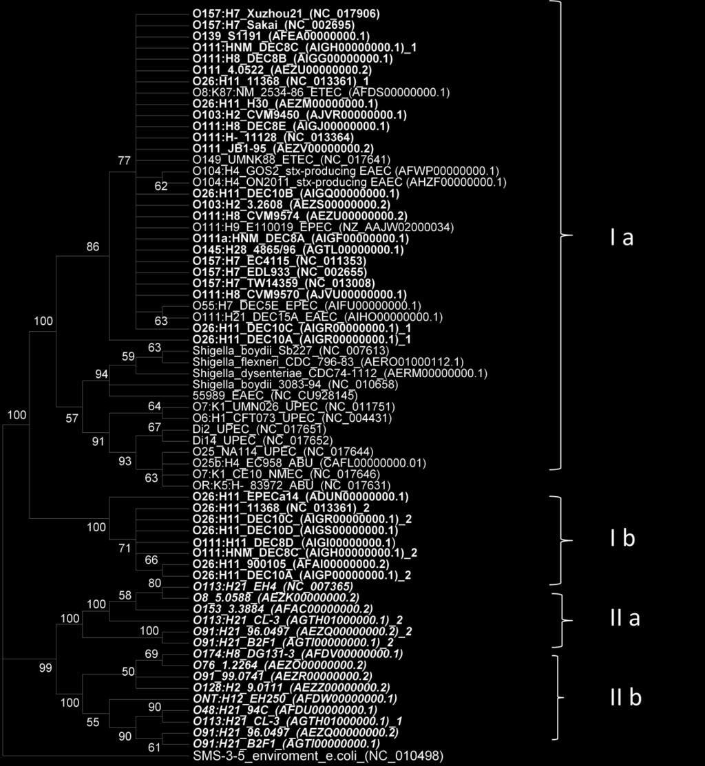
102 91
.")
103 FIG III-4. Phylogenetic tree based on pagc sequences from 34 pathogenic E. coli strains. pagc sequences were aligned and a tree was constructed using the maximum likelihood method with 2,000 iterations utilizing MEGA 5.05 (26). pagc sequences from eae-negative STEC were marked in bold and italic, and eae-positive STEC strains were marked in bold. pagc genes sequenced by this study were marked by black frames. EPEC: enteropathogenic E. coli, ETEC: enterotoxigenic E. coli. 92
104 CHAPTER IV: PHYLOGENETIC ANALYSIS OF NON- O157 SHIGA TOXIN-PRODUCING ESCHERICHIA COLI BY WHOLE GENOME SEQUENCING Abstract Non-O157 Shiga toxin-producing Escherichia coli (STEC) are emerging foodborne pathogens causing life threatening diseases and foodborne outbreaks. A better understanding of their evolution provides a framework for developing tools to control food safety. We obtained 15 genomes of non-o157 STEC strains, including O26, O111 and O103. Phylogenetic trees revealed close relationship between O26:H11 and O111:H11 and a scattered distribution of O111. We hypothesize that some STEC serotypes with same H antigens might share common ancestors. 93
105 Introduction Shiga toxin-producing Escherichia coli (STEC) are deadly pathogens, causing hemorrhagic colitis (HC) and hemolytic uremic syndrome (HUS) (1, 2). There are two categories of surface antigens (O somatic and H flagella), whose combinations are used to classify E. coli. O157:H7 has caused more outbreaks and HUS cases in the United States than any other STEC serotypes. However, there is a growing concern about the health risk of non-o157 STEC (3) as more than 470 serotypes of STEC are associated with human diseases (4). In the United States, non-o157 STEC cause an estimated 112,752 cases of illness each year, which is more than the number of cases (estimated 63,153) caused by E. coli O157:H7 (5). Worldwide, six serogroups including O26, O45, O103, O111, O121 and O145 are most commonly isolated from human patients. Among the non-o157 STEC, serogroups O26, O111 and O103 are considered the most clinically important and frequently identified non-o157 STEC in severe diseases and foodborne outbreaks (6-8). As emerging foodborne pathogens, the phylogentic relationship of STEC is not well understood. In this study, we used whole genome sequencing data to examine phylogenetic relationship of non-o157 STEC strains for a better understanding of the evolution history of these emerging pathogens. 94
106 Materials and Methods Bacteria Strains. Fifteen STEC strains representing different PFGE patterns, isolation years, hosts, and stx gene profiles, including O26:H11 (5), O111:H11 (3), five O111:H8 (5), O103:H2 (1), and O103:H25 (1) were used in this study (Table 1). In addition, 28 E. coli genomes downloaded from the GenBank were included for phylogenetic study. Pulsed field gel electrophoresis (PFGE). PFGE with XbaI was performed following a non-o157 PulseNet protocol ( _5%204_PNetStand_Ecoli_with_Sflexneri.pdf). Salmonella Braenderup H9812 was used as control for PFGE. All the PFGE gel pictures were analyzed using BioNumerics software (Applied Maths, Austin, TX). Unweighted pair group means with arithmetic averages (UPGMA) was used to construct a dendrogram with a 1.5% band position tolerance. eae subtyping. eae subtypes were determined using PCR-RFLP as described by Tramuta et al (9). The following strains were used as positive controls: EDL933 (gamma), TW06584 (kappa), E (alpha), TW07920 (epsilon), RDEC-1 (beta), TW10366 (rho), TW0350 (iota), TW07892 (eta), and TW01387 (theta). E. coli K12 was used as negative control. Multiplelocus sequence typing. Seven housekeeping genes (aspc, clpx, fadd, icda, lysp, mdh, and uida) extracted from genomes were selected for multilocus sequence typing (MLST) analysis as previously described for pathogenic 95
107 E. coli ( The MLST analysis was performed using MEGA 5.05 (10) with 2,000 iterations (model: maximum composite likelihood; substitution: transitions + transversions; Gamma). Whole genome sequencing. The whole genome DNA of selected STEC strains were extracted with the DNeasy Blood & Tissue Kit (Qiagen, Valencia, CA) according to the manufacturer s instructions. Roche 454 GS-FLX+ system (Roche, Branford, CT) was used to perform DNA shotgun sequencing to obtain ~ 20 coverage of draft genomes. The Roche Newbler (v 2.6) software package was used to conduct de novo assemblies. Multiple sequence alignment of all 43 genomes was performed using MAUVE (11). SNPs was extracted from the multiple sequence alignment and concatenated into pseudosequences by MAUVE. To further explore evolutionary relatedness, a parsimony phylogenetic tree based on whole genome wide SNPs was performed with 10,000 iterations by TNT (Tree analysis using New Technology) (12). 96
108 Results Pulsed field gel electrophoresis and eae subtyping. The 15 STEC strains were grouped into two main clusters (FIG IV-1) that separated H11 strains (O111:H11 and O26:H11) from H8 strains (O111:H8). However, PFGE was not able to differentiate O111:H11 and O26:H11. In addition, the O26:H11 and O111:H11 strains shared the same eae subtype (ß), while the O111:H8 strains contained. It appeared that O111:H11 and O26:H11 were more closely related to each other than either was to O111:H8, according to PFGE profiles and virulence genes associated elements in the genomes. Multi-locus sequence typing. In the MLST phylogenetic dendrogram, all the O111:H11, O26:H11 and O111:H8 strains formed one branch, with O26:H11 and O111:H11 clustering together in a lineage sister to the O111:H8 strains (FIG IV-2). It is interesting that O26:H11 strain DEC10B clustered with the O111:H11 strains, which also indicates the close relatedness between O26:H11 and O111:H11. Furthermore, five strains (12009, CVM9450, E22, DEC12C, and 03-EB-705) sharing the H2 antigen clustered together regardless of O serotypes. Genomic analysis revealed that O111:NM strain DEC12C carried flic for the H2 gene. Whole genome wide single nucleotide polymorphisms (SNPs) phylogenetic tree. The genome sizes of the 15 STEC strains ranged from 5.26 Mbp to 6.01 Mbp (Table IV-1). In order to analyze the phylogenetic relationship between non-o157 STEC, 28 E. coli genomes of various pathogenic E. coli including STEC, enteropathogenic E. coli (EPEC), enteroinvasive E. coli (EIEC), eneteroaggregative E. coli (EAEC), enterotoxigenic E. coli (ETEC), and Shiga toxin-producing EAEC 97
109 O104:H4 (from a German outbreak in 2011) downloaded from the GenBank were included for phylogenetic study. Multiple sequence alignment of all 43 genomes was performed using MAUVE (11) and approximately 183,470 single nucleotide polymorphisms (SNPs) were identified. Similarly as data shown by PFGE and MLST, the phylogenetic tree demonstrated that O26:H11 strains belonged to the same clade as the O111:H11 and O111:H8 strains, but grouped more closely with strains of the same H type (O111:H11) (FIG IV-3). All O111 strains but O111:H2 formed one clade (FIG IV-3), including O111 EPEC, O111:H8 and O111:H11. Additionally, we reconstructed maximum likelihood (ML) tree by Garli-2.0 (13) and Bio Neighbor Joining (BioNJ) (14) tree using SeaView 4 (15) (data not shown), displaying similar phylogenetic relationship with TNT tree with minor differences. For example, in the BioNJ tree, E. coli O26:H11 DEC10B grouped with the O26:H11 strains. The H2 strains including O103:H2, O45:H2 and O111:H2 strains were closely clustered together in all phylogenetic trees, including TNT, ML and BioNJ trees as shown in MLST dendrogram. The phylogenetic trees also supported that STEC O113:H21 and O91:H21 were closely related (FIG IV-3). 98
110 Discussion Shiga toxin-producing E. coli are among the most important emerging foodborne pathogens. Recently, more and more outbreaks and sporadic disease are related to diverse serotypes of STEC. Among all the non-o157 STEC, O26, O103 and O111 cause most known outbreaks and HUS diseases. In this study, 15 STEC of O26, O103 and O111 from human and animals were sequenced. The phylogenetic trees were constructed to determine the evolutionary relationship of non-o157 STEC. In this study, MLST, PFGE and whole genome wide SNP trees indicating that the H11 strains, including O26:H11 and O111:H11 (Figure IV-2 and IV-3) shared common ancestor; the H2 strains with different O antigens, namely, H2 group in Figure 3, shared common ancestor as well. Previous studies also have suggested that STEC strains with the same H antigens might share common ancestors. For example, Iguchi et al. (16) indicated that a close relatedness between STEC O103:H11 and O26:H11 by MLST analysis, which shared the same eae subtype (ß-eae) that was also inserted at the same t-rna locus (pheu-trna). The O111:H11 strains used in this study also carry ß-eae as well (Figure 1). In addition, Konczy et al. (17) and Ziebell et al. (18) demonstrated that STEC O69:H11 was found closely related to O26:H11. Additionally, Konczy et al. (17) reported that H25 STEC strains (O103:H25, O119:H25, and O98:H25) and H21 STEC strains (O91:H21, O113:H21, O146:H21 and ONT:H21) were clustered together, separately, based on MLST. These data and our findings provided strong evidence that some STEC strains with common H antigens appear to originate from common ancestors. It is interesting that the four H groups included the following serotypes, O26:H11, O111:H11, O111:NM, O111:H2, 99
111 O103:H2, O103:H25, O45:H2, O91:H21 and O113:H21, which have been identified among the most important non-o157 STEC associated with outbreaks and HUS. Thus, it is possible that some most clinical and epidemic STEC serotypes with same H antigens might have evolved from common ancestors, respectively. It is possible that ancestral strains of those H groups share similar or same genetic background and/or environmental niche that could facilitate acquisition of stx and other virulence genes essential to STEC pathogenesis. Our phylogenetic analyses demonstrated that the H groups were monophyletic while the serogroups were polyphyletic (FIG IV-3). Scattered distribution of different O111 strains suggested that strains from individual lineages might have acquired surface antigen genes independently in an ongoing parallel evolutionary process, such as E. coli O111:H21 DEC15E (O111 EPEC group), E. coli O111:H2 E22 (H2 group), E. coli O111:H8 CVM9634 (O111:H8 group) and E. coli O111:H11 CVM9545 (O111:H11 group) (Figure 3). Iguchi et al. (16) suggested that STEC O103:H2, O103:H11 and O103:H25 formed three different linages by MLST analysis and had distinct eae subtypes. The MLST and SNP phylogenetic trees in this study also supported that O103:H2 and O103:H25 were located at different linages (FIG IV-2 and FIG IV-3). Thus, just using serogroups may cause misleading conclusion about phylogenetic relatedness and health risk of STEC. Pairwise distance matrix analysis with 2,000 bootstrap iterations (substitution: transitions + transversions; complete-delete option) (TABLE IV-2) were conducted to determine number of SNPs differences (standard deviation) between different selected groups using MEGA 5.05 (10), including H7, eae-types, H2, O26:H11 and 100
112 O111 groups. The values of base differences per sequence from averaging over all sequence pairs between groups were shown. The smallest distance value was found between O111:H11 and O26:H11 strains, confirming their close relatedness. Previous studies indicated that O157:H7 evolved from O55:H7 in a series of steps acquiring O157 antigen gene cluster and other virulence genes through HGT (19-22). The distance between O157:H7 Sakai and O55:H7 CB9615 was 4215 SNPs with a standard deviation of 37. Because O26:H11 strains located in the O111 clade in phylogenetic trees and display closer relationship with O111:H11 than O111:H8, it is possible that O26:H11 evolved similarly from an ancestral O111:H11 by antigenic shift from O111 to O26 (distance between O26:H11 and O111:H11 groups was 3617, Table 2). Sharing the same niche with other O26 strains may facilitate this genetic exchange. Comparative genomics analysis of O26:H11, O111:H11 and other STEC is underway to reveal the possible mechanism. In conclusion, analyses based on whole genome wide SNPs, MLST, and PFGE suggest that in some occasions O serogroups appear not to track evolutionary relatedness among pathogenic E. coli. Instead, H antigens may be better markers of shared ancestry for some STEC serotypes. 101
113 References 1. Miliwebsky E, Deza N, Chinen I, Martinez Espinosa E, Gomez D, Pedroni E, Caprile L, Bashckier A, Manfredi E, Leotta G, Rivas M Prolonged fecal shedding of Shiga toxin-producing Escherichia coli among children attending day-care centers in Argentina. Rev Argent Microbiol 39: Caprioli A, Morabito S, Brugere H, Oswald E Enterohaemorrhagic Escherichia coli : emerging issues on virulence and modes of transmission. Vet Res 36: Bettelheim KA The non-o157 shiga-toxigenic (verocytotoxigenic) Escherichia coli ; under-rated pathogens. Crit Rev Microbiol 33: Blanco JE, Blanco M, Alonso MP, Mora A, Dahbi G, Coira MA, Blanco J Serotypes, virulence genes, and intimin types of Shiga toxin (verotoxin)-producing Escherichia coli isolates from human patients: prevalence in Lugo, Spain, from 1992 through J Clin Microbiol 42: Scallan E, Hoekstra RM, Angulo FJ, Tauxe RV, Widdowson MA, Roy SL, Jones JL, Griffin PM Foodborne illness acquired in the United States--major pathogens. Emerg Infect Dis 17: Brooks JT, Sowers EG, Wells JG, Greene KD, Griffin PM, Hoekstra RM, Strockbine NA Non-O157 Shiga toxin-producing Escherichia coli infections in the United States, J Infect Dis 192:
114 7. Ogura Y, Ooka T, Iguchi A, Toh H, Asadulghani M, Oshima K, Kodama T, Abe H, Nakayama K, Kurokawa K, Tobe T, Hattori M, Hayashi T Comparative genomics reveal the mechanism of the parallel evolution of O157 and non-o157 enterohemorrhagic Escherichia coli. Proc Natl Acad Sci U S A 106: Tozzi AE, Caprioli A, Minelli F, Gianviti A, De Petris L, Edefonti A, Montini G, Ferretti A, De Palo T, Gaido M, Rizzoni G Shiga toxinproducing Escherichia coli infections associated with hemolytic uremic syndrome, Italy, Emerg Infect Dis 9: Darling AC, Mau B, Blattner FR, Perna NT Mauve: multiple alignment of conserved genomic sequence with rearrangements. Genome Res 14: Tramuta C, Robino P, Oswald E, Nebbia P Identification of intimin alleles in pathogenic Escherichia coli by PCR-restriction fragment length polymorphism analysis. Vet Res Commun 32: Tamura K, Peterson D, Peterson N, Stecher G, Nei M, Kumar S MEGA5: molecular evolutionary genetics analysis using maximum likelihood, evolutionary distance, and maximum parsimony methods. Mol Biol Evol 28: Goloboff P FS, Nixon K TNT, a program for phylogenetic analysis. Cladistics 24:
115 13. Zwickl DJ Genetic algorithm approaches for the phylogenetic analysis of large biological sequence datasets under the maximum likelihood criterion. The University of Texas at Austin. 14. Gascuel O BIONJ: an improved version of the NJ algorithm based on a simple model of sequence data. Mol Biol Evol 14: Gouy M GS, Gascuel O SeaView version 4: a multiplatform graphical user interface for sequence alignment and phylogenetic tree building. Mol Biol Evol 27: Iguchi A, Iyoda S, Ohnishi M Molecular Characterization Reveals Three Distinct Clonal Groups among Clinical Shiga Toxin-Producing Escherichia coli Strains of Serogroup O103. J Clin Microbiol 50: Konczy P, Ziebell K, Mascarenhas M, Choi A, Michaud C, Kropinski AM, Whittam TS, Wickham M, Finlay B, Karmali MA Genomic O island 122, locus for enterocyte effacement, and the evolution of virulent verocytotoxin-producing Escherichia coli. J Bacteriol 190: Ziebell K, Konczy P, Yong I, Frost S, Mascarenhas M, Kropinski AM, Whittam TS, Read SC, Karmali MA Applicability of phylogenetic methods for characterizing the public health significance of verocytotoxinproducing Escherichia coli strains. Appl Environ Microbiol 74: Tarr PI, Schoening LM, Yea YL, Ward TR, Jelacic S, Whittam TS Acquisition of the rfb-gnd cluster in evolution of Escherichia coli O55 and O157. J Bacteriol 182:
116 20. Feng P, Lampel KA, Karch H, Whittam TS Genotypic and phenotypic changes in the emergence of Escherichia coli O157:H7. J Infect Dis 177: Feng PC, Monday SR, Lacher DW, Allison L, Siitonen A, Keys C, Eklund M, Nagano H, Karch H, Keen J, Whittam TS Genetic diversity among clonal lineages within Escherichia coli O157:H7 stepwise evolutionary model. Emerg Infect Dis 13: Rump LV, Meng J, Strain EA, Cao G, Allard MW, Gonzalez-Escalona N Complete DNA sequence analysis of enterohemorrhagic Escherichia coli plasmid po157_2 in beta-glucuronidase-positive E. coli O157:H7 reveals a novel evolutionary path. J Bacteriol 194:
117 TABLE IV-1. Serotypes, pathotypes, toxin genotypes, sources and genome sizes of Escherichia coli strains used in this study*. Strain Serotype Pathotype stx Source Size (Mbp) Accession No. CVM10021 O26:H11 STEC stx1 cow 5.5 AKAZ CVM9942 O26:H11 STEC stx1 cow 5.62 AJVW CVM10026 O26:H11 STEC stx1 cow 5.57 AJVX CVM10030 O26:H11 STEC stx1 cow 5.5 AKBA CVM9952 O26:H11 STEC stx1 pig 5.5 AKBC CVM9634 O111:H8 STEC stx1+stx2 cow 5.78 AKAW CVM9602 O111:H8 STEC stx1 human 5.1 AKAV CVM9574 O111:H8 STEC stx1+stx2 human 5.36 AJVV CVM9570 O111:H8 STEC stx1+stx2 cow 5.51 AJVU CVM9545 O111:H11 STEC stx1 cow 5.61 AJVT CVM9455 O111:H11 STEC stx2 unknown 6.01 AKAX CVM9534 O111:H11 STEC stx1 cow 5.46 AJVS CVM9553 O111:H11 STEC stx1 cow 5.6 AKAY CVM9340 O103:H25 STEC stx1 human 5.26 AJVQ CVM9450 O103:H2 STEC stx1 human 5.39 AJVR CFT073 O6:K2:H1 UPEC - unknown 5.23 AE SAKAI O157:H7 STEC stx1+stx2 human 5.59 BA CB9615 O55:H7 EPEC - human 5.39 NC_ /96 O145:H28 STEC stx2 human 5.23 AGTL O144:? EIEC - unknown 5.07 AAKB O-:H10 EAEC - human 4.98 AAMK MG1655 Unknown Commensal - unknown 4.64 NC_ H121:H19 STEC stx2 unknown 5.37 AEZX
118 TY-2482 O104:H4 EAEC+STEC stx2 human 5.29 AFOG CL-3 O113:H21 STEC stx2 human 5.05 AGTH B2F1 O91:H21 STEC stx2 human 5.01 AGTI E24377A O139:H28 ETEC - unknown 4.97 NC_ DEC12B O111:H2 STEC stx2 human 5.49 AIHB DEC12C O111:NM STEC stx2 human 5.45 AIHC E22 O103:H2 EPEC - unknown 5.53 AAJV EN-705 O45:H2 STEC stx1 human 5.3 AGTK O103:H2 STEC stx1+stx2 human 5.45 NC_ E O111:H9 EPEC - human 5.38 AAJW DEC15A O111:H21 EPEC - human 5.25 AIHO DEC15E O111:H21 EPEC - human 5.23 AIHS DEC8E O111:H8 STEC stx1 human 5.32 AIGJ DEC8B O111:H8 STEC stx1+stx2 human 5.37 AIGG O111:H- STEC stx1+stx2 human 5.37 NC_ DEC8D O111:H11 DEC - human 5.46 AIGI DEC8C O111:H11 STEC stx1 cow 5.91 AIGH DEC10B O26:H11 STEC stx1 human 5.58 AIGQ EPECa14 O26:H11 STEC stx1 unknown 5.44 ADUN O26:H11 STEC stx1 human 5.69 NC_ * Data on strains named with CVM were from this study; the rest were from GenBank. STEC: Shiga toxin-producing Escherichia coli ; EPEC: enteropathogenic Escherichia coli ; EIEC: enteroinvasive Escherichia coli ; ETEC: enterotoxigenic Escherichia coli ; EAEC: eneteroaggregative Escherichia coli ; DEC: diarrheagenic Escherichia coli. 107
119 TABLE IV-2. Pairwise distance matrix analysis of six selected groups. Group* H7 eae negative H2 O111 EPEC O111:H8 O111:H11 H7 eae negative (107) H (139) (77) O111 EPEC (105) (98) (113) O111:H (126) (75) (97) (83) O111:H (113) (98) (123) (84) 4324 (35) O26:H (108) (78) (107) (102) 6556 (42) 3617 (37) *groups as by FIG IV-3 108

120 Figure IV-1. Dendrogram of PFGE profiles of 15 O26, O103 and O111 STEC isolates. Similarity of the PFGE profiles was based on Dice algorithm with 1.5% tolerance. O26:H11 and O111:H11 strains showed close relationship and grouped in the same cluster, and shared the same eae subtype. 109
121 Figure IV-2. Dendrogram of MLST analyses using aspc, clpx, fadd, icda, lysp, mdh, and uida. Strains 4865/96 (O145:H28), 101-1(O-:H10), (O121:H19), DEC12B (O111:H2) and E (O111:H9) were not included in MLST study because at least one of selected gene alleles were either absent or only partially present. 110
 with 10,000 iterations.")
122 Figure IV-3. Parsimony phylogenetic tree of 43 E. coli from diverse pathotypes based on genome wide single nucleotide polymorphisms (SNPs) with 10,000 iterations. Strains sequenced in this study were marked within frame. A total of seven subgroups were labeled, as shown in pairwise distance matrix (Table IV-2). 111
Shiga Toxin producing E. coli (STEC) in food which serotypes are important? Lothar Beutin
 in food which serotypes are important? Lothar Beutin") BUNDESINSTITUT FÜR RISIKOBEWERTUNG Shiga Toxin producing E. coli (STEC) in food which serotypes are important? Lothar Beutin National Reference Laboratory for Escherichia coli Federal Institute for Risk
BUNDESINSTITUT FÜR RISIKOBEWERTUNG Shiga Toxin producing E. coli (STEC) in food which serotypes are important? Lothar Beutin National Reference Laboratory for Escherichia coli Federal Institute for Risk
Foodborne Outbreak of E. coli Infections and Hemolytic Uremic Syndrome in Germany, 2011
 Foodborne Outbreak of E. coli Infections and Hemolytic Uremic Syndrome in Germany, 2011 Kirk Smith, DVM, MS, PhD Supervisor Foodborne, Vectorborne and Zoonotic Diseases Unit Minnesota Department of Health
Shiga toxin-producing Escherichia coli
 Shiga toxin-producing Escherichia coli Terry Arthur Research Microbiologist Meat Safety and Quality Research Unit U.S. Meat Animal Research Center Use of product names by USDA implies no approval to the
51 st International Congress of Meat Science and Technology
 Use of Comparative Genomics as a Tool to Assess the Clinical and Public Health Significance of Emerging Shiga toxin Producing Escherichia coli Serotypes 51 st International Congress of Meat Science and
Applicability of Phylogenetic Methods for Characterizing the Public Health Significance of Verocytotoxin-Producing Escherichia coli Strains
 APPLIED AND ENVIRONMENTAL MICROBIOLOGY, Mar. 2008, p. 1671 1675 Vol. 74, No. 5 0099-2240/08/$08.00 0 doi:10.1128/aem.01619-07 Copyright 2008, American Society for Microbiology. All Rights Reserved. Applicability
E. coli 0157:H7. By Christopher Tong
 E. coli 0157:H7 By Christopher Tong The etiologic agent E. coli 0157:H7 have several transmissions that can be spread around to animals and humans. In humans this serotype of E. coli is transmitted to
Non-O157 Shiga Toxin Producing Escherichia coli in Foods
 1721 Journal of Food Protection, Vol. 73, No. 9, 2010, Pages 1721 1736 Copyright G, International Association for Food Protection Review Non-O157 Shiga Toxin Producing Escherichia coli in Foods EMILY C.
Discrimination of enterohemorrhagic E. coli (EHEC) from non-ehec strains. based on detection of various combinations of non-lee-encoded type III
 from non-ehec strains. based on detection of various combinations of non-lee-encoded type III") JCM Accepts, published online ahead of print on 24 July 2013 J. Clin. Microbiol. doi:10.1128/jcm.01471-13 Copyright 2013, American Society for Microbiology. All Rights Reserved. 1 2 3 Discrimination of
JCM Accepts, published online ahead of print on 24 July 2013 J. Clin. Microbiol. doi:10.1128/jcm.01471-13 Copyright 2013, American Society for Microbiology. All Rights Reserved. 1 2 3 Discrimination of
Key words : Verotoxin-producing Escherichia coli, VTEC, EHEC, imported meat
 Key words : Verotoxin-producing Escherichia coli, VTEC, EHEC, imported meat Table 2 Amounts of VT1, VT2 and VT2vh produced by the E. coli strains isolated from the beef and pork * Figure in parenthesis
Zoonosis from the ground
 Zoonosis from the ground Alex W. Friedrich Medical Microbiology University Medical Center Groningen alex.friedrich@umcg.nl Reported hospitalisation and case-fatality rates due to zoonoses in confirmed
Global food trade and emerging foodborne pathogens: The example of STEC O104
 Global food trade and emerging foodborne pathogens: The example of STEC O104 Stefano Morabito EU Reference Laboratory for E. coli Dipartimento di Sanità Pubblica Veterinaria e Sicurezza Alimentare Istituto
Enterovirulent Escherichia coli. Tom Cheasty Laboratory of Enteric Pathogens
 Enterovirulent Escherichia coli Tom Cheasty Laboratory of Enteric Pathogens Classes of Enterovirulent E. coli Urinary Tract Septicaemia / Meningitis Enteropathogenic Enteroinvasive Enterotoxigenic Vero
Update on infections with and clinical lab guidelines for Shiga toxin-producing E. coli (STEC) in the United States
 in the United States") Update on infections with and clinical lab guidelines for Shiga toxin-producing E. coli (STEC) in the United States Patricia M. Griffin, MD Enteric Diseases Epidemiology Branch Centers for Disease Control
Update on infections with and clinical lab guidelines for Shiga toxin-producing E. coli (STEC) in the United States Patricia M. Griffin, MD Enteric Diseases Epidemiology Branch Centers for Disease Control
Virulence profiles of Shiga toxin 2e-producing Escherichia coli isolated from healthy pig at slaughter
 Veterinary Microbiology 117 (2006) 328 332 Short communication Virulence profiles of Shiga toxin 2e-producing Escherichia coli isolated from healthy pig at slaughter C. Zweifel a, S. Schumacher a, L. Beutin
Gram-Negative rods Introduction to
 Lec 5 Oral Microbiology Dr. Chatin Gram-Negative rods Introduction to Enterobacteriaceae Characteristics: جامعة تكريت كلية طب االسنان Small gram-negative rods (2-5 by 0.5 microns) Most motile with peritrichous
2. REVIEW OF LITERATURE
 2. REVIEW OF LITERATURE 2.1 PROBLEM STATEMENT Diarrheal diseases are the major cause of death in children under 5 years of age in resourcepoor countries, resulting in approximately 2.5 million deaths each
3/08/2012. EHECO104: Lessons for Australia from the German outbreak. E. coli Pathotypes. EHEC Reservoirs & Transmission. EHEC Virulence Markers
 E. coli Pathotypes E. coli O157:H7 EHECO104: Lessons for Australia from the German outbreak Rowland Cobbold Senior Lecturer - Veterinary Public Health School of Veterinary Science University of Queensland
Escherichia coli diagnostics
 Escherichia coli diagnostics Workshop on Whole Genome Sequencing and Analysis, 19-21 Mar. 2018 Learning objective: After this lecture and exercise, you should be able to describe how the CGE methods for
Michael Weizenegger Laboratory Group Limbach, Heidelberg, Germany, Dept. of Microbiology and Molecular Genetic
 Michael Weizenegger Laboratory Group Limbach, Heidelberg, Germany, Dept. of Microbiology and Molecular Genetic The story of the E. coli out break in Germany A novel strain of Escherichia coli O104.H4 (the
Medical Microbiology Coursework Essay High Class 1 essay. North Germany. This outbreak caused the highest frequency of HUS cases caused by
 High Class 1 essay Discuss the new insights in the understanding of Haemolytic Uraemic Syndrome and its worldwide implications following the large scale outbreak of E.Coli O104:H4 diarrhea in Germany 2011
Toxins 2011, 3, manuscripts; doi: /toxins Short Note
 Toxins 2011, 3, 672-677 manuscripts; doi:10.3390/toxins3060672 Short Note OPEN ACCESS toxins ISSN 2072-6651 www.mdpi.com/journal/toxins Loss of vtx Genes after the First Subcultivation Step of Verocytotoxigenic
Surveillance and outbreak investigation of Shiga toxin-producing Escherichia coli using whole genome sequencing- time for a change!
 Surveillance and outbreak investigation of Shiga toxin-producing Escherichia coli using whole genome sequencing- time for a change! Dr Marie Anne Chattaway Deputy Head STEC Laboratory Gastrointestinal
True Pathogens of the Enterobacteriaceae: Salmonella, Shigella & Yersinia Salmonella
 Lec. 6 Oral Microbiology Dr. Chatin True Pathogens of the Enterobacteriaceae: Salmonella, Shigella & Yersinia Salmonella General Characteristics of Salmonella جامعة تكريت كلية طب االسنان Coliform bacilli
Title: The Operon Encoding SubAB a Novel Cytotoxin is Present in US STEC Isolates ACCEPTED
 JCM Accepts, published online ahead of print on February 00 J. Clin. Microbiol. doi:./jcm.000-0 Copyright 00, American Society for Microbiology and/or the Listed Authors/Institutions. All Rights Reserved.
Molecular Analysis as an Aid To Assess the Public Health Risk of Non-O157 Shiga Toxin-Producing Escherichia coli Strains
 APPLIED AND ENVIRONMENTAL MICROBIOLOGY, Apr. 2008, p. 2153 2160 Vol. 74, No. 7 0099-2240/08/$08.00 0 doi:10.1128/aem.02566-07 Copyright 2008, American Society for Microbiology. All Rights Reserved. Molecular
DR. HUDA ABO- ALEES GRAM-NEGATIVE BACILLI THE ENTERICS:
 DR. HUDA ABO- ALEES 214-2-15 GRAM-NEGATIVE BACILLI THE ENTERICS: Family Enterobacteriaceae: Genus Escherichia & Genus Klebsiella OBJECTIVES Describe the morphology & physiology for E.coli & Klebsiella
PT18 Identification and typing of STEC and other pathogenic E. coli
 12 th Annual Worksop of the National Reference Laboratories for E. coli Rome 12-13 October 2017 PT18 Identification and typing of STEC and other pathogenic E. coli Istituto Superiore di Sanità, Food Safety,
Understanding the Public Health Significance of Shiga Toxin-Producing E. coli. Betsy Booren, Ph.D. Director, Scientific Affairs
 Understanding the Public Health Significance of Shiga Toxin-Producing E. coli Betsy Booren, Ph.D. Director, Scientific Affairs June 21, 2011 A New Concern for the Meat Industry? The meat industry has long
3/18/ Update: STEC Diagnosis and Surveillance in Wisconsin. Objectives. Objectives. Shiga toxin-producing Escherchia coli (STEC)
") 2014 Update: STEC Diagnosis and Surveillance in Wisconsin Mike Rauch Tim Monson WI State Laboratory of Hygiene Communicable Disease Division WCLN Teleconference March 19, 2014 WISCONSIN STATE LABORATORY
2014 Update: STEC Diagnosis and Surveillance in Wisconsin Mike Rauch Tim Monson WI State Laboratory of Hygiene Communicable Disease Division WCLN Teleconference March 19, 2014 WISCONSIN STATE LABORATORY
2014 Update: STEC Diagnosis and Surveillance in Wisconsin
 2014 Update: STEC Diagnosis and Surveillance in Wisconsin Mike Rauch Tim Monson WI State Laboratory of Hygiene Communicable Disease Division WCLN Teleconference March 19, 2014 WISCONSIN STATE LABORATORY
GRAM-NEGATIVE BACILLI THE ENTERICS: Family Enterobacteriaceae: Genus Escherichia & Genus Klebsiella
 DR. HUDA ABO- ALEES 214-2-15 Obgectives: GRAM-NEGATIVE BACILLI THE ENTERICS: Family Enterobacteriaceae: Genus Escherichia & Genus Klebsiella Describe the morphology & physiology for E.coli & Klebsiella
Improved methodology for isolating Shiga toxin-producing E. coli (STEC) in food
 in food") Improved methodology for isolating Shiga toxin-producing E. coli (STEC) in food Oscar Cidon Sporrong Master Degree Project in Infection biology, 45 credits. Spring 2018 Department: Swedish National Food
Improved methodology for isolating Shiga toxin-producing E. coli (STEC) in food Oscar Cidon Sporrong Master Degree Project in Infection biology, 45 credits. Spring 2018 Department: Swedish National Food
Escherichia coli: Great Diversity around a Common Core
 Escherichia coli: Great Diversity around a Common Core Author G. Moriel, Danilo, Rosini, Roberto, L. Seib, Kate, Serino, Laura, Pizza, Mariagrazia, Rappuoli, Rino Published 2012 Journal Title mbio DOI
Robert Tauxe, MD, MPH
 Robert Tauxe, MD, MPH Deputy Director, Division of Foodborne, Waterborne and Environmental Diseases National Center for Emerging and Zoonotic Infectious Diseases Centers for Disease Control and Prevention
toxins ISSN
 Toxins 2010, 2, 954-962; doi:10.3390/toxins2050954 Review OPEN ACCESS toxins ISSN 2072-6651 www.mdpi.com/journal/toxins The Role of Lymphostatin/EHEC Factor for Adherence-1 in the Pathogenesis of Gram
Are all VTEC created Equal?
 PHL-HSE-Dublin Mid Leinster Are all VTEC created Equal? Anne Carroll Escherichia coli Commensal Microrganism but some strains are cause of infections in humans Syndromes associated to E. coli infections:
SHIGA-TOXIN PRODUCING ESCHERICHIA COLI STEC Update. Roshan Reporter, MD, MPH Rita Bagby, PS-PHN Leticia Martinez, PS-PHN
 SHIGA-TOXIN PRODUCING ESCHERICHIA COLI STEC Update Roshan Reporter, MD, MPH Rita Bagby, PS-PHN Leticia Martinez, PS-PHN Objectives At the conclusion of this presentation the participant should be able
NATIONAL REFERENCE CENTRE FOR SHIGA TOXIN/VEROTOXIN- PRODUCING ESCHERICHIA COLI (NRC STEC/VTEC)
") Laboratory of Microbiology and Infection Control, UZ Brussel NATIONAL REFERENCE CENTRE FOR SHIGA TOXIN/VEROTOXIN- PRODUCING ESCHERICHIA COLI (NRC STEC/VTEC) ANNUAL REPORT 2016 K. De Rauw Prof. Dr. D. Piérard
Laboratory of Microbiology and Infection Control, UZ Brussel NATIONAL REFERENCE CENTRE FOR SHIGA TOXIN/VEROTOXIN- PRODUCING ESCHERICHIA COLI (NRC STEC/VTEC) ANNUAL REPORT 2016 K. De Rauw Prof. Dr. D. Piérard
STEC Whole Genome Sequencing Project
 STEC Whole Genome Sequencing Project Eija Trees, PhD, DVM Chief, PulseNet Next Generation Subtyping Methods Unit 16 th Annual PulseNet Update Meeting August 29 th, 2012 National Center for Emerging and
In May 2011 there was a large scale outbreak of Haemolytic Uraemic Syndrome (HUS) in
 in") Discuss the new insights in the understanding of Haemolytic Uraemic Syndrome and its worldwide implications following the large scale outbreak of E.Coli O104:H4 diarrhea in Germany 2011 In May 2011 there
Discuss the new insights in the understanding of Haemolytic Uraemic Syndrome and its worldwide implications following the large scale outbreak of E.Coli O104:H4 diarrhea in Germany 2011 In May 2011 there
Drug Administration, 5100 Paint Branch Parkway, College Park, MD 20740, USA,
 JB Accepts, published online ahead of print on February 0 J. Bacteriol. doi:./jb.00- Copyright 0, American Society for Microbiology and/or the Listed Authors/Institutions. All Rights Reserved. 1 Draft
Improving the Detection of Shiga Toxin- Producing Escherichia coli (STEC)
") Improving the Detection of Shiga Toxin- Producing Escherichia coli (STEC) C a ra W i l d e r, P h. D. Te c h n i c a l Wr i t e r, ATC C A u g u s t 1 8, 2016 About ATCC Founded in 1925, ATCC is a non-profit
Improving the Detection of Shiga Toxin- Producing Escherichia coli (STEC) C a ra W i l d e r, P h. D. Te c h n i c a l Wr i t e r, ATC C A u g u s t 1 8, 2016 About ATCC Founded in 1925, ATCC is a non-profit
Ayman Musleh. Osama Hussein, Saba Massimi ... Dr.Anas
 14 Ayman Musleh Osama Hussein, Saba Massimi... Dr.Anas Enterobacteriaceae: *General properties: 1. Enterobacteriaceae are moderate-sized (0.3 to 1.0 1.0 to 6.0 μm). 2. non spore-forming. 3. gram-negative
Shiga Toxin Producing Escherichia coli
 Shiga Toxin Producing Escherichia coli Funded by The Beef Checkoff Shiga Toxin Producing Escherichia coli Contents Defining STEC... 1 Virulence Markers... 2 Diagnosis of Shiga Toxin-producing E. coli...
Subtype-Specific Suppression of Shiga Toxin 2 Released from Escherichia coli upon Exposure to Protein Synthesis Inhibitors
 JOURNAL OF CLINICAL MICROBIOLOGY, Sept. 2008, p. 2987 2991 Vol. 46, No. 9 0095-1137/08/$08.00 0 doi:10.1128/jcm.00871-08 Copyright 2008, American Society for Microbiology. All Rights Reserved. Subtype-Specific
Comparative genomics of E. coli and Shigella:
 Comparative genomics of E. coli and Shigella: Identification and characterization of pathogenic variants based on whole genome sequence analysis David A. Rasko PhD. University of Maryland School of Medicine
Human Infections with Non-O157 Shiga Toxin producing Escherichia
 RESEARCH Human Infections with Non-O157 Shiga Toxin producing Escherichia coli, Switzerland, 2000 2009 Ursula Käppeli, Herbert Hächler, Nicole Giezendanner, Lothar Beutin, and Roger Stephan We characterized
IPCVA, Buenos Aires - 7 December Infections with Shiga toxin producing E.coli (STEC): emerging issues and reflections on the global food trade
: emerging issues and reflections on the global food trade") IPCVA, Buenos Aires - 7 December 2012 Infections with Shiga toxin producing E.coli (STEC): emerging issues and reflections on the global food trade Alfredo Caprioli EU Reference Laboratory for Escherichia
IPCVA, Buenos Aires - 7 December 2012 Infections with Shiga toxin producing E.coli (STEC): emerging issues and reflections on the global food trade Alfredo Caprioli EU Reference Laboratory for Escherichia
Multi-clonal origin of macrolide-resistant Mycoplasma pneumoniae isolates. determined by multiple-locus variable-number tandem-repeat analysis
 JCM Accepts, published online ahead of print on 30 May 2012 J. Clin. Microbiol. doi:10.1128/jcm.00678-12 Copyright 2012, American Society for Microbiology. All Rights Reserved. 1 2 Multi-clonal origin
EXECUTIVE SUMMARY. The Significance of Non-O157 Shiga Toxinproducing Escherichia coli in Food
 EXECUTIVE SUMMARY The Significance of Non-O157 Shiga Toxinproducing Escherichia coli in Food MICHAEL A. Grant, 1 CRAIG Hedberg, 2 ROGER Johnson, 3 Janet Harris, 3 Catherine M. Logue, 4 JIANGHONG Meng,
One Health in Serbia-Scope of my talk
 One Health in Serbia-Scope of my talk Well, I have been living in Serbia little bit shorter of 3 years after my absence of 26 years My prospective of OH in Serbia will be assessed by the comparison of
Microbial Pathogenesis. How do bacteria cause disease? How do E.coli become pathogens? Commensal flora
 Microbial Pathogenesis How do E.coli become pathogens? Commensal flora Acquire genes that cause disease How do bacteria cause disease? 1- Direct toxic effects proteases flesh eating bacteria 2- Activation
Received 26 May 2005/Returned for modification 1 August 2005/Accepted 7 September 2005
 JOURNAL OF CLINICAL MICROBIOLOGY, Dec. 2005, p. 6098 6107 Vol. 43, No. 12 0095-1137/05/$08.00 0 doi:10.1128/jcm.43.12.6098 6107.2005 Copyright 2005, American Society for Microbiology. All Rights Reserved.
Nucleotide Polymorphisms within the O-antigen Gene Cluster of Escherichia coli O26,
 AEM Accepts, published online ahead of print on 13 July 2012 Appl. Environ. Microbiol. doi:10.1128/aem.01259-12 Copyright 2012, American Society for Microbiology. All Rights Reserved. 1 2 Nucleotide Polymorphisms
GI Bacterial Infections (part-1)
") GI Bacterial Infections (part-1) Mohammed Abdulla Mehdi FIBMS (internal medicine), FIBMS (Gastroenterology & Hepatology) Acute diarrhea and vomiting Acute diarrhea, sometimes with vomiting, is the predominant
GI Bacterial Infections (part-1) Mohammed Abdulla Mehdi FIBMS (internal medicine), FIBMS (Gastroenterology & Hepatology) Acute diarrhea and vomiting Acute diarrhea, sometimes with vomiting, is the predominant
Received 23 February 2011/Returned for modification 28 March 2011/Accepted 5 April 2011
 JOURNAL OF CLINICAL MICROBIOLOGY, June 2011, p. 2274 2278 Vol. 49, No. 6 0095-1137/11/$12.00 doi:10.1128/jcm.00386-11 Copyright 2011, American Society for Microbiology. All Rights Reserved. Characterization
Extra-intestinal pathogenic E. coli carrying the. shiga toxin gene stx2
 JCM Accepts, published online ahead of print on 9 October 2013 J. Clin. Microbiol. doi:10.1128/jcm.01349-13 Copyright 2013, American Society for Microbiology. All Rights Reserved. 1 2 3 4 5 6 7 8 9 10
overview Domain: Bacteria Kingdom: Bacteria Phylum: Proteobacteria Class: Gamma Proteobacteria Order: Enterobacteriales Family: Enterobacteriaceae
 E.coli overviwe E. coli is a Gram negative rod-shaped bacterium that is commonly found in the lower intestine of warm-blooded organisms. Most E. coli strains are harmless, but some, such as serotype O157:H7,
USDA s New Shiga Toxin- Producing Escherichia coli Policy. James Hodges Executive Vice President American Meat Institute
 USDA s New Shiga Toxin- Producing Escherichia coli Policy James Hodges Executive Vice President American Meat Institute June 20, 2012 Final Determination and Request for Comments (FDRC) September 20, 2011
EPIDEMIOLOGY OF SHIGA TOXIN-PRODUCING E. COLI (STEC) IN THE FINISHING PIGS AND HUMANS. Marion Tseng
 IN THE FINISHING PIGS AND HUMANS. Marion Tseng") EPIDEMIOLOGY OF SHIGA TOXIN-PRODUCING E. COLI (STEC) IN THE FINISHING PIGS AND HUMANS By Marion Tseng A DISSERTATION Submitted to Michigan State University in partial fulfillment of the requirements for
EPIDEMIOLOGY OF SHIGA TOXIN-PRODUCING E. COLI (STEC) IN THE FINISHING PIGS AND HUMANS By Marion Tseng A DISSERTATION Submitted to Michigan State University in partial fulfillment of the requirements for
Thorough identification of pathogenic Shigatoxin producing Escherichia coli (STEC) in Belgium
 in Belgium") Thorough identification of pathogenic Shigatoxin producing Escherichia coli (STEC) in Belgium Dr. Ir. Sarah Denayer NRL VTEC WIV-ISP, Brussels, Belgium 10 th Annual workshop of the NRLs for E. coli in
Thorough identification of pathogenic Shigatoxin producing Escherichia coli (STEC) in Belgium Dr. Ir. Sarah Denayer NRL VTEC WIV-ISP, Brussels, Belgium 10 th Annual workshop of the NRLs for E. coli in
Received 21 May 2003/Returned for modification 22 July 2003/Accepted 20 August 2003
 JOURNAL OF CLINICAL MICROBIOLOGY, Nov. 2003, p. 4930 4940 Vol. 41, No. 11 0095-1137/03/$08.00 0 DOI: 10.1128/JCM.41.11.4930 4940.2003 Copyright 2003, American Society for Microbiology. All Rights Reserved.
Food Microbiology. The good, the bad and the ugly 10/13/13. Good-bacteria are important in food production. Bad-some bacteria cause food poisoning
 Food Microbiology The good, the bad and the ugly Good-bacteria are important in food production Bad-some bacteria cause food poisoning Ugly-some bacteria cause food spoilage Some factors that influence
Is Whole Genome Sequencing Really Replacing Traditional Microbiology?
 Is Whole Genome Sequencing Really Replacing Traditional Microbiology? Peter Gerner-Smidt, MD, DSc Enteric Diseases Laboratory Branch InFORM II Phoenix, AZ, 18 November 2015 National Center for Emerging
7th annual workshop of the NRL for E. coli in the EU Rome, 8th -9th November 2012
 Prevalence of the 7 major serogroups of enterohemorrhagic Escherichia coli (EHEC) in fresh minced beef in France: A novel real-time PCR strategy for their early detection in food C.Mazuy-Cruchaudet 1,
Trends in Toxin Profiles of Human Shiga Toxin- Producing Escherichia Coli (STEC) O157 Strains, United States,
 O157 Strains, United States,") Georgia State University ScholarWorks @ Georgia State University Public Health Theses School of Public Health 4-23-2009 Trends in Toxin Profiles of Human Shiga Toxin- Producing Escherichia Coli (STEC)
Georgia State University ScholarWorks @ Georgia State University Public Health Theses School of Public Health 4-23-2009 Trends in Toxin Profiles of Human Shiga Toxin- Producing Escherichia Coli (STEC)
Pig digest: Bacteriology Manakorn Sukmak
 Pig digest: Bacteriology 24th International Pig Veterinary Society Congress 8th European Symposium of Porcine Health Management June 7th - 10th 2016Dublin, Ireland Manakorn Sukmak, DVM, MSc, PhD Dept.
Escherichia coli Verotoxigenic Infections
 Revision Dates Case Definition Reporting Requirements Epidemiology/Public Health Management March 2011 May 2018 March 2011 Includes O157:H7 Case Definition Confirmed Case Laboratory confirmation of infection
Bacteria Pathogen Virulence Primer
 A D V A N C E S I N P A T H O G E N R E D U C T I O N Bacteria Pathogen Virulence Primer GREGORY R. SIRAGUSA * Introduction The manner in which bacterial pathogens caused human disease has been and is
FOOD QUALITY AND STANDARDS - Methods of Detection and Characterization of Pathogenic Escherichia Coli - Peter Feng, Nancy Strockbine, Pina Fratamico
 METHODS OF DETECTION AND CHARACTERIZATION OF PATHOGENIC ESCHERICHIA COLI Peter Feng Division of Microbiology, U.S. Food and Drug Administration, College Park, MD, USA Nancy Strockbine Centers for Disease
Enterohemorrhagic Escherichia coli in Human Infection: In Vivo Evolution of a Bacterial Pathogen
 MAJOR ARTICLE Enterohemorrhagic Escherichia coli in Human Infection: In Vivo Evolution of a Bacterial Pathogen Alexander Mellmann, 1,a Martina Bielaszewska, 1,a Lothar B. Zimmerhackl, 4 Rita Prager, 3
SUMMARY OF FOODBORNE AND WATERBORNE DISEASE CHARACTERISTICS
 SUMMARY OF FOODBNE AND WATERBNE DISEASE CHARACTERISTICS BACTERIAL Bacillus cereus Vomiting toxin Diarrheal toxin Brucella species Campylobacter species Clostridium botulinum Clostridium perfringens 1-6
Campylobacter jejuni
 U.S. Food & Drug Administration Center for Food Safety & Applied Nutrition Foodborne Pathogenic Microorganisms and Natural Toxins Handbook Campylobacter jejuni 1. Name of the Organism: Campylobacter jejuni
coli (EHEC)Hemolysin Genes of a Shiga Toxin 1 (Stx1)- and Stx2Producing, Serotype 0128 Escherichia
Hemolysin Genes of a Shiga Toxin 1 (Stx1)- and Stx2Producing, Serotype 0128 Escherichia") Ada Me e log ica Bio tdica Vo l.2000 No. l, 18, 48, ll- The Enterohemorrhagic Escherichia coli (EHEC)Hemolysin Genes of a Shiga Toxin 1 (Stx1)- and Stx2Producing, Serotype 0128 Escherichia coli Strain
Ada Me e log ica Bio tdica Vo l.2000 No. l, 18, 48, ll- The Enterohemorrhagic Escherichia coli (EHEC)Hemolysin Genes of a Shiga Toxin 1 (Stx1)- and Stx2Producing, Serotype 0128 Escherichia coli Strain
I. Enterobacteriaceae (enteric = intestine) Enterics
 Enterics") I. Enterobacteriaceae (enteric = intestine) Enterics Enterics are ubiquitous in nature Except for few, most are present in the intestinal tract of animals and humans as commensal flora; therefore, they
I. Enterobacteriaceae (enteric = intestine) Enterics Enterics are ubiquitous in nature Except for few, most are present in the intestinal tract of animals and humans as commensal flora; therefore, they
Bacterial Enteric Pathogens: Clostridium difficile, Salmonella, Shigella, Escherichia coli, and others
 GUIDE TO INFECTION CONTROL IN THE HOSPITAL CHAPTER 48 Bacterial Enteric Pathogens: Clostridium difficile, Salmonella, Shigella, Escherichia coli, and others Authors Olivier Vandenberg, MD, PhD Michèle
Verocytotoxin (vtx)-producing Escherichia coli (VTEC),
-producing Escherichia coli (VTEC),") FOODBORNE PATHOGENS AND DISEASE Volume 8, Number 3, 2011 ª Mary Ann Liebert, Inc. DOI: 10.1089=fpd.2010.0693 Virulence Profiling and Quantification of Verocytotoxin- Producing Escherichia coli O145:H28
FOODBORNE PATHOGENS AND DISEASE Volume 8, Number 3, 2011 ª Mary Ann Liebert, Inc. DOI: 10.1089=fpd.2010.0693 Virulence Profiling and Quantification of Verocytotoxin- Producing Escherichia coli O145:H28
Enterohaemorrhagic Escherichia coli: emerging issues on virulence and modes of transmission
 Enterohaemorrhagic Escherichia coli: emerging issues on virulence and modes of transmission Alfredo Caprioli, Stefano Morabito, Hubert Brugère, Eric Oswald To cite this version: Alfredo Caprioli, Stefano
White Paper on Non-O157:H7 Shiga Toxin-Producing E. coli from Meat and Non-Meat Sources
 FRI FOOD SAFETY REVIEWS White Paper on Non-O157:H7 Shiga Toxin-Producing E. coli from Meat and Non-Meat Sources Charles Kaspar 1,2, M. Ellin Doyle 2, and John Archer 3 1 Department of Bacteriology, University
Key words: verotoxin-producing Escherichia coli, bloody diarrhea, intussusception, sorbitol-macconkey medium, PCR
 Key words: verotoxin-producing Escherichia coli, bloody diarrhea, intussusception, sorbitol-macconkey medium, PCR Fig. 1 Monthly distribution of enterocolitis with bloody stools seen at three hospitals
Shiga Toxin Producing. Escherichia Coli (STEC)
") Shiga Toxin Producing Escherichia Coli (STEC) Including E. Coli O157:H7 DISEASE REPORTABLE WITHIN 24 HOURS OF DIAGNOSIS Per N.J.A.C. 8:57, healthcare providers and administrators shall report by mail or
Shiga Toxin Producing Escherichia Coli (STEC) Including E. Coli O157:H7 DISEASE REPORTABLE WITHIN 24 HOURS OF DIAGNOSIS Per N.J.A.C. 8:57, healthcare providers and administrators shall report by mail or
Received 3 September 2010/Accepted 7 January 2011
 APPLIED AND ENVIRONMENTAL MICROBIOLOGY, Mar. 2011, p. 2035 2041 Vol. 77, No. 6 0099-2240/11/$12.00 doi:10.1128/aem.02089-10 Copyright 2011, American Society for Microbiology. All Rights Reserved. Detection
Understanding the Public Health Significance of Salmonella. Betsy Booren, Ph.D. Director, Scientific Affairs
 Understanding the Public Health Significance of Salmonella Betsy Booren, Ph.D. Director, Scientific Affairs June 18, 2012 2011 Salmonella Outbreaks Ground Beef Salmonella Typhimurium Kosher Broiled Chicken
Whole genome sequencing
 Downloaded from orbit.dtu.dk on: Dec 20, 2017 Whole genome sequencing Torpdahl, Mia; Löfström, Charlotta; Møller Nielsen, Eva Published in: Publication date: 2014 Document Version Publisher's PDF, also
Pathogenic mechanisms in VTEC-associated HUS
 Pathogenic mechanisms in VTEC-associated HUS Maurizio Brigotti Department of Experimental Pathology University of Bologna, Italy The main cause of acute renal failure in children under 2-3 years is the
Serotypes of verocytotoxigenic Escherichia coli isolated from cattle and buffalo calf diarrhoea
 FEMS Microbiology Letters 35 (1986) 261-265 261 Published by Elsevier FEM 02477 Serotypes of verocytotoxigenic Escherichia coli isolated from cattle and buffalo calf diarrhoea (Verocytotoxin; Shiga-like
Gram-negative rods: Enterobacteriaceae Part II Common Organisms. Escherichia coli. Escherichia coli. Escherichia coli. CLS 418 Clinical Microbiology I
 Gram-negative rods: Enterobacteriaceae Part II Common Organisms Karen Honeycutt, M.Ed., MLS(ASCP) CM SM CM Session Enterobacteriaceae Antigens O somatic, part of cell wall (serogroup) Stimulates earliest
Glycosphingolipid-mediated interaction of Shiga toxin with the human endothelium: status quo of receptor research
 Glycosphingolipid-mediated interaction of Shiga toxin with the human endothelium: status quo of receptor research Ivan U. Kouzel, Andreas Bauwens, Helge Karch and Johannes Müthing Institute for Hygiene,
Association of Enterohemorrhagic Escherichia coli Hemolysin with Serotypes of Shiga-Like-Toxin-Producing Escherichia coli of Human and Bovine Origins
 APPLIED AND ENVIRONMENTAL MICROBIOLOGY, Nov. 1998, p. 4134 4141 Vol. 64, No. 11 0099-2240/98/$04.00 0 Copyright 1998, American Society for Microbiology. All Rights Reserved. Association of Enterohemorrhagic
GENERAL CHARACTERS OF MEDICALLY IMPORTANT BACTERIA THE ENTEROBACTERIACEAE
 GENERAL CHARACTERS OF MEDICALLY IMPORTANT BACTERIA THE ENTEROBACTERIACEAE Presented by: Dr: lamiaa adel Assistant professor of Medical Microbiology and Immunology Intended Learning Outcomes By the end
Non-O157 Shiga toxin-producing E. coli: An emerging pathogen of public health importance
 Non-O157 Shiga toxin-producing E. coli: An emerging pathogen of public health importance Public Health Ontario Grand Rounds June 17, 2014 Vanessa G. Allen MD MPH Objectives Outline the microbiology and
OPINION 1 / 19. AFSSA Request No SA-0031 Related Request No SA Maisons-Alfort, 27 May 2010
 Maisons-Alfort, 27 May 2010 OPINION THE DIRECTOR GENERAL of the French Food Safety Agency on the advisability of revising the definition of pathogenic STEC, specified in AFSSA s Opinion of 15 July 2008.
Received 6 April 2004/Accepted 3 August 2004
 APPLIED AND ENVIRONMENTAL MICROBIOLOGY, Dec. 2004, p. 7173 7178 Vol. 70, No. 12 0099-2240/04/$08.00 0 DOI: 10.1128/AEM.70.12.7173 7178.2004 Prevalence and Characterization of Shiga Toxin-Producing Escherichia
Alberta Provincial Laboratory for Public Health, Edmonton, Alberta, Canada 1 ;
 JCM Accepts, published online ahead of print on 21 September 2011 J. Clin. Microbiol. doi:10.1128/jcm.05211-11 Copyright 2011, American Society for Microbiology and/or the Listed Authors/Institutions.
Enterobacteriaceae Vibrio د. حامد الزعبي
 Enterobacteriaceae Vibrio د. حامد الزعبي Enterobacteriaceae Escherichia coli, proteus and klebsiella... Shigella Salmonella Members of the Enterobacteriaceae are: small Gram negative straight rods. Some
Campylobacter: the actual status and control options
 Campylobacter: the actual status and control options Prof. Jaap A. Wagenaar, DVM, PhD Dept. Infectious Diseases and Immunology, Faculty of Veterinary Medicine, Utrecht University, Utrecht, the Netherlands
Received 4 August 2004/Accepted 6 January 2005
 APPLIED AND ENVIRONMENTAL MICROBIOLOGY, July 2005, p. 3405 3412 Vol. 71, No. 7 0099-2240/05/$08.00 0 doi:10.1128/aem.71.7.3405 3412.2005 Copyright 2005, American Society for Microbiology. All Rights Reserved.
Enterobacteriaceae Vibrio
 Enterobacteriaceae Vibrio حامد الزعبي د. Enterobacteriaceae Escherichia coli, proteus and klebsiella... Shigella Salmonella Members of the Enterobacteriaceae are: small Gram negative straight rods. Some
E. coli O157:H7 shedding in beef cattle. Jane Heller, Geraldine Lammers and Craig McConnel
 E. coli O157:H7 shedding in beef cattle Jane Heller, Geraldine Lammers and Craig McConnel Overview Background on E.coli O157:H7 Supershedding of E.coli O157:H7 Overview of collaborative study - MLA Future