Dual Function of CD81 in Influenza Virus Uncoating and Budding
|
|
|
- Morgan Morton
- 5 years ago
- Views:
Transcription
1 Dual Function of CD81 in Influenza Virus Uncoating and Budding The Harvard community has made this article openly available. Please share how this access benefits you. Your story matters. Citation Published Version Accessed Citable Link Terms of Use He, Jiang, Eileen Sun, Miriam V. Bujny, Doory Kim, Michael W. Davidson, and Xiaowei Zhuang Dual Function of CD81 in Influenza Virus Uncoating and Budding. PLoS Pathogens 9 (10): e doi: /journal.ppat doi: /journal.ppat August 22, :51:42 PM EDT This article was downloaded from Harvard University's DASH repository, and is made available under the terms and conditions applicable to Other Posted Material, as set forth at (Article begins on next page)
2 Dual Function of CD81 in Influenza Virus Uncoating and Budding Jiang He 1,2, Eileen Sun 2,3, Miriam V. Bujny 2, Doory Kim 2, Michael W. Davidson 4, Xiaowei Zhuang 2,5,6 * 1 Department of Molecular and Cellular Biology, Harvard University, Cambridge, Massachusetts, United States of America, 2 Department of Chemistry and Chemical Biology, Harvard University, Cambridge, Massachusetts, United States of America, 3 Program in Virology, Harvard Medical School, Harvard University, Boston, Massachusetts, United States of America, 4 National High Magnetic Field Laboratory and Department of Biological Science, The Florida State University, Tallahassee, Florida, United States of America, 5 Department of Physics, Harvard University, Cambridge, Massachusetts, United States of America, 6 Howard Hughes Medical Institute, Cambridge, Massachusetts, United States of America Abstract As an obligatory pathogen, influenza virus co-opts host cell machinery to harbor infection and to produce progeny viruses. In order to characterize the virus-host cell interactions, several genome-wide sirna screens and proteomic analyses have been performed recently to identify host factors involved in influenza virus infection. CD81 has emerged as one of the top candidates in two sirna screens and one proteomic study. The exact role played by CD81 in influenza infection, however, has not been elucidated thus far. In this work, we examined the effect of CD81 depletion on the major steps of the influenza infection. We found that CD81 primarily affected virus infection at two stages: viral uncoating during entry and virus budding. CD81 marked a specific endosomal population and about half of the fused influenza virus particles underwent fusion within the CD81-positive endosomes. Depletion of CD81 resulted in a substantial defect in viral fusion and infection. During virus assembly, CD81 was recruited to virus budding site on the plasma membrane, and in particular, to specific subviral locations. For spherical and slightly elongated influenza virus, CD81 was localized at both the growing tip and the budding neck of the progeny viruses. CD81 knockdown led to a budding defect and resulted in elongated budding virions with a higher propensity to remain attached to the plasma membrane. Progeny virus production was markedly reduced in CD81-knockdown cells even when the uncoating defect was compensated. In filamentous virus, CD81 was distributed at multiple sites along the viral filament. Taken together, these results demonstrate important roles of CD81 in both entry and budding stages of the influenza infection cycle. Citation: He J, Sun E, Bujny MV, Kim D, Davidson MW, et al. (2013) Dual Function of CD81 in Influenza Virus Uncoating and Budding. PLoS Pathog 9(10): e doi: /journal.ppat Editor: Wendy S. Barclay, Imperial College London, United Kingdom Received November 22, 2012; Accepted August 29, 2013; Published October 10, 2013 Copyright: ß 2013 He et al. This is an open-access article distributed under the terms of the Creative Commons Attribution License, which permits unrestricted use, distribution, and reproduction in any medium, provided the original author and source are credited. Funding: This work was in part funded by the National Institutes of Health (GM068518) to XZ. XZ is a Howard Hughes Medical Institute investigator. The funders had no role in study design, data collection and analysis, decision to publish, or preparation of the manuscript. Competing Interests: The authors have declared that no competing interests exist. * zhuang@chemistry.harvard.edu Current address: Crucell Vaccine Institute, Janssen Center of Excellence for Immunoprophylaxis, Leiden, the Netherlands. Introduction Influenza virus, the major causal agent of flu, is an enveloped, negative-sense RNA virus containing three viral membrane proteins: hemagglutinin (HA), neuraminidase (NA), and M2 proton channel. Encapsulated within the viral envelope is a layer of matrix protein (M1) and a segmented genome. The eight singlestranded RNAs package into viral ribonucleoprotein complexes (vrnps), each attached to a RNA-dependent RNA polymerase complex with three subunits: PA, PB1, and PB2 [1]. As an obligatory pathogen that encodes only 13 viral proteins, influenza virus must rely on host proteins and cellular machinery to complete its infection cycle. Influenza infection begins with virus binding to sialic acids on the plasma membrane [2]. Virusreceptor interaction subsequently triggers viral entry through multiple endocytic routes including clathrin-mediated endocytosis, a clathrin/caveolin-independent pathway, and macropinocytosis [3 7]. Upon internalization, virus particles are trafficked from early endosomes to maturing endosomes, where fusion between the virus and endosomal membranes results in release of vrnps into the cytoplasm followed by nuclear import of the vrnps [1, 8 10]. As replication proceeds, viral mrnas are exported out of the nucleus for protein translation, and viral components are trafficked to the plasma membrane, the site of virus assembly and progeny virion egress [11]. Recently, several genome-wide sirna screens identified host factors exploited by influenza virus [12 16]. CD81 emerged as a top candidate in two screens and was found to regulate early viral entry steps [15,16]. CD81 belongs to the family of tetraspanins and is expressed on both plasma and endosomal membranes [17 19]. It associates with other tetraspanins and tetraspanininteracting proteins to form tetraspanin-enriched microdomains [18,20,21]. Together, these proteins regulate many cellular processes such as cell adhesion, cell signaling, cell migration, and protein trafficking [18,19,22 24]. Tetraspanins are known to play an important role in different steps of viral infection [25]. For example, CD81 functions as a co-receptor for hepatitis C virus (HCV) [26 29]. CD81 interacts with HCV glycoprotein E2 to prime the virus for low-ph dependent fusion during entry [26,30,31]. In addition to mediating viral entry, CD81 is also potentially involved in viral assembly. CD81 is one of the cellderived components incorporated into purified influenza virus PLOS Pathogens 1 October 2013 Volume 9 Issue 10 e
3 Author Summary As a Trojan Horse that only encodes 13 viral proteins, influenza hijacks host cell machinery for productive infection. In this work, we studied the role of the host protein CD81 in influenza infection. We found that CD81 was important for influenza infection at two distinct stages: virus uncoating and virus budding. Specifically, during virus entry, more than half of internalized virus particles were trafficked into a specific CD81-positive endosomal population for virus uncoating. Depleting CD81 led to a significant defect in viral uncoating and infection. During virus egress, CD81 was recruited to virus assembly site, and incorporated into individual virions at specific sub-viral locations. CD81 depletion resulted in virions that failed to detach from the plasma membrane and a marked decrease in progeny virus production. particles [32]. It is however unknown how CD81 facilitates influenza viral entry, or whether CD81 plays a functional role in influenza virus assembly. We conducted a comprehensive study from viral entry to egress to examine the effect of CD81 depletion on influenza infection. Upon dissecting each of the major steps in influenza infection pathway, we found that CD81 was required for productive viral infection and that CD81 primarily functions at two stages: viral fusion within endosomes and virus budding. About half of the influenza virus particles that fused in cells underwent fusion within CD81+ endosomes, and CD81 depletion led to a decrease in virus fusion and infection. CD81 was highly enriched in the virus budding zones and recruited to specific sub-viral locations. During virus assembly, CD81 initially formed small clusters at the growing tip of assembling virus, and then localized at both the growing tip and budding neck of spherical or slightly elongated virions. CD81 depletion led to an increase in the propensity of budding virions to remain attached to the plasma membrane and a reduction in progeny virus production. These findings demonstrate a dual function of CD81 in both entry and budding of influenza viruses. Results CD81 is involved in both early and late stages of influenza virus infection To elucidate the role of CD81 in influenza virus infection, we used CD81 knockdown by sirna to probe the effect on three different influenza A virus strains: influenza A/WSN/33 (H1N1), a lab-adapted strain that mainly produces spherical virus particles; influenza A X-31, A/Aichi/68 (H3N2) which has a slightly elongated shape [33]; and influenza A/Udorn/72 (H3N2), which can produce long filamentous virus [34 37]. We screened six CD81 sirna constructs, including several previously reported ones [15,16], and found that CD81 sirna 1 gave the highest knockdown efficiency (Figure 1A, B and Figure S1A, B). After 48 hours, sirna 1 yielded 80 85% CD81 knockdown (Figure S1C). Treatment with sirna 1 specifically depleted CD81, whereas the expression levels of several known CD81-associating proteins, including CD9, CD82, CD63, integrin b1 (ITGB1), EGFR, and EWIF, were not affected (Figure S1D). In subsequent experiments, we used sirna 1 to deplete CD81 in A549 lung carcinoma cells. To assay the effect of CD81 depletion on the production of infectious viral progeny, sirna-treated A549 cells were infected with WSN, X-31 or Udorn virus at a MOI of,0.1 for 36 hours. The virus titer of the supernatant was determined using plaque assays. Compared to the non-targeting control sirna treated cells, the CD81-knockdown cells exhibited a substantial decrease in virus titer:,90% decrease for WSN,,75% decrease for X-31, and,70% decrease for Udorn (Figure 1C). These results are consistent with the previously published data [15,16], and indicate that multiple different influenza strains require CD81 for infection. Next, we proceeded to determine which stage(s) of the multistep influenza-infection process is CD81-dependent. To test whether CD81 affects early infection, we infected sirna-treated A549 cells and measured the expression of NP, the first viral protein expressed in influenza-infected cells [15,16,38]. We assayed the fraction of cells that express NP (NP+) as well as the level of NP expression in each NP+ cell using flow cytometry. For all three viral strains, CD81-knockdown cells had,50% fewer NP+ cells, as compared to non-targeting sirna treated control cells (Figure 1D). Among the NP+ cells, the NP expression level was similar between control and CD81-knockdown cells (data not shown). These results suggest that CD81 is involved in early infection either at the step of or prior to viral protein expression. In order to test whether CD81 directly affects viral protein expression, we next induced viral fusion (uncoating) at the plasma membrane through an acid-bypass treatment: treatment with a buffer of ph below 5, the ph value required for HA-induced membrane fusion. We then probed the expression of NP in these samples. The fraction of NP+ cells and the level of NP expression were similar between control and CD81-knockdown cells infected by the virus using the acid-bypass treatment (Figure 1E and data not shown). These results suggest that CD81 is not directly involved in the viral protein expression, and the inhibition of virus infection by CD81 knockdown was most likely due to inhibition of viral uncoating in endosomes or any step prior to uncoating. It is worth noting that this acid-bypass assay overcame the entry defect for the WSN and X-31 strain, but a similar acid bypass treatment did not work for the Udorn strain, likely because low ph causes fragmentation of filamentous influenza viruses [7]. To probe whether CD81 plays additional roles beyond viral uncoating, we induced influenza viral fusion at the plasma membrane using the acid-bypass treatment to overcome the entry defect, and determined the virus titer of the supernatant 17 hours post-infection. Notably, as compared to control sirna treated cells, the virus titer was decreased by more than 50% in the CD81- knockdown cells (Figure 1F). This defect did not result from a decrease in viral gene expression, as the percent of viral protein expressing cells and the expression level at 17 hours post-infection were unaffected upon CD81 knockdown (Figure 1E and data shown later). These results suggest that CD81 affects another step post viral gene expression. Taken together, the above results indicate that CD81 is important for two distinct stages of the influenza infection cycle: one during the early infection at or prior to viral uncoating and one during late infection after viral gene expression. In the following experiments, we aimed to identify the specific roles of CD81 in influenza virus infection. CD81 is not involved in virus binding, internalization or trafficking to early endosomes To identify the CD81-dependent entry step(s), we conducted a series of experiments to examine the effect of CD81 on virus binding, internalization, transport into early endosomes, and fusion. sirna-treated A549 cells were first incubated with fluorescently labeled X-31 virus for 30 minutes at 4uC, a temperature that inhibits endocytosis. The amount of surfacebound virus was analyzed through flow cytometry. CD81- knockdown cells exhibited no defect in binding with influenza PLOS Pathogens 2 October 2013 Volume 9 Issue 10 e
 A549 cells were treated with non-targeting control sirna, or CD81 sirna1 for 48 hours and immunostained with anti-cd81 antibody. Images are maximum projections of confocal z-stacks.")
 Influenza virus infection is impaired by CD81 depletion. A549 cells were treated with sirnas or mock treated for 48 hours and subsequently infected with X-31, WSN, or Udorn at a MOI of,0.")

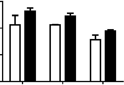
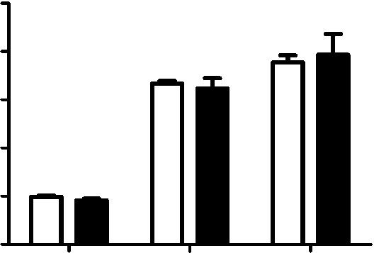
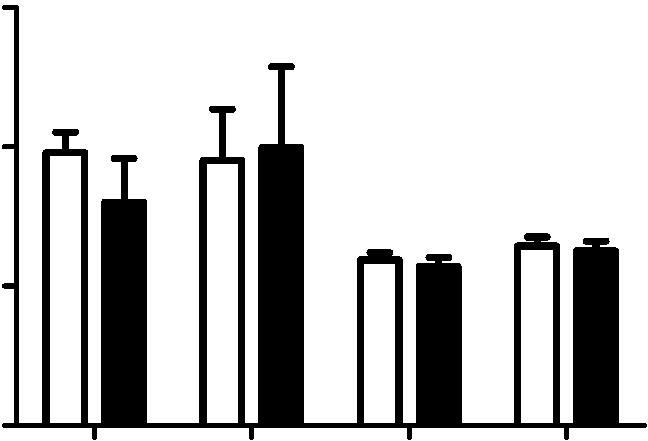
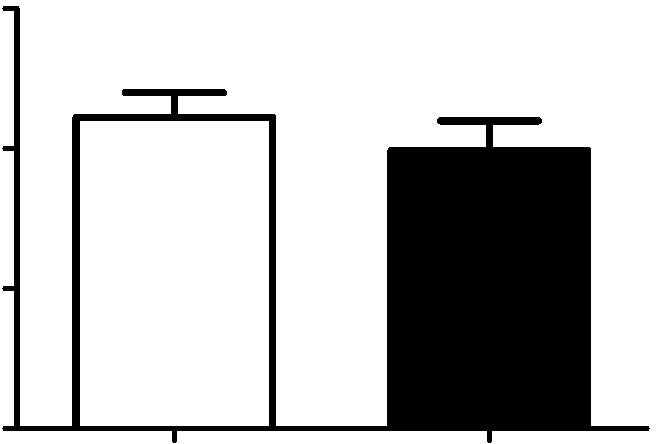
4 Figure 1. CD81 is involved in both early and late stages of influenza virus infection. A) A549 cells were treated with non-targeting control sirna, or CD81 sirna1 for 48 hours and immunostained with anti-cd81 antibody. Images are maximum projections of confocal z-stacks. B) A549 cells were treated with control or CD81 sirna for 48 hours. Cells were harvested for western blotting with indicated antibodies. Tubulin was used as a loading control. C) Influenza virus infection is impaired by CD81 depletion. A549 cells were treated with sirnas or mock treated for 48 hours and subsequently infected with X-31, WSN, or Udorn at a MOI of,0.1 for 36 hours. The viral titer in the supernatant was determined by plaque assays. The shaded bar indicates the infectivity measured in cells mock treated with a transfection solution that contains no sirna; the hollow bars indicated the infectivity measured in cells treated with control, non-targeting sirna; the black solid bars indicate the infectivity measured in cells treated with CD81 sirna. D) The number of infected cells expressing viral NP is reduced by about 50% upon CD81- knockdown. sirna-treated A549 cells were infected with WSN, X-31, or Udorn viruses with a MOI of,0.1 for 8 hours without acid bypass and the fraction of cells expressing NP was measured through flow cytometry. E) Viral NP expression is unaffected upon CD81 knockdown when influenza infection is induced by the acid-bypass treatment to eliminate the entry defect. sirna-treated A549 cells were allowed to bind with WSN or X-31 virus on ice for 1 hour, treated with warm low ph PBS buffer (ph 4.5) for 2 minutes. After 8 hours, cells were collected and stained against NP for flow cytometry analysis. F) Virus titer in the supernatant is reduced by,50% or more in CD81-knockdown cells infected by influenza viruses through the acid-bypass treatment. Briefly virus was allowed to bind with control or CD81-knockdown cells on ice for 1 hour. Unbound virus particles were washed out and low ph buffer was added in for 2 minutes to trigger virus fusion at the plasma membrane. At 17 hours post infection, supernatant was collected and the viral titer was assayed by plaque assay. For figure 1C 1F, the error bars are standard deviation derived from three independent experiments. A two-tailed student t-test was performed for all of the numerical data, and the p value of the data is shown. A p value smaller than 0.05 indicates there is a statistically significant difference. doi: /journal.ppat g001 viruses when compared to control cells treated by non-targeting sirna (Figure 2A). Next, influenza virus infection was allowed to proceed for 30 minutes at 37uC, and the number of internalized virus particles was quantified. As shown in Figure 2B, the number of internalized viruses per cell was similar between control and CD81-knockdown cells. Moreover, virus particles in both control and CD81-knockdown cells were delivered into early endosomes after internalization at 37uC. At 20 minutes post infection, the mean fraction of virus particles colocalized with early endosomes (marked by EEA1) was,50% and,53% for control and CD81- knockdown cells, respectively (Figure 2C). These results suggest that CD81 does not affect trafficking of influenza virus to early endosomes. Additionally, we tested the effect of CD81 depletion on two other viruses: respiratory syncytial virus (RSV) and GFP-encoding pseudotyped murine leukaemia virus (MLV), which are known to PLOS Pathogens 3 October 2013 Volume 9 Issue 10 e
5 Figure 2. CD81 is not required for virus binding, internalization or delivery into early endosomes. A) CD81-knockdown does not affect virus binding, as measured by flow cytometry. The magenta, blue and orange curves correspond to the intensity profiles measured for cells without adding viruses, control cells after influenza virus binding, and CD81-knockdown cells after influenza virus binding, respectively. B) The number of virus particles internalized is not affected by CD81 knockdown. The number of internalized virus particles was shown in a dot plot, with the middle line representing the mean value, and top/bottom line representing standard deviation. At least 40 randomly chosen cells were analyzed for each condition. C) The percent of virus particles colocalizing with early endosome is not affected by CD81 knockdown. Early endosomes were immunostained with anti-eea1 antibody. Data was plotted similarly as in (B). At least 40 randomly chosen cells were analyzed for each condition. D) CD81 depletion does not affect RSV or pseudo-typed MLV infection. sirna-treated A549 cells were infected with different doses of RSV and pseudotyped MLV virus for 24 hours. For RSV virus infection, RSV fusion protein expression was quantified by flow cytometry, while for pseudo-typed MLV virus, the GFP signal was analyzed. A two-tailed student t-test was performed for all of the numerical data, and the p value of the data is shown. doi: /journal.ppat g002 undergo virus fusion in early endosomes in a ph-independent manner [39,40]. Pseudotyped MLV infected cells express GFP, but cannot produce complete virions, allowing the quantification of MLV entry through measuring the GFP expression. For RSV, we measured the expression level of the fusion protein (F protein) after 24 hours of infection. As shown in Figure 2D, the fraction of infected cells was similar between control and CD81-knockdown cells with MLV and RSV infection. This data further corroborates the notion that CD81 does not affect virus trafficking into early endosomes. Taken together, the results presented above indicate that CD81 is not involved in influenza virus binding, internalization, or trafficking into early endosomes. Influenza virus particles are delivered to and fuse in CD81+ endosomes Next, we probed the role of CD81 in virus fusion. Influenza virus is trafficked from early endosomes to maturing endosomes, in which the low ph environment triggers conformational changes in HA that mediate viral fusion with the endosomal membrane [2,9,41]. In addition to being distributed on the plasma membrane, CD81 also showed substantial colocalization with early and maturing endosomes, which are Rab5 positive (Rab5+) (Figure 3A) [41]. About 30 35% Rab5+ endosomes contained CD81 (Figure 3A), suggesting that CD81 is enriched in a subpopulation of these endosomes. To probe whether influenza virus particles are delivered into CD81+ endosomes, we allowed Alexa Fluor 647-labeled X-31 to internalize for 15 minutes and immunostained the cells for CD81. As shown in Figure 3B, a substantial fraction (,54%) of virus colocalized with CD81+ endosomes. We then tracked individual influenza virus particles in living cells, a technique that has been previously established [5,10,41 43], to examine whether influenza viruses fuse in CD81+ endosomes. To this end, we expressed CD81-mEmerald in A549 cells. Similar to endogenous CD81, CD81-mEmerald was PLOS Pathogens 4 October 2013 Volume 9 Issue 10 e


 CD81 substantially")


 Influenza virus")


6 Figure 3. A major fraction of viruses are trafficked to and fuse in CD81-positive endosomes. A) CD81 substantially colocalizes with Rab5. A549 cells were electroporated with CD81-mEmerald and RFP-Rab5. At 24 hours, the cells were fixed and imaged. An enlarged image of the boxed region is shown on the right. Scale bar: 10 mm. B) Influenza virus particles traffick into CD81+ endosomes. A549 cells were cold bound with Alexa Fluor 647-labeled X-31 virus (red) on ice for 30 minutes and then chased for 15 minutes at 37uC. The samples were fixed and immunostained against CD81 (green). An enlarged image of the boxed region is shown on the right. All of the images are confocal XY cross sections. Scale bar: 10 mm. C) An PLOS Pathogens 5 October 2013 Volume 9 Issue 10 e
7 influenza virus particle enters and fuses within a CD81-positive endosome after entry. Live-cell confocal imaging of DiD-labeled X-31 added in situ to CD81-mEmearld expressing A549 cells maintained at 37uC. The images were collected with a 0.5 s interval. C-1) Several snapshots taken at different time points with the virus indicated by the white circles. C-2) The fluorescence signal of the indicated DiD-labeled virus as a function of time. Note that there is a sudden increase of DiD signal at 515 s, which indicates a viral fusion event. D) Influenza virus can also fuse in a CD81-negative endosome. D-1) Several snapshots taken at different time points with the virus indicated by the white circles. D-2) The fluorescence signal of the indicated DiD labeled virus as a function of time. The virus particle fused at 422 s. E) Among 61 virus particles tracked from binding to fusion, 5268% enter and fuse within CD81+ endosomes whereas the remaining 4868% fuse in CD81- endosomes. The results are taken for four independent experiments, and the 6error indicates the standard deviation derived from these experiments. F) Virus fusion is impaired upon CD81 depletion. DiDlabeled X-31 was allowed to bind with A549 cells on ice for 30 minutes, and then chased for the indicated times at 37uC. Cells were trypsinized and fixed immediately, and analyzed by flow cytometry. The increase in the DiD intensity versus the initial DiD intensity is plotted. The error bars are standard deviation derived from duplicate experiments. doi: /journal.ppat g003 localized on both plasma and endosomal membranes (Figure S2A) and the expression of CD81-mEmerald did not affect the fraction of endosomes that are CD81+ (Figure S2B D). Moreover, the expression of CD81 also did not affect influenza viral fusion or infectivity (Figure S2E, F). To facilitate tracking of individual virus particles, X-31 viruses were labeled with a saturating amount of DiD, a lipophilic dye, such that the fluorescence emission from the DiD molecules was low due to a self-quenching effect between neighboring dyes. Fusion between the virus envelope and endosomal membrane should lead to an increase in fluorescent intensity (dequenching), due to the diffusion of dyes from the virus into the lipid bilayer of the endosomes [42]. We added labeled viruses to the CD81-mEmerald expressing cells in situ at 37uC. The virus particles typically show restricted movement immediately after binding to the cell, followed by rapid and directed movement once the virus particles are internalized, similar to our previous observations [5,41,42]. We observed a proportion of virus particles entering into CD81+ endosomes soon after internalization, as illustrated by the example shown in Figure 3C and movie S1. These viruses remained colocalized with CD81 and eventually fused with the CD81+ endosomes, as reflected by the sudden increase of DiD fluorescence, presumably after the endosomes matured to acquire a sufficiently low ph (Figure 3C and movie S1). Among the 61 virus particles that we tracked from binding to fusion, about 5268% underwent viral fusion within CD81+ endosomes (Figure 3E). The remaining 4868% of virus particles fused in endosomes lacking CD81 (Figures 3D,E, and movie S2). To confirm that the fusion events indeed occurred in endosomes, we tracked individual DiD-labeled influenza virus particles in cells expressing RFP-Rab5. Similar to previous observations [41], nearly 90% of the viral fusion events occurred in Rab5+ endosomes (Figure S3). To investigate whether CD81 affects viral fusion, we next monitored the DiD fluorescent intensity in control and CD81- knockdown cells that were infected with DiD-labeled X-31 virus. In these experiments, cells were first incubated with DiD-labeled virus at 4uC and then the temperature was increased to 37uC to initiate viral entry. At specific time points after the temperature shift, infected cells were collected and the DiD fluorescence from these cells was quantified with flow cytometry. As expected, there was a consistent increase of DiD fluorescence with time due to viral fusion (Figure 3F). Notably, compared to control cells, CD81- knockdown cells exhibited a significant reduction in viral fusion (Figure 3F), suggesting that CD81 facilitates viral fusion. Most of the remaining viral fusion events in CD81-knockdown cells still occurred in Rab5+ endosomes (Figure S3). The reduction in viral fusion was, however, incomplete (Figure 3F), consistent with the observation that only about half of the virus particles fuse in CD81+ endosomes (Figure 3E), though the incomplete inhibition of viral fusion could also be in part due to the incomplete knockdown of CD81 (Figure S1). These data indicate that CD81 marks a specific population of Rab5+ endosomes that are responsible for half of viral fusion events. Because CD81-knockdown cells reduced viral infection (Figure 1C) and exhibited a higher reduction in virus titer when infected without acid bypass than when infected with acid bypass (Figures 1C,F), viral fusion within CD81+ endosomes likely leads to productive influenza infection. Taken together, our results indicate that half of virus particles are trafficked to and undergo viral fusion in CD81+ endosomes. CD81 could facilitate viral fusion by organizing endosomal membrane to assist viral fusion or helping virus traffick to fusion-competent endosomal compartments. CD81 is not involved in the expression or trafficking of viral proteins In the subsequent experiments, we aimed to determine which post-entry step(s) of the viral infection process are CD81 dependent. To this end, we examined the effect of CD81 on viral protein expression, viral protein trafficking, and the assembly and egress of progeny viruses. Our initial results in Figure 1E suggested that CD81 knockdown did not directly affect viral NP expression. This was further substantiated by infecting cells with various viral doses across different time points using the acid-bypass treatment. As shown in Figure 4A, the fraction of NP+ cells and NP expression level increased with the viral dose and infection time, while there was no significant difference between control and CD81-knockdown cells. To further validate the finding, we measured the expression of another cytosolic viral protein, M1. Similar to the results on NP, CD81-knockdown cells infected by influenza viruses using the acid-bypass treatment did not exhibit a difference in the fraction of M1+ cells or the level of M1 protein expression, when compared to the control cells (Figure 4B). These data suggest that CD81 does not play a role in viral gene expression for cytosolic viral proteins. To determine whether CD81 affects viral membrane protein expression, we probed M2 expression with acid bypass treatment. The fraction of M2+ cells and the M2 expression level in M2+ cells were similar between control and CD81-knockdown cells (Figure 4C). Furthermore, by only probing the surface M2 protein without permeabilizing the cells, we found that there was no difference in surface M2 protein expression level either (Figure 4D), indicating that CD81 knockdown also did not affect the trafficking of M2 to the cell surface. Similarly, the expression and trafficking of another viral membrane protein, NA, were also not affected upon CD81 depletion (Figure 4E). Taken together, these data indicate that CD81 does not play a direct role in the expression of influenza viral proteins or the trafficking of influenza membrane proteins to the plasma membrane. PLOS Pathogens 6 October 2013 Volume 9 Issue 10 e

8 PLOS Pathogens 7 October 2013 Volume 9 Issue 10 e
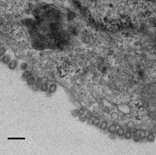
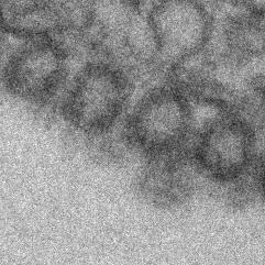
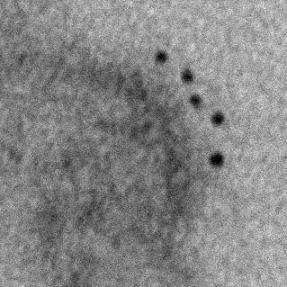
9 Figure 4. CD81 depletion does not affect viral protein expression and transport. A) CD81 knockdown does not affect the expression of viral NP protein in cells infected by influenza viruses with the acid-bypass treatment. Experiments were performed similarly as in 1E) except that the expression levels are evaluated at different time point post infection and with different dose of viruses. The percent of NP-expression cells and the NP expression level in NP+ cells are plotted. B) CD81 knockdown does not affect the expression of viral M1 protein in cells infected by influenza viruses with the acid-bypass treatment. Experiments were performed similarly as in (A) except cells were immunostained for M1. The percent of M1+ cells and the M1 expression level in the M1+ cells are plotted. C) CD81 knockdown does not affect the expression of viral M2 protein in cells infected by influenza viruses with the acid-bypass treatment. Experiments were performed similarly as in (A) except cells were immunostained for M2. The percent of M2+ cells and the M2 expression level in the M2+ cells are plotted. D) CD81 knockdown does not affect the amount of M2 protein trafficked to the cell surface in cells infected by influenza viruses with the acid-bypass treatment. Experiments were performed similarly as in (C) except cells were stained for M2 without permeabilization. The percent of M2+ cells and the surface M2 expression level in M2+ cells are plotted. E) CD81 knockdown does not affect the amount of NA protein trafficked to the cell surface in cells infected by influenza viruses with the acid-bypass treatment. The NA expression level was estimated from confocal images in control or CD81 sirna treated cells infected by X-31virus. A two-tailed student t-test was performed for all of the numerical data, and the p value of the data is shown. doi: /journal.ppat g004 CD81 is recruited to influenza virus budding sites on the plasma membrane Next, we probed the role of CD81 in virus assembly. To test whether CD81 is present at the viral assembly sites, A549 cells were infected with the three influenza strains and immunostained for CD81 and the viral protein. Notably, with X-31 infection, CD81 was mostly localized to a site concentrated with multiple viral proteins (Figures 5A and S4A, B). In contrast to uninfected cells, which showed a uniform distribution of CD81 on the plasma membrane, X-31-infected cells exhibited marked redistribution of CD81 into concentrated patches. All of the X-31 proteins that we could obtain specific immunofluorescence staining for, including PB1, NA and M2, were present in these patches. The CD81 patches were formed on the plasma membrane, as confirmed by immunofluorescence of non-permeabilized cells (Figure S4B). We note that there was only a modest decrease of CD81 expression upon viral infection (Figure S5A, B). For Udorn-infected cells, CD81 was enriched along the budding virus filaments marked by PB1 (Figure 5B). PB1 is a good filament marker that colocalized with Udorn HA and M2 in the budding filamentous virions (Figure S4C,D). We have also directly observed colocalization between CD81 and other Udorn proteins including HA, NA and an anti-udorn serum (Figure S4E G). Remarkably, upon sirna treatment, which depleted 80,85% of the endogenous CD81, the remaining CD81 was all concentrated in the budding viral filaments, whereas the cell body had little CD81 signal (Figures 5C and S4H). The average amount of CD81 per virus filament in the CD81-knockdown cells was reduced by more than 60% compared to that in control cells. Although our lack of WSN antibodies made it difficult to perform similar immunofluorescence experiments on WSN-infected cells, our EM images with CD81 labeled by immunogold showed that CD81 was also recruited to the WSN virus budding zones (Figure S6A). Taken together, these results indicate that CD81 is specifically recruited to the influenza virus assembly and budding sites. To probe which viral component may be responsible for recruiting CD81, we turned to a plasmid-based system that expresses only specific viral envelope proteins in cells [44]. We transiently transfected the plasmid containing HA or NA in A549 cells and immunostained the cells with CD81 and HA or NA. Interestingly, HA tends to form clusters on the cell surface even when expressed alone in A549 cells and CD81 accumulated substantially in the HA clusters (Figure S7A). About 46% of HA clusters colocalized with CD81. In contrast, NA when expressed alone did not form clusters but was distributed largely uniformly across the plasma membrane and there was no appreciable correlation between the CD81 distribution and NA distribution (Figure S7C). Mock transfection with plasmid that did not contain HA or NA did not yield any appreciable HA or NA staining (Figure S7B,D). These results suggest that HA is likely responsible for recruiting CD81 to the viral budding sites. CD81 is incorporated at specific sub-viral locations and facilitates influenza virus budding Although we observed a,50% or more decrease in virus titer in CD81-knockdown cells after acid-bypass treatment to overcome the CD81-dependent entry defects (Figure 1F), it remained unknown whether the defect in viral titer stems from a decrease in the number of budding virions assembled on the cell, the number of progeny virus particles released from the cell, or the specific infectivity per released virus particle. To distinguish between these possibilities, we first infected cells using the acidbypass treatment and then quantified the number of budding virions attached to the cells using transmission electron microscopy. After quantifying more than 250 cell cross-sections per condition, we performed statistical analysis and found no statistically significant difference in the number of assembling virus particles per cell cross-section in the control versus CD81- knockdown cells (Figure 6A). Next, we infected cells with WSN using the acid-bypass treatment, collected the virus particles in the supernatant, and then quantified the amount of viral matrix protein M1 using an ELISA assay and the number of released virus particles positive of both M1 and HA using an imaging assay (Figure 6B). Notably, compared to control cells treated by non-targeting sirna, CD81- knockdown cells exhibited 50% or more decrease in both the amount of viral M1 and the number of M1+ and HA+ virus particles released into the supernatant. These results suggest that the CD81-knockdown-induced reduction in viral titer in cells infected by the acid-bypass treatment stems from a defect in virus release. Given that the reduction in viral titer (Figure 1F) was similar to the reduction in the amount of released viral proteins or viral particles (Figure 6B), we did not further probe the change in specific infectivity per virus particle. To examine how CD81 may facilitate release of progeny virus particles, we next probed the distribution of CD81 within individual budding virions using immunogold electron microscopy. In X-31-infected cells, CD81 was readily observed in budding virions (Figure 6C, D). During early assembly stages, CD81 clusters located at the growing tip of budding virions (Figure 6C). Interestingly, when viruses grew into a mature, slightly elongated shape [33], CD81 was not only found on the growing tip, but also on the neck of budding virions (Figure 6D). The elongated morphology of X-31 allowed us to quantitatively analyze the CD81 distribution in virions by aligning the long axis of the virus particles and normalizing the position of immunogold-labeled CD81 to the total length of the virus. Remarkably, CD81 is highly enriched at the two ends of the budding virions (Figure 6D, E). PLOS Pathogens 8 October 2013 Volume 9 Issue 10 e
 and anti-pb1 antibody (red). Images are confocal XY crosssections. Scale bar: 10 mm.")
 Remaining CD81 in CD81-knockdown cells is incorporated into budding filamentous viruses of Udorn infected cells. Similar to (B) except that CD81-knockdown cells were used.")

.")

.")
. We performed similar experiments with the X-31 strain.")
10 Figure 5. CD81 is recruited to the virus budding sites. A) CD81 is recruited to the virus budding zone in X-31 infected cells. A549 cells were infected with X-31 for 16 hours. Cells were stained with anti-cd81 antibody (green) and anti-pb1 antibody (red). Images are confocal XY crosssections. Scale bar: 10 mm. B) CD81 is incorporated into budding filamentous virions of Udorn-infected cells. A549 cells were infected with Udorn virus for 16 hours, and stained with anti-cd81 antibody and anti-pb1 antibody. Scale bar: 10 mm. C) Remaining CD81 in CD81-knockdown cells is incorporated into budding filamentous viruses of Udorn infected cells. Similar to (B) except that CD81-knockdown cells were used. The CD81 expression level in Udorn-infected cells was calculated based on confocal images of more than 100 cells, and was found to be decreased by,88% upon CD81 depletion as compared to control cells. The amount of CD81 per viral filament was reduced by 63% compared to that in untreated cells. Scale bar: 10 mm. doi: /journal.ppat g005 Similarly, we also found CD81 to be enriched in budding WSN virus particles, but the quantity of immunogold detected per WSN virus is substantially lower than that in X-31 viruses, which made it difficult to determine the CD81 distribution in these viruses (Figure S6A). However, the nearly perfectly spherical shape of the WSN virus allowed us to detect an interesting morphological defect of budding viruses in CD81-knockdown cells. When we examined budding virions in cells infected by WSN, we found that most budding WSN viruses were spherical and completely enclosed by viral envelope in control cells treated by non-targeting sirna (Figure 6F). In stark contrast, budding WSN virus in CD81-knockdown cells appeared much more elongated (Figure 6G). The average length of budding virions in control and CD81-knockdown cells was,100 nm and,150 nm, respectively (Figure 6H). Furthermore, many budding viruses in CD81-knockdown cells did not have a fully enclosed envelope but remain attached to the plasma membrane through an open membrane neck (indicated by arrowheads in Figure 6G). We performed similar experiments with the X-31 strain. Again, we consistently observed many budding X-31 virions with the open budding neck defect upon CD81 depletion (Figure S6B, C), though characterizing whether the budding virions were further elongated was difficult due to the large variation of the virion length of the pleomorphic X-31. Taken together, the specific enrichment of CD81 at the neck of the budding virions, the defect in budding neck closure in CD81- knockdown cells, and the reduction in the number of released virus particles but not in the number of assembling virions upon CD81 knockdown suggests that CD81 plays a role in a late stage of the virus budding process, likely at the final scission step. CD81 may PLOS Pathogens 9 October 2013 Volume 9 Issue 10 e


11 Role of CD81 in Influenza Virus Infection PLOS Pathogens 10 October 2013 Volume 9 Issue 10 e
 CD81 knockdown does not change the number of budding virions attached to infected cells. sirna-treated cells were infected with WSN virus with the acid-bypass treatment for 15 hours.")
 CD81 knockdown causes a substantial reduction in the number of released virus particles.")


 Distribution of gold particles in budding X-31 viruses at 16 hour post infection.")
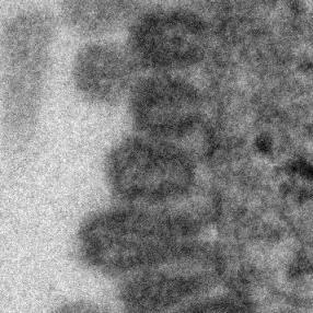
12 Figure 6. CD81 is enriched at specific sub-viral sites of budding virions and CD81 knockdown impairs virus scission. A) CD81 knockdown does not change the number of budding virions attached to infected cells. sirna-treated cells were infected with WSN virus with the acid-bypass treatment for 15 hours. Cells were directly fixed for transmission electron microscopy and the number of budding virus particles per cell cross-section is quantified for over 250 sections, and presented in the dot plot. A two-tailed student t-test was performed and the p value is provided. B) CD81 knockdown causes a substantial reduction in the number of released virus particles. sirna-treated cells were infected with WSN virus with the acid-bypass treatment for 17 hours. The amount of viral M1 protein in the supernatant was probed with ELISA. The number of M1 positive and HA positive virus particles in the supernatant was counted using immunofluorescence imaging. The error bar is standard deviation from three independent measurements. C) CD81 localizes at the tip of growing X-31 viruses during the early budding stages. Cells were infected with X-31 for 12 hours and CD81 was immunogold labeled for electron microscopy. An enlarged image of the area in the white box is shown in the upper right corner. Scale bar: 100 nm. D) CD81 mainly localizes at the tip and budding neck of the X-31 viruses during late budding stages. Similar to (C) except the infection time was 16 hours. Scale bar: 200 nm. E) Distribution of gold particles in budding X-31 viruses at 16 hour post infection. To align the virus particles, the length of each virus is normalized to 1, with its middle point assigned with coordinate value of 0. For individual gold particles on the budding virus, their coordinate values were calculated based on their relative distance to the middle point. Coordinates with negative values correspond to positions close to the plasma membrane. A total of 105 budding viruses were analyzed. F) Budding WSN viruses exhibit a spherical morphology with fully enclosed membrane envelope in control sirna-treated cells. A549 cells were infected with virus with the acid-bypass treatment for 13 hours. The region in the white box is magnified and shown in the upper right corner. Scale bar: 200 nm. G) Budding WSN viruses are more elongated in CD81 sirna treated A549 cells. A substantial fraction of budding viruses have an open membrane neck connected to the plasma membrane (indicated by arrowheads). The region in the white box is magnified and shown in the upper right corner. Scale bar: 200 nm. H) Budding WSN viruses are elongated upon CD81 depletion, as shown by the distribution of budding virus length in control or CD81-knockdown cells. doi: /journal.ppat g006 facilitate viral scission by directly participating in the scission process, by recruiting host or viral scission proteins, or by organizing the lipid domains and making it conducive to viral scission. Distribution of CD81 in budding filamentous Udorn virus Unlike WSN and X-31 strain, Udorn virus infection typically produces filaments that can reach 2 to 20 mm long [11,35,45,46]. We visualized immunogold-labeled CD81 distribution in A549 cells infected by Udorn virus with electron microscopy and observed CD81 clusters in budding viral filaments. Notably, CD81 appeared to be distributed along the entire Udorn filament (Figure 7A). As an alternative approach, we used a super-resolution fluorescence imaging technique, Stochastic Optical Reconstruction Microscopy (STORM), to measure the distribution of CD81 on the budding filament. STORM overcomes the diffraction limit of light microscopy by sequentially activating, imaging and localizing individual fluorescent photoswitchable molecules at high precision, thereby reconstituting images from the molecular localizations with nano-meter scale resolution [47,48]. Here, we used a singleobjective detection geometry and photoswitchable Alexa Fluor 647 and Atto 488 dyes to obtain a lateral resolution of nm and axial resolution of nm [48,49]. To visualize the localization of CD81 in filamentous Udorn, we immunostained CD81 and viral PB1, and performed two-color 3D STORM imaging. Consistent with the results from electron microscopy (Figure 7A), we found that CD81 formed small clusters evenly distributed along the entire filament (Figure 7B). Adjacent CD81 clusters were usually separated Figure 7. Scattered distribution of CD81 along budding filamentous Udorn virus. A) CD81 localizes along the filament of budding Udorn viruses. A549 cells were infected with Udorn virus for 18 hours and CD81 was immunogold labeled for electron microscopy. Shown here is a bundle of virus filament budding from the cell (the cell is not shown in order to magnify the virus filaments). Scale bar: 200 nm. B) CD81 and viral PB1 proteins appear to take an alternating distribution along filamentous Udorn virus. A549 cells were infected with Udorn virus for 16 hours and immunostained with anti-cd81 (green) and anti-pb1 (red) antibodies. CD81 was further probed with Alexa Fluor 405/Alexa Fluor 647-conjugated secondary antibody while PB1 was labeled with Atto 488 conjugated antibody for two-color 3D STORM imaging. Two example filamentous viruses were shown in B-1) and B-2). Left: xy projection images. Middle) xz projection images. Right) Localization distribution of CD81 and PB1 along the filament long axis. Scale bar: 500 nm. doi: /journal.ppat g007 PLOS Pathogens 11 October 2013 Volume 9 Issue 10 e
13 by about 150,200 nm. Curiously, CD81 appeared to be enriched between clusters of viral PB1 proteins (Figure 7B), but the significance of this alternating pattern is unclear. Discussion CD81, a cellular tetraspannin protein, is critical for influenza viral infection [15,16]. The infectivity of various strains of influenza viruses is strongly inhibited when cellular CD81 is depleted (Figure 1). In this work, we dissected the roles of CD81 on individual steps along the infection pathway from virus entry to egress. We found that CD81 plays functional roles in two separate steps of viral infection: viral fusion and virus budding. The role of CD81 in viral entry The specific role of CD81 during influenza viral entry was determined using a series of independent assays. First, knocking down CD81 by sirna led to,50% decrease in the percent of infected cells expressing viral proteins. The defect was not due to direct regulation of viral protein expression by CD81, since viral protein expression remained unchanged upon CD81 knockdown when influenza infection was induced by the acid-bypass treatment (Figure 1 and Figure 4). These results suggest that CD81 mediates influenza virus entry prior to viral gene expression. Next, CD81 knockdown did not affect virus binding, internalization or trafficking to early endosomes (Figure 2), but led to a significant defect in viral fusion (Figure 3). Furthermore, single-virus tracking experiments showed that half of internalized virus particles were trafficked into CD81-positive endosomes and underwent viral fusion within these endosomes, whereas the remaining half fused in CD81-negative endosomes (Figure 3). Notably, the fraction of viral fusion events occurring within CD81-positive endosomes correlated well with the 50% reduction in the percent of infected cells expressing viral proteins upon depletion of CD81, suggesting a role of CD81 in productive viral uncoating. Altogether, these results indicate that CD81 plays a role in influenza viral fusion. CD81 marks an endosomal route for productive virus uncoating process, though a parallel CD81- independent route also exists. Interestingly, the role of CD81 in influenza virus entry appears to differ from the role of CD81 previously observed in HCV and HIV entry. As an essential co-receptor for HCV, CD81 is important for the endocytosis of HCV [27,28,50,51]. Furthermore, CD81 interacts with HCV glycoprotein E2 and helps prime its fusogenic activity for low-ph dependent viral fusion [26,30]. Moreover, CD81 negatively regulates HIV-cell fusion [52]. Incorporation of CD81 and CD81-associated tetraspanins suppresses the HIV-mediated cellular fusion processes [52,53]. On the other hand, CD81 unlikely functions as a co-receptor or attachment factor for influenza viruses because the internalization of influenza viruses into cells does not require CD81. Influenza viral fusion does not need to be primed by CD81 either, as acid treatment is sufficient to trigger viral uncoating at the plasma membrane in CD81-depleted cells. Instead, our results suggest a role of CD81 in facilitating influenza virus fusion in endosomal compartments. Given that CD81 and CD81-associating proteins can organize membrane domains [17 20], CD81 may help organize the endosomal membrane for assisting influenza viral fusion. Alternatively, CD81 may play a role in trafficking influenza to fusion competent endosomal compartments. CD81 is highly enriched in multivesicular bodies (MVBs), an intermediate endosomal organelle on the maturation pathway of late endosomes and lysosomes [54]. CD81 depletion may inhibit the maturation of endosomes and thus compromise influenza virus fusion with endosomes. The role of CD81 in viral assembly In addition to its role in virus uncoating, CD81 also plays a functional role in a later stage of influenza infection post viral fusion. The requirement of CD81 in a post-fusion stage was evident from the finding that CD81 depletion led to a significant decrease in virus titer even when the acid-bypass treatment was used to induce viral uncoating at the plasma membrane, thereby eliminating entry defects (Figure 1F). The decrease did not result from a defect in expression of viral proteins or trafficking of viral proteins to the plasma membrane (Figure 4), suggesting that the perturbation likely occurred at the virus assembly stage. Furthermore, the average number of budding virions attached to each infected cell did not change upon CD81 knockdown, whereas the number of virus particles released into the supernatant markedly decreased (Figure 6A, B). These results further narrowed the involvement of CD81 to a relatively late stage of the budding process, likely the scission step that severs the virus particle from the host cells. Supporting this notion, CD81 was specifically recruited to viral budding sites (Figure 5), and among the viral proteins, HA is likely important for recruiting CD81 to the virus budding sites (Figure S7). Interestingly, CD81 was specifically enriched at the tip and budding neck of the spherical and slightly elongated viruses (Figure 6C E). Upon CD81 knockdown, the budding spherical viruses exhibited a consistent change in morphology: the budding virions appeared substantially elongated compared to their counterparts in control cells and failed to detach from the plasma membrane. Many budding viruses did not have their budding neck closed, indicating a defect in the final scission process (Figure 6F H). Taken together, our observations indicate a role of CD81 in scission process that severs the budding virions from the plasma membrane. CD81 could be directly participating in the scission process, recruiting other host or viral scission proteins for this purpose, or organizing the membrane domain at the budding site and making it conducive for viral scission. It is interesting to compare the role of CD81 in the assembly of influenza virus with that of other viruses. Previous studies have shown that HIV envelope proteins associate with a few tetraspanins, including CD81, and that HIV buds from the tetraspanin-enriched microdomains [55 57]. However, the exact role of CD81 in HIV egress remains unclear [53,58 61]. One study reports that HIV infection is significantly impaired upon CD81 depletion or treatment with anti-cd81 antibodies [61], whereas two other papers report that depletion of tetraspanins does not affect the efficiency of HIV release whereas overexpression of tetraspanins results in decreased infectivity in released virions [53,59]. Tetraspanins have also been proposed to facilitate cell-to-cell transmission of HTLV-1 infection [25]. The role of CD81 in the egress of influenza virus appears different from these previously reported roles of tetraspanins in HIV and HTLV-1 infection in that CD81 positively regulate viral scission. It has been previously shown that influenza virus scission is dependent on viral M2 protein [62]. During virus budding, M2 is localized at the neck of budding viruses and mutation of its amphiphilic tail at the C-terminus leads to a marked defect in virus budding [62]. Interestingly, M2 is known to localize at the interface between lipid rafts and non-rafts region, while CD81 is partitioned into tetraspanin-enriched microdomains, a platform that resembles lipid rafts [18]. Thus, it is possible that CD81 facilitates the recruitment of M2 to the budding neck of the viruses. Future studies on the interaction between CD81 and M2 during the viral scission process would be of interest to further PLOS Pathogens 12 October 2013 Volume 9 Issue 10 e
University of Groningen
 University of Groningen Mechanisms of Hemagglutinin Targeted Influenza Virus Neutralization Brandenburg, Boerries; Koudstaal, Wouter; Goudsmit, Jaap; Klaren, Vincent; Tang, Chan; Bujny, Miriam V.; Korse,
Quantifying Lipid Contents in Enveloped Virus Particles with Plasmonic Nanoparticles
 Quantifying Lipid Contents in Enveloped Virus Particles with Plasmonic Nanoparticles Amin Feizpour Reinhard Lab Department of Chemistry and the Photonics Center, Boston University, Boston, MA May 2014
Dr. Ahmed K. Ali Attachment and entry of viruses into cells
 Lec. 6 Dr. Ahmed K. Ali Attachment and entry of viruses into cells The aim of a virus is to replicate itself, and in order to achieve this aim it needs to enter a host cell, make copies of itself and
Figure S1. Western blot analysis of clathrin RNA interference in human DCs Human immature DCs were transfected with 100 nm Clathrin SMARTpool or
 Figure S1. Western blot analysis of clathrin RNA interference in human DCs Human immature DCs were transfected with 100 nm Clathrin SMARTpool or control nontargeting sirnas. At 90 hr after transfection,
Influenza viruses. Virion. Genome. Genes and proteins. Viruses and hosts. Diseases. Distinctive characteristics
 Influenza viruses Virion Genome Genes and proteins Viruses and hosts Diseases Distinctive characteristics Virion Enveloped particles, quasi-spherical or filamentous Diameter 80-120 nm Envelope is derived
Patricia Fitzgerald-Bocarsly
 FLU Patricia Fitzgerald-Bocarsly October 23, 2008 Orthomyxoviruses Orthomyxo virus (ortho = true or correct ) Negative-sense RNA virus (complementary to mrna) Five different genera Influenza A, B, C Thogotovirus
Supplemental information contains 7 movies and 4 supplemental Figures
 1 2 3 4 5 6 7 8 9 10 11 12 13 14 15 16 17 18 19 20 21 22 23 24 25 26 27 Supplemental information contains 7 movies and 4 supplemental Figures Movies: Movie 1. Single virus tracking of A4-mCherry-WR MV
Polyomaviridae. Spring
 Polyomaviridae Spring 2002 331 Antibody Prevalence for BK & JC Viruses Spring 2002 332 Polyoma Viruses General characteristics Papovaviridae: PA - papilloma; PO - polyoma; VA - vacuolating agent a. 45nm
Lecture Readings. Vesicular Trafficking, Secretory Pathway, HIV Assembly and Exit from Cell
 October 26, 2006 1 Vesicular Trafficking, Secretory Pathway, HIV Assembly and Exit from Cell 1. Secretory pathway a. Formation of coated vesicles b. SNAREs and vesicle targeting 2. Membrane fusion a. SNAREs
Virus Entry/Uncoating
 Virus Entry/Uncoating Delivery of genome to inside of a cell Genome must be available for first step of replication The Problem--barriers to infection Virion Barriers: Non-enveloped viruses capsid Enveloped
LESSON 1.4 WORKBOOK. Viral sizes and structures. Workbook Lesson 1.4
 Eukaryotes organisms that contain a membrane bound nucleus and organelles. Prokaryotes organisms that lack a nucleus or other membrane-bound organelles. Viruses small, non-cellular (lacking a cell), infectious
The Regulated Secretory Pathway in CD4 + T cells Contributes to Human Immunodeficiency Virus Type-1 Cell-to-Cell Spread at the Virological Synapse
 The Regulated Secretory Pathway in CD4 + T cells Contributes to Human Immunodeficiency Virus Type-1 Cell-to-Cell Spread at the Virological Synapse Clare Jolly 1 *, Sonja Welsch 2,3, Stefanie Michor 4,
LESSON 1.4 WORKBOOK. Viral structures. Just how small are viruses? Workbook Lesson 1.4 1
 Eukaryotes- organisms that contain a membrane bound nucleus and organelles Prokaryotes- organisms that lack a nucleus or other membrane-bound organelles Viruses-small acellular (lacking a cell) infectious
 http://nmhm.washingtondc.museum/collections/archives/agalleries/1918flu/ncp1603.jpg 1 https://assets-production-webvanta-com.s3-us-west- 2 2.amazonaws.com/000000/47/62/original/images/img_109_influenza/Spanish_flu_death_chart.jpg
Nature Medicine: doi: /nm.4322
 1 2 3 4 5 6 7 8 9 10 11 Supplementary Figure 1. Predicted RNA structure of 3 UTR and sequence alignment of deleted nucleotides. (a) Predicted RNA secondary structure of ZIKV 3 UTR. The stem-loop structure
SUPPLEMENTARY INFORMATION
 sirna pool: Control Tetherin -HA-GFP HA-Tetherin -Tubulin Supplementary Figure S1. Knockdown of HA-tagged tetherin expression by tetherin specific sirnas. HeLa cells were cotransfected with plasmids expressing
Supplementary Materials for
 advances.sciencemag.org/cgi/content/full/3/6/e1700338/dc1 Supplementary Materials for HIV virions sense plasma membrane heterogeneity for cell entry Sung-Tae Yang, Alex J. B. Kreutzberger, Volker Kiessling,
Supplemental Figures:
 Supplemental Figures: Figure 1: Intracellular distribution of VWF by electron microscopy in human endothelial cells. a) Immunogold labeling of LC3 demonstrating an LC3-positive autophagosome (white arrow)
Influenza virus exploits tunneling nanotubes for cell-to-cell spread
 Supplementary Information Influenza virus exploits tunneling nanotubes for cell-to-cell spread Amrita Kumar 1, Jin Hyang Kim 1, Priya Ranjan 1, Maureen G. Metcalfe 2, Weiping Cao 1, Margarita Mishina 1,
Influenza A H1N1 HA ELISA Pair Set
 Influenza A H1N1 HA ELISA Pair Set for H1N1 ( A/Puerto Rico/8/1934 ) HA Catalog Number : SEK11684 To achieve the best assay results, this manual must be read carefully before using this product and the
Fig. S1. Subcellular localization of overexpressed LPP3wt-GFP in COS-7 and HeLa cells. Cos7 (top) and HeLa (bottom) cells expressing for 24 h human
 and HeLa (bottom) cells expressing for 24 h human") Fig. S1. Subcellular localization of overexpressed LPP3wt-GFP in COS-7 and HeLa cells. Cos7 (top) and HeLa (bottom) cells expressing for 24 h human LPP3wt-GFP, fixed and stained for GM130 (A) or Golgi97
Fig. S1. Subcellular localization of overexpressed LPP3wt-GFP in COS-7 and HeLa cells. Cos7 (top) and HeLa (bottom) cells expressing for 24 h human LPP3wt-GFP, fixed and stained for GM130 (A) or Golgi97
Viral structure م.م رنا مشعل
 Viral structure م.م رنا مشعل Viruses must reproduce (replicate) within cells, because they cannot generate energy or synthesize proteins. Because they can reproduce only within cells, viruses are obligate
Lecture 2: Virology. I. Background
 Lecture 2: Virology I. Background A. Properties 1. Simple biological systems a. Aggregates of nucleic acids and protein 2. Non-living a. Cannot reproduce or carry out metabolic activities outside of a
Lysosomes and endocytic pathways 9/27/2012 Phyllis Hanson
 Lysosomes and endocytic pathways 9/27/2012 Phyllis Hanson General principles Properties of lysosomes Delivery of enzymes to lysosomes Endocytic uptake clathrin, others Endocytic pathways recycling vs.
Supplementary Figure 1. SC35M polymerase activity in the presence of Bat or SC35M NP encoded from the phw2000 rescue plasmid.
 1 2 3 4 5 6 7 8 9 10 11 12 13 14 15 16 17 18 19 20 21 22 23 24 25 26 27 Supplementary Figure 1. SC35M polymerase activity in the presence of Bat or SC35M NP encoded from the phw2000 rescue plasmid. HEK293T
Virus Entry. Steps in virus entry. Penetration through cellular membranes. Intracellular transport John Wiley & Sons, Inc. All rights reserved.
 Virus Entry Steps in virus entry Penetration through cellular membranes Intracellular transport Steps in virus entry How do virions get into cells? Viruses of bacteria, archaea, algae and plants use different
Coronaviruses. Virion. Genome. Genes and proteins. Viruses and hosts. Diseases. Distinctive characteristics
 Coronaviruses Virion Genome Genes and proteins Viruses and hosts Diseases Distinctive characteristics Virion Spherical enveloped particles studded with clubbed spikes Diameter 120-160 nm Coiled helical
Supplementary Figure S1. Venn diagram analysis of mrna microarray data and mirna target analysis. (a) Western blot analysis of T lymphoblasts (CLS)
 Western blot analysis of T lymphoblasts (CLS)") Supplementary Figure S1. Venn diagram analysis of mrna microarray data and mirna target analysis. (a) Western blot analysis of T lymphoblasts (CLS) and their exosomes (EXO) in resting (REST) and activated
Supplementary Figure S1. Venn diagram analysis of mrna microarray data and mirna target analysis. (a) Western blot analysis of T lymphoblasts (CLS) and their exosomes (EXO) in resting (REST) and activated
THE ROLE OF ALTERED CALCIUM AND mtor SIGNALING IN THE PATHOGENESIS OF CYSTINOSIS
 Research Foundation, 18 month progress report THE ROLE OF ALTERED CALCIUM AND mtor SIGNALING IN THE PATHOGENESIS OF CYSTINOSIS Ekaterina Ivanova, doctoral student Elena Levtchenko, MD, PhD, PI Antonella
Supplementary Figure 1. Prevalence of U539C and G540A nucleotide and E172K amino acid substitutions among H9N2 viruses. Full-length H9N2 NS
 Supplementary Figure 1. Prevalence of U539C and G540A nucleotide and E172K amino acid substitutions among H9N2 viruses. Full-length H9N2 NS nucleotide sequences (a, b) or amino acid sequences (c) from
Herpesviruses. Virion. Genome. Genes and proteins. Viruses and hosts. Diseases. Distinctive characteristics
 Herpesviruses Virion Genome Genes and proteins Viruses and hosts Diseases Distinctive characteristics Virion Enveloped icosahedral capsid (T=16), diameter 125 nm Diameter of enveloped virion 200 nm Capsid
Influenza A Virus Assembly Intermediates Fuse in the Cytoplasm
 Influenza A Virus Assembly Intermediates Fuse in the Cytoplasm Seema S. Lakdawala 1 *, Yicong Wu 2, Peter Wawrzusin 2, Juraj Kabat 3, Andrew J. Broadbent 1, Elaine W. Lamirande 1, Ervin Fodor 4, Nihal
Ralf Wagner Paul-Ehrlich-Institut
 www.pei.de Other Assays for the Detection of Neuraminidase (NA)-Specific Antibodies Ralf Wagner Paul-Ehrlich-Institut Overview to presented assays Assay principle based on: Chemical substrates: Protein
LESSON 4.4 WORKBOOK. How viruses make us sick: Viral Replication
 DEFINITIONS OF TERMS Eukaryotic: Non-bacterial cell type (bacteria are prokaryotes).. LESSON 4.4 WORKBOOK How viruses make us sick: Viral Replication This lesson extends the principles we learned in Unit
Reviewers' comments: Reviewer #1 (Remarks to the Author):
:") Reviewers' comments: Reviewer #1 (Remarks to the Author): Nature Communications manuscript number NCOMMS-16-15882, by Miyakawa et al. presents an intriguing analysis of the effects of the tumor suppressor
Reviewers' comments: Reviewer #1 (Remarks to the Author): Nature Communications manuscript number NCOMMS-16-15882, by Miyakawa et al. presents an intriguing analysis of the effects of the tumor suppressor
Coronaviruses cause acute, mild upper respiratory infection (common cold).
.") Coronaviruses David A. J. Tyrrell Steven H. Myint GENERAL CONCEPTS Clinical Presentation Coronaviruses cause acute, mild upper respiratory infection (common cold). Structure Spherical or pleomorphic enveloped
Coronaviruses David A. J. Tyrrell Steven H. Myint GENERAL CONCEPTS Clinical Presentation Coronaviruses cause acute, mild upper respiratory infection (common cold). Structure Spherical or pleomorphic enveloped
Plasmid-Driven Formation of Influenza Virus-Like Particles
 JOURNAL OF VIROLOGY, Jan. 2000, p. 547 551 Vol. 74, No. 1 0022-538X/00/$04.00 0 Copyright 2000, American Society for Microbiology. All Rights Reserved. Plasmid-Driven Formation of Influenza Virus-Like
Overview: Chapter 19 Viruses: A Borrowed Life
 Overview: Chapter 19 Viruses: A Borrowed Life Viruses called bacteriophages can infect and set in motion a genetic takeover of bacteria, such as Escherichia coli Viruses lead a kind of borrowed life between
Thursday, October 16 th
 Thursday, October 16 th Good morning. Those of you needing to take the Enzymes and Energy Quiz will start very soon. Students who took the quiz Wednesday: Please QUIETLY work on the chapter 6 reading guide.
SUPPLEMENTARY FIGURES
 SUPPLEMENTARY FIGURES Supplementary Figure 1. (A) Left, western blot analysis of ISGylated proteins in Jurkat T cells treated with 1000U ml -1 IFN for 16h (IFN) or left untreated (CONT); right, western
STRUCTURE, GENERAL CHARACTERISTICS AND REPRODUCTION OF VIRUSES
 STRUCTURE, GENERAL CHARACTERISTICS AND REPRODUCTION OF VIRUSES Introduction Viruses are noncellular genetic elements that use a living cell for their replication and have an extracellular state. Viruses
Reoviruses. Virion. Genome. Genes and proteins. Viruses and hosts. Diseases. Distinctive characteristics
 Reoviruses Virion Genome Genes and proteins Viruses and hosts Diseases Distinctive characteristics Virion Naked icosahedral capsid (T=13), diameter 60-85 nm Capsid consists of two or three concentric protein
BIOL*1090 Introduction To Molecular and Cellular Biology Fall 2014
 Last time... BIOL*1090 Introduction To Molecular and Cellular Biology Fall 2014 Lecture 3 - Sept. 15, 2014 Viruses Biological Membranes Karp 7th ed: Chpt. 4; sections 4-1, 4-3 to 4-7 1 2 VIRUS Non-cellular
Dynamic Partitioning of a GPI-Anchored Protein in Glycosphingolipid-Rich Microdomains Imaged by Single-Quantum Dot Tracking
 Additional data for Dynamic Partitioning of a GPI-Anchored Protein in Glycosphingolipid-Rich Microdomains Imaged by Single-Quantum Dot Tracking Fabien Pinaud 1,3, Xavier Michalet 1,3, Gopal Iyer 1, Emmanuel
Chapter 13B: Animal Viruses
 Chapter 13B: Animal Viruses 1. Overview of Animal Viruses 2. DNA Viruses 3. RNA Viruses 4. Prions 1. Overview of Animal Viruses Life Cycle of Animal Viruses The basic life cycle stages of animal viruses
number Done by Corrected by Doctor Ashraf
 number 4 Done by Nedaa Bani Ata Corrected by Rama Nada Doctor Ashraf Genome replication and gene expression Remember the steps of viral replication from the last lecture: Attachment, Adsorption, Penetration,
the world and viruses
 More than 5,450 viruses belonging to more than 2,000 species, 287 genera, 73 families and 3 orders are recognized in the 8th ICTVreport report. the world and viruses 1 1889 H2N2 Emerging viruses in the
Evidence for a biphasic mode of respiratory syncytial virus transmission in permissive HEp2 cell monolayers
 Huong et al. Virology Journal (2016) 13:12 DOI 10.1186/s12985-016-0467-9 RESEARCH Open Access Evidence for a biphasic mode of respiratory syncytial virus transmission in permissive HEp2 cell monolayers
Super-Resolution Microscopy Reveals Specific Recruitment of HIV-1 Envelope Proteins to Viral Assembly Sites Dependent on the Envelope C-Terminal Tail
 Super-Resolution Microscopy Reveals Specific Recruitment of HIV-1 Envelope Proteins to Viral Assembly Sites Dependent on the Envelope C-Terminal Tail Walter Muranyi 1, Sebastian Malkusch 2, Barbara Müller
number Done by Corrected by Doctor Ashraf Khasawneh
 number 3 Done by Mahdi Sharawi Corrected by Doctor Ashraf Khasawneh *Note: Please go back to the slides to view the information that the doctor didn t mention. Prions Definition: Prions are rather ill-defined
Chapter 19: Viruses. 1. Viral Structure & Reproduction. What exactly is a Virus? 11/7/ Viral Structure & Reproduction. 2.
 Chapter 19: Viruses 1. Viral Structure & Reproduction 2. Bacteriophages 3. Animal Viruses 4. Viroids & Prions 1. Viral Structure & Reproduction Chapter Reading pp. 393-396 What exactly is a Virus? Viruses
(a) Significant biological processes (upper panel) and disease biomarkers (lower panel)
 Significant biological processes (upper panel) and disease biomarkers (lower panel)") Supplementary Figure 1. Functional enrichment analyses of secretomic proteins. (a) Significant biological processes (upper panel) and disease biomarkers (lower panel) 2 involved by hrab37-mediated secretory
Supplementary Figure 1. Functional enrichment analyses of secretomic proteins. (a) Significant biological processes (upper panel) and disease biomarkers (lower panel) 2 involved by hrab37-mediated secretory
IFITM3 Inhibits Influenza A Virus Infection by Preventing Cytosolic Entry
 IFITM3 Inhibits Influenza A Virus Infection by Preventing Cytosolic Entry Eric M. Feeley 1, Jennifer S. Sims 1., Sinu P. John 1., Christopher R. Chin 1, Thomas Pertel 1, Li-Mei Chen 2, Gaurav D. Gaiha
genome edited transient transfection, CMV promoter
 Supplementary Figure 1. In the absence of new protein translation, overexpressed caveolin-1-gfp is degraded faster than caveolin-1-gfp expressed from the endogenous caveolin 1 locus % loss of total caveolin-1-gfp
10/13/11. Cell Theory. Cell Structure
 Cell Structure Grade 12 Biology Cell Theory All organisms are composed of one or more cells. Cells are the smallest living units of all living organisms. Cells arise only by division of a previously existing
VIRUSES. 1. Describe the structure of a virus by completing the following chart.
 AP BIOLOGY MOLECULAR GENETICS ACTIVITY #3 NAME DATE HOUR VIRUSES 1. Describe the structure of a virus by completing the following chart. Viral Part Description of Part 2. Some viruses have an envelope
Last time we talked about the few steps in viral replication cycle and the un-coating stage:
 Zeina Al-Momani Last time we talked about the few steps in viral replication cycle and the un-coating stage: Un-coating: is a general term for the events which occur after penetration, we talked about
Chapter 19: Viruses. 1. Viral Structure & Reproduction. 2. Bacteriophages. 3. Animal Viruses. 4. Viroids & Prions
 Chapter 19: Viruses 1. Viral Structure & Reproduction 2. Bacteriophages 3. Animal Viruses 4. Viroids & Prions 1. Viral Structure & Reproduction Chapter Reading pp. 393-396 What exactly is a Virus? Viruses
Supplementary Figure 1
 Supplementary Figure 1 AAV-GFP injection in the MEC of the mouse brain C57Bl/6 mice at 4 months of age were injected with AAV-GFP into the MEC and sacrificed at 7 days post injection (dpi). (a) Brains
Viruses. Rotavirus (causes stomach flu) HIV virus
 HIV virus") Viruses Rotavirus (causes stomach flu) HIV virus What is a virus? A virus is a microscopic, infectious agent that may infect any type of living cell. Viruses must infect living cells in order to make more
Viruses Rotavirus (causes stomach flu) HIV virus What is a virus? A virus is a microscopic, infectious agent that may infect any type of living cell. Viruses must infect living cells in order to make more
AP Biology. Viral diseases Polio. Chapter 18. Smallpox. Influenza: 1918 epidemic. Emerging viruses. A sense of size
 Hepatitis Viral diseases Polio Chapter 18. Measles Viral Genetics Influenza: 1918 epidemic 30-40 million deaths world-wide Chicken pox Smallpox Eradicated in 1976 vaccinations ceased in 1980 at risk population?
Size nm m m
 1 Viral size and organization Size 20-250nm 0.000000002m-0.000000025m Virion structure Capsid Core Acellular obligate intracellular parasites Lack organelles, metabolic activities, and reproduction Replicated
VIROLOGY. Engineering Viral Genomes: Retrovirus Vectors
 VIROLOGY Engineering Viral Genomes: Retrovirus Vectors Viral vectors Retrovirus replicative cycle Most mammalian retroviruses use trna PRO, trna Lys3, trna Lys1,2 The partially unfolded trna is annealed
19/06/2013. Viruses are not organisms (do not belong to any kingdom). Viruses are not made of cells, have no cytoplasm, and no membranes.
. Viruses are not made of cells, have no cytoplasm, and no membranes.") VIRUSES Many diseases of plants and animals are caused by bacteria or viruses that invade the body. Bacteria and viruses are NOT similar kinds of micro-organisms. Bacteria are classified as living organisms,
VIRUSES Many diseases of plants and animals are caused by bacteria or viruses that invade the body. Bacteria and viruses are NOT similar kinds of micro-organisms. Bacteria are classified as living organisms,
Morphogenesis of Influenza Virus Particle
 !!! Morphogenesis of Influenza Virus Particle ( )!!!! CONTENTS! PREFACE 3 Chapter I The Native Morphology of Influenza Virions I.1 ABSTRACT 9 I.2 INTRODUCTION 11 I.3 MATERIALS AND METHODS 13 I.4 RESULTS
Unit 2: Lesson 2 Case Studies: Influenza and HIV LESSON QUESTIONS
 1 Unit 2: Lesson 2 Case Studies: Influenza and HIV LESSON QUESTIONS What steps are involved in viral infection and replication? Why are some kinds of influenza virus more deadly than others? How do flu
PDGF Receptor-α Does Not Promote HCMV Entry into Epithelial and Endothelial Cells but Increased Quantities Stimulate Entry by an Abnormal Pathway
 PDGF Receptor-α Does Not Promote HCMV Entry into Epithelial and Endothelial Cells but Increased Quantities Stimulate Entry by an Abnormal Pathway The Harvard community has made this article openly available.
Virus Structure. Characteristics of capsids. Virus envelopes. Virion assembly John Wiley & Sons, Inc. All rights reserved.
 Virus Structure Characteristics of capsids Virus envelopes Virion assembly Capsids package viral genomes and transmit them to a new host cell Capsid rigid, symmetrical container composed of viral protein
Molecular Trafficking
 SCBM 251 Molecular Trafficking Assoc. Prof. Rutaiwan Tohtong Department of Biochemistry Faculty of Science rutaiwan.toh@mahidol.ac.th Lecture outline 1. What is molecular trafficking? Why is it important?
Respiratory Viruses. Respiratory Syncytial Virus
 Adam Ratner, MD Respiratory Viruses Respiratory viruses are among the most common causes of disease throughout life. Often mild and self-limited, they are still associated with tremendous economic and
Virion Genome Genes and proteins Viruses and hosts Diseases Distinctive characteristics
 Hepadnaviruses Virion Genome Genes and proteins Viruses and hosts Diseases Distinctive characteristics Hepatitis viruses A group of unrelated pathogens termed hepatitis viruses cause the vast majority
October 26, Lecture Readings. Vesicular Trafficking, Secretory Pathway, HIV Assembly and Exit from Cell
 October 26, 2006 Vesicular Trafficking, Secretory Pathway, HIV Assembly and Exit from Cell 1. Secretory pathway a. Formation of coated vesicles b. SNAREs and vesicle targeting 2. Membrane fusion a. SNAREs
Tanimoto et al., http ://www.jcb.org /cgi /content /full /jcb /DC1
 Supplemental material JCB Tanimoto et al., http ://www.jcb.org /cgi /content /full /jcb.201510064 /DC1 THE JOURNAL OF CELL BIOLOGY Figure S1. Method for aster 3D tracking, extended characterization of
Protein Trafficking in the Secretory and Endocytic Pathways
 Protein Trafficking in the Secretory and Endocytic Pathways The compartmentalization of eukaryotic cells has considerable functional advantages for the cell, but requires elaborate mechanisms to ensure
19 Viruses BIOLOGY. Outline. Structural Features and Characteristics. The Good the Bad and the Ugly. Structural Features and Characteristics
 9 Viruses CAMPBELL BIOLOGY TENTH EDITION Reece Urry Cain Wasserman Minorsky Jackson Outline I. Viruses A. Structure of viruses B. Common Characteristics of Viruses C. Viral replication D. HIV Lecture Presentation
Human Immunodeficiency Virus type 1 (HIV-1) p24 / Capsid Protein p24 ELISA Pair Set
 p24 / Capsid Protein p24 ELISA Pair Set") Human Immunodeficiency Virus type 1 (HIV-1) p24 / Capsid Protein p24 ELISA Pair Set Catalog Number : SEK11695 To achieve the best assay results, this manual must be read carefully before using this product
Human Immunodeficiency Virus type 1 (HIV-1) p24 / Capsid Protein p24 ELISA Pair Set Catalog Number : SEK11695 To achieve the best assay results, this manual must be read carefully before using this product
J. Cell Sci. 129: doi: /jcs : Supplementary information
 Movie 1. AgLDL is contained in small sub-regions of the lysosomal synapse that are acidic. J774 cells were incubated with agldl dual labeled with a ph sensitive and a ph insensitive fluorophore for 1 hr.
The Zombies of the Scientific Community Viruses and Other Acellular Infectious Agents. Acellular Agents
 viruses protein and nucleic acid viroids RNA virusoids RNA prions proteins The Zombies of the Scientific Community Viruses and Other Acellular Infectious Agents Acellular Agents Viruses major cause of
Viral reproductive cycle
 Lecture 29: Viruses Lecture outline 11/11/05 Types of viruses Bacteriophage Lytic and lysogenic life cycles viruses viruses Influenza Prions Mad cow disease 0.5 µm Figure 18.4 Viral structure of capsid
Overview of virus life cycle
 Overview of virus life cycle cell recognition and internalization release from cells progeny virus assembly membrane breaching nucleus capsid disassembly and genome release replication and translation
Chapter 6- An Introduction to Viruses*
 Chapter 6- An Introduction to Viruses* *Lecture notes are to be used as a study guide only and do not represent the comprehensive information you will need to know for the exams. 6.1 Overview of Viruses
Influenza B Hemagglutinin / HA ELISA Pair Set
 Influenza B Hemagglutinin / HA ELISA Pair Set Catalog Number : SEK11053 To achieve the best assay results, this manual must be read carefully before using this product and the assay is run as summarized
Chapter 13 Viruses, Viroids, and Prions. Biology 1009 Microbiology Johnson-Summer 2003
 Chapter 13 Viruses, Viroids, and Prions Biology 1009 Microbiology Johnson-Summer 2003 Viruses Virology-study of viruses Characteristics: acellular obligate intracellular parasites no ribosomes or means
Medical Virology. Herpesviruses, Orthomyxoviruses, and Retro virus. - Herpesviruses Structure & Composition: Herpesviruses
 Medical Virology Lecture 2 Asst. Prof. Dr. Dalya Basil Herpesviruses, Orthomyxoviruses, and Retro virus - Herpesviruses Structure & Composition: Herpesviruses Enveloped DNA viruses. All herpesviruses have
Dynamic Interaction of Stress Granule, DDX3X and IKK-α Mediates Multiple Functions in
 Dynamic Interaction of Stress Granule, and Mediates Multiple Functions in Hepatitis C Virus Infection Véronique Pène, Qisheng Li#, Catherine Sodroski, Ching-Sheng Hsu, T. Jake Liang# Liver Diseases Branch,
Attachment and Entry. Lecture 5 Biology W3310/4310 Virology Spring Who hath deceived thee so o-en as thyself? --BENJAMIN FRANKLIN
 Attachment and Entry Lecture 5 Biology W3310/4310 Virology Spring 2016 Who hath deceived thee so o-en as thyself? --BENJAMIN FRANKLIN Viruses are obligate intracellular parasites Virus particles are too
University of Groningen. Computational studies of influenza hemagglutinin Boonstra, Sander
 University of Groningen Computational studies of influenza hemagglutinin Boonstra, Sander IMPORTANT NOTE: You are advised to consult the publisher's version (publisher's PDF) if you wish to cite from it.
Human Genome Complexity, Viruses & Genetic Variability
 Human Genome Complexity, Viruses & Genetic Variability (Learning Objectives) Learn the types of DNA sequences present in the Human Genome other than genes coding for functional proteins. Review what you
LEC 2, Medical biology, Theory, prepared by Dr. AYAT ALI
 General Characteristics, Structure and Taxonomy of Viruses Viruses A virus is non-cellular organisms made up of genetic material and protein that can invade living cells. They are considered both a living
Nucleic acid: singled stranded, double stranded, RNA, or DNA, linear or circular. Capsid: protein coat that is most of the mass of the virus.
 Viruses General Characteristics of Viruses 1. Depending on view may be regarded as exceptionally complex aggregates of nonliving chemicals or as exceptionally simple living microbes. 2. Contain a single
SUPPLEMENTARY INFORMATION
 DOI: 10.1038/ncb2988 Supplementary Figure 1 Kif7 L130P encodes a stable protein that does not localize to cilia tips. (a) Immunoblot with KIF7 antibody in cell lysates of wild-type, Kif7 L130P and Kif7
Fayth K. Yoshimura, Ph.D. September 7, of 7 HIV - BASIC PROPERTIES
 1 of 7 I. Viral Origin. A. Retrovirus - animal lentiviruses. HIV - BASIC PROPERTIES 1. HIV is a member of the Retrovirus family and more specifically it is a member of the Lentivirus genus of this family.
Influenza A H7N9 (A/Anhui/1/2013) Hemagglutinin / HA ELISA Pair Set
 Hemagglutinin / HA ELISA Pair Set") Influenza A H7N9 (A/Anhui/1/2013) Hemagglutinin / HA ELISA Pair Set Catalog Number : SEK40103 To achieve the best assay results, this manual must be read carefully before using this product and the assay
Influenza A H7N9 (A/Anhui/1/2013) Hemagglutinin / HA ELISA Pair Set Catalog Number : SEK40103 To achieve the best assay results, this manual must be read carefully before using this product and the assay
Supplementary Information
 Supplementary Information Supplementary Figure 1. EBV-gB 23-431 mainly exists as trimer in HEK 293FT cells. (a) Western blotting analysis for DSS crosslinked FLAG-gB 23-431. HEK 293FT cells transfected
Part I. Content: History of Viruses. General properties of viruses. Viral structure. Viral classifications. Virus-like agents.
 Viruses Part I Content: History of Viruses. General properties of viruses. Viral structure. Viral classifications. Virus-like agents. History Through the 1800s, many scientists discovered that something
Supplementary information. MARCH8 inhibits HIV-1 infection by reducing virion incorporation of envelope glycoproteins
 Supplementary information inhibits HIV-1 infection by reducing virion incorporation of envelope glycoproteins Takuya Tada, Yanzhao Zhang, Takayoshi Koyama, Minoru Tobiume, Yasuko Tsunetsugu-Yokota, Shoji
Virology Introduction. Definitions. Introduction. Structure of virus. Virus transmission. Classification of virus. DNA Virus. RNA Virus. Treatment.
 DEVH Virology Introduction Definitions. Introduction. Structure of virus. Virus transmission. Classification of virus. DNA Virus. RNA Virus. Treatment. Definitions Virology: The science which study the
Instructions. Fuse-It-mRNA easy. Shipping and Storage. Overview. Kit Contents. Specifications. Note: Important Guidelines
 Membrane fusion is a highly efficient method for transfecting various molecules and particles into mammalian cells, even into sensitive and primary cells. The Fuse-It reagents are cargo-specific liposomal
F-actin VWF Vinculin. F-actin. Vinculin VWF
 a F-actin VWF Vinculin b F-actin VWF Vinculin Supplementary Fig. 1. WPBs in HUVECs are located along stress fibers and at focal adhesions. (a) Immunofluorescence images of f-actin (cyan), VWF (yellow),
TFEB-mediated increase in peripheral lysosomes regulates. Store Operated Calcium Entry
 TFEB-mediated increase in peripheral lysosomes regulates Store Operated Calcium Entry Luigi Sbano, Massimo Bonora, Saverio Marchi, Federica Baldassari, Diego L. Medina, Andrea Ballabio, Carlotta Giorgi