Excellent Network Courses. Department of Neurology Affiliated hospital of Jiangsu University
|
|
|
- Lee Randall Fletcher
- 5 years ago
- Views:
Transcription


1 Excellent Network Courses Department of Neurology Affiliated hospital of Jiangsu University
2 Agnosia
3 Visual Agnosia Lissauer (1890) described 2 types: a) Apperceptive Cannot see objects b) Associative Does not know what the object is
4 1. Apperceptive Agnosia classic form ; most severe Visual processing intact; but no ability to distinguish between shapes, cannot copy, cannot match shapes May be due to extensive damage to the occipital lobe as a result of carbon monoxide poisoning, mercury intoxication, cardiac arrest, bilateral strokes, bilateral posterior cortical atrophy
5 1. Apperceptive Agnosia Case Study Mr. X, 25 years old man with carbon monoxide poisoning blind until he was found navigating the hall with his wheelchair Name colors, follow visual stimuli with his eyes Able to determine relative size, some movement Able to identify objects tactically Memory, speech comprehension, repetition were intact
6 1. Apperceptive Agnosia Case Study He could not match, copy, recognize objects, photos, body parts, letters, numbers. Apperceptive Agnosia
7 Types of apperceptive agnosia a) Dorsal simultanagnosia b) Ventral simultanagnosia
8 Types of apperceptive agnosia a) Dorsal simultanagnosia Wolpert (1924): unable to appreciate the meaning of a whole picture but able to recognize individual parts. Luria: complex, perceptuomotor breakdown of the active feature-by-feature analysis necessary for processing elements of a visual scene. Cannot count objects presented together. Due to bilateral parietooccipital damage.
9 Types of apperceptive agnosia a) Dorsal simultanagnosia Case Study Ms. S, who was developed visual problems after basal artery occlusion. She could see only one object at a time. She said she could only see bits and fragments. Lost her place easily. Made very few long saccades that relate one part of the picture to another.

10 Types of apperceptive agnosia a) Dorsal simultanagnosia Case Study
11 Types of apperceptive agnosia b) Ventral simultanagnosia Due to lesions in the left occipitotemporal junction. Unable to relate small portions of what they see to the remainder of the object. Cannot perceive more than one object at a time. Milder form than dorsal simultanagosia.
12 Types of apperceptive agnosia b) Ventral simultanagnosia
13 2. Associative Agnosia Able to group objects and copy drawings. Unable to draw from memory. Cannot appreciate the entire form of a picture or object. Deficits more pronounced if the object becomes degraded. Makes visual similarity errors. Poor at matching novel or complex objects.
14 2. Associative Agnosia
15 3. Special Types of Agnosia 1 Prosopagnosia: visual agnosia for faces only ---- Bilateral or right hemispheric damage to fusiform gyrus or occipitotemporal area.
16 3. Special Types of Agnosia Prosopagnosia informs us of the following for facial processing: Posterior right hemisphere is important Anterior fusiform gyrus and parahippocampal gyrus important for facial identification and retrieval of biographical information Superior temporal sulcus is sensitive to facial gestures and facial orientation Fusiform gyrus can be modified by experience

17 3. Special Types of Agnosia
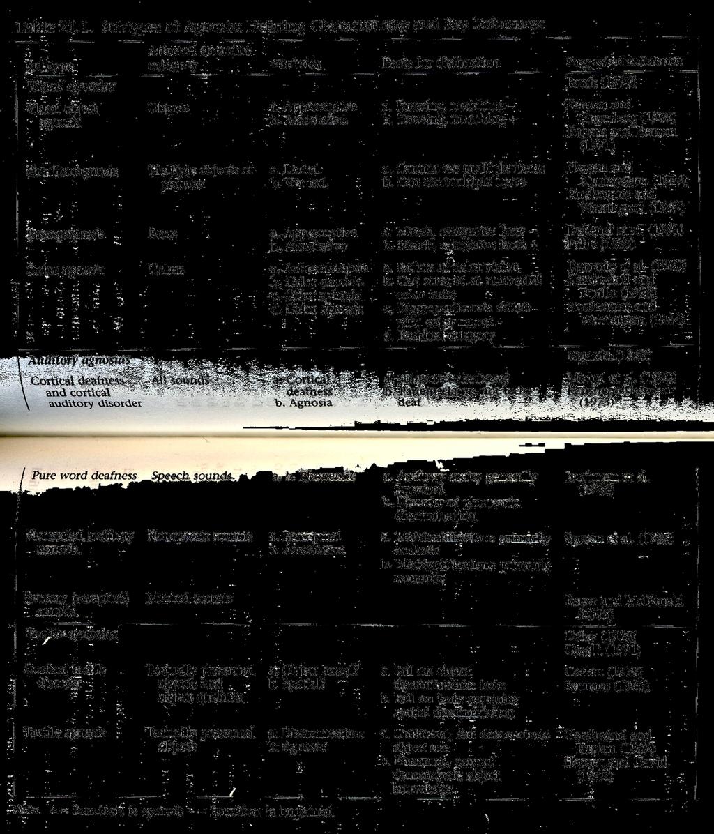
18 3. Special Types of Agnosia 2 Auditory Agnosia: Impaired capacity to recognize sound but adequate hearing. 3 types: a) Verbal auditory agnosia or pure word deafness cannot understand speech sounds b) Nonverbal auditory agnosia cannot understand non-speech sounds c) Mixed auditory agnosia cannot attach meaning to speech and non-speech sounds
19 Case Study Mr. M, 65 year old seen for nerves and headaches 3 years after stroke. No aphasia but unable to recognize common sounds.
20 Case Study Somatosensory or Tactile Agnosia Also called astereognosis. Unable to identify objects by touch in the absence of sensory deficits, naming problems, or intellectual deterioration. 2 types proposed: Unable to use tactile information to form a percept. Tactile asymbolia: unable to link percept to symbolic meaning. May be due to damage to left inferior parietal area.

21


22
Sensorimotor Functioning. Sensory and Motor Systems. Functional Anatomy of Brain- Behavioral Relationships
 Sensorimotor Functioning Sensory and Motor Systems Understanding brain-behavior relationships requires knowledge of sensory and motor systems. Sensory System = Input Neural Processing Motor System = Output
Disorders of Object and Spatial perception. Dr John Maasch Brain Injury Rehabilitation Service Burwood Hospital.
 Disorders of Object and Spatial perception Dr John Maasch Brain Injury Rehabilitation Service Burwood Hospital. Take Home Message 1 Where there are lesions of the posterior cerebrum and posterior temporal
Perceptual Disorders. Agnosias
 Perceptual Disorders Agnosias Disorders of Object Recognition AGNOSIA : a general term for a loss of ability to recognize objects, people, sounds, shapes, or smells. Agnosias result from damage to cortical
FAILURES OF OBJECT RECOGNITION. Dr. Walter S. Marcantoni
 FAILURES OF OBJECT RECOGNITION Dr. Walter S. Marcantoni VISUAL AGNOSIA -damage to the extrastriate visual regions (occipital, parietal and temporal lobes) disrupts recognition of complex visual stimuli
Higher Cortical Function
 Emilie O Neill, class of 2016 Higher Cortical Function Objectives Describe the association cortical areas processing sensory, motor, executive, language, and emotion/memory information (know general location
Learning Objectives.
 Emilie O Neill, class of 2016 Learning Objectives 1. Describe the types of deficits that occur with lesions in association areas including: prosopagnosia, neglect, aphasias, agnosia, apraxia 2. Discuss
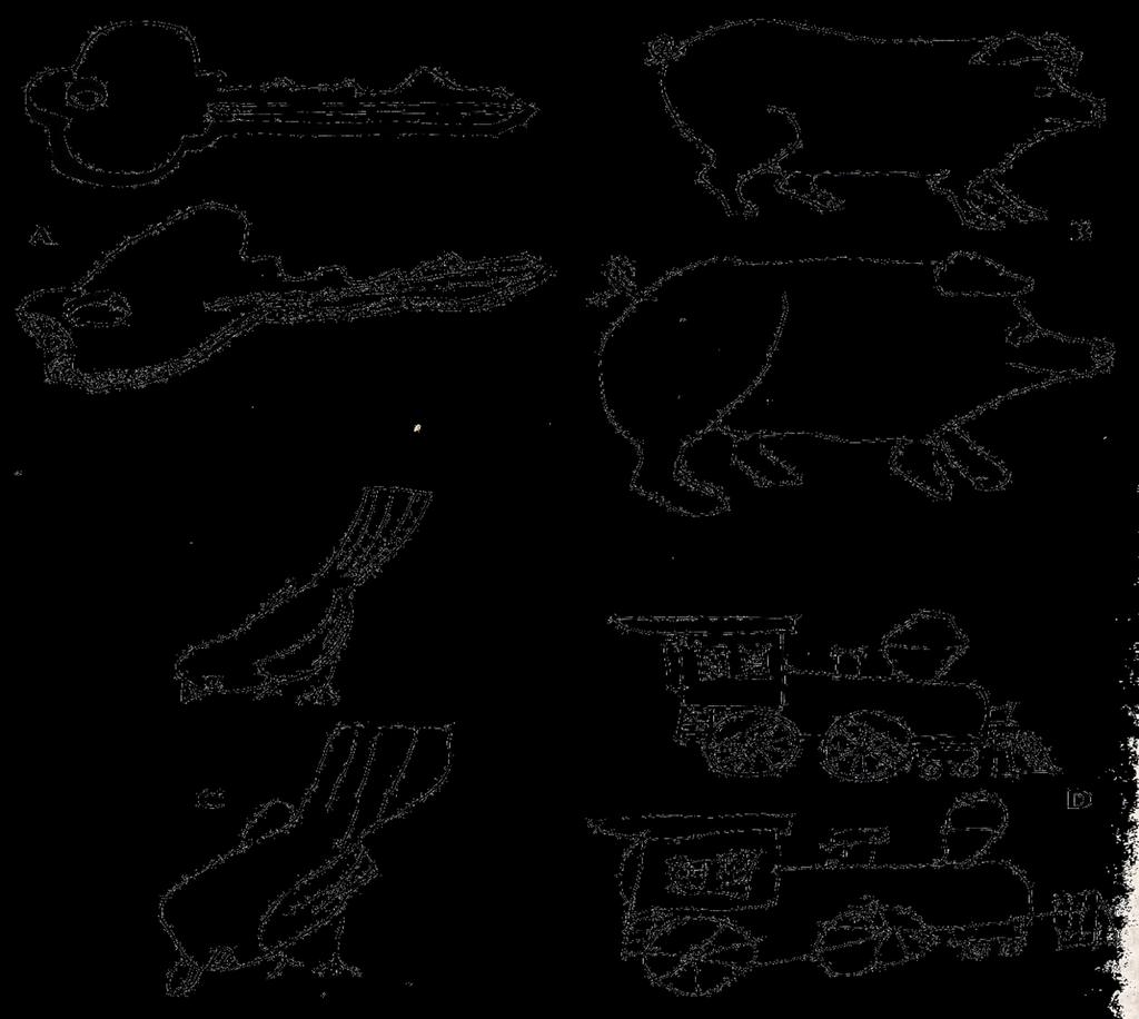
Identify these objects
 Pattern Recognition The Amazing Flexibility of Human PR. What is PR and What Problems does it Solve? Three Heuristic Distinctions for Understanding PR. Top-down vs. Bottom-up Processing. Semantic Priming.
Cortical Visual Symptoms
 대한안신경의학회지 : 제 6 권 Supplement 2 ISSN: 2234-0971 Jeong-Yoon Choi Department of Neurology, Seoul National University Bundang Hospital, Seongnam, Korea Jeong-Yoon Choi. MD. PhD. Department of Neurology, Seoul
Topic 11 - Parietal Association Cortex. 1. Sensory-to-motor transformations. 2. Activity in parietal association cortex and the effects of damage
 Topic 11 - Parietal Association Cortex 1. Sensory-to-motor transformations 2. Activity in parietal association cortex and the effects of damage Sensory to Motor Transformation Sensory information (visual,
FRONTAL LOBE. Central Sulcus. Ascending ramus of the Cingulate Sulcus. Cingulate Sulcus. Lateral Sulcus
 FRONTAL LOBE Central Ascending ramus of the Cingulate Cingulate Lateral Lateral View Medial View Motor execution and higher cognitive functions (e.g., language production, impulse inhibition, reasoning
Cortical Organization. Functionally, cortex is classically divided into 3 general types: 1. Primary cortex:. - receptive field:.
 Cortical Organization Functionally, cortex is classically divided into 3 general types: 1. Primary cortex:. - receptive field:. 2. Secondary cortex: located immediately adjacent to primary cortical areas,
Human Paleoneurology and the Evolution of the Parietal Cortex
 PARIETAL LOBE The Parietal Lobes develop at about the age of 5 years. They function to give the individual perspective and to help them understand space, touch, and volume. The location of the parietal
Fundamentals of Cognitive Psychology, 3e by Ronald T. Kellogg Chapter 2. Multiple Choice
 Multiple Choice 1. Which structure is not part of the visual pathway in the brain? a. occipital lobe b. optic chiasm c. lateral geniculate nucleus *d. frontal lobe Answer location: Visual Pathways 2. Which
Psy /16 Human Communication. By Joseline
 Psy-302 11/16 Human Communication By Joseline Lateralization Left Hemisphere dominance in speech production in 95% of right handed and 70% of left handed people Left -> Timing, Sequence of events Right
Key questions about attention
 Key questions about attention How does attention affect behavioral performance? Can attention affect the appearance of things? How does spatial and feature-based attention affect neuronal responses in
Inside Your Patient s Brain Michelle Peterson, APRN, CNP Centracare Stroke and Vascular Neurology
 Inside Your Patient s Brain Michelle Peterson, APRN, CNP Centracare Stroke and Vascular Neurology Activity Everyone stand up, raise your right hand, tell your neighbors your name 1 What part of the brain
CEREBRUM. Dr. Jamila EL Medany
 CEREBRUM Dr. Jamila EL Medany Objectives At the end of the lecture, the student should be able to: List the parts of the cerebral hemisphere (cortex, medulla, basal nuclei, lateral ventricle). Describe
Exam 1 PSYC Fall 1998
 Exam 1 PSYC 2022 Fall 1998 (2 points) Briefly describe the difference between a dualistic and a materialistic explanation of brain-mind relationships. (1 point) True or False. George Berkely was a monist.
Pathologies of postchiasmatic visual pathways and visual cortex
 Pathologies of postchiasmatic visual pathways and visual cortex Optic radiation: anatomy Pathologies of the postchiamsatic visual pathways and visual cortex Characterized by homonymous hemianopsia. This
Clinical and Experimental Neuropsychology. Lecture 3: Disorders of Perception
 Clinical and Experimental Neuropsychology Lecture 3: Disorders of Perception Sensation vs Perception Senses capture physical energy from environment that are converted into neural signals and elaborated/interpreted
Chapter 2 Test. 1. Evolutionary structures within the are the most primitive. *a. hindbrain b. thalamus c. forebrain d. midbrain e.
 Cognitive Psychology In and Out of the Laboratory 5th Edition Galotti TEST BANK Full clear download (no formatting errors) at: https://testbankreal.com/download/cognitive-psychology-laboratory-5thedition-galotti-test-bank/
Agnosia. Introduction
 Agnosia L A Baugh, L Desanghere, and J J Marotta, The University of Manitoba, Winnipeg, MB, Canada ª 2010 Elsevier Ltd. All rights reserved. Glossary Apperceptive agnosia A form of visual agnosia in which
Neocortex. Hemispheres 9/22/2010. Psychology 472 Pharmacology of Psychoactive Drugs. Structures are divided into several section or lobes.
 Neocortex Psychology 472 Pharmacology of Psychoactive Drugs 1 Is the most developed in Humans Has many folds and fissures The folds of tissue are called gyri or a gyrus (single) The fissures or valleys
Cognitive Neuroscience Cortical Hemispheres Attention Language
 Cognitive Neuroscience Cortical Hemispheres Attention Language Based on: Chapter 18 and 19, Breedlove, Watson, Rosenzweig, 6e/7e. Cerebral Cortex Brain s most complex area with billions of neurons and
C:\Documents and Settings\sstensaas\Desktop\dental visual 2010\VisualPath dental 2010.docVisualPath dental 2010.doc
 Neuroanatomy Suzanne Stensaas April 8, 2010, 10:00-12:00 p.m. Reading: Waxman Ch. 15, Computer Resources: HyperBrain Ch 7 THE VISUAL PATHWAY Objectives: 1. Describe the pathway of visual information from
Language Speech. Speech is the preferred modality for language.
 Language Speech Speech is the preferred modality for language. Outer ear Collects sound waves. The configuration of the outer ear serves to amplify sound, particularly at 2000-5000 Hz, a frequency range
MULTI-CHANNEL COMMUNICATION
 INTRODUCTION Research on the Deaf Brain is beginning to provide a new evidence base for policy and practice in relation to intervention with deaf children. This talk outlines the multi-channel nature of
OBJECTIVES. At the end of the lecture, students should be able to: List the cerebral arteries.
 DR JAMILA EL MEDANY OBJECTIVES At the end of the lecture, students should be able to: List the cerebral arteries. Describe the cerebral arterial supply regarding the origin, distribution and branches.
Stroke School for Internists Part 1
 Stroke School for Internists Part 1 November 4, 2017 Dr. Albert Jin Dr. Gurpreet Jaswal Disclosures I receive a stipend for my role as Medical Director of the Stroke Network of SEO I have no commercial
Neuroscience Tutorial
 Neuroscience Tutorial Brain Organization : cortex, basal ganglia, limbic lobe : thalamus, hypothal., pituitary gland : medulla oblongata, midbrain, pons, cerebellum Cortical Organization Cortical Organization
Nervous System. 1. What N.S. division controls skeletal muscles? 3. What kind of neuroglia myelinates axons in the PNS?
 . What N.S. division controls skeletal muscles? Nervous System SRS Review %. Central nervous system %. Peripheral nervous system %. Afferent division %. Somatic division %. Autonomic division %. Sympathetic
Agnosia, Epilepsy, Sleep Walking, and DID. D. Kiper
 Agnosia, Epilepsy, Sleep Walking, and DID D. Kiper 6.12.2018 Agnosia A-gnosis (absence of knowledge); Seelenblindheit; agnosia (Freud) Akinetopsia Achromatopsia Capgras syndrome DF: A Visual Agnostic DF
Homework Week 2. PreLab 2 HW #2 Synapses (Page 1 in the HW Section)
") Homework Week 2 Due in Lab PreLab 2 HW #2 Synapses (Page 1 in the HW Section) Reminders No class next Monday Quiz 1 is @ 5:30pm on Tuesday, 1/22/13 Study guide posted under Study Aids section of website
Homework Week 2 Due in Lab PreLab 2 HW #2 Synapses (Page 1 in the HW Section) Reminders No class next Monday Quiz 1 is @ 5:30pm on Tuesday, 1/22/13 Study guide posted under Study Aids section of website
Introduction to Physiological Psychology Review
 Introduction to Physiological Psychology Review ksweeney@cogsci.ucsd.edu www.cogsci.ucsd.edu/~ksweeney/psy260.html n Learning and Memory n Human Communication n Emotion 1 What is memory? n Working Memory:
Chapter 3: 2 visual systems
 Chapter 3: 2 visual systems Overview Explain the significance of the turn to the brain in cognitive science Explain Mishkin and Ungerleider s hypothesis that there are two distinct visual systems Outline
Lecture 35 Association Cortices and Hemispheric Asymmetries -- M. Goldberg
 Lecture 35 Association Cortices and Hemispheric Asymmetries -- M. Goldberg The concept that different parts of the brain did different things started with Spurzheim and Gall, whose phrenology became quite
Hemispheric Specialization (lateralization) Each lobe of the brain has specialized functions (Have to be careful with this one.)
 Each lobe of the brain has specialized functions (Have to be careful with this one.)") Cerebral Cortex Principles contralaterality the right half of your brain controls the left half of your body and vice versa. (contralateral control.) Localization of function Specific mental processes
Cerebral Cortex Principles contralaterality the right half of your brain controls the left half of your body and vice versa. (contralateral control.) Localization of function Specific mental processes
THE COCHLEA AND AUDITORY PATHWAY
 Dental Neuroanatomy Suzanne S. Stensaas, PhD February 23, 2012 Reading: Waxman, Chapter 16, Review pictures in a Histology book Computer Resources: http://www.cochlea.org/ - Promenade around the Cochlea
Layered organization of cortex: Paleocortex 3 layers hippocampal formation / ventral & medial cortex closest to brainstem
 Layered organization of cortex: Paleocortex 3 layers hippocampal formation / ventral & medial cortex closest to brainstem Archicortex 3-4 layers hippocampal formation / amygdala Neocortex 6 layers more
CISC 3250 Systems Neuroscience
 CISC 3250 Systems Neuroscience Levels of organization Central Nervous System 1m 10 11 neurons Neural systems and neuroanatomy Systems 10cm Networks 1mm Neurons 100μm 10 8 neurons Professor Daniel Leeds
CEREBRUM Dr. Jamila Elmedany Dr. Essam Eldin Salama
 CEREBRUM Dr. Jamila Elmedany Dr. Essam Eldin Salama Objectives At the end of the lecture, the student should be able to: List the parts of the cerebral hemisphere (cortex, medulla, basal nuclei, lateral
PsychoBrain. 31 st January Dr Christos Pliatsikas. Lecturer in Psycholinguistics in Bi-/Multilinguals University of Reading
 PsychoBrain 31 st January 2018 Dr Christos Pliatsikas Lecturer in Psycholinguistics in Bi-/Multilinguals University of Reading By the end of today s lecture you will understand Structure and function of
Assessing the Stroke Patient. Arlene Boudreaux, MSN, RN, CCRN, CNRN
 Assessing the Stroke Patient Arlene Boudreaux, MSN, RN, CCRN, CNRN Cincinnati Pre-Hospital Stroke Scale May be done by EMS o One of many o F facial droop on one side o A arm drift (hold a pizza box, close
International Encyclopedia of Rehabilitation
 International Encyclopedia of Rehabilitation Copyright 2010 by the Center for International Rehabilitation Research Information and Exchange (CIRRIE). All rights reserved. No part of this publication may
Our senses provide us with wonderful capabilities. If you had to lose one, which would it be?
 Our senses provide us with wonderful capabilities. If you had to lose one, which would it be? Neurological disorders take away sensation without a choice! http://neuroscience.uth.tmc.edu/s2/chapter04.html
Impaired face discrimination in acquired prosopagnosia is associated with abnormal response to individual faces in the right middle fusiform gyrus
 Impaired face discrimination in acquired prosopagnosia is associated with abnormal response to individual faces in the right middle fusiform gyrus Christine Schiltz Bettina Sorger Roberto Caldara Fatima
Title:Atypical language organization in temporal lobe epilepsy revealed by a passive semantic paradigm
 Author's response to reviews Title:Atypical language organization in temporal lobe epilepsy revealed by a passive semantic paradigm Authors: Julia Miro (juliamirollado@gmail.com) Pablo Ripollès (pablo.ripolles.vidal@gmail.com)
shows syntax in his language. has a large neocortex, which explains his language abilities. shows remarkable cognitive abilities. all of the above.
 Section: Chapter 14: Multiple Choice 1. Alex the parrot: pp.529-530 shows syntax in his language. has a large neocortex, which explains his language abilities. shows remarkable cognitive abilities. all
CEREBRUM & CEREBRAL CORTEX
 CEREBRUM & CEREBRAL CORTEX Seonghan Kim Dept. of Anatomy Inje University, College of Medicine THE BRAIN ANATOMICAL REGIONS A. Cerebrum B. Diencephalon Thalamus Hypothalamus C. Brain Stem Midbrain Pons
PS3019 Cognitive and Clinical Neuropsychology
 PS3019 Cognitive and Clinical Neuropsychology Carlo De Lillo HWB Room P05 Consultation time: Tue & Thu 2.30-3.30 E-mail: CDL2@le.ac.uk Overview Neuropsychology of visual processing Neuropsychology of spatial
Does Wernicke's Aphasia necessitate pure word deafness? Or the other way around? Or can they be independent? Or is that completely uncertain yet?
 Does Wernicke's Aphasia necessitate pure word deafness? Or the other way around? Or can they be independent? Or is that completely uncertain yet? Two types of AVA: 1. Deficit at the prephonemic level and
LISC-322 Neuroscience. Visual Field Representation. Visual Field Representation. Visual Field Representation. Visual Field Representation
 LISC-3 Neuroscience THE VISUAL SYSTEM Central Visual Pathways Each eye sees a part of the visual space that defines its visual field. The s of both eyes overlap extensively to create a binocular. eye both
The Brain and Cranial Nerves Pg Three Main Regions of the Brain. Forebrain
 The Brain and Cranial Nerves Pg. 129 Three Main Regions of the Brain Forebrain Cerbral hemispheres Diencephalon Midbrain Brain stem Hindbrain Pons Cerebellum Medulla oblongata Interprets sensory inputs
The Brain and Cranial Nerves Pg. 129
 The Brain and Cranial Nerves Pg. 129 Three Main Regions of the Brain Forebrain Cerbral hemispheres Diencephalon Midbrain Brain stem Hindbrain Pons Cerebellum Medulla oblongata Forebrain Interprets sensory
b. The groove between the two crests is called 2. The neural folds move toward each other & the fuse to create a
 Chapter 13: Brain and Cranial Nerves I. Development of the CNS A. The CNS begins as a flat plate called the B. The process proceeds as: 1. The lateral sides of the become elevated as waves called a. The
It Doesn t Take A Lot of Brains to Understand the Brain: Functional Neuroanatomy Made Ridiculously Simple
 It Doesn t Take A Lot of Brains to Understand the Brain: Functional Neuroanatomy Made Ridiculously Simple 6 th Annual Northern Kentucky TBI Conference March 23, 2012 www.bridgesnky.org James F. Phifer,
Cerebral Cortex Structure, Function, Dysfunction Reading Ch 10 Waxman Dental Neuroanatomy Lecture. Suzanne Stensaas, Ph.D.
 Cerebral Cortex Structure, Function, Dysfunction Reading Ch 10 Waxman Dental Neuroanatomy Lecture Suzanne Stensaas, Ph.D. March 7, 2012 Anatomy Review Lobes and layers Brodmann s areas Vascular Supply
Human Brain. Lateralization of Function. Cortex. Cerebral Hemispheres. An extension of the spinal cord. Dr. Coulson Cognitive Science Department UCSD
 Lateralization of Function Human Brain An extension of the spinal cord Dr. Coulson Cognitive Science Department UCSD Cerebral Hemispheres Two millimeters thick and has area of 1.5 square meters Corpus
PSY 310: Sensory and Perceptual Processes 1
 Processing streams PSY 310 Greg Francis Neurophysiology We are working under the following hypothesis What we see is determined by the pattern of neural activity in the brain This leads to several interesting
The origins of localization
 Association Cortex, Asymmetries, and Cortical Localization of Affective and Cognitive Functions Michael E. Goldberg, M.D. The origins of localization The concept that different parts of the brain did different
49a A&P: Nervous System -! Synaptic Transmission and Central Nervous System
 49a A&P: Nervous System -! Synaptic Transmission and Central Nervous System 49a A&P: Nervous System -! Synaptic Transmission and Central Nervous System! Class Outline" 5 minutes" "Attendance, Breath of
THE COCHLEA AND AUDITORY PATHWAY
 Dental Neuroanatomy Suzanne S. Stensaas, PhD April 14, 2010 Reading: Waxman, Chapter 16, Review pictures in a Histology book Computer Resources: http://www.cochlea.org/ - Promenade around the Cochlea HyperBrain
Association Cortex, Asymmetries, and Cortical Localization of Affective and Cognitive Functions. Michael E. Goldberg, M.D.
 Association Cortex, Asymmetries, and Cortical Localization of Affective and Cognitive Functions Michael E. Goldberg, M.D. The origins of localization The concept that different parts of the brain did different
Specific Sulci/Fissures:
 Specific Sulci/Fissures: Central Sulcus Longitudinal Fissure Sylvian/Lateral Fissure Transverse Fissure http://www.bioon.com/book/biology/whole/image/1/1-8.tif.jpg http://www.dalbsoutss.eq.edu.au/sheepbrains_me/human_brain.gif
Required Slide. Session Objectives
 Vision: CNS 2018 Required Slide Session Objectives Visual system: CNS At the end of this session, students will be able to: 1. Understand how axons from the eyes travel through the optic nerves and tracts
Cerebrum-Cerebral Hemispheres. Cuneyt Mirzanli Istanbul Gelisim University
 Cerebrum-Cerebral Hemispheres Cuneyt Mirzanli Istanbul Gelisim University The largest part of the brain. Ovoid shape. Two incompletely separated cerebral hemispheres. The outer surface of the cerebral
The Frontal Lobes. Anatomy of the Frontal Lobes. Anatomy of the Frontal Lobes 3/2/2011. Portrait: Losing Frontal-Lobe Functions. Readings: KW Ch.
 The Frontal Lobes Readings: KW Ch. 16 Portrait: Losing Frontal-Lobe Functions E.L. Highly organized college professor Became disorganized, showed little emotion, and began to miss deadlines Scores on intelligence
Pathway from the eye to the cortex
 Vision: CNS 2017 Pathway from the eye to the cortex Themes of this lecture Visual information is analyzed in more complicated ways than in the retina. One major pathway from the eye leads to the striate
Introduction to Physiological Psychology Learning and Memory II
 Introduction to Physiological Psychology Learning and Memory II ksweeney@cogsci.ucsd.edu cogsci.ucsd.edu/~ksweeney/psy260.html Memory Working Memory Long-term Memory Declarative Memory Procedural Memory
Human Brain. Lateralization of Function. An extension of the spinal cord. Dr. Coulson Cognitive Science Department UCSD
 Lateralization of Function Human Brain An extension of the spinal cord Dr. Coulson Cognitive Science Department UCSD Cerebral Hemispheres Corpus Callosum Cerebral Lobes Neurons Brain composed of neurons
Brain anatomy tutorial. Dr. Michal Ben-Shachar 459 Neurolinguistics
 Brain anatomy tutorial Dr. Michal Ben-Shachar 459 Neurolinguistics The human brain Left hemisphere Right hemisphere http://www.brainmuseum.org/ Zoom out Zoom in Types of Brain Tissue Gray Matter: Cell
Auditory and Vestibular Systems
 Auditory and Vestibular Systems Objective To learn the functional organization of the auditory and vestibular systems To understand how one can use changes in auditory function following injury to localize
 Peripheral facial paralysis (right side). The patient is asked to close her eyes and to retract their mouth (From Heimer) Hemiplegia of the left side. Note the characteristic position of the arm with
PROPERTY OF ELSEVIER SAMPLE CONTENT - NOT FINAL. Gross Anatomy and General Organization of the Central Nervous System
 3 Gross Anatomy and General Organization of the Central Nervous System C h a p t e r O u t l i n e The Long Axis of the CNS Bends at the Cephalic Flexure Hemisecting a Brain Reveals Parts of the Diencephalon,
Cerebral Cortex: Association Areas and Memory Tutis Vilis
 97 Cerebral Cortex: Association Areas and Memory Tutis Vilis a) Name the 5 main subdivisions of the cerebral cortex. Frontal, temporal, occipital, parietal, and limbic (on the medial side) b) Locate the
A CONVERSATION ABOUT NEURODEVELOPMENT: LOST IN TRANSLATION
 A CONVERSATION ABOUT NEURODEVELOPMENT: LOST IN TRANSLATION Roberto Tuchman, M.D. Chief, Department of Neurology Nicklaus Children s Hospital Miami Children s Health System 1 1 in 6 children with developmental
M555 Medical Neuroscience Lab 1: Gross Anatomy of Brain, Crainal Nerves and Cerebral Blood Vessels
 M555 Medical Neuroscience Lab 1: Gross Anatomy of Brain, Crainal Nerves and Cerebral Blood Vessels Anatomical Directions Terms like dorsal, ventral, and posterior provide a means of locating structures
Brain. Cerebral white matter. Brain cortex. Frontal lobe. Frontal lobe Brain cortex
 Brain Brain cortex Layer (stratum) of grey matter which cover hemisphers Longitudinal fissure - 2 hemispheres Enlargement of neocortex folding the brain surface into convolutions (gyri) separated by groves
The Central Nervous System I. Chapter 12
 The Central Nervous System I Chapter 12 The Central Nervous System The Brain and Spinal Cord Contained within the Axial Skeleton Brain Regions and Organization Medical Scheme (4 regions) 1. Cerebral Hemispheres
Supplementary Digital Content
 Supplementary Digital Content Contextual modulation of pain in masochists: involvement of the parietal operculum and insula Sandra Kamping a, Jamila Andoh a, Isabelle C. Bomba a, Martin Diers a,b, Eugen
Thalamus and Sensory Functions of Cerebral Cortex
 Thalamus and Sensory Functions of Cerebral Cortex I: To describe the functional divisions of thalamus. II: To state the functions of thalamus and the thalamic syndrome. III: To define the somatic sensory
PARIETAL LOBE. Vasilios A. Zerris MD, MPH, MSc, FAANS
 PARIETAL LOBE Vasilios A. Zerris MD, MPH, MSc, FAANS Diplomate of the American Board of Neurological Surgery Fellow of the American Association of Neurological Surgeons Professor of Neurosurgery, European
Reading Words and Non-Words: A Joint fmri and Eye-Tracking Study
 Reading Words and Non-Words: A Joint fmri and Eye-Tracking Study A N N - M A R I E R A P H A I L S R E B C S, S K I D M O R E C O L L E G E D R. J O H N H E N D E R S O N U N I V E R S I T Y O F S O U
The effects of sensory deprivation on sensory processing. Ione Fine, University of Washington
 The effects of sensory deprivation on sensory processing Ione Fine, University of Washington Molyneux s question "Suppose a man born blind, and now adult, and taught by his touch to distinguish between
Why does language set up shop where it does?
 Why does language set up shop where it does? Does modality affect the functional neuroanatomy of language? For example, the M350 and the N400 localize in the vicinity of auditory cortex. Is that just an
Overview of Brain Structures
 First Overview of Brain Structures Psychology 470 Introduction to Chemical Additions Steven E. Meier, Ph.D. All parts are interrelated. You need all parts to function normally. Neurons = Nerve cells Listen
Outline.! Neural representation of speech sounds. " Basic intro " Sounds and categories " How do we perceive sounds? " Is speech sounds special?
 Outline! Neural representation of speech sounds " Basic intro " Sounds and categories " How do we perceive sounds? " Is speech sounds special? ! What is a phoneme?! It s the basic linguistic unit of speech!
Test Bank. Multiple Choice
 Chapter 2: The Brain: An Overview of Structure and Function Test Bank Multiple Choice 1. Evolutionary structures within the are the most primitive. a. hindbrain b. thalamus c. forebrain d. midbrain Answer
Spatial memory deficits in patients with lesions to the right hippocampus and to the right parahippocampal cortex.
 IN PRESS: NEUROPSYCHOLOGIA Volume 36, no 6, September 1998. Spatial memory deficits in patients with lesions to the right hippocampus and to the right parahippocampal cortex. Veronique D. Bohbot1* 1, Miroslav
Brain Structures. Some scientists divide the brain up into three parts. Hindbrain Midbrain Forebrain
 The Brain Phineas Gage Play The Frontal Lobes and Behavior: The Story of Phineas Gage (12:03) Module #25 from The Brain: Teaching Modules (2 nd edition). http://www.learner.org/resources/series1 42.html
Text to brain: predicting the spatial distribution of neuroimaging observations from text reports (submitted to MICCAI 2018)
") 1 / 22 Text to brain: predicting the spatial distribution of neuroimaging observations from text reports (submitted to MICCAI 2018) Jérôme Dockès, ussel Poldrack, Demian Wassermann, Fabian Suchanek, Bertrand
1 / 22 Text to brain: predicting the spatial distribution of neuroimaging observations from text reports (submitted to MICCAI 2018) Jérôme Dockès, ussel Poldrack, Demian Wassermann, Fabian Suchanek, Bertrand
Gives few collaterals, it is mainly a single process surrounded by a myelin sheath
 Lecture 1 - Nerve fiber refers to both axons and dendrites, the dendrites are the afferent fibers (sensory); they receive impulses from neighbouring neurons, and the axon is the efferent fiber (motor);
correlates with social context behavioral adaptation.
 REVIEW OF FRONTAL LOBE STRUCTURES Main organization of frontal cortex: 1. Motor area (precentral gyrus). 2. Premotor & supplementary motor areas (immediately anterior to motor area). Includes premotor,
Biological Bases of Behavior. 6: Vision
 Biological Bases of Behavior 6: Vision Sensory Systems The brain detects events in the external environment and directs the contractions of the muscles Afferent neurons carry sensory messages to brain
The occipital lobe is involved in many aspects of
 728 Distribution of the Occipital Branches of the Posterior Cerebral Artery Correlation With Occipital Lobe Infarcts Slobodan V. Marinkovic", MD, Milan M. Milisavljevic", MD, Vera Lolid-Draganid, MD, and
Verbal Working Memory. The left temporoparietal junction in verbal working memory: Storage or attention. Baddelely s Multiple-Component Model
 Verbal Working Memory The left temporoparietal junction in verbal working memory: Storage or attention Susan Ravizza LTM vs WM Focusing on the storage component of WM Maintenance of words, pictures, goals
LISC-322 Neuroscience Cortical Organization
 LISC-322 Neuroscience Cortical Organization THE VISUAL SYSTEM Higher Visual Processing Martin Paré Assistant Professor Physiology & Psychology Most of the cortex that covers the cerebral hemispheres is
Principles Arteries & Veins of the CNS LO14
 Principles Arteries & Veins of the CNS LO14 14. Identify (on cadaver specimens, models and diagrams) and name the principal arteries and veins of the CNS: Why is it important to understand blood supply
The Nervous System. Nerves, nerves everywhere!
 The Nervous System Nerves, nerves everywhere! Purpose of the Nervous System The information intake and response system of the body. Coordinates all body functions, voluntary and involuntary! Responds to
Prosopagnosia q. Background. EHF De Haan, University of Amsterdam, Amsterdam, Netherlands
 Prosopagnosia q EHF De Haan, University of Amsterdam, Amsterdam, Netherlands Ó 2017 Elsevier Inc. All rights reserved. Background 1 Prosopagnosia and Sensory Status 2 Prosopagnosia and Other Face Processing