|
|
|
- Madeleine Ray
- 6 years ago
- Views:
Transcription
1 Question 1: Briefly describe the structure of the following: (a) Brain (b) Eye (c) Ear (A) Brain: Brain is the main coordinating centre of the body. It is a part of nervous system that controls and monitors every organ of the body. It is well protected by cranial meninges that are made up of an outer layer called dura mater, a thin middle layer called arachnoid, and an inner layer called pia mater. It is divided into three regions forebrain, midbrain, and hindbrain. Forebrain: It is the main thinking part of the brain. It consists of cerebrum, thalamus, and hypothalamus. (a) Cerebrum: Cerebrum is the largest part of the brain and constitutes about four-fifth of its weight. Cerebrum is divided into two cerebral hemispheres by a deep longitudinal cerebral fissure. These hemispheres are joined by a tract of nerve fibre known as corpus callosum. The cerebral hemispheres are covered by a layer of cells known as cerebral cortex or grey matter. Cerebrum has sensory regions known as association areas that receive sensory impulses from various receptors as well as from motor regions that control the movement of various muscles. The innermost part of cerebrum gives an opaque white appearance to the layer and is known as the white matter. (b) Thalamus: Thalamus is the main centre of coordination for sensory and motor signalling. It is wrapped by cerebrum. (c) Hypothalamus: It lies at the base of thalamus and contains a number of centres that regulate body temperature and the urge for eating and drinking. Some regions of cerebrum, along with hypothalamus, are involved in the regulation of sexual behaviour and expression of emotional reactions such as excitement, pleasure, fear, etc.
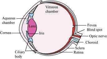
2 Midbrain: It is located between the thalamus region of the forebrain and pons region of hindbrain. The dorsal surface of midbrain consists of superior and inferior corpora bigemina and four round lobes called corpora quadrigemina. A canal known as cerebral aqueduct passes through the midbrain. Midbrain is concerned with the sense of sight and hearing. Hindbrain: It consists of three regions pons, cerebellum, and medulla oblongata. (a) Pons is a band of nerve fibre that lies between medulla oblongata and midbrain. It connects the lateral parts of cerebellar hemisphere together. (b) Cerebellum is a large and well developed part of hindbrain. It is located below the posterior sides of cerebral hemispheres and above medulla oblongata. It is responsible for maintaining posture and equilibrium of the body. (c) Medulla oblongata is the posterior and simplest part of the brain. It is located beneath the cerebellum. Its lower end extends in the form of spinal cord and leaves the skull through foramen magnum. (B) Eye: Eyes are spherical structures that consist of three layers. (a) The outer layer is composed of sclera and cornea. (i) Sclera is an opaque tissue that is usually known as white of the eye. It is composed of a dense connective tissue. (ii) Cornea is a transparent anterior portion of eye that lacks blood vessels and is nourished by lymph from the nearby area. It is slightly bulged forward and helps in focusing light rays with the help of lens. (b) The middle layer of eye is vascular in nature and contains choroid, ciliary body, and iris. (i) Choroid lies next to the sclera and contains numerous blood vessels that provide nutrients and oxygen to the retina and other tissues. (ii) Ciliary body: The choroid layer is thin over posterior region and gets thickened in the anterior portion to form ciliary body. It contains blood vessels, ciliary muscles, and ciliary processes.
3 (iii) Iris: At the junction of sclera and cornea, the ciliary body continues forward to form thin coloured partition called iris. It is the visible coloured portion of eye. The eye contains a transparent, biconvex, and elastic structure just behind the iris. It is known as lens. The lens is held in position by suspensory ligaments attached to the ciliary body. The lens divides the eye ball into two chambers an anterior aqueous and posterior vitreous chamber. (c) The innermost nervous coat of eye contains retina. Retina is the innermost layer. It contains three layers of cells inner ganglion cells, middle bipolar cells, and outermost photoreceptor cells. The receptor cells present in the retina are of two types rod cells and cone cells. (a) Rod cells The rods contain the rhodopsin pigment (visual purple) that is highly sensitive to dim light. It is responsible for twilight vision. (b) Cone cells The cones contain the iodopsin pigment (visual violet) and are highly sensitive to high intensity light. They are responsible for daylight and colour visions. The innermost ganglionic cells give rise to optic nerve fibre that forms optic nerve in each eye and is connected with the brain. (C) Ear: Ear is the sense organ for hearing and equilibrium. It consists of three portions external ear, middle ear, and internal ear. 1. External ear: It consists of pinna, external auditory meatus, and a tympanic membrane. (a) Pinna is a sensitive structure that collects and directs the vibrations into the ear to produce sound. (b) External auditory meatus is a tubular passage supported by cartilage in external ear. (c) Tympanic membrane is a thin membrane that lies close to the auditory canal. It separates the middle ear from external ear. 2. Middle ear: It is an air-filled tympanic cavity that is connected with pharynx through eustachian tube. Eustachian tube helps to equalize air pressure in both sides of tympanic membrane. The middle ear contains a flexible chain of three middle bones called ear
4 ossicles. The three ear ossicles are malleus, incus, and stapes that are attached to each other. 3. Internal ear: It is also known as labyrinth. Labyrinth is divided into bony labyrinth and a membranous labyrinth. Bony labyrinth is filled with perilymph while membranous labyrinth is filled with endolymph. Membranous labyrinth is divided into 2 parts. (a) Vestibular apparatus Vestibular apparatus is a central sac-like part that is divided into utriculus and sacculus. A special group of sensory cells called macula are present in sacculus and utriculus. Vestibular apparatus also contains three semi-circular canals. The lower end of each semi-circular canal contains a projecting ridge called crista ampularis. Each ampulla has a group of sensory cells called crista. Crista and macula are responsible for maintaining the balance of body and posture. (b) Cochlea: Cochlea is a long and coiled outgrowth of sacculus. It is the main hearing organ. Cochlea consists of three membranes. The organ of corti, a hearing organ, is located on the basilar membrane that has hair cells. Question 2: Compare the following: (a) Central neural system (CNS) and Peripheral neural system (PNS) (b) Resting potential and action potential (c) Choroid and retina (a) Central neural system (CNS) and Peripheral neural system (PNS) Central neural system Peripheral neural system 1. It is the main 1. It is not the main coordinating centre of the
5 coordinating centre of the body. body. 2. It includes brain and spinal cord. 2. It includes cranial and spinal nerves that connect central nervous system to different parts of the body. (b) Resting potential and action potential Resting potential Action potential 1. It is the potential difference across 1. It is the potential difference across the nerve fibre when there is no nerve fibre when there is conduction of nerve impulse. conduction of nerve impulse. 2. The membrane is more permeable to K + ions than to Na + ions. 2. The membrane is more permeable to Na + ions than to K + ions. (c) Choroid and retina Choroid Retina 1. Choroid is the middle vascular layer of eye. 1. Retina is the innermost nervous coat of eye. 2. It contains numerous blood vessels that provide nutrients and oxygen to retina and other tissues. 2. It contains photoreceptor cells, rods and cones that are associated with twilight and colour vision respectively. Question 3: Explain the following processes: (a) Polarisation of the membrane of a nerve fibre
6 (b) Depolarisation of the membrane of a nerve fibre (c) Conduction of a nerve impulse along a nerve fibre (d) Transmission of a nerve impulse across a chemical synapse (a) Polarisation of the membrane of a nerve fibre During resting condition, the concentration of K + ions is more inside the axoplasm while the concentration of Na + ions is more outside the axoplasm. As a result, the potassium ions move faster from inside to outside as compared to sodium ions. Therefore, the membrane becomes positively charged outside and negatively charged inside. This is known as polarization of membrane or polarized nerve. (b) Depolarisation of the membrane of a nerve fibre When an electrical stimulus is given to a nerve fibre, an action potential is generated. The membrane becomes permeable to sodium ions than to potassium ions. This results into positive charge inside and negative charge outside the nerve fibre. Hence, the membrane is said to be depolarized. (c) Conduction of a nerve impulse along a nerve fibre There are two types of nerve fibres myelinated and non-myelinated. In myelinated nerve fibre, the action potential is conducted from node to node in jumping manner. This is because the myelinated nerve fibre is coated with myelin sheath. The myelin sheath is impermeable to ions. As a result, the ionic exchange and depolarisation of nerve fibre is not possible along the whole length of nerve fibre. It takes place only

7 at some point, known as nodes of Ranvier, whereas in non-myelinated nerve fibre, the ionic exchange and depolarization of nerve fibre takes place along the whole length of the nerve fibre. Because of this ionic exchange, the depolarized area becomes repolarised and the next polarized area becomes depolarized. (d) Transmission of a nerve impulse across a chemical synapse Synapse is a small gap that occurs between the last portion of the axon of one neuron and the dendrite of next neuron. When an impulse reaches at the end plate of axon, vesicles consisting of chemical substance or neurotransmitter, such as acetylcholine, fuse with the plasma membrane. This chemical moves across the cleft and attaches to chemo-receptors present on the membrane of the dendrite of next neuron. This binding of chemical with chemo-receptors leads to the depolarization of membrane and generates a nerve impulse across nerve fibre. The chemical, acetylcholine, is inactivated by enzyme acetylcholinestrase. The enzyme is present in the post synaptic membrane of the dendrite. It hydrolyses acetylcholine and this allows the membrane to repolarise. Question 4: Draw labelled diagrams of the following: (a) Neuron (b) Brain (c) Eye (d) Ear (a) Neuron
 Eye")
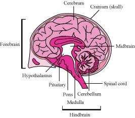
8 (b) Brain (c) Eye

9 (d) Ear Question 5: Write short notes on the following: (a) Neural coordination (b) Forebrain (c) Midbrain (d) Hindbrain (e) Retina (f) Ear ossicles (g) Cochlea (h) Organ of Corti (i) Synapse (a) Neural coordination The neural system provides rapid coordination among the organs of the body. This coordination is in the form of electric impulses and is quick and short lived. All the physiological processes in the body are closed linked and dependent upon each other. For example, during exercise, our body requires more oxygen and food. Hence, the breathing rate increases automatically and the heart beats faster. This leads to a faster supply of oxygenated blood to the muscles. Moreover, the cellular functions require regulation continuously. These functions are carried out by the hormones. Hence, the neural system along with the endocrine system control and coordinate the physiological processes. (b) Forebrain It is the main thinking part of the brain. It consists of cerebrum, thalamus, and hypothalamus.
10 (i) Cerebrum: Cerebrum is the largest part of the brain and constitutes about four-fifth of its weight. Cerebrum is divided into two cerebral hemispheres by a deep longitudinal cerebral fissure. These hemispheres are joined by a tract of nerve fibres known as corpus callosum. The cerebral hemispheres are covered by a layer of cells known as cerebral cortex or grey matter. Cerebrum has sensory regions known as association areas that receive sensory impulses from various receptors as well as from motor regions that control the movement of various muscles. The innermost part of cerebrum gives an opaque white appearance to the layer and is known as the white matter. (ii) Thalamus: Thalamus is the main centre of coordination for sensory and motor signalling. It is wrapped by cerebrum. (iii) Hypothalamus: It lies at the base of thalamus and contains a number of centres that regulate body temperature and the urge for eating and drinking. Some regions of cerebrum, along with hypothalamus, are involved in the regulation of sexual behaviour and expression of emotional reactions such as excitement, pleasure, fear, etc. (c) Midbrain It is located between the thalamus region of the forebrain and pons region of hindbrain. The dorsal surface of midbrain consists of superior and inferior corpora bigemina and four round lobes called corpora quadrigemina. A canal known as cerebral aqueduct passes through the midbrain. Midbrain is concerned with the sense of sight and hearing. (d) Hindbrain It consists of three regions pons, cerebellum, and medulla oblongata. (i) Pons is a band of nerve fibres that lies between medulla oblongata and midbrain. It connects the lateral parts of cerebellar hemisphere together.
11 (ii) Cerebellum is a large and well developed part of hindbrain. It is located below the posterior sides of cerebral hemispheres and above the medulla oblongata. It is responsible for maintaining posture and equilibrium of the body. (iii) Medulla oblongata is the posterior and simplest part of the brain. It is located beneath the cerebellum. Its lower end extends in the form of spinal cord and leaves the skull through foramen magnum. (e) Retina Retina is the innermost layer. It contains three layers of cells inner ganglion cells, middle bipolar cells, and outermost photoreceptor cells. The receptor cells present in the retina are of two types rod cells and cone cells. (i) Rod cells The rods contain rhodopsin pigment (visual purple), which is highly sensitive to dim light. It is responsible for twilight vision. (ii) Cone cells The cones contain iodopsin pigment (visual violet) and are highly sensitive to high intensity light. They are responsible for daylight and colour visions. The innermost ganglionic cells give rise to optic nerve fibre that forms optic nerve in each eye and is connected with the brain. In this region, the photoreceptor cells are absent. Hence, it is known as the blind spot. At the posterior part, lateral to blind spot, there is a pigmented spot called macula lutea. This spot has a shallow depression at its middle known as fovea. Fovea has only cone cells. They are devoid of rod cells. Hence, it is the place of most distinct vision. (f) Ear ossicles The middle ear contains a flexible chain of three middle bones called ear ossicles. The three ear ossicles are as follows. (i) Malleus (ii) Incus (iii) Stapes The malleus is attached to tympanic membrane on one side and to incus on the other side. The incus is connected with stapes. Stapes, in turn, are attached with an oval membrane, fenestra ovalis, of internal ear. The ear ossicles act as a lever that transmits sound waves from external ear to internal ear.
12 (g) Cochlea Cochlea is a long, coiled outgrowth of sacculus. It is the main hearing organ. The cochlea forms three chambers. (i) Upper scala vestibule (ii) Middle scala media (iii) Lower scale tympani The floor of the scala media is basilar membrane while its roof is Reissner s membrane. Reissner s membrane gives out a projection called tectorial membrane. The organ of corti, a hearing organ, is located on the basilar membrane. Organ of corti contains receptor hair cells. The upper scala vestibule and lower scala tympani contain perilymph. (h) Organ of corti Organ of corti is the hearing organ. It is located on the basilar membrane that contains hair cells. Hair cells act as auditory receptors. They are present on the internal side of organ of corti. (i) Synapse Synapse is a junction between the axon terminal of one neuron and the dendrite of next neuron. It is separated by a small gap known as synaptic cleft. There are two types of synapses. (a) Electrical synapse (b) Chemical synapse
13 In electrical synapses, the pre and post synaptic neurons lie in close proximity to each other. Hence, the impulse can move directly from one neuron to another across the synapse. This represents a faster method of impulse transmission. In chemical synapses, the pre and post synaptic neurons are not in close proximity. They are separated by a synaptic cleft. The transmission of nerve impulses is carried out by chemicals such as neurotransmitters. Question 6: Give a brief account of: (a) Mechanism of synaptic transmission (b) Mechanism of vision (c) Mechanism of hearing (a) Mechanism of synaptic transmission Synapse is a junction between two neurons. It is present between the axon terminal of one neuron and the dendrite of next neuron separated by a cleft. There are two ways of synaptic transmission. (1) Chemical transmission (2) Electrical transmission 1. Chemical transmission When a nerve impulse reaches the end plate of axon, it releases a neurotransmitter (acetylcholine) across the synaptic cleft. This chemical is synthesized in cell body of the neuron and is transported to the axon terminal. The acetylcholine diffuses across the cleft and binds to the receptors present on the membrane of next neuron. This causes depolarization of membrane and initiates an action potential. 2. Electrical transmission In this type of transmission, an electric current is formed in the neuron. This electric current generates an action potential and leads to transmission of nerve impulse across the nerve fibre. This represents a faster method of nerve conduction than the chemical method of transmission.
14 (b) Mechanism of vision Retina is the innermost layer of eye. It contains three layers of cells inner ganglion cells, middle bipolar cells, and outermost photoreceptor cells. A photoreceptor cell is composed of a protein called opsin and an aldehyde of vitamin A called retinal. When light rays are focused on the retina through cornea, it leads to the dissociation of retinal from opsin protein. This changes the structure of opsin. As the structure of opsin changes, the permeability of membrane changes, generating a potential difference in the cells. This generates an action potential in the ganglionic cells and is transmitted to the visual cortex of the brain via optic nerves. In the cortex region of brain, the impulses are analysed and image is formed on the retina. (c) Mechanism of hearing The pinna of the external region collects the sound waves and directs it towards ear drum or external auditory canal. These waves strike the tympanic membrane and vibrations are created. Then, these vibrations are transmitted to the oval window, fenestra ovalis, through three ear ossicles, named as malleus, incus, and stapes. These ear ossicles act as lever and transmit the sound waves to internal ear. These vibrations from fenestra ovalis are transmitted into cochlear fluid. This generates sound waves in the lymph. The formation of waves generates a ripple in the basilar membrane. This movement bends the sensory hair cells present on the organ of corti against tectorial membrane. As a result of this, sound waves are converted into nerve impulses. These impulses are then carried to auditory cortex of brain via auditory nerves. In cerebral cortex of brain, the impulses are analysed and sound is recognized.
 How does the eye regulate the amount of light that falls on the retina? (a) Photoreceptors are cells that are sensitive to light. They are of two types rods and cones.")
15 Question 7: briefly: (a) How do you perceive the colour of an object? (b) Which part of our body helps us in maintaining the body balance? (c) How does the eye regulate the amount of light that falls on the retina? (a) Photoreceptors are cells that are sensitive to light. They are of two types rods and cones. These are present in the retina. Cones help in distinguishing colours. There are three types of cone cells those responding to green light, those responding to blue light, and those responding to red light. These cells are stimulated by different lights, from different sources. The combinations of the signals generated help us see the different colours. (b) Vestibular apparatus is located in the internal ear, above the cochlea and helps in maintaining body balance. Crista and macula are the sensory spots of the vestibular apparatus controlling dynamic equilibrium. (c) Pupil is the small aperture in the iris that regulates the amount of light entering the eye. Cornea, aqueous humour, lens, and vitreous humour act together and refract light rays, focussing them onto the photoreceptor cells of the retina.
16 Question 8: Explain the following: (a) Role of Na + in the generation of action potential. (b) Mechanism of generation of light-induced impulse in the retina. (c) Mechanism through which a sound produces a nerve impulse in the inner ear. (a) Sodium ions play an important role in the generation of action potential. When a nerve fibre is stimulated, the membrane potential decreases. The membrane becomes more permeable to Na + ions than to K + ions. As a result, Na + diffuses from the outside to the inside of the membrane. This causes the inside of the membrane to become positively-charged, while the outer membrane gains a negatively charge. This reversal of polarity across the membrane is known as depolarisation. The rapid inflow of Na + ions causes the membrane potential to increase, thereby generating an action potential. (b) Retina is the innermost layer of the eye. It contains three layers of cells inner ganglion cells, middle bipolar cells, and outermost photoreceptor cells. Photoreceptor cells are composed of a protein called opsin and an aldehyde of vitamin A called
17 retinal. When light rays are focused on the retina through the cornea, retinal gets dissociated from opsin. As a result, the structure of opsin gets changed. This in turn causes the permeability of the membrane to change, thereby generating a potential difference in the cells. Consequently, an action potential is generated in the ganglion cells and is transmitted to the visual cortex of the brain via the optic nerves. In the cortex region of the brain, the impulses are analysed and the image is formed on the retina. (c) The pinna of the external ear collects the sound waves and directs them to the tympanic membrane (ear drum) via the external auditory canal. The ear drum then vibrates the sound waves and conducts them to the internal ear through the ear ossicles. The ear ossicles increase the intensity of the sound waves. These vibrating sound waves are conducted through the oval window to the fluid in the cochlea. Consequently, a movement is created in the lymph. This movement produces vibrations in the basilar membrane, which in turn stimulate the auditory hair cells. These cells generate a nerve impulse, conducting it to the auditory cortex of the brain via afferent fibres. The auditory cortex region interprets the nerve impulse and sound is recognised. Question 9: Differentiate between: (a) Myelinated and non-myelinated axons (b) Dendrites and axons (c) Rods and cones (d) Thalamus and Hypothalamus (e) Cerebrum and Cerebellum
18 (a) Myelinated and non-myelinated axons Myelinated axons Non-myelinated axons 1. Transmission of nerve impulse is faster 1. Transmission of nerve impulse is slower 2. Myelinated axon has a myelin sheath. 2. Myelin sheath is absent 3. Node of Ranvier is present between adjacent myelin sheaths. 4. Found in the brain, the spinal cord, the cranial and spinal nerves 5. Schwann cells are observed inside the myelin sheath (b) Dendrites and axons Dendrites 3. Node of Ranvier is absent 4. Found in autonomous and somatic neural systems 5. Schwann cells are not observed inside the myelin sheath Axons 3. Dendrites are always non- 3. Axons can be myelinated or non 1. Dendrite is a small projection arising from the neuron. It conducts the nerve impulse toward the cell body. 1. Axon is a single, long projection that conducts the nerve impulse away from cell body to the next neuron. 2. Nissl s granules are present in dendrites. 2. Nissl s granules are absent from axons.
19 myelinated. myelinated. (c) Rods and cones Rods Cones 1. Rods help in twilight vision. 1. Cones help in colour vision. 2. They have visual purple pigment called rhodopsin. 2. They have visual violet pigment called iodopsin. 3. Rods are the photoreceptor cells of the retina that are sensitive to dim light. 3. Cones are the photoreceptor cells of the retina that are sensitive to bright light. (d) Thalamus and Hypothalamus Thalamus Hypothalamus Thalamus is the part of the Hypothalamus is the part of the forebrain that receives nerve forebrain that controls involuntary impulses of pain, temperature, functions such as hunger, thirst, touch, etc., and conducts them to the cerebral hemisphere. sweating, sleep, fatigue, sexual desire, temperature regulation, etc. (e) Cerebrum and Cerebellum Cerebrum Cerebellum It is the part of the forebrain that controls voluntary functions. It is the place where intelligence, will power, memory, etc., reside. It is the part of the hindbrain that controls voluntary functions and controls the equilibrium.
20 Question 10: the following: (a) Which part of the ear determines the pitch of a sound? (b) Which part of the human brain is the most developed? (c) Which part of our central neural system acts as a master clock? (a) Cochlea determines the pitch of a sound. (b) Forebrain is largest and the most developed part of the human brain. (c) Hypothalamus acts as a master clock in the human body. Question 11: The region of the vertebrate eye, where the optic nerve passes out of the retina, is called the (a) fovea (b) iris (c) blind spot (d) optic chaisma : (c) Blind spot Blind spot is the part where the optic nerve passes out of the retina. Photoreceptors are absent from this region. Question 12: Distinguish between: (a) afferent neurons and efferent neurons (b) impulse conduction in a myelinated nerve fibre and unmyelinated nerve fibre (c) aqueous humor and vitreous humor (d) blind spot and yellow spot (f) cranial nerves and spinal nerves.
21 (a) Afferent neurons and efferent neurons Afferent neurons Efferent neurons Afferent neuron conducts nerve impulses toward the brain or the spinal cord. Efferent neuron conducts nerve impulses from t brain or spinal cord to the effector organs such muscles or glands. (b) Impulse conduction in a myelinated nerve fibre and an unmyelinated nerve fibre Impulse conduction in a myelinated nerve fibre In a myelinated nerve fibre, the action potential 1. is conducted from one 1. node to another. 2. The conduction of impulses is faster. 2. (c) Aqueous humour and vitreous humour Aqueous humour Impulse conduction in an unmyelinate nerve fibre In an unmyelinated nerve fibre, the acti potential is not conducted from node to node. is carried along the whole length of the ner fibre. The conduction of impulses is slower. Vitreous humour It is a thin, watery fluid present between the cornea and the lens. It is a transparent gel present between t lens and the retina. (d) Blind spot and yellow spot Blind spot Yellow spot Blind spot is a spot on the Yellow spot is a small area on the reti 1. retina present at the point of 1. present at the posterior pole of the ey origin of the optic nerve. lateral to the blind spot.
22 2. Photoreceptor cells are absent from this region. 2. Only cones are present in this region. They are insensitive to light as They are sensitive to bright light as con 3. both rods and cones are 3. are present. absent. (f) Cranial nerves and spinal nerves Cranial nerves Spinal nerves Cranial nerves arise from the brain. There are 12 pairs of cranial nerves Spinal nerves arise from the spinal cord. There are 31 pairs of spinal nerves.
Chapter 21 Neural Control and Coordination 1. Briefly describe the structure of the following: (A) Brain (B) Eye (C) Ear Ans. (A) Brain: Brain is the
 Brain (B) Eye (C) Ear Ans. (A) Brain: Brain is the") Chapter 21 Neural Control and Coordination Briefly describe the structure of the following: (A) Brain (B) Eye (C) Ear Ans. (A) Brain: Brain is the main coordinating center of the body. It controls and
Chapter 21 Neural Control and Coordination Briefly describe the structure of the following: (A) Brain (B) Eye (C) Ear Ans. (A) Brain: Brain is the main coordinating center of the body. It controls and
Visit For All NCERT solutions, CBSE sample papers, Question papers, Notes for Class 6 to 12. Chapter-21
 Chapter-21 NEURAL CONTROL AND COORDINATION POINTS TO REMEMBER Action potential : A sudden change in the electrical charges in the plasma membrane of a nerve fibre. Aqueous humour : The thin watery fluid
CHAPTER 21 NEURAL CONTROL AND COORDINATION
 NEURAL CONTROL AND COORDINATION 315 CHAPTER 21 NEURAL CONTROL AND COORDINATION 21.1 Neural System 21.2 Human Neural System 21.3 Neuron as Structural and Functional Unit of Neural System 21.4 Central Neural
Points To Remember. Neural Control and Coordination
 Points To Remember Coordination : Process through which two or more organs interact and complement the functions of one another surrounding the brain. Action potential : A sudden change in the electrical
a) Central sulcus- shallow groove that runs across brain sagitally
 Central sulcus- shallow groove that runs across brain sagitally") KEY BRAIN Brain Gross Anatomy Terms 1) Explain each of the following in terms of structure of the brain a) Central sulcus- shallow groove that runs across brain sagitally b) Lateral fissure- deep groove
KEY BRAIN Brain Gross Anatomy Terms 1) Explain each of the following in terms of structure of the brain a) Central sulcus- shallow groove that runs across brain sagitally b) Lateral fissure- deep groove
[CHAPTER 12: THE NERVOUS SYSTEM] [ANSWER KEY]
![[CHAPTER 12: THE NERVOUS SYSTEM] [ANSWER KEY]](/thumbs/71/65616244.jpg "[CHAPTER 12: THE NERVOUS SYSTEM] [ANSWER KEY]") WORDBANK: Cholinesterase Dopamine Axon Choroid layer Cochlea Incus Action Potential Cataract Cornea Astigmatism Dendrite Malleus Alzheimer s Disease Central Excitatory Response Fovea Centralis Acetylcholine
WORDBANK: Cholinesterase Dopamine Axon Choroid layer Cochlea Incus Action Potential Cataract Cornea Astigmatism Dendrite Malleus Alzheimer s Disease Central Excitatory Response Fovea Centralis Acetylcholine
Copyright 2009 Pearson Education, Inc.
 Outline Nervous System Sensory Systems I. II. III. IV. V. VI. Biol 105 Lecture 11 Chapter 9 Senses Sensory receptors Touch Vision Hearing and balance Smell Senses Sensory receptor cells Sensory receptors
Biology 3201 The Nervous System Test
 Biology 3201 The Nervous System Test 1. The central nervous system consists of: a. Nerves and neurons c. spinal chord and nerves b. brain and neurons d. brain and spinal chord 2. This part of the brain
Presentation On SENSATION. Prof- Mrs.Kuldeep Kaur
 Presentation On SENSATION Prof- Mrs.Kuldeep Kaur INTRODUCTION:- Sensation is a specialty area within Psychology that works at understanding how are senses work and how we perceive stimuli in the environment.
1 BEYOND THE SENSES CONTENT. Photo receptors[rod, Cone] Optic nerve Auditory receptors Auditory nerve
![1 BEYOND THE SENSES CONTENT. Photo receptors[rod, Cone] Optic nerve Auditory receptors Auditory nerve](/thumbs/94/120813514.jpg "1 BEYOND THE SENSES CONTENT. Photo receptors[rod, Cone] Optic nerve Auditory receptors Auditory nerve") 1 BEYOND THE SENSES CONTENT EYE Protection, Structure, Photo receptors, Image formation NEURON Structure, Transmission of impulse through synapse Brain structure, features and functions Mode of vision
1 BEYOND THE SENSES CONTENT EYE Protection, Structure, Photo receptors, Image formation NEURON Structure, Transmission of impulse through synapse Brain structure, features and functions Mode of vision
Taste buds Gustatory cells extend taste hairs through a narrow taste pore
 The Special Senses Objectives Describe the sensory organs of smell, and olfaction. Identify the accessory and internal structures of the eye, and explain their function. Explain how light stimulates the
1. BEYOND THE SENSES. SKIN Various reetor
 1. BEYOND THE SENSES Major Idea Sense is possible only when impulses from sense organs [eye, ear, nose, tongue and skin] reach at the brain through the sensory nerves. Photoreceptors Audtory receptors
Biology. A Guide to the Natural World. Chapter 27 Lecture Outline Communication and Control 1: The Nervous System. Fifth Edition.
 Biology A Guide to the Natural World Chapter 27 Lecture Outline Communication and Control 1: The Nervous System Fifth Edition David Krogh The Nervous System Nervous tissue is composed of two kinds of cells:
SPECIAL SENSES PART I: OLFACTION & GUSTATION
 SPECIAL SENSES PART I: OLFACTION & GUSTATION 5 Special Senses Olfaction Gustation Vision Equilibrium Hearing Olfactory Nerves Extend through cribriform plate into nasal cavity on both sides of nasal septum
The white of the eye and the part that maintains its shape is know n as the:
 Scrub In The white of the eye and the part that maintains its shape is know n as the: a. Cornea b. Pupil c. Retina d. Sclera The structure that is found in the ear and contains the organ of hearing is
For this lab you will use parts of Exercise #18 in your Wise lab manual. Please be sure to read those sections before coming to lab
 Bio 322 Human Anatomy Objectives for the laboratory exercise The Eye and Ear Required reading before beginning this lab: Saladin, KS: Human Anatomy 5 th ed (2017) Chapter 17 For this lab you will use parts
2. WINDOWS OF KNOWLEDGE
 CONTENT 2. WINDOWS OF KNOWLEDGE Vision - The protective measures of eyes. - Structure of human eye, Working of eye lens, - Photo receptors in the retina, Sense of vision. - Disorders & diseases of eyes,
The Sense Organs 10/13/2016. The Human Eye. 1. Sclera 2. Choroid 3. Retina. The eye is made up of three layers:
 The human body gathers information from the outside world by using the five senses of: The Sense Organs 12.3 Sight Hearing Taste Smell Touch This information is essential in helping the body maintain homeostasis.
Chapter 9. Nervous System
 Chapter 9 Nervous System Central Nervous System (CNS) vs. Peripheral Nervous System(PNS) CNS Brain Spinal cord PNS Peripheral nerves connecting CNS to the body Cranial nerves Spinal nerves Neurons transmit
Page 1. Neurons Transmit Signal via Action Potentials: neuron At rest, neurons maintain an electrical difference across
 Chapter 33: The Nervous System and the Senses Neurons: Specialized excitable cells that allow for communication throughout the body via electrical impulses Neuron Anatomy / Function: 1) Dendrites: Receive
-Detect heat or cold and help maintain body temperature
 Sensory Receptors -Transduce stimulus energy and transmit signals to the central nervous system -Reception occurs when a receptor detectd a stimulus -Perception occurs in the brain as this information
Nervous System Integumentary System Skeletal System Muscular System Circulatory System
 Nervous System Integumentary System Skeletal System Muscular System Circulatory System Respiratory System Digestive System Excretory System Endocrine System Reproductive System Lymphatic/Immune Systems
action potential afferent neuron Weblike; specifically, the weblike middle layer of the three meninges. arachnoid astrocytes autonomic nervous system
 action potential A large transient depolarization event, including polarity reversal, that is conducted along the membrane of a muscle cell or a nerve fiber. afferent neuron Nerve cell that carries impulses
The neurvous system senses, interprets, and responds to changes in the environment. Two types of cells makes this possible:
 NERVOUS SYSTEM The neurvous system senses, interprets, and responds to changes in the environment. Two types of cells makes this possible: the neuron and the supporting cells ("glial cells"). Neuron Neurons
Senses and Sense Organs
 Senses and Sense Organs SENSORY SYSTEMS Human experience is effected by both internal and external stimuli. Humans are able to distinguish among many different types of stimuli by means of a highly developed
4. Which letter in figure 9.1 points to the fovea centralis? Ans: b
 Chapter 9: The Sensory System 1. Proprioceptors are involved in the sense of A) pain. B) temperature. C) pressure. D) movement of limbs. 2. Which are chemoreceptors? A) taste B) olfactory C) proprioceptors
BIO 115 Anatomy & Physiology II Practice Assignment 4: The Nervous System & The Senses This is not a required assignment but it is recommended.
 BIO 115 Anatomy & Physiology II Practice Assignment 4: The Nervous System & The Senses This is not a required assignment but it is recommended. 1. This figure depicts a typical neuron. What structures
Bio11 schedule. Chapter 13 and 14. The Nervous System. The Nervous System. Organization of Nervous Systems. Nerves. Nervous and Sensory Systems
 Bio11 schedule Lecture Nervous system and senses Lab Current events reports (10 pts) Urinalysis Lecture exam 2 Thursday Feb 24 Same format as before Study guide will be posted Your total points so far
Essential questions. What are the structures of the sensory system? 3.03 Remember the structures of the sensory system 2
 Essential questions What are the structures of the sensory system? 3.03 Remember the structures of the sensory system 2 The Senses Eyes Sight Ears Hearing Nose Smell Tongue Taste Skin Touch 3.03 Remember
Chapter 14: Nervous System Guided Notes (A-day)
") Chapter 14: Nervous System Guided Notes (A-day) Nervous System Overview Major Function: Control the body's and. Divided into the Nervous System (CNS=Brain and Spinal Cord) and the Nervous System (PNS=Cranial
Chapter 14: Nervous System Guided Notes (A-day) Nervous System Overview Major Function: Control the body's and. Divided into the Nervous System (CNS=Brain and Spinal Cord) and the Nervous System (PNS=Cranial
o A cushion of fat surrounds most of the eye
 Name Period SPECIAL SENSES The Senses of touch o Temperature o Pressure o Pain o Smell o Taste o Sight o Hearing o Equilibrium The Eye and Vision are in the eyes has over a o Most of the eye is enclosed
Activity 1: Anatomy of the Eye and Ear Lab
 Activity 1: Anatomy of the Eye and Ear Lab 1. Launch the view! Launch Human Anatomy Atlas. Navigate to Quizzes/Lab Activities, find the Eye and Ear Lab section. Launch Augmented Reality mode and scan the
Chapter 38 Active Reading Guide Nervous and Sensory Systems
 Name: AP Biology Mr. Croft Chapter 38 Active Reading Guide Nervous and Sensory Systems Section 1 1. This concept begins with a look at the evolution of nervous systems. You will want to study this to tie
The Nervous System: General and Special Senses Pearson Education, Inc.
 18 The Nervous System: General and Special Senses Introduction Sensory information arrives at the CNS Information is picked up by sensory receptors Sensory receptors are the interface between the nervous
Introduction. Senses our perception of what is out there 2 groups. General senses Special senses
 Introduction Senses our perception of what is out there 2 groups General senses Special senses Central Processing and Adaptation Adaptation the loss of sensitivity after continuous stimulation Tonic receptors
Chapter 10. The Senses
 Chapter 10 The Senses 1 Introduction A. Sensory receptors detect changes in the environment and stimulate neurons to send nerve impulses to the brain. B. A sensation is formed based on the sensory input.
Special Senses. Accessory Structures of the Eye. The Eye and Vision. Accessory Structures of the Eye. Accessory Structures of the Eye
 8 PART A Special Senses PowerPoint Lecture Slide Presentation by Jerry L. Cook, Sam Houston University ESSENTIALS OF HUMAN ANATOMY & PHYSIOLOGY EIGHTH EDITION ELAINE N. MARIEB The Senses General senses
TASTE: Taste buds are the sense organs that respond to gustatory stimuli. Chemoreceptors that respond to chemicals broken down from food in the saliva
 UNIT 5: Nervous System- Senses Somatic Senses Somatic senses are associated with receptors in the skin, muscles, joints, and viscera (organs of the body) Include senses of touch, pressure, temperature,
The Nervous System. Chapter 35: Biology II
 The Nervous System Chapter 35: Biology II Anatomy and Physiology Anatomy: the study of structure Physiology: The study of how living organisms function, including such processes as nutrition, movement,
Nervous System C H A P T E R 2
 Nervous System C H A P T E R 2 Input Output Neuron 3 Nerve cell Allows information to travel throughout the body to various destinations Receptive Segment Cell Body Dendrites: receive message Myelin sheath
Lesson 14. The Nervous System. Introduction to Life Processes - SCI 102 1
 Lesson 14 The Nervous System Introduction to Life Processes - SCI 102 1 Structures and Functions of Nerve Cells The nervous system has two principal cell types: Neurons (nerve cells) Glia The functions
Five Levels of Organization Cell Tissue Organ Organ System Organism
 28.1 35.1 Levels Human of Body Organization Systems Five Levels of Organization Cell Tissue Organ Organ System Organism ORGANS ORGAN SYSTEM ORGANISM 28.1 35.1 Levels Human of Body Organization Systems
THE VERTEBRATE NERVOUS SYSTEM
 NAME: DATE: PARTNER: THE VERTEBRATE NERVOUS SYSTEM The vertebrate nervous system includes sensation, integration, and motor output. Sensation includes specialized senses (vision, taste, hearing, etc.)
Chapter 15 Lecture Outline
 Chapter 15 Lecture Outline See separate PowerPoint slides for all figures and tables preinserted into PowerPoint without notes. Copyright 2016 McGraw-Hill Education. Permission required for reproduction
The Senses. Chapter 10 7/8/11. Introduction
 Chapter 10 The Senses Introduction A. Sensory receptors detect changes in the environment and stimulate neurons to send nerve impulses to the brain. B. A sensation is formed based on the sensory input.
8.3 The Central Nervous System. SBI4U Ms. Ho-Lau
 8.3 The Central Nervous System SBI4U Ms. Ho-Lau The Central Nervous System the structural and functional centre for the entire nervous system the site of neural integration and processing The Central
Special Senses. Mechanoreception Electroreception Chemoreception Others
 Special Senses Mechanoreception Electroreception Chemoreception Others Recall our receptor types Chemically regulated: Respond to particular chemicals Voltage regulated: respond to changing membrane potential
Nervous System. Chapter Structure of the Nervous System. Neurons
 33.1 Structure of the Neurons Neurons are specialized nerve cells that help you gather information about your environment, interpret the information, and react to it. Neurons consist of three main regions:
Special Senses: The Eye
 Unit 4 Special Senses: The Eye ESSENTIALS OF HUMAN ANATOMY & PHYSIOLOGY The Senses General senses of touch Temperature Pressure Pain Special senses Smell Taste Sight Hearing Equilibrium The Eye and Vision
Organs of the Nervous System: brain, spinal cord, and nerves
 Nervous System The Nervous System functions as a control center and coordinates all actions and reactions, sending immediate and specific information as electrical impulses. Organs of the Nervous System:
Axon Nerve impulse. Axoplasm Receptor. Axomembrane Stimuli. Schwann cell Effector. Myelin Cell body
 Nervous System Review 1. Explain a reflex arc. 2. Know the structure, function and location of a sensory neuron, interneuron, and motor neuron 3. What is (a) Neuron Axon Nerve impulse Axoplasm Receptor
Good Morning! Take out your notes and vocab 1-10! Copyright 2003 Pearson Education, Inc. publishing as Benjamin Cummings
 Good Morning! Take out your notes and vocab 1-10! Functions of the Nervous System 1. Sensory input gathering information To monitor changes occurring inside and outside the body (changes = stimuli) 2.
Biology 3201 Quiz on Nervous System. Total 33 points
 Biology 3201 Quiz on Nervous System Total 33 points Name: Circle the best response to the following: (33 points) 1. What do we call the long fibre that carries impulses away from the nerve cell body? A.
Functional Organization of the Central Nervous System
 Functional Organization of the Central Nervous System Hierarchical orgnization CNS consists of the brain and the spinal cord The brain analyzes and interprets the information Response messages are
Primary Functions. Monitor changes. Integrate input. Initiate a response. External / internal. Process, interpret, make decisions, store information
 NERVOUS SYSTEM Monitor changes External / internal Integrate input Primary Functions Process, interpret, make decisions, store information Initiate a response E.g., movement, hormone release, stimulate/inhibit
20-20,000 Hertz range of human hearing
 20-20,000 Hertz range of human hearing accommodation automatic adjustment in focal length of the lens of the eye; changing the shape of the lens aqueous humor Watery fluid in the anterior chambers of the
Biology. Slide 1 of 49. End Show. Copyright Pearson Prentice Hall
 Biology 1 of 49 2 of 49 Sensory Receptors Neurons that react directly to stimuli from the environment are called sensory receptors. Sensory receptors react to stimuli by sending impulses to other neurons
NOTES CHAPTER 9 (Brief) The Nervous System LECTURE NOTES
 The Nervous System LECTURE NOTES") NOTES CHAPTER 9 (Brief) The Nervous System LECTURE NOTES I. Divisions of the Nervous System two major divisions A. Central Nervous System (CNS) 1. brain 2. spinal cord B. Peripheral Nervous System (PNS)
NOTES CHAPTER 9 (Brief) The Nervous System LECTURE NOTES I. Divisions of the Nervous System two major divisions A. Central Nervous System (CNS) 1. brain 2. spinal cord B. Peripheral Nervous System (PNS)
The nervous system regulates most body systems using direct connections called nerves. It enables you to sense and respond to stimuli
 The nervous system regulates most body systems using direct connections called nerves. It enables you to sense and respond to stimuli The basic function of nervous system are: Receive sensory input internal
Surgical Anatomy Ear and Eye. Presenters: Dr. Jim Hurrell and Dr. Dennis McCurnin
 Surgical Anatomy Ear and Eye Presenters: Dr. Jim Hurrell and Dr. Dennis McCurnin A Warm Welcome from My Faculty TEAM and Me!!! 2 The Pledge of Allegiance 3 The Senses 4 Hearing 3 Layers of Ear EXTERNAL
Will s Pre-Test for Exam IV
 Will s Pre-Test for Exam IV 1) The brain and spinal cord comprise the. (a) autonomic nervous system (b) peripheral nervous system (c) central nervous system (d) efferent nervous system (e) afferent nervous
is the clear, transparent part at the front of the eye. It allows light to enter the eye and it also refracts (focuses) the light onto the retina.
 the light onto the retina.") Senses- Vision Light is a small part (1/70th) of the total electromagnetic (EM) spectrum. The EM band extends from radio waves at one extreme to x-rays at the other. The eye detects light and converts
Senses- Vision Light is a small part (1/70th) of the total electromagnetic (EM) spectrum. The EM band extends from radio waves at one extreme to x-rays at the other. The eye detects light and converts
Biology. Slide 1 of 37. End Show. Copyright Pearson Prentice Hall
 Biology 1 of 37 35-3 Divisions of the Nervous 2 of 37 The Nervous The human nervous system has two major divisions: central nervous system peripheral nervous system 3 of 37 The Central Nervous The Central
Nervous System - PNS and CNS. Bio 105
 Nervous System - PNS and CNS Bio 105 Outline I. Central Nervous System vs Peripheral Nervous System II. Peripheral Nervous System A. Autonomic Nervous Systems B. Somatic Nervous Systems III. Autonomic
The Nervous System. Functions of the Nervous System input gathering To monitor occurring inside and outside the body Changes =
 The Nervous System Functions of the Nervous System input gathering To monitor occurring inside and outside the body Changes = To process and sensory input and decide if is needed output A response to integrated
3/15/17. Outline. Nervous System - PNS and CNS. Two Parts of the Nervous System
 Nervous System - PNS and CNS Bio 105 Outline I. Central Nervous System vs Peripheral Nervous System II. Peripheral Nervous System A. Autonomic Nervous Systems B. Somatic Nervous Systems III. Autonomic
Structural Organization of Nervous System
 Nervous System Structural Organization of Nervous System Myelinated Neuron Myelin White, fatty material which covers nerve fibers(axons) Protects and insulates fiber Increases the rate of transmission
Question 1: Solution 1: Question 2: Question 3: Class X Chapter 9 The Nervous System Biology
 EXERCISE 1 MULTIPLE CHOICE TYPE (Select the most appropriate option in each case). Question 1: The insulating sheath covering the neural axon is called. (a) plasmalemma (c) dura mater Solution 1: (b) neurolemma
Unit Three. The brain includes: cerebrum, diencephalon, brain stem, & cerebellum. The brain lies within the cranial cavity of the skull.
 Human Anatomy & Physiology 11 Divisions of the Nervous System Karen W. Smith, Instructor Unit Three BRAIN & SPINAL CORD Refer to the following URLs. Be sure to study these along with your book. http://www.sirinet.net/~jgjohnso/nervous.html
AUDITORY APPARATUS. Mr. P Mazengenya. Tel 72204
 AUDITORY APPARATUS Mr. P Mazengenya Tel 72204 Describe the anatomical features of the external ear Describe the tympanic membrane (ear drum) Describe the walls of the middle ear Outline the structures
SOME BASIC TERMINOLOGY CNS: Central Nervous System: Brain + Spinal Cord
 SOME BASIC TERMINOLOGY CNS: Central Nervous System: Brain + Spinal Cord CEREBROSPINAL FLUID (CSF): The fluid filling the ventricles, cerebral aqueduct, central canal, and subarachnoid space. It is a filtrate
The Nervous System. Overview. Phylogenetic Development
 The Nervous System Juliana Paz Bio 490 Dr. Smith May 20, 2010 Overview Irritability: The capacity of cells and the whole organism to respond in a characteristic fashion to stimuli. Specific responses for
Chapter 12 Nervous System Review Assignment
 Name: Class: Date: Chapter 12 Nervous System Review Assignment Multiple Choice Identify the choice that best completes the statement or answers the question. 1. Which part of a neuron receives an impulse
Chap Senses. 1. Give an example of something a general sensory receptor would detect.
 Carl Christensen, PhD Chap. 17 - Senses Bio. 2304 Human Anatomy 1. Give an example of something a general sensory receptor would detect. 2. Classification of Sensory Receptors a. mechanoreceptors b. thermoreceptors
Chapter 35. Nervous System
 Chapter 35 Nervous System Objectives You will be able to describe the function of the nervous system, relate the structure of a neuron to its function and explain the changes that occur across a neuron
Biology Unit 1: Nervous System
 Biology 3201 Unit 1: Nervous System Nervous System Pre-Lesson Work: - Create a concept map about the human nervous system. 1. Brain What are the major parts of the 2. Spinal Cord Nervous System? 3. A series
Sensory Systems. BIOLOGY OF HUMANS Concepts, Applications, and Issues. Judith Goodenough Betty McGuire
 BIOLOGY OF HUMANS Concepts, Applications, and Issues Fifth Edition Judith Goodenough Betty McGuire 9 Sensory Systems Lecture Presentation Anne Gasc Hawaii Pacific University and University of Hawaii Honolulu
Neural Basis of Motor Control
 Neural Basis of Motor Control Central Nervous System Skeletal muscles are controlled by the CNS which consists of the brain and spinal cord. Determines which muscles will contract When How fast To what
Special Senses. Unit 6.7 (6 th Edition) Chapter 7.7 (7 th Edition)
 Chapter 7.7 (7 th Edition)") Special Senses Unit 6.7 (6 th Edition) Chapter 7.7 (7 th Edition) 1 Learning Objectives Identify the five special senses. Identify the four general senses. Trace the pathway of light rays as they pass
Special Senses Unit 6.7 (6 th Edition) Chapter 7.7 (7 th Edition) 1 Learning Objectives Identify the five special senses. Identify the four general senses. Trace the pathway of light rays as they pass
Otoconia: Calcium carbonate crystals Gelatinous mass. Cilia. Hair cells. Vestibular nerve. Vestibular ganglion
 VESTIBULAR SYSTEM (Balance/Equilibrium) The vestibular stimulus is provided by Earth s, and. Located in the of the inner ear, in two components: 1. Vestibular sacs - gravity & head direction 2. Semicircular
o A cushion of fat surrounds most of the eye
 Name Period SPECIAL SENSES The Senses General senses of touch o Temperature o Pressure o Pain Special senses o Smell o Taste o Sight o Hearing o Equilibrium The Eye and Vision 70 percent of all sensory
Biology 12 Human Biology - The Nervous System Name. Main reference: Biology Concepts and Connects Sixth edition Chapter 28
 Biology 12 Human Biology - The Nervous System Name Main reference: Biology Concepts and Connects Sixth edition Chapter 28 Vocabulary acetylcholine (ACh), acetylcholinesterase (AChE), action potential,
ANATOMY & PHYSIOLOGY: ( Division B) Boyceville Invite Dec. 3, 2016 (50pts.)
 Boyceville Invite Dec. 3, 2016 (50pts.)") ANATOMY & PHYSIOLOGY: ( Division B) Boyceville Invite Dec. 3, 2016 (50pts.) Multiple Choice Identify the choice that best completes the statement or answers the question. 1. Which of the following statements
ANATOMY & PHYSIOLOGY: ( Division B) Boyceville Invite Dec. 3, 2016 (50pts.) Multiple Choice Identify the choice that best completes the statement or answers the question. 1. Which of the following statements
THE NERVOUS SYSTEM (CHAPTER 36)
") THE NERVOUS SYSTEM (CHAPTER 36) I) The Central Nervous System (DIVISION 1) A) A nerve impulse traveling in your body must first go to the brain (Control Center) for processing 1) Brain and spinal cord
THE NERVOUS SYSTEM (CHAPTER 36) I) The Central Nervous System (DIVISION 1) A) A nerve impulse traveling in your body must first go to the brain (Control Center) for processing 1) Brain and spinal cord
Neurology study of the nervous system. nervous & endocrine systems work together to maintain homeostasis
 Nervous System Neurology study of the nervous system nervous & endocrine systems work together to maintain homeostasis Nervous System works very fast Uses electrical signals called nerve impulses Short-lived
Biology (2) conductors: carry information from sensors to modulators or from modulators to effectors (nerves)
 conductors: carry information from sensors to modulators or from modulators to effectors (nerves)") 1. Unit 1- Maintaining Dynamic Equilibrium II Ch. 12 The Nervous System (pp. 390-419) 12.1 Structure of the Nervous System Biology 3201 nervous system: a high-speed communication system which delivers
1. Unit 1- Maintaining Dynamic Equilibrium II Ch. 12 The Nervous System (pp. 390-419) 12.1 Structure of the Nervous System Biology 3201 nervous system: a high-speed communication system which delivers
BIOLOGY Schwaan cell - cells that help to make up the myelin sheath.
 BIOLOGY 3201 TERMINOLOGY NERVOUS/ENDOCRINE SYSTEMS 1. Dendrite - hairlike receptors on the ends of neurons. 2. Neuron - Basic unit of the nervous system. 3. Soma - Cell body. 4. Axon - long extension leading
Special Senses PART A
 8 Special Senses PART A PowerPoint Lecture Slide Presentation by Jerry L. Cook, Sam Houston University ESSENTIALS OF HUMAN ANATOMY & PHYSIOLOGY EIGHTH EDITION ELAINE N. MARIEB The Senses General senses
biological psychology, p. 40 The study of the nervous system, especially the brain. neuroscience, p. 40
 biological psychology, p. 40 The specialized branch of psychology that studies the relationship between behavior and bodily processes and system; also called biopsychology or psychobiology. neuroscience,
Chapter 7. Objectives
 Chapter 7 The Nervous System: Structure and Control of Movement Objectives Discuss the general organization of the nervous system Describe the structure & function of a nerve Draw and label the pathways
Nervous System. Part Two
 Nervous System Part Two CNS: Spinal Cord Protected by bone, fluid, & membranes Composed of gray and white matter Serves as conduction pathway btwn brain and peripheral nerves Extends from base of brain
Chapter 12 Nervous System Written Assignment KEY
 Chapter 12 Nervous System Written Assignment KEY 1. Describe, in correct order, the events that occur during the transmission of a nerve impulse (action potential) as it travels from point X to point Y.
Nervous System. Human Anatomy & Physiology P. Wilson
 Nervous System Human Anatomy & Physiology P. Wilson 1 2 Types of cells in the nervous system: Neurons & Neuroglial cells Neuroglial (aka glial) cells perform functions that are vital to neurons by filling
The cochlea: auditory sense. The cochlea: auditory sense
 Inner ear apparatus 1- Vestibule macula and sacculus sensing acceleration of the head and direction of gravity 2- Semicircular canals mainly for sensing direction of rotation of the head 1 3- cochlea in
Chapter 18 Senses SENSORY RECEPTION 10/21/2011. Sensory Receptors and Sensations. Sensory Receptors and Sensations. Sensory Receptors and Sensations
 SENSORY RECEPTION Chapter 18 Senses s convert stimulus energy to action potentials s 1. Are specialized cells, or 2. Specialized endings that detect stimuli All stimuli are forms of energy s in eyes detect
TABLE OF CONTINENTS. PSYC1002 Notes. Neuroscience.2. Cognitive Processes Learning and Motivation. 37. Perception Mental Abilities..
 TABLE OF CONTINENTS Neuroscience.2 Cognitive Processes...21 Learning and Motivation. 37 Perception.....54 Mental Abilities.. 83 Abnormal Psychology....103 1 Topic 1: Neuroscience Outline 1. Gross anatomy
Chapter 7 Nervous System
 Chapter 7 Nervous System Two message centers: Functions of these systems: 1. * 2. * Overview of the Nervous System Parts: General Functions: Functions Sensory input: Sensation via nerves Integration: interpretation
Sensory system. Dr. Carmen E. Rexach Anatomy 35 Mt San Antonio College
 Sensory system Dr. Carmen E. Rexach Anatomy 35 Mt San Antonio College Sensory receptors Detect stimuli Classified by structure Origin Distribution Modality Structural Classification naked nerve endings
Chapter 7. The Nervous System: Structure and Control of Movement
 Chapter 7 The Nervous System: Structure and Control of Movement Objectives Discuss the general organization of the nervous system Describe the structure & function of a nerve Draw and label the pathways
13031_ch 10 8/15/08 10:01 AM Page 152. Overview
 13031_ch 10 8/15/08 10:01 AM Page 152 Overview The sensory system enables us to detect changes taking place both internally and externally. These changes are detected by specialized structures called receptors.