The Temporal Bone Anatomy & Pathology
|
|
|
- Cuthbert Clarke
- 6 years ago
- Views:
Transcription

1 Department of Radiology University of California San Diego The Temporal Bone Anatomy & Pathology John R. Hesselink, M.D.


2 Temporal Bone Axial View


3 Temporal Bone Coronal View
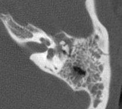
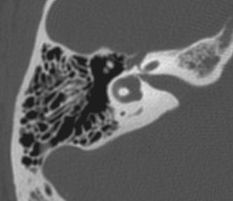
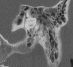
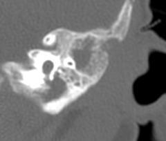
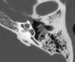
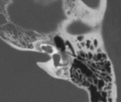
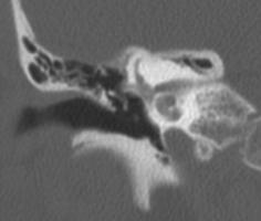
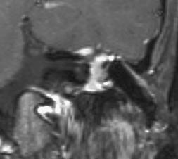
4 Longitudinal Fracture
5 The Temporal Bone Longitudinal fractures Most common fracture (80%) Pass through EAC, mastoid & middle ear High incidence of ossicular derangement Inner ear usually spared Facial paralysis in about 15% CSF otorrhea or rhinorrhea


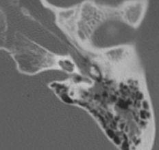
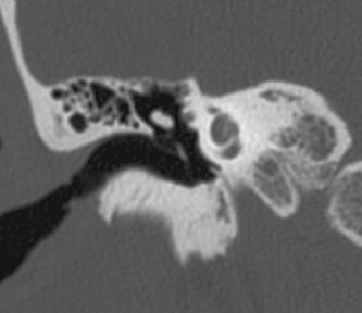
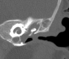
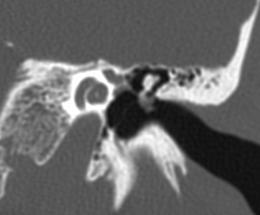
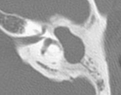
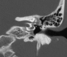
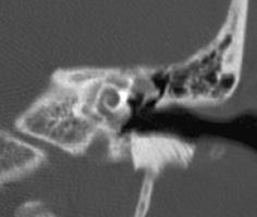
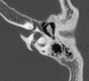
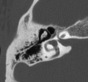
6 History: 40 y/o man with hearing loss following a 40 foot fall Dx: Transverse fracture 525
7 The Temporal Bone Transverse Fractures Commonly involve the inner ear Cochlear fracture - sensorineural hearing loss Labyrinthine fracture - severe vertigo Facial palsy in 50% Perilymph fistula Injury to carotid artery
8 Diseases of the Mastoid and Middle Ear Acute otomastoiditis Chronic otomastoiditis Acquired cholesteatoma Paraganglioma Glomus jugulare Glomus tympanicum


9 Temporal Bone Inflammation Otitis Media / Mastoiditis


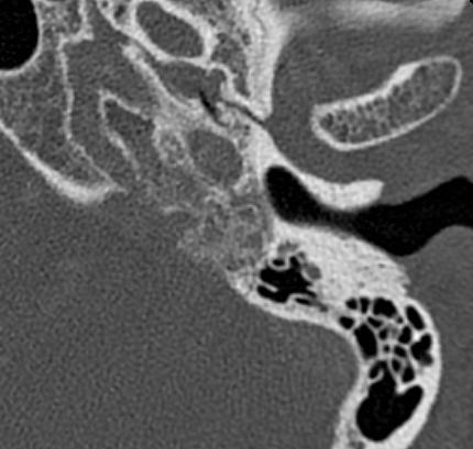
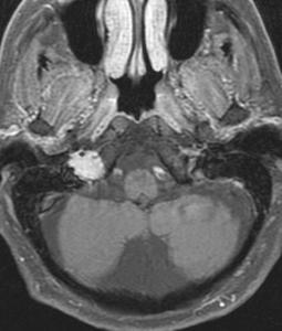
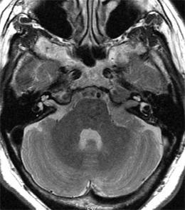
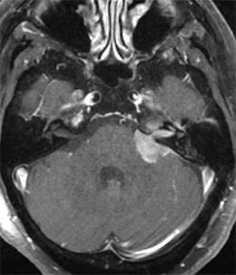
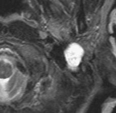
10 History: 5 month old girl with left ear pain Dx: Otomastoiditis and external otitis 384


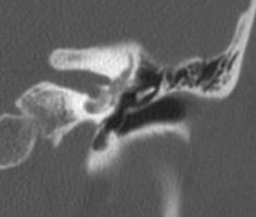
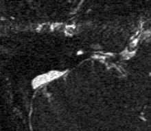
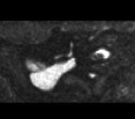
11 History: 24 y/o man with a left conductive hearing loss 665 Dx: Cholesteatoma
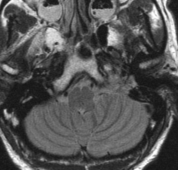
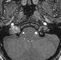
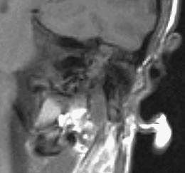
12 Acquired Cholesteatoma Critical Imaging Findings Erosion of scutum and ossicles Integrity of lateral semicircular canal Facial nerve involvement Erosion through tegmen or sigmoid sinus plate


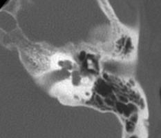
13 History: 33 y/o male with left ear deafness 177


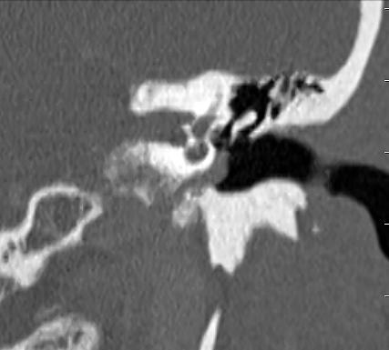
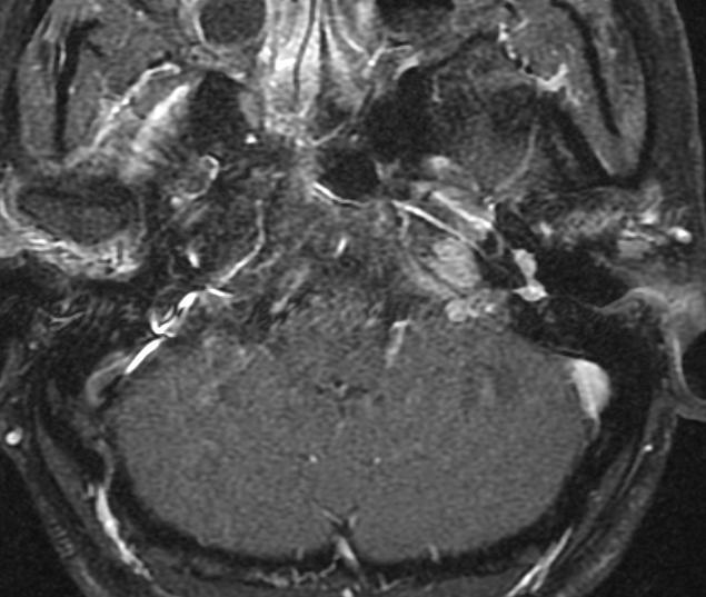
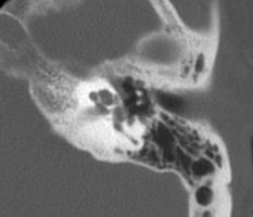
14 History: 48 y/o woman with sensorineural hearing loss
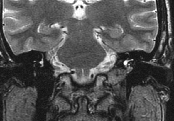
15 {Page 2}


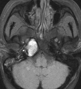
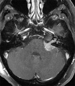
16 {Page 3} Dx: Glomus jugulare (paraganglioma)
17 Paragangliomas Origin: paraganglia along CN 9 and 10 Glomus jugulare - Nerve of Arnold (X) Glomus tympanicum - Jacobson's nerve (IX) Presentation: pulsatile tinnitus, conductive hearing loss, retrotympanic mass, and cranial nerve deficits Biological behavior - slow growing but locally invasive

18 Location of Glomus Tissue


19 History: 60 y/o man with polycystic kidney disease, tinnitus, hoarseness & paretic right vocal cord 409


20 {Page 2} Dx: Glomus jugulare


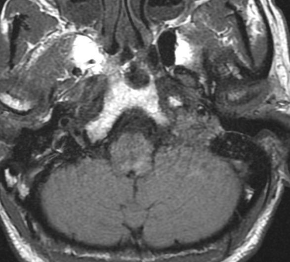
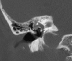
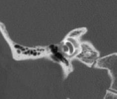
21 Aberrant Carotid Artery Q 49-53

22 Aberrant Carotid Artery B 5,6


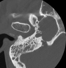
23 History: 13 y/o girl with pulsatile tinnitus 411


24 {Page 2} Dx: High dehiscent jugular bulb
25 Middle Ear Masses Differential Diagnosis Paraganglioma High & exposed jugular bulb Aberrant carotid artery Persistent stapedial artery Schwannoma Primary & metastatic bone tumors Other middle ear tumors - cholesteatoma, squamous cell carcinoma
26 Diseases of the Inner Ear Labyrinthitis Otosclerosis Acoustic schwannoma Endolymphatic sac tumors

27 Dr. Harris "The ringing in your ears - - I think I can help."


28 History: 55 y/o woman with acute hearing loss in the right ear 10 days ago Dx: Viral labyrinthitis on steroids for 8 days 619
29 Labyrinthitis Presentation: sensorineural hearing loss and vertigo Viral most common, but other agents possible Source: tympanic, meningeal, hematogenous, posttraumatic or postsurgical Imaging features: Acute & subacute: enhancement with gadolinium Chronic: fibrosis and ossification (labyrinthitis ossificans)
30 Otosclerosis Acute phase: otospongiosis - resorption of bone Chronic phase: otosclerosis - sclerosis of bone Resorption of enchondral bone around otic capsule & replacement with spongy vascular new bone 65% females & 80% of cases are bilateral Onset during the 2nd and 3rd decades Two types: Fenestral & Cochlear


31 History: 58 y/o woman with progressive bilateral hearing loss for 8 years 699

32 {Page 2} Dx: Cochlear otosclerosis
33 Retrofenestral Otosclerosis (Cochlear Otospongiosis) Sensorineural hearing loss CT findings: Acute phase - patchy band-like lucency around the cochlear Chronic phase - no visible abnormalities Cochlear implantation does not work


34 History: 58 y/o woman with vertigo & hearing loss 700

35 {Page 2} Dx: Fenestral otosclerosis
36 Fenestral Otosclerosis Most common form Conductive hearing loss Fixation of the foot plate of the stapes CT findings: lucent focus just anterior to oval window dense bone encroaches on oval window Surgical correction possible


37 History: Right sensorineural hearing loss & facial palsy Courtesy Mahmood Mafee,
38 Causes of Sensorineural Hearing Loss Vestibular Schwannoma Other CP angle & petrous tumors Brain stem lesions Labyrinthitis Cochlear otosclerosis Bone dysplasias Leptomeningeal processes Congenital Trauma (transverse fx's)
39 Diseases of the Petrous Apex Cholesterol granuloma (cyst) Primary cholesteatoma Petrous apicitis (Gradenigo's syndrome) Intrapetrous carotid artery aneurysm Chondrosarcoma Metastatic tumors
40 Diseases of the Petrous Apex Cholesterol Granuloma Chronic inflammatory cysts Contain blood products & cholesterol crystals Cyst capsule lined by fibrous tissue Hyperintense on T1 & T2-weighted images
41 Diseases of the Petrous Apex Primary Cholesteatoma Also called epidermoid tumor or cyst Arise from epithelial rests Capsule lined by stratified squamous epithelial Cyst contents are primarily desquamated keratin Usually high T2 signal & low T1 signal
42 Cholesterol Granuloma 568
")

43 Temporal Bone Inflammation Petrous Apicitis (Gradinego's Syndrome) Schwartz & Harnsberger, Imaging of the Temporal Bone, Thieme, 1992, p. 334


44 History: 62 y/o female with right ear deafness & pain 596


45 Dx: Multiple myeloma MGUS for several years {Page 2}


46 History: 60 y/o woman with vertigo, left sensorineural hearing loss & left facial palsy

47 Dx: AML - Chloroma {Page 2}

48 The Facial Nerve Normal Anatomy Schuknecht HF, Pathology of the Ear, Lea & Febiger, 1993, pp45-66
49 The Facial Nerve Inflammatory Disease Bell's palsy Ramsey Hunt syndrome Herpes zoster oticus Lyme disease Syphilis Imaging - gadolinium enhancement of the 7th nerve


50 History: 37 HIV + male with 7th nerve deficits Dx: Bell's palsy 414


51 History: 81 y/o woman with chronic lymphocytic leukemia and a right 7 th nerve palsy Dx: Bell s palsy Herpes zoster 711
52 Facial Nerve Tumors Differential Diagnosis Schwannoma Hemangioma Epidermoid tumor Acquired cholesteatoma Tympanic segment - glomus tympanicum, cholesteatoma, persistent stapedial artery Stylomastoid segment - glomus jugulare

53 PD Facial Nerve Schwannoma


54 History: 21 y/o woman with progressive left facial weakness & hearing loss for 11 months 670

55 Dx: Plexiform neurofibroma {Page 2}
56 Congenital Anomalies External & Middle Ear Associated with facial, cervical & skeletal dysplasias External ear deformities Conductive hearing loss More common anomalies Atresia of external ear First branchial arch dysplasia Second branchial arch dysplasia
57 Atresia of External Auditory Canal Critical Imaging Information Thickness of atresia plate Size of tympanic cavity Status of the ossicles and oval window Presence of any congenital cholesteatoma


58 Atresia of External Auditory Canal PB 8-10


59 Membranous Labyrinth Superior semicircular duct Superior ampulla Saccule Utricle Lateral ampulla Lateral semicircular duct Cochlear duct Endolymphatic duct Endolymphatic sac Ductus reuniens Utriculosaccular duct Posterior semicircular duct Posterior ampulla Swartz et al, AJNR 17:17-21, 1996

60 Inner Ear Anatomy Som & Curtin, Head & Neck Imaging, Mosby, 1996, p1412
61 Inner Ear Anomalies Semicircular canals & vestibule Cochlear anomalies Labyrinthine aplasia (Michel's deformity) Incomplete partition (Mondini malformation) Atresia/stenosis of the internal auditory canal Perilymphatic hydrops

62 Cochlear Aplasia with Labyrinthine Dysgenesis Hasso et al., in Som & Curtin, Head & Neck Imaging, Mosby, 1996, p1367


63 History: 17 y/o male was partially deaf in the left ear Dx: 192
64 Perilymphatic Hydrops Increased pressure in the inner ear due to CSF fistula Can lead to perforation of the stapes foot plate Etiology: Defective lamina cribrosa Congenitally wide cochlear aqueduct Trauma most common


65 History: 27 y/o man with deaf right ear & progressive sensorineural hearing loss in left ear


66 {Page 2} Dx: Endolymphatic hydrops Vestibular aqueduct syndrome
67 Endolymphatic Hydrops Vestibular Aqueduct Syndrome Sensorineural hearing loss & vertigo Deficient absorption of endolymph Can be congenital, acquired, or idiopathic Meniere's disease: Idiopathic form Imaging: Dilated endolymphatic sac and vestibular aqueduct < 1.5 mm No larger than semicircular canal


68 History: 77 y/o man developed vertigo with loud noises Dx: Dehiscence SSC 651
69 Superior Canal Dehiscence Syndrome Caused by dehiscence of bone overlying the superior semicircular canal Causes vertigo & oscillopsia with sound or pressure stimuli Diagnosed with thin-section coronal CT Rx: Surgical plugging of the SSC
Pediatric Temporal Bone
 Pediatric Temporal Bone Suresh K. Mukherji, MD, FACR Professor and Chief of Neuroradiology Professor of Radiology, Otolaryngology Head Neck Surgery, Radiation Oncology and Periodontics & Oral Medicine
Arastoo Vossough, M.D., Ph.D. Associate Professor of Radiology
 Disorders of the Temporal Bone Arastoo Vossough, M.D., Ph.D. Associate Professor of Radiology 1st Branchial Cleft Cyst Remnant of 1 st branchial apparatus Rare Cystic lesions: I- around pinna II- connecting
Modern Imaging & Current Controversies
 Temporal Bone: Modern Imaging & Current Controversies Suresh K. Mukherji, MD, FACR Professor and Chief of Neuroradiology Professor of Radiology, Otolaryngology Head Neck Surgery, Radiation i Oncology,
Cholesteatoma and Non-cholesteatomatous Inflammatory Disease. Cholesteatoma. Disclosures. Overview EAC. Cholesteatoma. None
 Disclosures Cholesteatoma and Non-cholesteatomatous Inflammatory Disease None Amy F Juliano, MD Staff Radiologist, Massachusetts Eye and Ear Infirmary Assistant Professor of Radiology, Harvard Medical
CE Directed Reading. Temporal Bone CT: Anatomy, Technique, and Associated Pathophysiology
 Temporal Bone CT: Anatomy, Technique, and Associated Pathophysiology Chris Young, MS, R.R.A., R.T.(R) Computed tomography (CT) of the temporal bone is performed to evaluate trauma, tumors, sinuses, the
Anatomy of the Ear Region. External ear Middle ear Internal ear
 Ear Lecture Objectives Make a list of structures making the external, middle, and internal ear. Discuss the features of the external auditory meatus and tympanic membrane. Describe the shape, position,
COCHLEAR IMPLANTS Aetiology of Deafness. Bruce Black MD
 COCHLEAR IMPLANTS Aetiology of Deafness Heterochromia iridis. Cases may be healthy or associated with a variety of conditions, e.g. Waardenburg syndrome. Waardenburg syndrome. Note the snowy lock of hair
RADIOLOGY TEACHING CONFERENCE
 RADIOLOGY TEACHING CONFERENCE John Athas, MD Monica Tadros, MD Columbia University, College of Physicians & Surgeons Department of Otolaryngology- Head & Neck Surgery September 27, 2007 CT SCAN IMAGING
AUDITORY APPARATUS. Mr. P Mazengenya. Tel 72204
 AUDITORY APPARATUS Mr. P Mazengenya Tel 72204 Describe the anatomical features of the external ear Describe the tympanic membrane (ear drum) Describe the walls of the middle ear Outline the structures
Temporal bone anatomy and imaging features of common conditions causing hearing loss: A pictorial review
 Temporal bone anatomy and imaging features of common conditions causing hearing loss: A pictorial review Poster No.: C-1892 Congress: ECR 2012 Type: Educational Exhibit Authors: A. Masukawa, H. Takeuchi,
Radiologic Evaluation of Petrous Apex Masses. Pavan Kavali, MS-IV Morehouse School of Medicine November 16, 2009
 Radiologic Evaluation of Petrous Apex Masses Pavan Kavali, MS-IV Morehouse School of Medicine November 16, 2009 Roadmap Petrous Apex Anatomy Patient D.S.: Clinical Presentation Differential diagnosis of
Ear. Utricle & saccule in the vestibule Connected to each other and to the endolymphatic sac by a utriculosaccular duct
 Rahaf Jreisat *You don t have to go back to the slides. Ear Inner Ear Membranous Labyrinth It is a reflection of bony labyrinth but inside. Membranous labyrinth = set of membranous tubes containing sensory
Middle ear CT imaging: Review of anatomy and common pathology
 Middle ear CT imaging: Review of anatomy and common pathology Poster No.: C-0665 Congress: ECR 2017 Type: Educational Exhibit Authors: M. R. Campos Arenas, M. C. Sánchez-Porro, J. Garrido Rull ; 1 1 2
Petrous Bone Normal anatomy
 Petrous Bone Normal anatomy By Mamdouh Mahfouz MD Prof. of Radiology Cairo University ssregypt.com Axial Coronal Petrous bone External ear Middle ear Inner ear External ear Cartilaginous part Bony part
Case Studies in CPA/IAC
 Outline Case Studies in CPA/IAC Atul K Mallik MD PhD Department of Radiology and Imaging Sciences University of Utah Health Sciences Center Salt Lake City, Utah, USA Case based review of cerebellopontine
The Ear. Dr. Heba Kalbouneh Assistant Professor of Anatomy and Histology
 The Ear Dr. Heba Kalbouneh Assistant Professor of Anatomy and Histology The Ear The ear consists of the external ear; the middle ear (tympanic cavity); and the internal ear (labyrinth), which contains
Injury retrotympanic white, blue and red. Clinicalradiological
 Injury retrotympanic white, blue and red. Clinicalradiological correlation Poster No.: C-0211 Congress: ECR 2013 Type: Educational Exhibit Authors: R. Esteban Saiz, R. Castañón Martinez, M. Rebolledo Vicente,
Chronic Ear Disease. Daekeun Joo Resident Lecture Series 11/18/09
 Chronic Ear Disease Daekeun Joo Resident Lecture Series 11/18/09 ETD URIs Viral-induced damage to ET lining resulting in decreased mucociliary clearance Viral invasion of ME mucosa results in inflamm Reflux
Correlation of HRCT mastoid with clinical presentation and operative findings in ear diseases
 International Journal of Otorhinolaryngology and Head and Neck Surgery Chintale SG et al. Int J Otorhinolaryngol Head Neck Surg. 2017 Jul;3(3):656-660 http://www.ijorl.com pissn 2454-5929 eissn 2454-5937
High resolution computed tomography of temporal bone in the evaluation of otologic diseases
 International Journal of Otorhinolaryngology and Head and Neck Surgery Handi PS et al. Int J Otorhinolaryngol Head Neck Surg. 2018 Jan;4(1):87-92 http://www.ijorl.com pissn 2454-5929 eissn 2454-5937 Original
Pediatric Ear Diseases
 Pediatric Ear Diseases Yasushi Naito Pediatric Ear Diseases Diagnostic Imaging Atlas and Case Reports 242 figures, 7 in color and 5 tables, 2013 Basel Freiburg Paris London New York New Delhi Bangkok
Anatomy of the ear: Lymphatics
 Anatomy of the ear: 1. External ear which consist of auricle and external auditory canal. The auricle has a framework of cartilage except the lobule, the skin is closely adherent to perichonderium at the
Imaging of Hearing Loss
 Contemporary Imaging of Sensorineural Hearing Loss Imaging of Hearing Loss Discussion Outline (SNHL) Imaging Approaches Anatomic Relationships Lesions: SNHL KL Salzman, MD University of Utah School of
High-resolution Computed Tomography Study of Temporal Bone Pathologies
 Original Article Print ISSN: 2321-6379 Online ISSN: 2321-595X DOI: 10.17354/ijss/2016/375 High-resolution Computed Tomography Study of Temporal Bone Pathologies Manjit Bagul Senior Resident, Department
Imaging of Petrous Apex: Anatomy and Pathology
 University of Utah Head and Neck Conference 2018 Petrous apex Imaging of Petrous Apex: Anatomy and Pathology Philip Chapman MD University of Alabama, Birmingham Good News PAs tend to be symmetric A quick
Advanced Vascular Imaging: Pulsatile Tinnitus. Disclosures. Pulsatile Tinnitus: Differential Diagnosis. Pulsatile Tinnitus
 Advanced Vascular Imaging: Pulsatile Tinnitus Patrick Turski MD, Zach Clark MD, Tabby Kennedy MD The Objectives of this presentation are to: Review the differential diagnosis of pulsatile tinnitus Discuss
Unit VIII Problem 9 Physiology: Hearing
 Unit VIII Problem 9 Physiology: Hearing - We can hear a limited range of frequency between 20 Hz 20,000 Hz (human hearing acuity is between 1000 Hz 4000 Hz). - The ear is divided into 3 parts. Those are:
Dr. Sami Zaqout Faculty of Medicine IUG
 Auricle External Ear External auditory meatus The Ear Middle Ear (Tympanic Cavity) Auditory ossicles Internal Ear (Labyrinth) Bony labyrinth Membranous labyrinth External Ear Auricle External auditory
Bony and membranous labyrinth. Vestibular system. János Hanics M.D.
 Bony and membranous labyrinth. Vestibular system. János Hanics M.D. The position of the inner ear The labyrinthes of the inner ear - Continuous cavity system in the petrous part of temporal bone - Cavity
Congenital Absence of the Oval Window: Radiologic Diagnosis and Associated Anomalies
 AJNR Am J Neuroradiol 21:322 327, February 2000 Congenital Absence of the Oval Window: Radiologic Diagnosis and Associated Anomalies Barbara Zeifer, Paul Sabini, and Jonathan Sonne BACKGROUND AND PURPOSE:
ICD10 CODES CODE DESCRIPTION R Abnormal auditory function study H Abnormal auditory perception, bilateral H Abnormal auditory
 ICD10 CODES CODE DESCRIPTION R94.120 Abnormal auditory function study H93.293 Abnormal auditory perception, bilateral H93.292 Abnormal auditory perception, left ear H93.291 Abnormal auditory perception,
J.P.S. Bakshi Manual of Ear, Nose and Throat
 J.P.S. Bakshi Manual of Ear, Nose and Throat Reading excerpt Manual of Ear, Nose and Throat of J.P.S. Bakshi Publisher: B. Jain http://www.narayana-publishers.com/b5603 Copying excerpts is not permitted.
Facial Paralysis: Objectives: Discuss the anatomy of the facial nerve. Look at common patterns of facial nerve palsy
 Facial Paralysis: Objectives: Discuss the anatomy of the facial nerve Look at common patterns of facial nerve palsy Discuss imaging appearance of lesions that lead to facial paralysis. Lindell R. Gentry,
Imaging in patients undergoing cochlear implant: CT and MR technique
 Imaging in patients undergoing cochlear implant: CT and MR technique Poster No.: C-0102 Congress: ECR 2012 Type: Educational Exhibit Authors: G. Posillico Keywords: Ear / Nose / Throat, CT, MR, Comparative
Imaging of the Temporal Bone: A Symptom-Based Approach
 Imaging of the Temporal Bone: A Symptom-Based Approach Tadesse Eshetu, MD, and Nafi Aygun, MD Some of the symptoms associated with the temporal bone diseases are nonspecific, whereas others overlap with
1. Axial view, left temporal bone. Plane through the upper antrum (A), superior semicircular canal (SSC) and IAC.
, superior semicircular canal (SSC) and IAC.") PA IAC SSC A 1. Axial view, left temporal bone. Plane through the upper antrum (A), superior semicircular canal (SSC) and IAC. IAC VII M I LSC Plane through the IAC, malleus head and incus and the lateral
PA IAC SSC A 1. Axial view, left temporal bone. Plane through the upper antrum (A), superior semicircular canal (SSC) and IAC. IAC VII M I LSC Plane through the IAC, malleus head and incus and the lateral
OTOLOGY. 1. BRIEF DESCRIPTION OF OTOLOGIC TRAINING Rotations that include otologic training are a component of each of the four years of training.
 OTOLOGY 1. BRIEF DESCRIPTION OF OTOLOGIC TRAINING Rotations that include otologic training are a component of each of the four years of training. Longwood Rotation PGY-2 through PGY-5 years o Clinic experience
Middle and Inner Ear: Improved Depiction with Multiplanar Reconstruction of Volumetric CT Data 1
 Note: This copy is for your personal non-commercial use only. To order presentation-ready copies for distribution to your colleagues or clients, contact us at www.rsna.org/rsnarights. EDUCATION EXHIBIT
The Ear The ear consists of : 1-THE EXTERNAL EAR 2-THE MIDDLE EAR, OR TYMPANIC CAVITY 3-THE INTERNAL EAR, OR LABYRINTH 1-THE EXTERNAL EAR.
 The Ear The ear consists of : 1-THE EXTERNAL EAR 2-THE MIDDLE EAR, OR TYMPANIC CAVITY 3-THE INTERNAL EAR, OR LABYRINTH 1-THE EXTERNAL EAR Made of A-AURICLE B-EXTERNAL AUDITORY MEATUS A-AURICLE It consists
Acquired Deafness Loss of hearing that occurs or develops sometime in the course of a lifetime, but is not present at birth.
 Page 1 of 5 URMC» Audiology Glossary of Terms A Acoustic Neuroma A tumor, usually benign, which develops on the hearing and balance nerves and can cause gradual hearing loss, tinnitus, and dizziness. Acquired
Gross Anatomy of the. TEMPORAL BONE, EXTERNAL EAR, and MIDDLE EAR. Assignment: Head to Toe Temporomandibular Joint (TMJ)
") Gross Anatomy the TEMPORAL BONE, EXTERNAL EAR, and MIDDLE EAR M1 Gross and Developmental Anatomy 9:00 AM, December 11, 2008 Dr. Milton M. Sholley Pressor Anatomy and Neurobiology Assignment: Head to Toe
Gross Anatomy the TEMPORAL BONE, EXTERNAL EAR, and MIDDLE EAR M1 Gross and Developmental Anatomy 9:00 AM, December 11, 2008 Dr. Milton M. Sholley Pressor Anatomy and Neurobiology Assignment: Head to Toe
Expand The Scope of Temporal Bone Reporting: Why, What and Where to Look For.
 Expand The Scope of Temporal Bone Reporting: Why, What and Where to Look For. Award: Certificate of Merit Poster No.: C-1521 Congress: ECR 2014 Type: Educational Exhibit Authors: P. Mundada, B. S. Purohit,
Gross Anatomy of the. TEMPORAL BONE, EXTERNAL EAR, and MIDDLE EAR
 Gross Anatomy of the TEMPORAL BONE, EXTERNAL EAR, and MIDDLE EAR M1 Gross and Developmental Anatomy 9:00 AM, December 11, 2008 Dr. Milton M. Sholley Professor of Anatomy and Neurobiology Assignment: Head
Control of eye movement
 Control of eye movement Third Nerve Palsy Eye down and out Trochlear Nerve Palsy Note: Right eye Instead of intorsion and depression action of superior oblique See extorsion and elevation Observe how
Temporal bone imaging in osteogenesis imperfecta patients with hearing loss
 Temporal bone imaging in osteogenesis imperfecta patients with hearing loss F. Swinnen 1, J. Casselman 2, P. Coucke 3, C. Cremers 4, E. De Leenheer 1, I. Dhooge 1 (1) Departement of Otorhinolaryngology,
The ear: some applied basic science
 Chapter 1 The ear: some applied basic science The pinna The external ear or pinna is composed of cartilage with closely adherent perichondrium and skin. It is developed from six tubercles of the first
Educational Exhibit Authors: P. Digge, R. K. N. Solanki, S. M. Paruthikunnan, D. S. Shah ; 1
 High-field MRI versus high-resolution CT of temporal bone in inner ear pathologies of children with bilateral profound sensorineural hearing loss: A pictorial essay. Poster No.: C-0867 Congress: ECR 2015
The close anatomic relationship between the cochlea and
 ORIGINAL RESEARCH R.J. Young D.R. Shatzkes J.S. Babb A.K. Lalwani The Cochlear-Carotid Interval: Anatomic Variation and Potential Clinical Implications BACKGROUND AND PURPOSE: A temporal bone CT study
Sasan Dabiri, MD, Assistant Professor
 Sasan Dabiri, MD, Assistant Professor Department of Otorhinolaryngology Head & Neck Surgery Amir A lam hospital Tehran University of Medical Sciences October 2015 Outlines Anatomy of Vestibular System
Paraganglioma of the Skull Base. Ross Zeitlin, MD Medical College of Wisconsin Milwaukee, WI
 Paraganglioma of the Skull Base Ross Zeitlin, MD Medical College of Wisconsin Milwaukee, WI Case Presentation 63-year-old female presents with right-sided progressive conductive hearing loss for several
UC SF. Safe Surgery Rule #1. Cholesteatoma. It s hard to have a surgical complication when you are not operating
 UC SF Cholesteatoma Chronic Ear Surgery: Staying Out of Trouble! Lawrence R. Lustig, MD Department of Oto-HNS University of California San Francisco Ligaments and folds Spaces NU Epitympanic Cholesteatoma
1. GOAL 2. OBJECTIVES a) KNOWLEDGE b) SKILLS c) INTEGRATION
 KNOWLEDGE b) SKILLS c) INTEGRATION") 1. GOAL The broad goal of the teaching of undergraduate students in Otorhinolaryngology is that the undergraduate student have acquired adequate knowledge and skills for optimally dealing with common disorders
1. GOAL The broad goal of the teaching of undergraduate students in Otorhinolaryngology is that the undergraduate student have acquired adequate knowledge and skills for optimally dealing with common disorders
For the following questions, indicate the letter that corresponds to the SINGLE MOST APPROPRIATE ANSWER
 GROSS ANATOMY EXAMINATION May 15, 2000 For the following questions, indicate the letter that corresponds to the SINGLE MOST APPROPRIATE ANSWER 1. Pain associated with an infection limited to the middle
High Signal from the Otic Labyrinth on Onenhanced Magnetic Resonance Imaging
 High Signal from the Otic Labyrinth on Onenhanced Magnetic Resonance Imaging JaneL. Weissman, 1 Hugh D. Curtin, 1 3 Barry E. Hirsch, 2 and William L. Hirsch, Jr. 1 Summary: High signal from the otic labyrinth
Chapter 17, Part 2! The Special Senses! Hearing and Equilibrium!
 Chapter 17, Part 2! The Special Senses! Hearing and Equilibrium! SECTION 17-5! Equilibrium sensations originate within the inner ear, while hearing involves the detection and interpretation of sound waves!
Chapter 17, Part 2! Chapter 17 Part 2 Special Senses! The Special Senses! Hearing and Equilibrium!
 Chapter 17, Part 2! The Special Senses! Hearing and Equilibrium! SECTION 17-5! Equilibrium sensations originate within the inner ear, while hearing involves the detection and interpretation of sound waves!
Gerard J. Gianoli, MD, FACS The Ear and Balance Institute Baton Rouge, Louisiana
 Gerard J. Gianoli, MD, FACS The Ear and Balance Institute Baton Rouge, Louisiana SSCD is defined anatomically as the absence of bone between the SSC and the middle fossa dura PSCD is a defect of the PSC
Dr. Vitthalrao Vikhe Patil Foundation s Medical College & Hospital, Ahmednagar, Maharashtra, India
 DOI: 10.21276/sjams.2017.5.3.17 Scholars Journal of Applied Medical Sciences (SJAMS) Sch. J. App. Med. Sci., 2017; 5(3B):770-779 Scholars Academic and Scientific Publisher (An International Publisher for
Imaging the Spinal Cord & Intradural Disease
 Department of Radiology University of California San Diego Imaging the Spinal Cord & Intradural Disease John R. Hesselink, M.D. Spinal Cord Diseases Tumors Syringohydromyelia Trauma Ischemia / Infarction
Modifying radiology protocols for cochlear implant surgery in a government sponsored scheme: Need of the hour
 Available online at www.ijmrhs.com ISSN No: 2319-5886 International Journal of Medical Research & Health Sciences, 2016, 5, 6:151-157 Modifying radiology protocols for cochlear implant surgery in a government
Ear, Nose, and Throat Disorders
 Health Reference Series Second Edition Basic Consumer Health Information about Disorders of the Ears, Hearing Loss, Vestibular Disorders, Nasal and Sinus Problems, Throat and Vocal Cord Disorders, and
Fracture mimics on temporal bone CT - a guide for the radiologist
 Fracture mimics on temporal bone CT - a guide for the radiologist Award: Certificate of Merit Poster No.: C-0158 Congress: ECR 2012 Type: Educational Exhibit Authors: Y. Kwong, D. Yu, J. Shah; Nottingham/UK
Unit VIII Problem 9 Anatomy of The Ear
 Unit VIII Problem 9 Anatomy of The Ear - The ear is an organ with 2 functions: Hearing. Maintenance of equilibrium/balance. - The ear is divided into 3 parts: External ear. Middle ear (which is also known
Anomalous Facial Nerve Canal with Cochlear Malformations
 AJNR Am J Neuroradiol 22:838 844, May 2001 Anomalous Facial Nerve Canal with Cochlear Malformations Laura Vitale Romo and Hugh D. Curtin BACKGROUND AND PURPOSE: Anteromedial migration of the first segment
Rory Attwood MBChB,FRCS
 Hearing loss Overview Rory Attwood MBChB,FRCS Division of Otorhinolaryngology Faculty of Health Sciences Tygerberg Campus, University of Stellenbosch Not deafness Deaf is a total lack of hearing Deafness
Abnormal direction of internal auditory canal and vestibulocochlear nerve
 Medicine Otorhinolaryngology fields Okayama University Year 2004 Abnormal direction of internal auditory canal and vestibulocochlear nerve Shin Kariya kazunori Nishizaki Hirofumi Akagi Michael M. Paparella
Cholesteatoma-Pathogenesis and Surgical Management. Grand Rounds Presentation February 24, 1999 Kyle Kennedy, M.D. Jeffrey Vrabec,, M.D.
 Cholesteatoma-Pathogenesis and Surgical Management Grand Rounds Presentation February 24, 1999 Kyle Kennedy, M.D. Jeffrey Vrabec,, M.D. Introduction Cholesteatoma (keratoma)-essentially an accumulation
Skull Base Course. Dissection with fresh temporal bones and half heads
 Skull Base Course Dissection with fresh temporal bones and half heads 711 November 2016 Gruppo Otologico Via Emmanueli 42 Piacenza 29122 t +39 0523 754 362 fax +39 0523 453 708 www.gruppootologico.com
Assisting in Otolaryngology
 Assisting in Otolaryngology Learning Objectives Identify the structures and explain the functions of the external, middle, and internal ear. Describe the conditions that can lead to hearing loss, including
Real role of MRI in ORL diagnosis protocol in hearing loss and vertiginous syndrome: A daily challenge for the clinician and the radiologist
 Real role of MRI in ORL diagnosis protocol in hearing loss and vertiginous syndrome: A daily challenge for the clinician and the radiologist Poster No.: C-0848 Congress: ECR 2012 Type: Scientific Exhibit
Bilateral temporal bone fractures: a case report
 International Journal of Otorhinolaryngology and Head and Neck Surgery Natarajan K et al. Int J Otorhinolaryngol Head Neck Surg. 2018 Jan;4(1):271-275 http://www.ijorl.com pissn 2454-5929 eissn 2454-5937
THE EAR AND HEARING Be sure you have read and understand Chapter 16 before beginning this lab. INTRODUCTION: hair cells outer ear tympanic membrane
 BIOLOGY 211: HUMAN ANATOMY & PHYSIOLOGY ****************************************************************************************************** THE EAR AND HEARING ******************************************************************************************************
Chapter 143: Otosclerosis (OS) Sameer Ahmed 2/23/2011
 Sameer Ahmed 2/23/2011") Chapter 143: Otosclerosis (OS) Sameer Ahmed 2/23/2011 Intro Disorder of fibrous osteodystrophy of the human otic capsule Abnormal resorption and deposition of bone Primarily causes CHL SNHL and MHL are
Chapter 143: Otosclerosis (OS) Sameer Ahmed 2/23/2011 Intro Disorder of fibrous osteodystrophy of the human otic capsule Abnormal resorption and deposition of bone Primarily causes CHL SNHL and MHL are
CT and MR Imaging In Patients Undergoing Evaluation for Cochlear Implantation
 CT and MR Imaging In Patients Undergoing Evaluation for Cochlear Implantation Poster No.: C-1219 Congress: ECR 2015 Type: Educational Exhibit Authors: S. S. Ghuman, T. Buxi, S. Sud, A. Sud, A. Yadav, K.
Management of Ear, Hearing and Balance Disorders: Fact, Fiction, and Future
 Management of Ear, Hearing and Balance Disorders: Fact, Fiction, and Future George W. Hicks, M,D. 7440 N. Shadeland Avenue, Suite 150 Indianapolis, IN 46250 904 N. Samuel Moore Parkway Mooresville, IN
Bruce Black MD EAC TRAUMA
 EAC TRAUMA Bruising in the deep canal due to cotton bud/q-tip selfcleaning attempts. No action required. A granuloma of the deep Lt. EAC. Superficial trauma has become secondarily infected. Clean thoroughly,
Role of high resolution computed tomography in the evaluation of temporal bone lesions: our experience
 International Journal of Otorhinolaryngology and Head and Neck Surgery Jyothi AC et al. Int J Otorhinolaryngol Head Neck Surg. 2016 Jul;2(3):135-139 http://www.ijorl.com pissn 2454-5929 eissn 2454-5937
Interpretation of Computed Tomography of the Petrous Temporal Bone
 Systematic Review Article & Research Interpretation of Computed Tomography of the Petrous Temporal Bone Abstract This review will familiarise the reader with the normal radiological anatomy of the temporal
Cone Beam CT Atlas of the Normal Suspensory Apparatus of the Middle Ear Ossicles
 Cone Beam CT Atlas of the Normal Suspensory Apparatus of the Middle Ear Ossicles Poster No.: C-2036 Congress: ECR 2013 Type: Authors: Educational Exhibit B. Smet 1, I. De Kock 2, P. Gillardin 2, M. Lemmerling
Section 1 EAR. Section Outlines
 Section 1 EAR Section Outlines Development of the Ear Anatomy of the Ear Physiology of the Ear History Taking with Symptomatology of Ear Diseases Examination of the Ear Congenital Diseases of the External
Clinical Course of Pediatric Congenital Inner Ear Malformations
 The Laryngoscope Lippincott Williams & Wilkins, Inc., Philadelphia 2000 The American Laryngological, Rhinological and Otological Society, Inc. Clinical Course of Pediatric Congenital Inner Ear Malformations
CT as first diagnostic approach in non-traumatic conditions of temporal bone
 CT as first diagnostic approach in non-traumatic conditions of temporal bone Award: Certificate of Merit Poster No.: C-1408 Congress: ECR 2016 Type: Educational Exhibit Authors: M. Bernabéu Rodríguez,
Eye and Ocular Adnexa, Auditory Systems
 Eye and Ocular Adnexa, Auditory Systems CPT copyright 2011 American Medical Association. All rights reserved. Fee schedules, relative value units, conversion factors and/or related components are not assigned
SPECIAL SENSES PART I: OLFACTION & GUSTATION
 SPECIAL SENSES PART I: OLFACTION & GUSTATION 5 Special Senses Olfaction Gustation Vision Equilibrium Hearing Olfactory Nerves Extend through cribriform plate into nasal cavity on both sides of nasal septum
Otosclerosis. Diagnosis, Evaluation, Pathology, Surgical Techniques, and Outcomes
 Otosclerosis Diagnosis, Evaluation, Pathology, Surgical Techniques, and Outcomes Chris de Souza, MS, DORL, DNB, FACS Marcos V. Goycoolea, MD, MS, PhD, MD, FACS Contents List of Videos Introduction Contributors
Taste buds Gustatory cells extend taste hairs through a narrow taste pore
 The Special Senses Objectives Describe the sensory organs of smell, and olfaction. Identify the accessory and internal structures of the eye, and explain their function. Explain how light stimulates the
Chapter 1: Applied Anatomy, Physiology and Embryology of the Ear. Anatomy and Physiology. The Outer Ear. The Pinna. The External Ear Canal
 Chapter 1: Applied Anatomy, Physiology and Embryology of the Ear The ear contains two specialized sensory organs, the cochlea and the vestibular apparatus, enclosed in the extremely hard protective casing
Major Anatomic Components of the Orbit
 Major Anatomic Components of the Orbit 1. Osseous Framework 2. Globe 3. Optic nerve and sheath 4. Extraocular muscles Bony Orbit Seven Bones Frontal bone Zygomatic bone Maxillary bone Ethmoid bone Sphenoid
Activity 1: Anatomy of the Eye and Ear Lab
 Activity 1: Anatomy of the Eye and Ear Lab 1. Launch the view! Launch Human Anatomy Atlas. Navigate to Quizzes/Lab Activities, find the Eye and Ear Lab section. Launch Augmented Reality mode and scan the
Complications of At t i c o a n t ral Cholesteato ma:
 Complications of t t i c o a n t ral Cholesteato ma: MR Manifestations 1 Jeong Hyun Lee, M.D., Ho Kyu Lee, M.D., Soo Mi Lim, M.D., Ji Hoon Shin, M.D., Choong Gon Choi, M.D., Dae Chul Suh, M.D., Kwang Sun
Scrub In. What is the function of cerumen? Which part of the ear collects sound waves and directs them into the auditory canal?
 Scrub In What is the function of cerumen? a. Keeps the ear canal from collapsing b. Helps transmit sound waves c. Protection d. Lubrication Which part of the ear collects sound waves and directs them into
15 Marzo 2014 Aspetti radiologici dei disordini vestibolari: approccio multidisciplinare
 15 Marzo 2014 Aspetti radiologici dei disordini vestibolari: approccio multidisciplinare MR Imaging of inner ear endo-perilymphatic spaces at 3T after intratympanic contrast agent administration in Definite
Visualization of the membranous structures of the inner ear
 Published April 3, 2008 as 10.3174/ajnr.A1036 REVIEW ARTICLE J.I. Lane R.J. Witte B. Bolster M.A. Bernstein K. Johnson J. Morris State of the Art: 3T Imaging of the Membranous Labyrinth SUMMARY: This article
Advanced ENT Imaging. Objectives. Sinus Disease 3/18/2014. Tanya J. Rath, MD. Review what studies to order for common clinical scenarios
 Advanced ENT Imaging Tanya J. Rath, MD Director of head and Neck Imaging Assistant Professor University of Pittsburgh Medical Center University of Pittsburgh School of Medicine Fourth Annual ENT ENT for
DIZZINESS Varieties. : Fainting, hypotension : Rotatory, spinning. : Muscular incoordination : Collapse without LOC: ELH : Disturbed awareness
 DIZZINESS Varieties head Syncope Vertigo Dysequilibrium Ataxia Drop attacks Confusion Panic Attacks Non-organic : Fainting, hypotension : Rotatory, spinning : Unsteadiness on moving : Muscular incoordination
Normal membranous labyrinth. Dilated membranous labyrinth in Meniere's disease (Hydrops)
") Meniere s Disease Normal membranous labyrinth Dilated membranous labyrinth in Meniere's disease (Hydrops) Normal membranous labyrinth Dilated membranous labyrinth in Meniere's disease (Hydrops) DEFINITION
Meniere s Disease Normal membranous labyrinth Dilated membranous labyrinth in Meniere's disease (Hydrops) Normal membranous labyrinth Dilated membranous labyrinth in Meniere's disease (Hydrops) DEFINITION
Structure, Energy Transmission and Function. Gross Anatomy. Structure, Function & Process. External Auditory Meatus or Canal (EAM, EAC) Outer Ear
 Outer Ear") Gross Anatomy Structure, Energy Transmission and Function IE N O ME 1 Structure, Function & Process 4 External Auditory Meatus or Canal (EAM, EAC) Outer third is cartilaginous Inner 2/3 is osseous Junction
Gross Anatomy Structure, Energy Transmission and Function IE N O ME 1 Structure, Function & Process 4 External Auditory Meatus or Canal (EAM, EAC) Outer third is cartilaginous Inner 2/3 is osseous Junction
Vertigo. Definition Important history questions Examination Common vertigo cases and management Summary
 Vertigo Vertigo Definition Important history questions Examination Common vertigo cases and management Summary Cases 1) 46 year old man presents two weeks after knocking his head with recurrent episodes
Cochlear Implant Failure: Imaging Evaluation of the Electrode Course
 Clinical Radiology (2003) 58: 288 293 doi:10.1016/s0009-9260(02)00523-8, available online at www.sciencedirect.com Pictorial Review Cochlear Implant Failure: Imaging Evaluation of the Electrode Course