Histology of Ear. Histology Department Medical Faculty University of Sumatera Utara 2008
|
|
|
- Bartholomew Bell
- 5 years ago
- Views:
Transcription
1 Histology of Ear Histology Department Medical Faculty University of Sumatera Utara 2008
2 References: Gartner LP, Hiatt JL. Color textbook of histology. 2 nd ed. Philadelphia. WB Saunders company Kierszenbaum AL. Histology and cell biology, Kierszenbaum AL. Histology and cell biology, an introduction to pathology. 2 nd ed. Philadelphia. Mosby Elsevier. 2007
3 Ear 1. External Ear 2. Middle Ear 3. Inner Ear
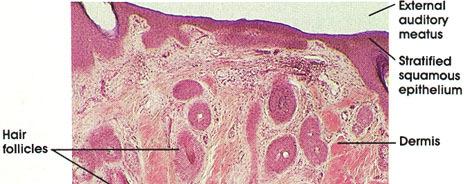
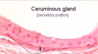
4 External Ear Pinna (Auricle) Irregularly shaped plate o/t elastic cartilage covered by thin skin Meatus acusticus externa The canal that extends f/t pinna into the temporal bone to the external surface o/t tympanic membrane Superficial portion is composed of elastic cartilage, which is continuous with the cartilage o/t pinna. Temporal bone replaces the cartilage as support in the inner 2/3 o/t canal Is covered with skin containing hair follicles, sebaceous glands, ceruminous glands (modified sweat glands)


5 Meatus Acusticus Externus Pinna


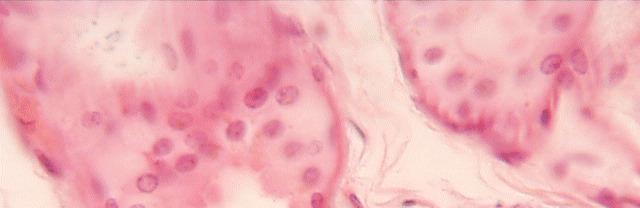
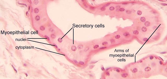
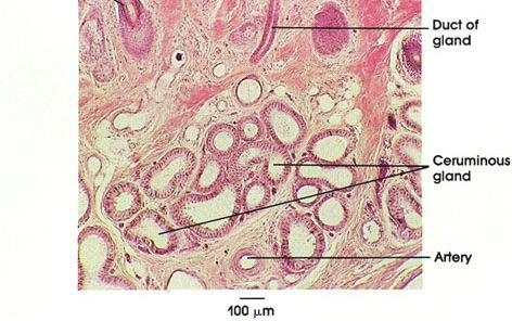
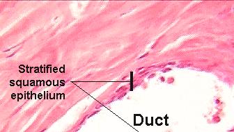
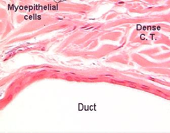
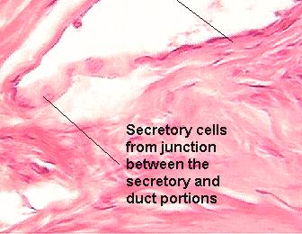
6 Ceruminous glands Modification of apocrine sweat gland Tubular glands; Produce cerumen (earwax) Myoepithelial cells surrounded the secretory portion of ceruminous glands
7
8
9 Middle Ear Tympanic membrane Tympanic cavity Auditory ossicles malleus (hammer) incus (anvil) stapes (stirrup) Auditory (Eustachian) tube Muscle Tensor tympani muscle Stapedius muscle
10 Tympanic membrane External surface is covered by epidermis; Collagen and elastic fibers, fibroblasts interposed btw 2 epithelial layers Internal surface is covered by simple Internal surface is covered by simple squamous to cuboidal epithelium
11 Tympanic cavity Is an air filled space located i/t petrous portion o/t temporal bone Posterior: mastoid air cell Anterior: auditory (eustachian) tube Medial wall: oval window and round window Lateral wall: tympanic membrane Bony ossicles spans the distance btw tympanic membrane and the membrane o/t oval window. Is lined mostly by simple squamous epithelium, and pseudostratified ciliated columnar ep (near auditory tube) Lamina propria Bony wall: Adheres to bony wall and has no glands Overlaying cartilage portions: has many mucous glands whose ducts open into tympanic cavity Muscles M tensor tympani: movement o/t tympanic membrane M stapedius: movement o/t bony ossicles
12 Auditory Ossicles Malleus Is attached to tympanic membrane Incus Interposed btw malleus and stapes Stapes Is attached to the oval window Are articulated in series by synovial joints lined with simple squamous ep.
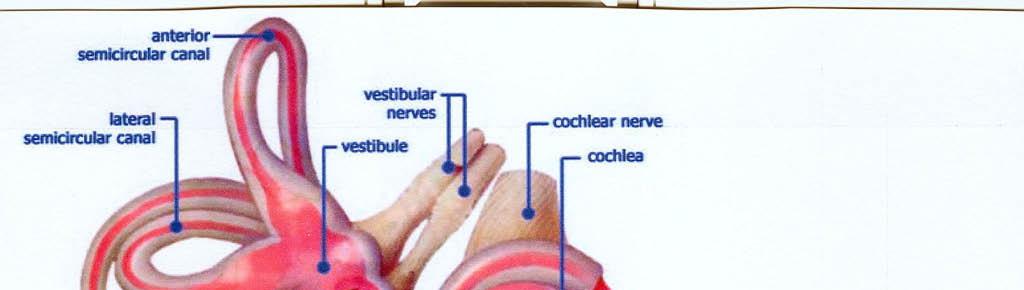
13 Inner Ear Bony labirynth Semicircular canals Vestibule Cochlea Membranous labyrinth Semicircular ducts Saccule and Utricle Cochlear duct and Organ of Corti
14 Bony Labyrinth Semicircular canals Vestibule Cochlea Membranous Labyrinth Semicircular ducts Utricle Saccule Endolymphatic sac Cochlear duct (Scalae media)
15
16
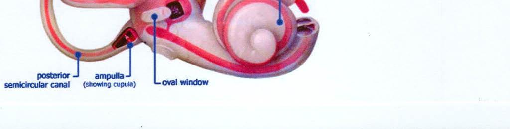
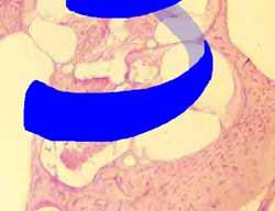
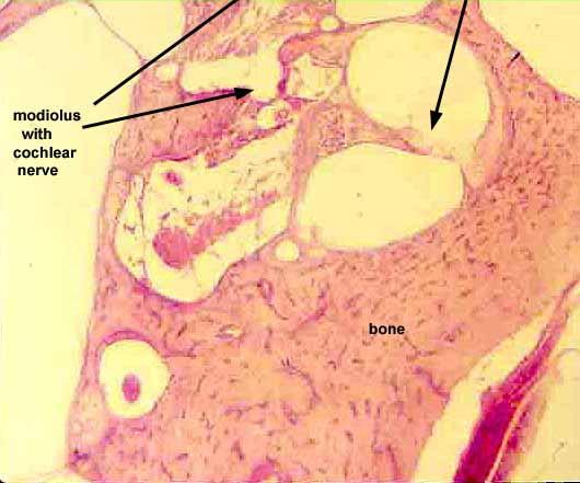
17 Bony labyrinth Is lined w/ endosteum and is separated f/t membranous labyrinth by perilymphatic space that is filled w/ perilymph. Semicircular canals are oriented at 90 o to one another One end of each canal is an enlarged end called ampulla Semicircular duct (membranous labyrinth) Vestibule is the central region o/t bony labyrinth Oval window (fenestra vestibuli) Round window (fenestra cochlea) Utricle and saccule (membranous labyrinth) Cochle arises as a hollow bony spiral that turns upon itself Modiolus: central bony column Osseous spiral lamina
18
19 Membranous labyrinth Is composed of an epithelium from embryonic ectoderm derived f/t embryonic ectoderm, which invades the developing temporal bone and gives rise to saccule and utricle, semicircular duct, and cochlea Circulating throught the entire membranous labyrinth is endolymph Thin strands of connective tissue from endosteum pass through the perilymph to be inserted into membranous labyrinth. The connective tissue strands carry blood vessels.
20
21 Saccule and utricle Ducts o/t saccule and utricle Ductus utriculosaccularis connects saccule and utricule Endolymphatic duct endolymphatic sac Ductus reuniens joins the saccule with cochlear duct The walls are composed of A thin outer vascular layer of connective tissue Simple squamous to low cuboidal epithelium Non receptor epithelium Light cells Dark cells Receptor epithelium (neuroepithelium) maculae
22 Maculae Are located so that they are perpendicular to each other Macula o/t saccule is located in the wall detecting linear vertical acceleration Macula o/t utricle is located in the floor detecting linear horizontal acceleration Are thickened areas of epithelium, 2 3mm in diameter. Are composed of 2 types of neuroepithelial cells (Type I and Type II hair cells) supporting cells Are covered by and embedded in a thick, gelatinous, glycoprotein mass, the otolithic membrane. The surface of this membrane contains otolith (otoconia), small calcium carbonate.
23 Type I hair cells Has a single kinocilium and stereocilias arranged in rows according to length Each stereocilium is anchored in terminal web Bending can occur only in the neck region of stereocilia Are plump cells w/ a rounded base that narrows toward the neck Cytoplasm: Occasional RER A supranuclear golgi complex Numerous small vesicles
24 Type II hair cells Has a single kinocilium and stereocilias arranged in rows according to length Each stereocilium is anchored in terminal web Bending can occur only in the neck region of stereocilia Is more columnar than type I hair cells Cytoplasm A larger golgi complex and more vesicles
25 Supporting cells Are interposed btw hair cells Structure Have a few micovilli Junctional complexes bind these cells to each other Junctional complexes bind these cells to each other and hair cells Exhibit well-developed golgi complex and secretory granules Functions: Maintain the hair cells Contribute to the production of endolymph
26 Innervation from vestibular portion o/t vestibulocochlear nerve Base of type I hair cells are entirely surrounded afferent nerve fiber Type II hair cells exhibit many afferent fibers synapsing on the basal area of the cell. Synaptic ribbons Type II synapses w/ efferent nerves for increasing efficiency of synaptic release
27
28
29 Semicircular ducts Continuation o/t membranous labyrinth from utricle Each of 3 ducts is dilated at its lateral end (near the utricle) ampullae Cristae ampularis: specialized receptor areas Is composed of a ridge whose free surface is covered sensory epithelium Type I and II hair cells Supporting cells Cupula: similar to otolithic membrane but it s cone-shaped and does not contain otoliths
30
31
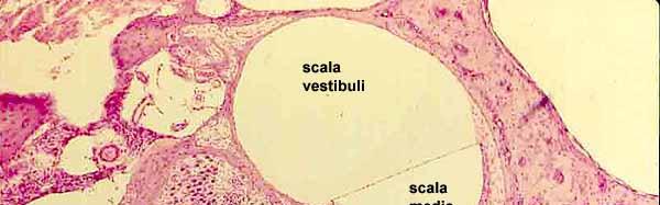
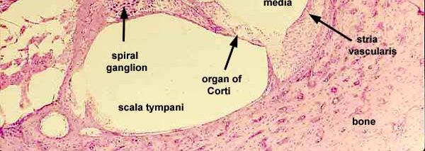
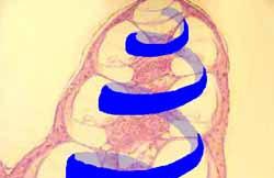
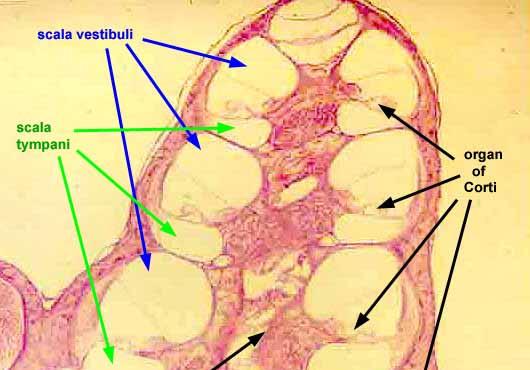
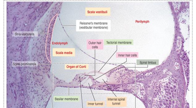
32 Cochlear duct and organ of corti Cochlear duct is a diverticulum of the saccule. Wedge-shaped receptor organ housed in the bony cochlea Surrounded on 2 sides by perilymph but separated from it by 2 membranes The roof is vestibular (Reissner s membrane) whereas the The roof is vestibular (Reissner s membrane) whereas the floor is the basilar membrane The perilymph-filled compartment lying above the vestibular membrane is scala vestibuli whereas the perilymph-filled compartment lying below the basilar membrane is scala tympani. These 2 compartments communicate at the helicotrema, near the apex o/t cochlea.
33
34 Vestibular and Basilar membrane Vestibular membrane Is composed of 2 layers of squamous epithelium separated each other by a basal lamina Inner layer: from scala media Outer layer: from scala vestibuli Tight junction seal both layers of cells Basilar membrane Extends from the spiral lamina at modiolus to the lateral wall Is composed of 2 zones: Zona arcuata Thinner, medial, supports the organ of corti Zona pectinata Fibrous meshwork containing few fibroblast


35 Vestibular Membrane Basilar Membrane
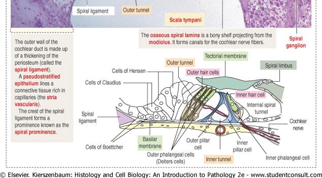
36 Stria Vascularis Is a pseudostratified epithelium located in the lateral wall o/t cochlear duct, extends btw vestibular membrane and spiral prominence contains an intraepithelial plexus of capillaries which are surrounded by basal processes of marginal cells ascending processof basal and intermediate cells Is composed of 3 cell types: Marginal cells Dark staining dense cytoplasm contain mitochondria and small vesicles abudant microvilli on their free surfaces Basal cells Less dense cytoplasm Its cytoplasmic process interdigitate w/ other cells Intermediate cells Less dense cytoplasm
37
38 Capillary


39 MC= marginal cells IC= Intermediate cells BC = basal cells ZO= zonula occludens ZA = zonula adherens D= desmosome CAP= capillary
40 Spiral Prominence Located on the inferior wall of cochlear duct Is a small protuberance that just out from periosteum of cochlea The basal cells and vascular layer covers the prominence. Inferiorly, these cells are reflected into spiral sulcus, where they become cuboidal and continue onto basilar lamina as cells of Claudius, which lies overlie the smaller cells of Böttcher
41 Limbus Located at narrowest portion o/t cochlear duct, where the vestibular and basilar membranes meet Is composed of periosteum Part of limbus projects over the internal spiral sulcus (tunnel) Vestibular lip: the upper portion Tympanic lip: the lower portion Accomodates branches o/t accoustic nerve Interdental cells secrete the tectorial membrane
42 Reissner's membrane (RM), spiral ligament (SL), stria vascularis(sv), spiral prominence (SP),external sulcus(es), basilar membrane (BM), pars arcuata (PA), pars pectinata (PP), Boettcher's cells (B), Claudius' cells (C), organ of Corti (OC), tectorial membrane (TM), inner sulcus cells (IS), spiral limbus (L), habenula perforata (circled), and osseous spiral lamina (OSL).
43 Organ of Corti Specialized receptor organ for hearing Lies on basilar membrane Is composed of neuroepithelial hair cells supporting cells Pillar cells Phalangeal cells Border cells Cells of Hensen Tectorial membrane Inner tunnel
44
45 Supporting Cells Inner and outer pillar cells Tall cells with wide bases and apical ends that are attacehed to basilar membrane The central portions are deflected to form the walls of inner tunnel; apical portion contact each other. Phalangeal cells Outer phalangeal cells Tall columnar cells that are attached to basilar membrane Apical portions are cup-shaped to support the basilar portions of outer hair cells along with efferent and afferent nerve fibers Do not reach the free surface of organ of corti Space of Nuel: a fluid-filled gap around unsupported regions o/t outer hair cells Communicates w/ inner tunnel Inner phalangeal cells Located deep to the innel pillar cells Completely surround the inner hair cells
46 Supporting Cells Border cells Delineate the inner border o/t organ of corti Slender cells that support inner aspects o/t organ of corti Cells of Hensen Define the outer border Located btw outer phalangeal cells and cells of Claudius
47
48 Neuroepithelial (Hair) cells o/t Organ of Corti Inner hair cells Single row of cells supported by inner phalangeal cells Are short and exhibit a centrally nucleus, numerous mitochondria located below terminal web, RER, SER, and small vesicles. Apical surface contains stereocilia arranged in V shape stereocilia: contains microfilamen, cross-linked w/ fimbrin Basal aspects contains microtubules and synapses w/ afferent fibers o/t cochlear portion o/t vestibulocochlear nerve
49 Neuroepithelial (Hair) cells o/t Organ of Corti Outer hair cells Are supported by outer phalangeal cells Are arranged in rows of 3 (or 4) Are elongated cylindrical cells whose Apical portion: 100 stereocilia in W shape Lateral portion Cortical lattice: filaments that support cell Basal portion: Nuclei and mitochondria afferent and efferent fibers synapses
50
51
52 Tectorial Membrane Contains α- and β-tectorin proteins Extends outward over the sensory epithelium from the spiral lamina Is in close contact w/ taller stereocilia of hair cell When the basilar mebrane and organ of corti are displaced, stereocilia hit the tectorial membrane and depolarization of hair cells occurs
53 Thank You
Chapter 17, Part 2! The Special Senses! Hearing and Equilibrium!
 Chapter 17, Part 2! The Special Senses! Hearing and Equilibrium! SECTION 17-5! Equilibrium sensations originate within the inner ear, while hearing involves the detection and interpretation of sound waves!
Chapter 17, Part 2! Chapter 17 Part 2 Special Senses! The Special Senses! Hearing and Equilibrium!
 Chapter 17, Part 2! The Special Senses! Hearing and Equilibrium! SECTION 17-5! Equilibrium sensations originate within the inner ear, while hearing involves the detection and interpretation of sound waves!
Anatomy of the Ear Region. External ear Middle ear Internal ear
 Ear Lecture Objectives Make a list of structures making the external, middle, and internal ear. Discuss the features of the external auditory meatus and tympanic membrane. Describe the shape, position,
AUDITORY APPARATUS. Mr. P Mazengenya. Tel 72204
 AUDITORY APPARATUS Mr. P Mazengenya Tel 72204 Describe the anatomical features of the external ear Describe the tympanic membrane (ear drum) Describe the walls of the middle ear Outline the structures
Ear. Utricle & saccule in the vestibule Connected to each other and to the endolymphatic sac by a utriculosaccular duct
 Rahaf Jreisat *You don t have to go back to the slides. Ear Inner Ear Membranous Labyrinth It is a reflection of bony labyrinth but inside. Membranous labyrinth = set of membranous tubes containing sensory
Structure, Energy Transmission and Function. Gross Anatomy. Structure, Function & Process. External Auditory Meatus or Canal (EAM, EAC) Outer Ear
 Outer Ear") Gross Anatomy Structure, Energy Transmission and Function IE N O ME 1 Structure, Function & Process 4 External Auditory Meatus or Canal (EAM, EAC) Outer third is cartilaginous Inner 2/3 is osseous Junction
Gross Anatomy Structure, Energy Transmission and Function IE N O ME 1 Structure, Function & Process 4 External Auditory Meatus or Canal (EAM, EAC) Outer third is cartilaginous Inner 2/3 is osseous Junction
Chapter 15 Hearing & Equilibrium
 Chapter 15 Hearing & Equilibrium ANATOMY OF THE OUTER EAR EAR PINNA is the outer ear it is thin skin covering elastic cartilage. It directs incoming sound waves to the EXTERNAL AUDITORY CANAL, which is
The cochlea: auditory sense. The cochlea: auditory sense
 Inner ear apparatus 1- Vestibule macula and sacculus sensing acceleration of the head and direction of gravity 2- Semicircular canals mainly for sensing direction of rotation of the head 1 3- cochlea in
Bony and membranous labyrinth. Vestibular system. János Hanics M.D.
 Bony and membranous labyrinth. Vestibular system. János Hanics M.D. The position of the inner ear The labyrinthes of the inner ear - Continuous cavity system in the petrous part of temporal bone - Cavity
Unit VIII Problem 9 Anatomy of The Ear
 Unit VIII Problem 9 Anatomy of The Ear - The ear is an organ with 2 functions: Hearing. Maintenance of equilibrium/balance. - The ear is divided into 3 parts: External ear. Middle ear (which is also known
20 The ear (Vestibulo-acoustic Organs)
") 20 The ear (Vestibulo-acoustic Organs) Median line Sella turcica Tuba auditiva Cavum tympani A. carotis int. Superior border of petrous part Membrana tympani Cochlea N. facialis Meatus acusticus internus
20 The ear (Vestibulo-acoustic Organs) Median line Sella turcica Tuba auditiva Cavum tympani A. carotis int. Superior border of petrous part Membrana tympani Cochlea N. facialis Meatus acusticus internus
For more information about how to cite these materials visit
 Author(s): Matthew Velkey, 2009 License: Unless otherwise noted, this material is made available under the terms of the Creative Commons Attribution Non-commercial Share Alike 3.0 License: http://creativecommons.org/licenses/by-nc-sa/3.0/
The Ear. Dr. Heba Kalbouneh Assistant Professor of Anatomy and Histology
 The Ear Dr. Heba Kalbouneh Assistant Professor of Anatomy and Histology The Ear The ear consists of the external ear; the middle ear (tympanic cavity); and the internal ear (labyrinth), which contains
Hearing. By: Jimmy, Dana, and Karissa
 Hearing By: Jimmy, Dana, and Karissa Anatomy - The ear is divided up into three parts - Sound enters in through the outer ear and passes into the middle where the vibrations are received and sent to the
Taste buds Gustatory cells extend taste hairs through a narrow taste pore
 The Special Senses Objectives Describe the sensory organs of smell, and olfaction. Identify the accessory and internal structures of the eye, and explain their function. Explain how light stimulates the
Dr. Sami Zaqout Faculty of Medicine IUG
 Auricle External Ear External auditory meatus The Ear Middle Ear (Tympanic Cavity) Auditory ossicles Internal Ear (Labyrinth) Bony labyrinth Membranous labyrinth External Ear Auricle External auditory
The Ear The ear consists of : 1-THE EXTERNAL EAR 2-THE MIDDLE EAR, OR TYMPANIC CAVITY 3-THE INTERNAL EAR, OR LABYRINTH 1-THE EXTERNAL EAR.
 The Ear The ear consists of : 1-THE EXTERNAL EAR 2-THE MIDDLE EAR, OR TYMPANIC CAVITY 3-THE INTERNAL EAR, OR LABYRINTH 1-THE EXTERNAL EAR Made of A-AURICLE B-EXTERNAL AUDITORY MEATUS A-AURICLE It consists
Otoconia: Calcium carbonate crystals Gelatinous mass. Cilia. Hair cells. Vestibular nerve. Vestibular ganglion
 VESTIBULAR SYSTEM (Balance/Equilibrium) The vestibular stimulus is provided by Earth s, and. Located in the of the inner ear, in two components: 1. Vestibular sacs - gravity & head direction 2. Semicircular
What is the effect on the hair cell if the stereocilia are bent away from the kinocilium?
 CASE 44 A 53-year-old man presents to his primary care physician with complaints of feeling like the room is spinning, dizziness, decreased hearing, ringing in the ears, and fullness in both ears. He states
Unit VIII Problem 9 Physiology: Hearing
 Unit VIII Problem 9 Physiology: Hearing - We can hear a limited range of frequency between 20 Hz 20,000 Hz (human hearing acuity is between 1000 Hz 4000 Hz). - The ear is divided into 3 parts. Those are:
Auditory System. Barb Rohrer (SEI )
") Auditory System Barb Rohrer (SEI614 2-5086) Sounds arise from mechanical vibration (creating zones of compression and rarefaction; which ripple outwards) Transmitted through gaseous, aqueous or solid medium
Auditory System Barb Rohrer (SEI614 2-5086) Sounds arise from mechanical vibration (creating zones of compression and rarefaction; which ripple outwards) Transmitted through gaseous, aqueous or solid medium
Anatomy and Physiology of Hearing
 Anatomy and Physiology of Hearing The Human Ear Temporal Bone Found on each side of the skull and contains the organs for hearing and balance Divided into four major portions: - squamous - mastoid - tympanic
SENSORY SYSTEM VII THE EAR PART 1
 SENSORY SYSTEM VII THE EAR PART 1 Waves Sound is a compression wave The Ear Ear Outer Ear Pinna Outer ear: - Made up of the pinna and the auditory canal Auditory Canal Outer Ear Pinna (also called the
THE COCHLEA AND AUDITORY PATHWAY
 Dental Neuroanatomy Suzanne S. Stensaas, PhD February 23, 2012 Reading: Waxman, Chapter 16, Review pictures in a Histology book Computer Resources: http://www.cochlea.org/ - Promenade around the Cochlea
ENT 318 Artificial Organs Physiology of Ear
 ENT 318 Artificial Organs Physiology of Ear Lecturer: Ahmad Nasrul Norali The Ear The Ear Components of hearing mechanism - Outer Ear - Middle Ear - Inner Ear - Central Auditory Nervous System Major Divisions
Activity 1: Anatomy of the Eye and Ear Lab
 Activity 1: Anatomy of the Eye and Ear Lab 1. Launch the view! Launch Human Anatomy Atlas. Navigate to Quizzes/Lab Activities, find the Eye and Ear Lab section. Launch Augmented Reality mode and scan the
THE COCHLEA AND AUDITORY PATHWAY
 Dental Neuroanatomy Suzanne S. Stensaas, PhD April 14, 2010 Reading: Waxman, Chapter 16, Review pictures in a Histology book Computer Resources: http://www.cochlea.org/ - Promenade around the Cochlea HyperBrain
A&P 1. Ear, Hearing & Equilibrium Lab. Basic Concepts. These notes follow Carl s Talk at the beginning of lab
 A&P 1 Ear, Hearing & Equilibrium Lab Basic Concepts These notes follow Carl s Talk at the beginning of lab In this "Lab Exercise Guide", we will be looking at the basics of hearing and equilibrium. NOTE:
Auditory Physiology Richard M. Costanzo, Ph.D.
 Auditory Physiology Richard M. Costanzo, Ph.D. OBJECTIVES After studying the material of this lecture, the student should be able to: 1. Describe the morphology and function of the following structures:
Anatomy of the ear: Lymphatics
 Anatomy of the ear: 1. External ear which consist of auricle and external auditory canal. The auricle has a framework of cartilage except the lobule, the skin is closely adherent to perichonderium at the
The Senses. Chapter 10 7/8/11. Introduction
 Chapter 10 The Senses Introduction A. Sensory receptors detect changes in the environment and stimulate neurons to send nerve impulses to the brain. B. A sensation is formed based on the sensory input.
The ear: some applied basic science
 Chapter 1 The ear: some applied basic science The pinna The external ear or pinna is composed of cartilage with closely adherent perichondrium and skin. It is developed from six tubercles of the first
Printable version - Hearing - OpenLearn - The Open University
 Skip to content Accessibility Sign in Contact Search the OU The Open University Study at the OU Research at the OU OU Community About the OU Hearing Printable page generated Saturday, 12 November 2011,
MECHANISM OF HEARING
 MECHANISM OF HEARING Sound: Sound is a vibration that propagates as an audible wave of pressure, through a transmission medium such as gas, liquid or solid. Sound is produced from alternate compression
VESTIBULAR SYSTEM ANATOMY AND PHYSIOLOGY. Professor.Dr. M.K.Rajasekar MS., DLO.,
 VESTIBULAR SYSTEM ANATOMY AND PHYSIOLOGY Professor.Dr. M.K.Rajasekar MS., DLO., Life is hard for those who don t have a VOR During a walk I found too much motion in my visual picture of the surroundings
THE EAR AND HEARING Be sure you have read and understand Chapter 16 before beginning this lab. INTRODUCTION: hair cells outer ear tympanic membrane
 BIOLOGY 211: HUMAN ANATOMY & PHYSIOLOGY ****************************************************************************************************** THE EAR AND HEARING ******************************************************************************************************
Auditory System Feedback
 Feedback Auditory System Feedback Using all or a portion of the information from the output of a system to regulate or control the processes or inputs in order to modify the output. Central control of
Chapter 3: Anatomy and physiology of the sensory auditory mechanism
 Chapter 3: Anatomy and physiology of the sensory auditory mechanism Objectives (1) Anatomy of the inner ear Functions of the cochlear and vestibular systems Three compartments within the cochlea and membranes
Tissues. tissue = many cells w/ same structure and function. cell shape aids its function tissue shape aids its function
 Tissues tissue = many cells w/ same structure and function cell shape aids its function tissue shape aids its function Histology = study of tissues 4 types of tissues Epithelial coverings contact openings
Kingdom of Bahrain Arabian Gulf University College of Medicine and Medical Sciences Year 6 ENT SMC Otitis Media (Dr.
 Kingdom of Bahrain Arabian Gulf University College of Medicine and Medical Sciences Year 6 ENT SMC Otitis Media (Dr. Jalal Almarzooq) - Anatomy of the ear: The ear is divided into 3 parts: External ear.
For this lab you will use parts of Exercise #18 in your Wise lab manual. Please be sure to read those sections before coming to lab
 Bio 322 Human Anatomy Objectives for the laboratory exercise The Eye and Ear Required reading before beginning this lab: Saladin, KS: Human Anatomy 5 th ed (2017) Chapter 17 For this lab you will use parts
Auditory and vestibular system
 Auditory and vestibular system Sensory organs on the inner ear inner ear: audition (exteroceptor) and vestibular apparatus (proprioceptor) bony and membranous labyrinths within the temporal bone (os temporale)
Vestibular Function and Anatomy. UTMB Grand Rounds April 14, 2004 Gordon Shields, MD Arun Gadre, MD
 Vestibular Function and Anatomy UTMB Grand Rounds April 14, 2004 Gordon Shields, MD Arun Gadre, MD System of balance Membranous and bony labyrinth embedded in petrous bone 5 distinct end organs 3 semicircular
The white of the eye and the part that maintains its shape is know n as the:
 Scrub In The white of the eye and the part that maintains its shape is know n as the: a. Cornea b. Pupil c. Retina d. Sclera The structure that is found in the ear and contains the organ of hearing is
A&P 1. Ear, Hearing & Equilibrium Lab. Basic Concepts. Pre-lab Exercises
 A&P 1 Ear, Hearing & Equilibrium Lab Basic Concepts Pre-lab Exercises In this "Lab Exercise Guide", we will be looking at the basics of hearing and equilibrium. NOTE: these notes do not follow the order
Introduction. Senses our perception of what is out there 2 groups. General senses Special senses
 Introduction Senses our perception of what is out there 2 groups General senses Special senses Central Processing and Adaptation Adaptation the loss of sensitivity after continuous stimulation Tonic receptors
Νευροφυσιολογία και Αισθήσεις
 Biomedical Imaging & Applied Optics University of Cyprus Νευροφυσιολογία και Αισθήσεις Διάλεξη 11 Ακουστικό και Αιθουσιαίο Σύστημα (Auditory and Vestibular Systems) Introduction Sensory Systems Sense of
SPECIAL SENSES: THE AUDITORY SYSTEM
 SPECIAL SENSES: THE AUDITORY SYSTEM REVISION OF PHYSICS: WAVES A wave is an oscillation of power, sound waves have two main characteristics: amplitude, which is the maximum displacement or the power of
The Nervous System: General and Special Senses Pearson Education, Inc.
 18 The Nervous System: General and Special Senses Introduction Sensory information arrives at the CNS Information is picked up by sensory receptors Sensory receptors are the interface between the nervous
Specialists in the Field Ophthalmology:
 The Eye and Ear Outline of class lecture After studying this chapter you should be able to: 1. List and describe the accessory structures of the eye. 2. Describe the structures and functions of the fibrous
Sensory system. Dr. Carmen E. Rexach Anatomy 35 Mt San Antonio College
 Sensory system Dr. Carmen E. Rexach Anatomy 35 Mt San Antonio College Sensory receptors Detect stimuli Classified by structure Origin Distribution Modality Structural Classification naked nerve endings
to vibrate the fluid. The ossicles amplify the pressure. The surface area of the oval window is
 Page 1 of 6 Question 1: How is the conduction of sound to the cochlea facilitated by the ossicles of the middle ear? Answer: Sound waves traveling through air move the tympanic membrane, which, in turn,
Chapter Fourteen. The Hearing Mechanism. 1. Introduction.
 Chapter Fourteen The Hearing Mechanism 1. Introduction. 2. Hearing. 3. The Ear. 4. The External Ear. 5. The Inner Ear. 6. Frequency Discrimination. 7. The Organ of Corti. 8. Tests and Exrecises. 9. References.
o A cushion of fat surrounds most of the eye
 Name Period SPECIAL SENSES The Senses of touch o Temperature o Pressure o Pain o Smell o Taste o Sight o Hearing o Equilibrium The Eye and Vision are in the eyes has over a o Most of the eye is enclosed
Vestibular physiology
 Vestibular physiology 2017 Utricle A flat epithelium: horizontal in the upright head Utricle Hair cells: no axons hair cells Utricle Hair cells synapse onto 8th nerve afferents. 8th nerve afferents Hair
Chapter 18. The Nervous System. General and Special Senses. Lecture Presentation by Steven Bassett Southeast Community College
 Chapter 18 The Nervous System General and Special Senses Lecture Presentation by Steven Bassett Southeast Community College Introduction Every plasmalemma functions as a receptor for the cell Plasmalemma
Special Senses. Accessory Structures of the Eye. The Eye and Vision. Accessory Structures of the Eye. Accessory Structures of the Eye
 8 PART A Special Senses PowerPoint Lecture Slide Presentation by Jerry L. Cook, Sam Houston University ESSENTIALS OF HUMAN ANATOMY & PHYSIOLOGY EIGHTH EDITION ELAINE N. MARIEB The Senses General senses
9.01 Introduction to Neuroscience Fall 2007
 MIT OpenCourseWare http://ocw.mit.edu 9.01 Introduction to Neuroscience Fall 2007 For information about citing these materials or our Terms of Use, visit: http://ocw.mit.edu/terms. 9.01 Recitation (R02)
The Special Senses. Smell, taste, vision, hearing and equilibrium Housed in complex sensory organs
 The Special Senses Smell, taste, vision, hearing and equilibrium Housed in complex sensory organs Chemical Senses Interaction of molecules with receptor cells Olfaction (smell) and gustation (taste) Both
Intro to Audition & Hearing
 Intro to Audition & Hearing Lecture 16 Chapter 9, part II Jonathan Pillow Sensation & Perception (PSY 345 / NEU 325) Fall 2017 1 Sine wave: one of the simplest kinds of sounds: sound for which pressure
Required Slide. Session Objectives
 Auditory Physiology Required Slide Session Objectives Auditory System: At the end of this session, students will be able to: 1. Characterize the range of normal human hearing. 2. Understand the components
Chapter 1: Applied Anatomy, Physiology and Embryology of the Ear. Anatomy and Physiology. The Outer Ear. The Pinna. The External Ear Canal
 Chapter 1: Applied Anatomy, Physiology and Embryology of the Ear The ear contains two specialized sensory organs, the cochlea and the vestibular apparatus, enclosed in the extremely hard protective casing
Chapter 15 Lecture Outline
 Chapter 15 Lecture Outline See separate PowerPoint slides for all figures and tables preinserted into PowerPoint without notes. Copyright 2016 McGraw-Hill Education. Permission required for reproduction
Deafness and hearing impairment
 Auditory Physiology Deafness and hearing impairment About one in every 10 Americans has some degree of hearing loss. The great majority develop hearing loss as they age. Hearing impairment in very early
Chapter 10. The Senses
 Chapter 10 The Senses 1 Introduction A. Sensory receptors detect changes in the environment and stimulate neurons to send nerve impulses to the brain. B. A sensation is formed based on the sensory input.
Overview of Sensory Receptors
 Sensory Systems Chapter 45 Overview of Sensory Receptors Sensory receptors provide information from our internal and external environments that is crucial for survival and success -Exteroceptors sense
Olfaction. The Special Senses. The Special Senses. Olfaction. The Ethmoid. Olfactory Receptors. The five special senses are
 The Special Senses The Special Senses Chapter 14 in Open Stax Chapter 17 in Martini The five special senses are Olfaction Gustation Equilibrium Hearing Vision Olfaction Olfaction The sense of smell, or
Essential questions. What are the structures of the sensory system? 3.03 Remember the structures of the sensory system 2
 Essential questions What are the structures of the sensory system? 3.03 Remember the structures of the sensory system 2 The Senses Eyes Sight Ears Hearing Nose Smell Tongue Taste Skin Touch 3.03 Remember
SPECIAL SENSES PART I: OLFACTION & GUSTATION
 SPECIAL SENSES PART I: OLFACTION & GUSTATION 5 Special Senses Olfaction Gustation Vision Equilibrium Hearing Olfactory Nerves Extend through cribriform plate into nasal cavity on both sides of nasal septum
Auditory system. Organ of hearing
 Auditory system Organ of hearing Auditory systems is an engineering masterpiece Size of a pea Detects vibrations as small as the size of an atom Responds 1000 times faster than the visual photoreceptors
يراهظلا( يئلاطلا جيسنلا
 Epithelium النسيج الطالئي )الظهاري( Features of Epithelium Epithelium occurs in the body as a sheet of cells that covers a body surface, lines a cavity, or forms a gland. Coverings, linings, glands. Derived
Chapter 7. Audition, the Body Senses, and the Chemical Senses. Copyright Allyn & Bacon 2004
 Chapter 7 Audition, the Body Senses, and the Chemical Senses This multimedia product and its contents are protected under copyright law. The following are prohibited by law: any public performance or display,
The Organs of Special Senses
 8 The Organs of Special Senses Special senses are those other than touch, pain, temperature, and proprioception. Vision, hearing, and equilibrium are the special senses discussed in this chapter. The Eye
Tissues. tissue = many cells w/ same structure and function. cell shape aids function tissue shape aids function. Histology = study of tissues
 Tissues tissue = many cells w/ same structure and function cell shape aids function tissue shape aids function Histology = study of tissues 4 types of tissues Epithelial coverings contact openings Connective
Before we talk about the auditory system we will talk about the sound and waves
 The Auditory System PHYSIO: #3 DR.LOAI ZAGOUL 24/3/2014 Refer to the slides for some photos. Before we talk about the auditory system we will talk about the sound and waves All waves have basic characteristics:
Gathering information the sensory systems; Vision
 Visual System Gathering information the sensory systems; Vision The retina is the light-sensitive receptor layer at the back of the eye. - Light passes through the cornea, the aqueous chamber, the lens,
Chapter 18 Senses SENSORY RECEPTION 10/21/2011. Sensory Receptors and Sensations. Sensory Receptors and Sensations. Sensory Receptors and Sensations
 SENSORY RECEPTION Chapter 18 Senses s convert stimulus energy to action potentials s 1. Are specialized cells, or 2. Specialized endings that detect stimuli All stimuli are forms of energy s in eyes detect
Cochlear anatomy, function and pathology I. Professor Dave Furness Keele University
 Cochlear anatomy, function and pathology I Professor Dave Furness Keele University d.n.furness@keele.ac.uk Aims and objectives of these lectures Introduction to gross anatomy of the cochlea Focus (1) on
Sound. Audition. Physics of Sound. Properties of sound. Perception of sound works the same way as light.
 Sound Audition Perception of sound works the same way as light. Have receptors to convert a physical stimulus to action potentials Action potentials are organized in brain structures You apply some meaning
Audition. Sound. Physics of Sound. Perception of sound works the same way as light.
 Audition Sound Perception of sound works the same way as light. Have receptors to convert a physical stimulus to action potentials Action potentials are organized in brain structures You apply some meaning
Epithelial Tissue. Functions include: 1. Protection 4. Absorption 2. Secretion 5. Filtration 3. Sensory reception
 Tissues There are 4 primary tissue types in the human body: 1. Epithelial (covering/lining) 2. Connective (support) 3. Muscle (movement) 4. Nervous (control) Epithelium Epithelial Tissue Covers the surface
4. Which letter in figure 9.1 points to the fovea centralis? Ans: b
 Chapter 9: The Sensory System 1. Proprioceptors are involved in the sense of A) pain. B) temperature. C) pressure. D) movement of limbs. 2. Which are chemoreceptors? A) taste B) olfactory C) proprioceptors
Histology = the study of tissues. Tissue = a complex of cells that have a common function
 { EPITHELIAL TISSUE Histology = the study of tissues Tissue = a complex of cells that have a common function The Four Primary Tissue Types: Epithelium (epithelial tissue) covers body surfaces, lines body
Special Senses. Mechanoreception Electroreception Chemoreception Others
 Special Senses Mechanoreception Electroreception Chemoreception Others Recall our receptor types Chemically regulated: Respond to particular chemicals Voltage regulated: respond to changing membrane potential
HEARING. Structure and Function
 HEARING Structure and Function Rory Attwood MBChB,FRCS Division of Otorhinolaryngology Faculty of Health Sciences Tygerberg Campus, University of Stellenbosch Analyse Function of auditory system Discriminate
BIOH111. o Cell Biology Module o Tissue Module o Integumentary system o Skeletal system o Muscle system o Nervous system o Endocrine system
 BIOH111 o Cell Biology Module o Tissue Module o Integumentary system o Skeletal system o Muscle system o Nervous system o Endocrine system Endeavour College of Natural Health endeavour.edu.au 1 Textbook
Presentation On SENSATION. Prof- Mrs.Kuldeep Kaur
 Presentation On SENSATION Prof- Mrs.Kuldeep Kaur INTRODUCTION:- Sensation is a specialty area within Psychology that works at understanding how are senses work and how we perceive stimuli in the environment.
The olfactory epithelium is located at the roof of the nasal cavity. Nasal conchae cause turbulance of incoming air
 Special Senses I. Olfaction II. Gustation A. Anatomy and general info The olfactory epithelium is located at the roof of the nasal cavity Nasal conchae cause turbulance of incoming air Olfactory glands
Histology of the Eye
 Histology of the Eye Objectives By the end of this lecture, the student should be able to describe: The general structure of the eye. The microscopic structure of:»cornea.»retina. EYE BULB Three coats
Lecture Overview. Chapter 4 Epithelial Tissues Lecture 9. Introduction to Tissues. Epithelial Tissues. Glandular Epithelium
 Visual Anatomy & Physiology First Edition Martini & Ober Chapter 4 Lecture 9 Lecture Overview Introduction to Tissues Location General characteristics Functions Classification Glandular Epithelium 2 Where
The Physiology of the Senses Lecture 10 - Balance
 The Physiology of the Senses Lecture 10 - Balance www.tutis.ca/senses/ Contents Objectives... 1 The sense of balance originates from the labyrinth... 2 The auditory and vestibular systems have a common
Equilibrium (Balance) *
 *") OpenStax-CNX module: m63740 1 Equilibrium (Balance) * Steven Telleen Based on Sensory Perception by OpenStax This work is produced by OpenStax-CNX and licensed under the Creative Commons Attribution License
OpenStax-CNX module: m63740 1 Equilibrium (Balance) * Steven Telleen Based on Sensory Perception by OpenStax This work is produced by OpenStax-CNX and licensed under the Creative Commons Attribution License
Lecture Overview. Marieb s Human Anatomy and Physiology. Chapter 4 Tissues: The Living Fabric Epithelial Tissues Lecture 9. Introduction to Tissues
 Marieb s Human Anatomy and Physiology Marieb Hoehn Chapter 4 Tissues: The Living Fabric Epithelial Tissues Lecture 9 Lecture Overview Introduction to Tissues Epithelial Tissues Location General characteristics
Biology 218 Human Anatomy
 Chapter 22 Adapted form Tortora 10 th ed. LECTURE OUTLINE A. Special Senses 1. Olfaction: Sense of Smell (p. 672) i. The olfactory epithelium is located in the superior portion of the nasal cavity and
Receptors / physiology
 Hearing: physiology Receptors / physiology Energy transduction First goal of a sensory/perceptual system? Transduce environmental energy into neural energy (or energy that can be interpreted by perceptual
College of Medicine Dept. of Medical physics Physics of ear and hearing /CH
 College of Medicine Dept. of Medical physics Physics of ear and hearing /CH 13 2017-2018 ***************************************************************** o Introduction : The ear is the organ that detects
Sound and the auditory system
 978--521-68889-5 - Auditory Perception: An Analysis and Synthesis, Third Edition 1 Sound and the auditory system This chapter provides a brief introduction to the physical nature of sound, the manner in
Hearing: the function of the outer, the middle and inner ear. Hearing tests. The auditory pathways
 Hearing: the function of the outer, the middle and inner ear. Hearing tests. The auditory pathways Dr. Gabriella Kékesi 74. Hearing: the function of the outer, the middle and inner ear. Hearing tests.
Dr. Abeer.c.Yousif. Histology -2 nd stage. What is histology?
 What is histology? Histology is the science of microscopic anatomy of cells and tissues, in Greek language Histo= tissue and logos = study and it's tightly bounded to molecular biology, physiology, immunology
Surgical Anatomy Ear and Eye. Presenters: Dr. Jim Hurrell and Dr. Dennis McCurnin
 Surgical Anatomy Ear and Eye Presenters: Dr. Jim Hurrell and Dr. Dennis McCurnin A Warm Welcome from My Faculty TEAM and Me!!! 2 The Pledge of Allegiance 3 The Senses 4 Hearing 3 Layers of Ear EXTERNAL