高雄醫學大學 口腔醫學院 口腔病理影像科 牙科 X 光影像判讀 教學範例
|
|
|
- Eric Norris
- 5 years ago
- Views:
Transcription
1 高雄醫學大學 口腔醫學院 口腔病理影像科 牙科 X 光影像判讀 教學範例
2 Content Image No. 001 Dentigerous cyst over left upper embedded canine 頁 1 Image No 頁 2 Radicular cyst over upper anterior teeth Image No 頁 3 (1) Calcifying odontogenic cyst over right maxilla (2) Osteosclerosis over left lower retromolar area Image No 頁 4 Dentigerous cyst with adenomatoid odontogenic tumor over left maxilla Image No 頁 5 (1) Odontogenic keratocyst, left mandibular body, OR (2) Dentigerous cyst, left lower third molar, OR (3) Unicystic amelblastoma, left mandibular body Image No 頁 6 (1) Odontogenic keratocyst, mandibular body, ramus, OR (2) Ameloblastoma, OR (3) Odontogenic myxoma Image No 頁 8 Cementoblastoma associated with the apical portion of tooth 36 Image No 頁 9 (1) Dentigerous cyst over embedded horizontal tooth, left mandible body, OR (2) Ameloblastoma, OR (3) Residual cyst with embedded horizontal tooth Image No 頁 10 Sialolithiasis, anterior mouth floor 陳玉昆老師
3 Image No 頁 11 (1) Radicular cyst (periapical granuloma) in the apex of the tooth 13 and 14 and tooth 44 and 45 (2) Stafne s cyst in the lower left cortical border near mandibular angle 陳玉昆老師
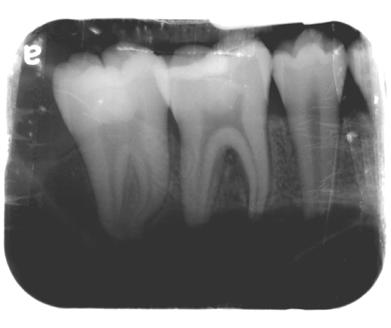
 There is a well-defined unilocular round shaped circumcoronal radiolucence with a scalloped margin")
4 Image No. 001 Dentigerous cyst over left upper embedded canine Main X-ray findings (Panoramic radiography) There is a well-defined unilocular round shaped circumcoronal radiolucence with a scalloped margin over the embedded tooth 13 extending from distal side of root of tooth 21 right to distal root of tooth 16, and from alveolar crest between tooth 12, 14 up to apex of embedded tooth 13, measuring approximately 3 2.5cm in diameter. Tooth 11, 12, 14, 15 are displaced. 陳玉昆老師頁 1
 There is a well-defined, unilocular half-round shaped")

5 Image No. 002 Radicular cyst over upper anterior teeth Main X-ray findings (Panoramic radiography) There is a well-defined, unilocular half-round shaped radiolucency with a thin corticated margin, extending from tooth 13 apex to tooth 23 apex and from one-third of intermaxillary suture up to palatal surface, measuring approximately cm in diameter. Main X-ray findings (Periapical radiography) Root canal partially enlargement over tooth 11 and 21, especially upper portion, and filling material at lingual side of tooth 11 and 21 is observed. Loss of lamina dura over apex of tooth 11, 21, 22. 陳玉昆老師頁 2

6 Image No. 003 (1) Calcifying odontogenic cyst over right maxilla (2) Osteosclerosis over left lower retromolar area Main X-ray findings (Panoramic radiography) There are three major abnormalities. One is a well-defined ovoid, irregular border unilocular radiolucency over right upper maxilla with a focus of calcification and part of corticated margin around root apex of tooth 11, 12. This lesion extends from the distal side of tooth 15 to the apex of tooth 21 and from middle third of root of tooth 13 superior to the lower part of right sinus, which may be extended to the nasal cavity, measuring approximately 3 4cm in diameter. It contains a focus of calcification causing root resorption and loss of lamina dura of tooth 15, 13, 12, 11. There are also two well-defined round shaped radiopaque shadows over left posterior mandible. One is near the apex of distal root of tooth 36, measured about cm, whereas another is just beneath the left oblique ridge, measured cm. The degree of radiopacity is equivalent to the cortical bone of mandible border and, it does not cause any effect to the surrounding tissue. 陳玉昆老師頁 3
 There is a well-defined unilocular round shaped circumcoronal radiolucence with a")

7 Image No. 004 Dentigerous cyst with adenomatoid odontogenic tumor over left maxilla Main X-ray findings (Panoramic radiography) There is a well-defined unilocular round shaped circumcoronal radiolucence with a corticated margin over the impacted tooth 23 extending from apex of tooth 21 left to distal aspect of tooth 26 and from apex of retained tooth 63 up to cementoenamel junction of impacted tooth 23, measuring approximately 3 2.5cm in diameter. Root of impacted 23 is well-developed. Main X-ray findings (Occlusal radiography) There is a well-defined unilocular round shaped circumcoronal radiolucence with a corticated margin over the impacted tooth 23 extending from palatal aspect of tooth 15 left to apex of 25 and from apex of tooth 63 posterior to cementoenamel junction of impacted tooth 23, measuring approximately 3 4cm in diameter. 陳玉昆老師頁 4
 Odontogenic keratocyst, left mandibular body, OR (2) Dentigerous cyst, left lower third molar, OR (3) Unicystic amelblastoma, left mandibular body Main X-ray findings (Panoramic radiography)")
8 Image No. 005 (1) Odontogenic keratocyst, left mandibular body, OR (2) Dentigerous cyst, left lower third molar, OR (3) Unicystic amelblastoma, left mandibular body Main X-ray findings (Panoramic radiography) There is a well-defined unilocular scalloped shaped circumcoronal radiolucence with a smooth and thin corticated margin over the submerged well-developed tooth 38 extending from left retromolar area down to mandibular body and from distal root of tooth 37 up to half of left ramus area, measuring approximately 3.5 3cm in diameter. Root of tooth 37 is displaced toward mesial side. Tooth 37 is tilted to distal side by the lesion. Left mandibular canal cannot be identified with the lesion. Thinning of left lower cortical border as well as external oblique ridge is noted. 陳玉昆老師頁 5
 Odontogenic keratocyst, mandibular body, ramus, OR (2) Ameloblastoma, OR (3) Odontogenic myxoma Main X-ray findings Panoramic radiography There is a well-defined multilocular, soap bubble")
9 Image No. 006 (1) Odontogenic keratocyst, mandibular body, ramus, OR (2) Ameloblastoma, OR (3) Odontogenic myxoma Main X-ray findings Panoramic radiography There is a well-defined multilocular, soap bubble appearance radiolucent lesion with a corticated margin over right posterior mandibular area. The lesion extends from distal aspect of tooth 43 to condylar neck and from inferior border of mandible up to tooth 44, 45, 46, 47 root apex and sigmoid notch, measuring approximately cm in diameter. This mass involve mandibular canal and cause bony expansion of right mandibular body. Periapical radiography This mass involves adjacent tooth 45, 46, and the resorption of the root of the teeth is noted, but it doesn t cause movement of the teeth. Occlusal radiography There is a well-defined multilocular, soap bubble appearance radiolucent lesion with a corticated margin over the buccal side of the tooth, 33, 34, 35, 36, 37, and it causes bony expansion of right mandibular body, but there is no movement of the teeth. P-A radiography The lesion extends from lateral aspect of the lower teeth to the mesial aspect of mandibular border and from mandibular border up to condylar neck, measuring approximately cm in diameter. 陳玉昆老師頁 6
10 陳玉昆老師頁 7

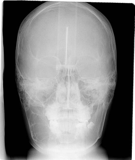
11 Image No. 007 Cementoblastoma associated with the apical portion of tooth 36 Main X-ray findings There is a well-defined, homogenous, bulbous, radiopaque mass associated with the distal buccal root apical portion of the mandibular left first molar. A radiolucence rim appears to be seen surrounding the mass. The mass extends from the distal buccal root apex of tooth 36 to the superior border of mandibular canal, and from the furcation area of tooth 36 to the mesial buccal root apex of tooth 37, measuring approximately 1 1.4cm in diameter. Root resorption of mandibular first molar has occurred. Genioplasty has been performed and the chin was fixed with wires. 陳玉昆老師頁 8
 Dentigerous cyst over embedded horizontal tooth, left mandible body, OR (2) Ameloblastoma, OR (3) Residual cyst with embedded horizontal tooth Main X-ray findings Panoramic")

12 Image No. 008 (1) Dentigerous cyst over embedded horizontal tooth, left mandible body, OR (2) Ameloblastoma, OR (3) Residual cyst with embedded horizontal tooth Main X-ray findings Panoramic radiography There is a well-defined unilocular ovoid shaped radiolucence with a corticated margin over an embedded horizontal tooth over the left mandible body, extending from root apex of tooth 35 and 38 to cortical bone border displaced mandibular canal and from tooth 35 to tooth 38, approximately 5 3cm in maximum diameter. Periapical radiography It showed a well-defined unilocular radiolucence involved the horizontal embedded tooth, with distance from the inferior alveolar bone surface to the superior border of radiolucence about 0.8cm. 陳玉昆老師頁 9

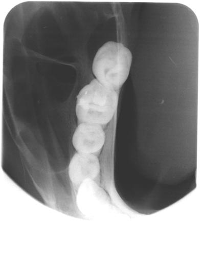
13 Image No. 009 Sialolithiasis, anterior mouth floor Main X-ray findings This is an occlusal projection of lower anterior mouth floor. There is a well-defined oval-shaped radiopaque lesion near the mildline of mouth floor. The size is measured about 3 5mm in diameter. The density is homogenous and slightly lower than tooth structure. 陳玉昆老師頁 10
 Radicular cyst (periapical granuloma) in the apex of the tooth 13 and 14 and tooth 44 and 45 (2) Stafne s cyst in the lower left cortical border near mandibular angle Main X-ray findings")
14 Image No. 010 (1) Radicular cyst (periapical granuloma) in the apex of the tooth 13 and 14 and tooth 44 and 45 (2) Stafne s cyst in the lower left cortical border near mandibular angle Main X-ray findings There are two solitary well-defined non-corticated unilocular radiolucent shadows; one is located in the mesial surface of the apex of the tooth 14, measured about cm in maximum diameter; another is located in the mesial surface of the middle root of the tooth 13, measured about cm in maximum diameter. Lamina dura of tooth 12 is disappeared due to the radiolucency of the tooth 13. In the apex of the tooth 44 and 45 reveal two radiolucent shadows and there is a well-defined unilocular radiolucent image with corticated border in the lower left cortical border near mandibular angle beneath the left inferior alveolar canal, measured about cm in maximum diameter. The adjacent structures reveal normal. 陳玉昆老師頁 11
Disclosure. Educational Objectives. Terminology. Odontogenic Cysts. Terminology
 Disclosure Lisa J. Koenig BChD, DDS, MS Professor & Program Director, Oral Medicine and Oral Radiology Marquette University School of Dentistry Consultant to Soredex for the Scanora 3D and 3Dx Author/Editor
Dr.Sepideh Falah-kooshki
 Dr.Sepideh Falah-kooshki MAXILLA Premaxillary/median palatal suture (radiolucent). Incisive fossa and foramen (radiolucent). Nasal passages (radiolucent). Nasal septum (radiopaque). Anterior nasal spine
PACIFIC JOURNAL OF MEDICAL SCIENCES ISSN:
 PACIFIC JOURNAL OF MEDICAL SCIENCES {Formerly: Medical Sciences Bulletin} ISSN: 2072 1625 Pac. J. Med. Sci. (PJMS) www.pacjmedsci.com. Email: pacjmedsci@gmail.com. ADENOMATOID ODONTOGENIC TUMOR WITH RARE
IMAGING OF CYSTS OF THE JAWS July 2002 N. Serman
 IMAGING OF CYSTS OF THE JAWS July 2002 N. Serman This is an area where radiology plays an important role in assisting with the diagnosis, determining the size of the lesion and the relationship to adjacent
Pre-reading - radiolucencies
 Pre-reading - radiolucencies Multiple radiolucencies o Suggests a systemic cause o Most likely: cherubism or KCOT s of nevoid basal cell carcinoma syndrome o Sometimes: florid osseous dysplasia (if limited
Clinical details: Details of scan: CONE BEAM CT REPORT: Name: H. B. Gender: Reason for referral: Referred by:
 Name: H. B. Gender: Male DOB: 11/12/1950 Age: 64 Date taken: 16/11/2015 Date reported: 19/11/2015 Clinical details: Reason for referral: Referred by: Investigate symptoms related to left TMJ. Reconstructed
Large Dentigerous Cyst
 Volume 16.2.1 Feb 2016 This Lecture Series qualifies for 0.5 Informal CPD Learning Hours Large Dentigerous Cyst By Dr Hassem Geha A 55 year-old male presented with a painless swelling in the right mandible.
Radiology. & supporting structures. Lec. 14 Common diseases of teeth Dr. Areej
 Radiology Lec. 14 Common diseases of teeth Dr. Areej & supporting structures A radiograph is only one part of the diagnostic process. Usually one does NOT make a diagnosis solely from a radiograph. A diagnosis
A Case Report of Odontogenic Keratocyst in Anterior Mandibule Position
 A Case Report of Odontogenic Keratocyst in Anterior Mandibule Position Malihe Moeini 1, Seyed Ehsan Anvar 2, Rasool Barzegari Bafghi 3* 1.Resident of Oral and Maxillofacial Radiology, Faculty of Dentistry,
Common/Important Radiolucencies. B. Most Common Location Apex of permanent first molar, rare in primary teeth.
 Cincinnati Dental Association Breakfast at Tiffany s: The Jewels and Gems of Oral Pathology November 17, 2010 John A. Svirsky, DDS, MEd Virginia Commonwealth University 804-828-0547 FAX: 804-828-6234 EMAIL:
Name:XXX Sex: 男 Age:17 y/o Marital status: 未婚 Occupation: 學生
 General data Name:XXX Sex: 男 Age:17 y/o Marital status: 未婚 Occupation: 學生 Chief complaint A swelling mass over upper left arch for 3~4 months Present illness This 17 y/o male found an swelling over upper
Upper arch. 1Prosthodontics. Dr.Bassam Ali Al-Turaihi. Basic anatomy & & landmark of denture & mouth
 1Prosthodontics Lecture 2 Dr.Bassam Ali Al-Turaihi Basic anatomy & & landmark of denture & mouth Upper arch Palatine process of maxilla: it form the anterior three quarter of the hard palate. Horizontal
Maxilla and mandible benign lesions: Radiologic Findings and Differential Diagnosis in CT
 Maxilla and mandible benign lesions: Radiologic Findings and Differential Diagnosis in CT Poster No.: C-0964 Congress: ECR 2012 Type: Scientific Exhibit Authors: N. Lopez 1, E. Marcos Naranjo 2, M. D.
TRAUMATIC BONE CYST OF IDIOPATHIC ORIGIN? A REPORT OF TWO CASES
 Traumatic Bone Cyst Kumar S. et al 183 CASE REPORT TRAUMATIC BONE CYST OF IDIOPATHIC ORIGIN? A REPORT OF TWO CASES Kumar Satish 1, S. Padmashree 1, Jayalekshmy Rema 1 ABSTRACT BACKGROUND: Traumatic bone
DENTAL RADIOGRAPH INTERPRETATION
 DENTAL RADIOGRAPH INTERPRETATION Brook A. Niemiec, DVM Diplomate, American Veterinary Dental College Fellow, Academy of Veterinary Dentistry www.vetdentaltraning.com www.vetdentalrad.com Interpreting dental
INTERPRETATION RADIOGRAPHIC INTERPRETATION. Law of Symmetry. We will be reviewing: 7/30/16
 RADIOGRAPHIC INTERPRETATION Pam Wood, CDA, RDH, M.Ed, CAGS Community College of Rhode Island pwood@ccri.edu INTERPRETATION the ability to read what is revealed on a dental radiograph any dental professional
Pericoronal radiolucency associated with incomplete crown
 Imaging Science in entistry 2013; 43: 295-301 http://dx.doi.org/10.5624/isd.2013.43.4.295 Pericoronal radiolucency associated with incomplete crown Kyung-Soo Nah 1, * 1 epartment of Oral and Maxillofacial
The clinical appearance and diagnosis of odontogenic cysts. SE Arc-Állcsont-Szájsebészeti és Fogászati Klinika BUDAPEST
 The clinical appearance and diagnosis of odontogenic cysts SE Arc-Állcsont-Szájsebészeti és Fogászati Klinika BUDAPEST DEFINITION A cyst is a sac with walls of connective tissue, lined by epithelium, containing
Fundamental & Preventive Curvatures of Teeth and Tooth Development. Lecture Three Chapter 15 Continued; Chapter 6 (parts) Dr. Margaret L.
 Dr. Margaret L.") Fundamental & Preventive Curvatures of Teeth and Tooth Development Lecture Three Chapter 15 Continued; Chapter 6 (parts) Dr. Margaret L. Dennis Proximal contact areas Contact areas are on the mesial and
Fundamental & Preventive Curvatures of Teeth and Tooth Development Lecture Three Chapter 15 Continued; Chapter 6 (parts) Dr. Margaret L. Dennis Proximal contact areas Contact areas are on the mesial and
RADIOGRAPHIC INTERPRETATION Differential Diagnosis
 RADIOGRAPHIC INTERPRETATION Differential Diagnosis MODULE 1: The Introduction. Chief complaint Demographics Age Sex Race Historical findings Physical findings Clinical Radiographic Location Maxilla/mandible
Case Report An Extrafollicular Adenomatoid Odontogenic Tumor Mimicking a Periapical Cyst
 Hindawi Case Reports in Radiology Volume 2018, Article ID 6987050, 5 pages https://doi.org/10.1155/2018/6987050 Case Report An Extrafollicular Adenomatoid Odontogenic Tumor Mimicking a Periapical Cyst
Medical NBDE-II. Dental Board Exams Part I.
 Medical NBDE-II Dental Board Exams Part I http://killexams.com/exam-detail/nbde-ii Question: 149 Anatomically, the term "clinical root" can be defined as which of the following: A. The space in the tooth
Dental Morphology and Vocabulary
 Dental Morphology and Vocabulary Palate Palate Palate 1 2 Hard Palate Rugae Hard Palate Palate Palate Soft Palate Palate Palate Soft Palate 4 Palate Hard Palate Soft Palate Maxillary Arch (Maxilla) (Uppers)
Normal Radiographic Anatomy Maxillary Central Area
 VARIA Normal Radiographic Anatomy Maxillary Central Area Carmen Elena Georgescu 1, Gabriela Tãnase 2, Augustin Mihai 3 Bucharest, Romania Summary The radiopgraphic recognition of disease requires knowledge
Study of maxillary and mandibular cystic lesions
 Study of maxillary and mandibular cystic lesions Poster No.: C-1428 Congress: ECR 2013 Type: Educational Exhibit Authors: M. L. Rozas Rodríguez, M. E. Banegas Illescas, M. Y. Torres Sousa, R. M. Fernández
Periodontal Disease. Radiology of Periodontal Disease. Periodontal Disease. The Role of Radiology in Assessment of Periodontal Disease
 Radiology of Periodontal Disease Steven R. Singer, DDS srs2@columbia.edu 212.305.5674 Periodontal Disease! Includes several disorders of the periodontium! Gingivitis! Marginal Periodontitis! Localized
Advanced radiographic techniques for the detection of lesions in bone
 Endodontic Topics 2004, 7, 52 72 Printed in Denmark. All rights reserved Copyright r Blackwell Munksgaard ENDODONTIC TOPICS 2004 Advanced radiographic techniques for the detection of lesions in bone ELISABETTA
Elevators. elevators:- There are three major components of the elevator are:-
 Elevators Elevators:- Are exo-levers, instrument designed to elevate or luxate the teeth or roots from their bony socket in close or surgical method of extraction to force a tooth or root along the line
Dental panoramic tomography: An approach for the general radiologist
 Pictorial Essay Australasian Radiology (2006) 50, 526 533 Dental panoramic tomography: An approach for the general radiologist R Boeddinghaus and A Whyte Perth Radiological Clinic, Perth, Western Australia,
Differential Diagnosis of Radiolucent Lesions of the Jaws
 Differential Diagnosis of Radiolucent Lesions of the Jaws Multilocular Multilocular Radiolucencies Odontogenic Keratocyst Botryoid Odontogenic Cyst Glandular odontogenic Cyst Invasive Ameloblastoma Central
[ 06-10] Dr. B. Siva Reddy, Dr. B. Ajay Reginald, Dr. D. Sireesha, Dr. Meda Samatha India Abstract: Keywords ARTICLE 20/07/ /09/2018
![[ 06-10] Dr. B. Siva Reddy, Dr. B. Ajay Reginald, Dr. D. Sireesha, Dr. Meda Samatha India Abstract: Keywords ARTICLE 20/07/ /09/2018](/thumbs/87/95905171.jpg "[ 06-10] Dr. B. Siva Reddy, Dr. B. Ajay Reginald, Dr. D. Sireesha, Dr. Meda Samatha India Abstract: Keywords ARTICLE 20/07/ /09/2018") Dentigerous Cyst Associated with an Impacted Mesiodens: A Rare Case Report with Review of Literature [PP: 06-10] Dr. B. Siva Reddy, Dr. B. Ajay Reginald, Dr. D. Sireesha, Dr. Meda Samatha, Department of
Dentigerous Cyst Associated with an Impacted Mesiodens: A Rare Case Report with Review of Literature [PP: 06-10] Dr. B. Siva Reddy, Dr. B. Ajay Reginald, Dr. D. Sireesha, Dr. Meda Samatha, Department of
Arrangement of the artificial teeth:
 Lecture Prosthodontic Dr. Osama Arrangement of the artificial teeth: It s the placement of the teeth on a denture with definite objective in mind or it s the setting of teeth on temporary bases. Rules
Permanent 2 nd Maxillary Molars
 Permanent 2 nd Maxillary Molars In comparison to the first max molar First molars appears in the oral cavity at the age of 6 years old.. While 2 nd molar 3 rd molar Max. 2 nd molar have long roots (sometimes
Unusual transmigration of canines report of two cases in a family
 ISSN: Electronic version: 1984-5685 RSBO. 2014 Jan-Mar;11(1):88-92 Case Report Article Unusual transmigration of canines report of two cases in a family Sulabha A. Narsapur 1 Sameer Choudhari 2 Shrishal
Oral Surgery. Basic Techniques of Dental Local Anesthesia. A variety of techniques used in administration and deposition of local anesthesia:
 Oral Surgery Lecture: 9 Dr. Saif Saadedeen Basic Techniques of Dental Local Anesthesia A variety of techniques used in administration and deposition of local anesthesia: 1. Topical anesthesia 2. Infiltration
Malignant Lesions Steven R. Singer, DDS
 Definitions Malignant Lesions Steven R. Singer, DDS srs2@columbia.edu 212.305.5674 Malignancies are uncontrolled growths of tissue Primary tumors represent de novo tumors in their initial site Metastatic
Keratocystic Odontogenic Tumor : What radiologist needs to know?
 Keratocystic Odontogenic Tumor : What radiologist needs to know? Poster No.: C-0444 Congress: ECR 2014 Type: Authors: Keywords: DOI: Educational Exhibit K. El Karzazi, J. M. Villanueva Rincón, R. Corrales,
Inter-radicular Radiolucencies
 Inter-radicular Radiolucencies Differential Diagnosis Laterally Displaced Radicular Cyst Accessory canals Root fracture Lateral Periodontal Cyst Botryoid variant Odontogenic Keratocyst Incisive Canal Cyst
Ossifying fibromas of the jaw bone: 20 cases
 (2010) 39, 57 63 2010 The British Institute of Radiology http://dmfr.birjournals.org CASE REPORT Ossifying fibromas of the jaw bone: 20 cases Y Liu 1, M You 1, H Wang*,1, Z Yang 1, J Miao 1, K Shimizutani
Normal Radiographic Anatomy Maxillary Lateral Area. Carmen Elena Georgescu1, Gabriela Tãnase 2, Augustin Mihai 3. Objectives.
 Normal Radiographic Anatomy Maxillary Lateral Area Carmen Elena Georgescu1, Gabriela Tãnase 2, Augustin Mihai 3 Bucharest, Romania Summary Intraoral examinations are the backbone of dental radiography.
Dentigerous cyst associated with an impacted mesiodens: report of 2 cases
 Imaging Science in Dentistry 2012; 42 : 255-60 http://dx.doi.org/10.5624/isd.2012.42.4.255 Dentigerous cyst associated with an impacted mesiodens: report of 2 cases Neha Khambete, Rahul Kumar*, Mukund
Problem diagnoses. Current issues in Anatomic pathology. Problem Diagnoses in Tumors of the Oral Cavity 5/29/2009
 Current issues in Anatomic pathology Problem Diagnoses in Tumors of the Oral Cavity Richard Jordan DDS PhD FRCPath Professor of Oral Pathology & Pathology Director, UCSF Oral Pathology Diagnostic Laboratory
Radiographic features of cysts and benign tumors of the jaws. Cyst. Effects on adjacent structures. Types. Odontogenic Cysts. Non-Odontogenic cysts
 Radiographic features of cysts and benign tumors of the jaws Cyst A Cyst is a benign pathologic cavity filled with fluid, lined by epithelium, and surrounded by a connective tissue wall Steven R. Singer,
Role of MDCT and VR reconstructions in the diagnosis and characterization of maxillary cystic lesions.
 Role of MDCT and VR reconstructions in the diagnosis and characterization of maxillary cystic lesions. Poster No.: C-0704 Congress: ECR 2011 Type: Scientific Exhibit Authors: M. Coronella 1, P. V. Foti
Reconstruction of large mandibular defects
 Immediate Reconstruction of a Large Mandibular Defect of Locally Invasive Benign Lesions (A New Method) Gholamreza Shirani, OMFS, DDS, MS,* Mahnaz Arshad, DDS, 1 Farnoush Mohammadi, OMFS, DDS, MS* Tehran,
An unusual site of Adenomatoid Odontogenic Tumor: A rare case report
 J. Int Oral Health 2010 Case Report All right reserved An unusual site of Adenomatoid Odontogenic Tumor: A rare case report Sapna Panjwani*, Anjana Bagewadi**, Vaishali Keluskar*** *Post Graduate Student
Intraosseous Transmigration of Impacted Canines: Report of Five Cases Sulabha AN, Sachin Deshpande, Sameer C
 International Journal of Oral & Maxillofacial Pathology. 2012;3(3):56-60 ISSN 2231 2250 Available online at http://www.journalgateway.com or www.ijomp.org Case Report Intraosseous Transmigration of Impacted
Vascular. Extravasated blood. Melanocytic. Tattoo. Epidermolysis bullosa. Lichen planus. Pemphigoid Pemphigus Lupus. Candidosis. Surface Epithelial
 Oral Soft Tissue Pathology Epithelial Thickening (white) Combination Erythema migrans Epithelial atrophy (red) Surface Lesions Clinical Impression Enlargements Surface Debris Pigmented Vesicular Ulcerated
Cone Beam Computed Tomography Findings in Calcifying Cystic Odontogenic Tumor Associated with Odontome: A Case Report
 Phulambrikar T., et al. Dent Shiraz Univ Med Sci., December 2015; 16(4): 374-379. Case Report Cone Beam Computed Tomography Findings in Calcifying Cystic Odontogenic Tumor Associated with Odontome: A Case
Digital Imaging from a new perspective
 TREATMENT CENTRES HANDPIECES HYGIENE SYSTEMS X-RAY SYSTEMS CEREC TREATMENT CENTRES HANDPIECES HYGIENE SYSTEMS X-RAY SYSTEMS CEREC SIRONA CREATING AND MAINTAINING VALUE. You are right to expect a great
AMERICAN JOURNAL OF BIOLOGICAL AND PHARMACEUTICAL RESEARCH
 AMERICAN JOURNAL OF BIOLOGICAL AND PHARMACEUTICAL RESEARCH e-issn - 2348-2184 Print ISSN - 2348-2176 Journal homepage: www.mcmed.us/journal/ajbpr SOLID AND MULTICYSTIC FOLLICULAR AMELOBLASTOMA - A CASE
ANATOMY OF THE PERIODONTIUM. Dr. Fatin Awartani
 ANATOMY OF THE PERIODONTIUM Part II Cementum and Alveolar bone Associate Professor Periodontal division King Saud university Cementum Calcified mesenchymal tissue that forms the outer covering of the anatomic
This lecture is sponsored by a grant from the Delta Dental of Iowa Foundation IDA Annual Conference Guest Lecture Series.
 This lecture is sponsored by a grant from the Delta Dental of Iowa Foundation IDA Annual Conference Guest Lecture Series. Radiology: Back to the Classroom! Juan F. Yepes DDS, MD, MPH, MS, DrPH Associate
Oral cavity landmarks
 By: Dr. Ahmed Rabah Oral cavity landmarks The knowledge of oral anatomy and physiology will help the operator and provides enough landmarks to act as positive guide during denture construction. This subject
Odontogenic Cysts - An Overview
 Namita V Nayyer Michaelina Macluskey and William Keys Odontogenic Cysts - An Overview Abstract: This article aims to discuss the clinical features, radiological assessment, histopathology and management
Periapical Radiography
 Periapical Radiography BARBARA E. DIXON B.D.S., M.Sc., D.P.D.S. Main Indications Detection of Apical infection/inflammation Assessment of the periodontal status After trauma Assessment of Unerupted teeth
IMPACTED CANINES. Unfortunately, this important tooth is the second most common tooth to be impacted after third molars
 IMPACTED CANINES After we talked about impacted third molars, today we ll discuss about maxillary impacted canines in upper dental arch, how to manage these cases as a dental surgeon. You will study about
Incidental finding of dentigerous cyst - a case report
 Case Report Incidental finding of dentigerous cyst - a case report Pulivarthi Sushma 1, Sowbhagya M.B 2, Balaji P 3, Mahesh Kumar T.S 4 1 Postgraduate, 2 Reader, 3 Professor and Head of department, 4 Senior
Dentalelle Tutoring - Faulty Radiographs
 Dentalelle Tutoring - Faulty Radiographs Errors in improperly exposing or processing dental films can produce undesirable dental radiographs of nondiagnostic quality. These are known as faulty radiographs.
Comprehensive AOCMF Classification System. Cornelius CP, Kunz C, Prein J, Audigé L. Mandibular fractures Level-2 system (cases 1 to 18)
") Comprehensive AOCMF Classification System Cornelius CP, Kunz C, Prein J, Audigé L Mandibular fractures Level-2 system (cases 1 to 18) Case 1: Body fracture traversing anterior transition zone Imaging:
Comprehensive AOCMF Classification System Cornelius CP, Kunz C, Prein J, Audigé L Mandibular fractures Level-2 system (cases 1 to 18) Case 1: Body fracture traversing anterior transition zone Imaging:
Case Report Basal Cell Ameloblastoma of Mandible: A Rare Case Report with Review
 Case Reports in Dentistry Volume 2013, Article ID 187820, 4 pages http://dx.doi.org/10.1155/2013/187820 Case Report Basal Cell Ameloblastoma of Mandible: A Rare Case Report with Review Hemant Shakya, 1
4/2/17. Panoramic Radiography: Normal Variants and Pathology. Composite of in-focused and blurred images. It s a type of Tomogram.
 Fundamentals Panoramic Radiography: Normal Variants and Pathology It s a type of Tomogram Jimmie L. Harper D.D.S., M.S. Cincinnati Oral and Maxillofacial Surgery, Inc. Volunteer Assistant Professor, Division
Annals and Essences of Dentistry
 doi: 10.5958/0976-156X.2014.00005.7 LARGE RADICULAR CYST OF THE POSTERIOR MAXILLA A CASE REPORT 1 Manoj Meena 2 Nigel R Figueiredo 3 Ajit D Dinkar 4 Ena Mathur 5 Arum Khatkar 6 Sonam Malik 1 Senior lecturer
An Expansile Large Odontogenic Keratocyst Maxilla: A Case Report.
 RESEARCH AND REVIEWS: JOURNAL OF DENTAL SCIENCES An Expansile Large Odontogenic Keratocyst Maxilla: A Case Report. Nasib Chand Khabra 1, Ish Pandhi 1 *, Kiran DN 2, Sunil Alipuria 1, Bhawna Gulati 1, and
You know you would like to stop swearing at the computer after each shot. Troubleshooting oral radiography
 You know you would like to stop swearing at the computer after each shot Troubleshooting oral radiography Goals of oral radiology Achieve diagnostic images of the teeth and surrounding bone. Images should
AMELOBLASTIC FIBROMA: A RARE CASE REPORT
 Case Report International Journal of Dental and Health Sciences Volume 04, Issue 03 AMELOBLASTIC FIBROMA: A RARE CASE REPORT Namratha Patil 1 1.Sr lecturer, dept of oral medicine and radiology, KAHES VK
DEVELOPING ANALOGUE/SUBTITUTE FOR THE MANDIBULAR DENTURE BEARING AREA. Dr Muhammad Rizwan Memon FCPS Assistant Professor
 DEVELOPING ANALOGUE/SUBTITUTE FOR THE MANDIBULAR DENTURE BEARING AREA Dr Muhammad Rizwan Memon FCPS Assistant Professor Crest of Residual Ridge Buccal Shelf Shape of supporting structure Mylohyoid Ridge
Arun V Subramaniam et al. / International Journal of Biopharmaceutics. 2014; 5(3): International Journal of Biopharmaceutics
: International Journal of Biopharmaceutics") 225 e- ISSN 0976-1047 Print ISSN 2229-7499 International Journal of Biopharmaceutics Journal homepage: www.ijbonline.com IJB ODONTOGENIC KERATOCYSTS: VARIOUS RADIOGRAPHIC APPEARANCES Arun Subramaniam*
225 e- ISSN 0976-1047 Print ISSN 2229-7499 International Journal of Biopharmaceutics Journal homepage: www.ijbonline.com IJB ODONTOGENIC KERATOCYSTS: VARIOUS RADIOGRAPHIC APPEARANCES Arun Subramaniam*
CENTRAL GIANT CELL GRANULOMA PRESENTING AS UNILOCULAR RADIOLUCENCY IN POSTERIOR MANDIBLE A CASE REPORT
 IJCRR Section: Healthcare Sci. Journal Impact Factor 4.016 Case Report CENTRAL GIANT CELL GRANULOMA PRESENTING AS UNILOCULAR RADIOLUCENCY IN POSTERIOR MANDIBLE A CASE REPORT S. Aruleena Shaminey 1, G.
Arrangement of posterior artificial teeth Standardized parameters Curve of Wilson Curve of Spee
 . Arrangement of posterior artificial teeth Posterior teeth are set up in tight centric occlusion. The mandibular teeth are set in the wax occlusion rim over the residual ridge in their ideal buccolingual
Advanced Probing Techniques
 Module 21 Advanced Probing Techniques MODULE OVERVIEW The clinical periodontal assessment is one of the most important functions performed by dental hygienists. This module begins with a review of the
 www.oralradiologists.com CONE BEAM CT REPORT CASE XXXX Patient information Patient Name: - Referring Doctor: - Patient DOB: - Scan Date: [Start date] Reason for Exam: Maxillary facial pain Doctor Notes:
CASE REPORT. CBCT-Assisted Treatment of the Failing Long Span Bridge with Staged and Immediate Load Implant Restoration
 Computer Aided Implantology Academy Newsletter - Newsletter 20 - July 2009 CASE REPORT CBCT-Assisted Treatment of the Failing Long Span Bridge with Staged and Immediate Load Implant Restoration Case Report
Radiology in Periodontics
 10.5005/jp-journals-10011-1334 Geetha Vijay, Vijay Raghavan REVIEW ARTICLE Geetha Vijay, Vijay Raghavan ABSTRACT The aim is to give a brief account of how to image the periodontal tissues and to describe
Techniques of local anesthesia in the mandible
 Techniques of local anesthesia in the mandible The technique of choice for anesthesia of the mandible is the block injection and this is attributed to the absence of the advantages which are present in
Autologous Bone Augmentation in Combination with an Ameloblastoma in the Maxillary Region- A Case Report?
 Archives of Clinical and Medical Case Reports doi: 10.26502/acmcr.96550023 Volume 2, Issue 2 Case Report Autologous Bone Augmentation in Combination with an Ameloblastoma in the Maxillary Region- A Case
Calcifying Cystic Odontogenic Tumor A Case Report and Review on Diverse Presentation of the Tumor
 : J Dentistry and Otolaryngology Volume 14 Issue 3 Version 1.0 Year 2014 Type: Double Blind Peer Reviewed International Research Journal Publisher: Global Journals Inc. (USA) Online ISSN: 2249-4618 & Print
Only 40% of the Story
 X-RAY, X-RAY, READ ALL ABOUT IT! The Use and Utility of Dental Radiographs in Practice Lisa Fink, DVM, DAVDC Dentistry & Oral Surgery Service October 4, 2015 Only 40% of the Story Radiographs of teeth
Lec. 11 & 12 Dr. Ali H. Murad Dental pulp 1- Coronal pulp
 Lec. 11 & 12 Dr. Ali H. Murad Dental pulp Is the soft connective tissue located in the central portion of each tooth. All pulps have similar morphologic characteristic, such as a soft, gelatinous consistency
Course Description 343 DDS- Clinical Oral and Maxillofacial Radiology II ( )
") King Saud University College of Dentistry Dept. of Oral Medicine & Diagnostic Sciences Division of Oral & Maxillofacial Radiology Course Description 343 DDS- Clinical Oral and Maxillofacial Radiology II
King Saud University College of Dentistry Dept. of Oral Medicine & Diagnostic Sciences Division of Oral & Maxillofacial Radiology Course Description 343 DDS- Clinical Oral and Maxillofacial Radiology II
IN THE NAME OF GOD. Dr.kheirandish DDS,MSC Oral and maxillofacial pathology
 IN THE NAME OF GOD Dr.kheirandish DDS,MSC Oral and maxillofacial pathology ODONTOGENIC CYSTS AND TUMORS Chapter 15 I. DENTIGEROUS CYST II. III. IV. ERUPTION CYST ODONTOGENIC KERATOCYST Orthokeratinized
European Veterinary Dental College
 European Veterinary Dental College EVDC Training Support Document Preparation of Radiograph Sets (Cat and Dog) Document version : evdc-tsd-radiograph_positioning_(dog_and_cat)-20120121.docx page 1 of 13
10/23/2014. features to image interpretation what to look for and what it means. interpretation vs. diagnosis. science or art? image investigation
 features to image interpretation what to look for and what it means interpretation vs. diagnosis ERNEST LAM, DMD, MSc, PhD, FRCD(C) PROFESSOR AND THE DR. LLOYD & MRS. KAY CHAPMAN CHAIR IN CLINICAL SCIENCES
Prosthetic Options in Implant Dentistry. Hakimeh Siadat, DDS, MSc Associate Professor
 Prosthetic Options in Dentistry Hakimeh Siadat, DDS, MSc Associate Professor Dental Research Center, Department of Prosthodontics & Dental s Faculty of Dentistry, Tehran University of Medical Sciences
Case Report An Unusual Case of Tooth in the Floor of the Orbit: The Libyan Experience
 Case Reports in Dentistry Volume 2012, Article ID 954789, 5 pages doi:10.1155/2012/954789 Case Report An Unusual Case of Tooth in the Floor of the Orbit: The Libyan Experience Y. Naresh Shetty, Irfan Adil
Infratemporal fossa: Tikrit University college of Dentistry Dr.Ban I.S. head & neck Anatomy 2 nd y.
 Infratemporal fossa: This is a space lying beneath the base of the skull between the lateral wall of the pharynx and the ramus of the mandible. It is also referred to as the parapharyngeal or lateral pharyngeal
Squamous cell carcinoma of the maxillary sinus mimicking periodontitis
 Squamous cell carcinoma of the maxillary sinus mimicking periodontitis 저자저널명발행기관 NDSL URL Na, Ji Yeon ; Kang, Joo Hyun ; Choi, Seong-Ho ; Jeong, Ho-Gul ; Han, Sang-Sun 大韓齒科醫師協會誌 = The journal of the Korean
Jaws: Cysts and Odontogenic Neoplasms
 Topic 10: Jaw Cysts General Features of Jaw Cysts Sources of Epithelium in Cysts Radiographic Features of Jaw Cysts Microscopic Features of Jaw Cysts Treatment and Prognosis of Jaw Cysts Classification
INFLAMMATORY DENTIGEROUS CYST OR INFLAMMATORY CYSTIC LESIONS OF MIXED DENTITION?: A REPORT OF THREE CASES
 Case Report International Journal of Dental and Health Sciences Volume 03, Issue 03 INFLAMMATORY DENTIGEROUS CYST OR INFLAMMATORY CYSTIC LESIONS OF MIXED DENTITION?: A REPORT OF THREE CASES Pritam K Mankapure
Case Study. Case # 1 Author: Dr. Suheil Boutros (USA) 2013 Zimmer Dental, Inc. All rights reserved. 6557, Rev. 03/13.
 2013 Zimmer Dental, Inc. All rights reserved. 6557, Rev. 03/13.") Placement of a Zimmer Trabecular Metal Dental Implant with Simultaneous Ridge Augmentation and Immediate Non-Functional Loading Following Tooth Extraction and Orthodontic Treatment for Implant Site Development
Placement of a Zimmer Trabecular Metal Dental Implant with Simultaneous Ridge Augmentation and Immediate Non-Functional Loading Following Tooth Extraction and Orthodontic Treatment for Implant Site Development
6. Timing for orthodontic force
 6. Timing for orthodontic force Orthodontic force is generally less than 300gm, so early mechanical stability is enough for immediate orthodontic force. There is no actually difference in success rate
Surgical Uprighting Is a Successful Procedure for Management of Impacted Mandibular Second Molars
 DENTOALVEOLAR SURGERY Surgical Uprighting Is a Successful Procedure for Management of Impacted Mandibular Second Molars Bonnie L. Padwa, DMD, MD,* Rushil R. Dang, BDS, DMD,y and Cory M. Resnick, DMD, MDz
Sample Case #1. Disclaimer
 ABO Sample Cases Disclaimer Sample Case #1 The following sample questions and answers were composed and vetted by a panel of experts in orthodontics and are intended to provide an example of the types
Conservative management of a dentigerous cyst associated with eruption of teeth in a 7-year-old girl: a case report
 SE REPORT https://doi.org/10.5125/jkaoms.2017.43.s1.s1 pissn 2234-7550 eissn 2234-5930 onservative management of a dentigerous cyst associated with eruption of teeth in a 7-year-old girl: a case report
Origin of Odontogenic Cysts & Tumors
 Origin of Odontogenic Cysts & Tumors Odontogenic Apparatus Origin of Odontogenic Cysts & Tumors Odontogenic Apparatus Remnants of dental lamina Reduced enamel epithelium Odontogenic rests Basal cell layer
Course Description 343 DDS- Clinical Oral and Maxillofacial Radiology II ( )
") King Saud University College of Dentistry Dept. of Oral Medicine & Diagnostic Sciences Division of Oral & Maxillofacial Radiology Course Description 343 DDS- Clinical Oral and Maxillofacial Radiology II
King Saud University College of Dentistry Dept. of Oral Medicine & Diagnostic Sciences Division of Oral & Maxillofacial Radiology Course Description 343 DDS- Clinical Oral and Maxillofacial Radiology II
Relation between the complex diagnostics and the complications of the wisdom-tooth surgery
 Relation between the complex diagnostics and the complications of the wisdom-tooth surgery Changes of the "surgical profile" at the Dental Surgery Department of the Oral & Maxillofacial Surgery Clinic,
Bones Ethmoid bone Inferior nasal concha Lacrimal bone Maxilla Nasal bone Palatine bone Vomer Zygomatic bone Mandible
 splanchnocranium - Consists of part of skull that is derived from branchial arches - The facial bones are the bones of the anterior and lower human skull Bones Ethmoid bone Inferior nasal concha Lacrimal
Intrusion of Incisors to Facilitate Restoration: The Impact on the Periodontium
 Note: This is a sample Eoster. Your EPoster does not need to use the same format style. For example your title slide does not need to have the title of your EPoster in a box surrounded with a pink border.
Technique. Simpo PDF Merge and Split Unregistered Version -
 extraoral radiographic examinations both ~.~ x-ray source and image receptor (film or electronic sensor) are placed outside the patient's mouth. This chapter describes the most common extraoral radiographic