VistaVox S 3D from Dürr Dental
|
|
|
- Dale Malone
- 6 years ago
- Views:
Transcription


1 VistaVox S 3D from Dürr Dental 3D and 2D X-ray images with exceptional image quality COMPRESSED AIR SUCTION IMAGING DENTAL CARE HYGIENE

2 Taking diagnostics to the next level VistaVox S combines diagnostic reliability with efficiency and lower radiation doses Key features: Ideal 3D imaging volume matched to the shape of the jaw Excellent 2D and 3D images with just one unit Simplest face-to-face positioning with one positioning line for 3D images and three positioning lines for 2D images Reduced radiation dose thanks to the anatomically adapted volume Dose reduced by up to 62% in SQ mode Reduced volume height for image acquisition in children preventing unnecessary exposure of the eye Metal artifact reduction in 3D and 2D image 7" touch-display for intuitive operation Modern, ergonomic image processing software VistaSoft 02 VistaVox S

3 Ideal imaging volume, easy positioning, high image quality: the new VistaVox S represents a milestone in the field of 3D X-ray systems. Thanks to its unique technology, the 3D images generated with this system cover everything you need for reliable diagnosis, well-founded treatment decisions and convincing patient communication. In addition, the S-Pan technology of the VistaVox S also enables pinpoint accurate OPG image acquisitions in the best Dürr Dental quality. All of these things make VistaVox S not only a highly efficient solution for dental practices, but also a safe investment. 3D diagnostics: the key indications With VistaVox S 3D images you can increase diagnostic reliability and enable accurate treatment planning. The key indications at a glance: Tooth Hyperplasia or dysplasia development Retained or impacted teeth Fractures Root or jaw fractures Implant Augmentation/bone formation technology For planning In the event of complications Endodontics Periapical examinations Complex anomalies of the root canal system Fractured root canal instruments within the root canal Foreign bodies Suspected perforation, in particular pin perforation Localisation of foreign bodies in the mouth and jaw area Salivary stones Localisation of salivary stones Pathological Maxillary sinus area changes Jawbone Cysts, tumours, osteonecrosis 03

4 See what you want to see VistaVox S offers an ideal 3D volume that is adapted to the shape of the jaw Almost universal fit At Ø 100 x 85 mm, the jaw-shaped field of view of the VistaVox S is visibly larger than the most commonly used volume of Ø 80 x 80 mm. The advantage: thanks to this changed volume shape, VistaVox S also completely covers the region of the rear molars an essential requirement for diagnostics, e.g. for an impacted wisdom tooth. By rendering the image in the shape of the jaw, the entire dental region is reliably covered, as the VistaVox S volume displays the diagnostically relevant details of a Ø 130 mm volume. SQ mode The SQ mode (Standard Quality mode) offers a further option for reducing the radiation dose. In this setting, the dose is reduced by 62% in comparison to HQ mode (Highest Quality mode). SQ mode can be used e.g. for implant planning, determination of the apical bone supply, for investigation of the sinuses or for the localisation of impacted or excess teeth. SQ mode can be used in all programs. The special feature of VistaVox S is that its imaging volume is based on the human anatomy, representing precisely the region you need covered for diagnostics in the dental region. The ideal jaw-shaped volume is achieved with the aid of a special curved path with 540 rotation, for which the VistaVox S requires just 18 seconds. In conjunction with a tightly collimated fan beam and the highly sensitive Csl sensor, this path allows a particularly low dose of radiation to be used. The VistaVox S reconstruction algorithms allow the 3D volume to be displayed in the shortest possible time. 04 VistaVox S

5 In order to visualise the FoV of VistaVox S (blue) in the axial view, the conventional standard volume of Ø 80 x 80 mm (red) has also been marked for comparison purposes. The jaw-shaped volume measuring Ø 100 x 85 mm even displays the diagnostic range of a Ø 130 volume. H x D for all patients 8,5 = 100 % 8,0 < 100 % < 90 % 1,020 patients were examined in a study from Dr Johannes Krause. The study shows that a volume with a height of 85 mm and diameter of 110 mm is required for 100% coverage of the dental region. With a volume of Ø 80 x 80 mm, only around 1.4% of all patients can be covered in full. By contrast, the adapted, jaw-shaped volume of the VistaVox S covers the dental region of all patients.* Height in cm 7,5 < 80 % < 70 % 7,0 < 60 % 6,5 < 50 % < 40 % 6,0 < 30 % 5,5 < 20 % < 10 % 5,0 7,0 7,5 8,0 8,5 9,0 9,5 10,0 10,5 11,0 Ø in cm *Source and graphic bottom right: Dissertation conclusions, Dr Johannes Krause, Investigations into the required field of view for imaging 3D diagnostics in dental medicine, 1 January 2013 VistaVox S 05

6 2D images with exceptional image quality VistaVox S offers not only excellent value for money, but will also help you and your surgery team to increase your flexibility. In addition to DVT images, you can also use VistaVox S to generate brilliant OPGs, which set new standards in the image sharpness of extraoral images. Thanks to this versatility, the new VistaVox S will really add value to your surgery. The unit also raises the bar in terms of efficiency. It enables the scan of a complete OPG image in a very short time of just seven seconds with an exceptionally low radiation dose. This will save you valuable time not only in comparison to conventional X-ray solutions. Key features: S-Pan technology for easier diagnostics CsI sensor for improved image quality and reduced radiation exposure Extremely fast: OPG images in 7 seconds Simplest face-to-face positioning with 3 positioning light lines Tolerant of typical positioning errors thanks to the S-Pan technology Panoramic X-ray programs With a total of 17 X-ray programs, you are well equipped for every diagnostic requirement. In addition to the standard panorama program, VistaVox S also offers: Half-side recordings of right, left and front 4 child programs: an imaging mode with smaller exposure area; a 45 56% reduction in the dose without any loss of diagnostic information 5 programs for orthogonal X-ray images 2 programs for temporomandibular imaging (functional diagnosis) 2 programs for sinus X-ray images to display paranasal sinuses 06 VistaVox S


 for 2D images and one light line (sagittal) for 3D images make")
7 Image taken with S-Pan technology S-Pan technology: extremely sharp images for even more reliable diagnostics With S-Pan technology, the image regions that best correspond to the actual patient anatomy are automatically selected from a large number of parallel layers. These image parts are merged to form a panoramic image, which focuses on the actual anatomy of the patient. Deviations from the average dentition are taken into account, as are individually-angled teeth. The result is an image of impressive clarity, in which you will be able to immediately and effortlessly locate all anatomically relevant structures. Since the reconstruction is aligned to the actual position of the bite, incorrect positioning is compensated for to a certain extent. This saves time for the surgery and prevents the patient from having to have repeat images taken. The display: all of the functions at a glance The innovative 7" touch-display of the VistaVox S guides the operator reliably and clearly through the necessary steps. Handling and navigation are exceptionally intuitive, ensuring smooth processes while taking X-rays. Simple and efficient patient positioning Three light lines (sagittal, Frankfurt plane and Canine) for 2D images and one light line (sagittal) for 3D images make positioning a pleasant, easy and efficient task. Fits in every dental practice The streamlined design of the new VistaVox S allows it to be positioned in many different places in your surgery rooms. Thanks to its design it blends in extremely well and is an attractive, space-saving eyecatcher wherever you place it. VistaVox S 07

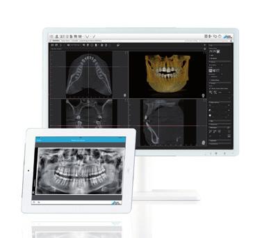
8 VistaSoft the diagnostics centre for your surgery Dürr Dental VistaSoft combines 3D and 2D X-ray images along with camera images and images imported from third-party hardware or from referring doctors. Network capable, with intuitive operation: VistaSoft represents a particularly efficient solution for the acquisition, display and editing of digital images. For reliable diagnostics, the contrast and sharpness of the images can also be edited with diagnostic filters to further assist the diagnosis. The software supports exports of DICOM data and various interfaces to all standard practice management software packages. The new design of VistaSoft has been optimised for professional diagnostics so that it offers you the best possible support. It is supplemented with an extremely simple and fast user guide it takes just one click of the mouse to access all of the functions you will need on a daily basis. This will make your work faster than ever before. Automated panoramic reconstruction is just one click away The rendered OPG view makes it easier to navigate in the 3D volume. The panoramic curve required for this is automatically positioned by VistaSoft. A slider is used to select the required layer thickness. A further plus point: VistaSoft is compatible with all current X-ray, scanner and camera systems from Dürr Dental. Thanks to the optional Imaging App, the image data can be called up at any time and in any place via an ipad. Easy image comparisons on the virtual lightbox VistaSoft enables the reproduction of video, X-ray and 3D images on a shared light table. This allows you to consult images from different sources in your diagnostics. All 3D views can be rotated and tilted for optimum alignment. With the aid of a navigation head, which always displays the current position, orientation is very simple in the different views. All notes created in each layer can be quickly located with the aid of a list: with just one click, the view will jump to the corresponding layer, dispensing with the need for laborious searches. The visualisation of implants is also an important function for implantology and an essential part of patient communication. VistaSoft further advantages and potential applications: Three different 3D views (Panoramic, TSA, MPR) Easy to draw the nerve channel into the image Easy measurements in the 3D volume Implant planning Export of 3D DICOM data 08

.")
9 With VistaSoft you can comfortably display the mandibular canal and check its correct course via the transversal layer images (TSA view). Implant planning with VistaSoft VistaVox S 09
10 Figures, data and facts at a glance VistaVox S X-ray HV generator Current Rated power Tubes Focal point Total fi ltration Image detector Type Pixel size Active sensor surface Scanning times Scanning times Panoramic programs Panoramic image Image acquisition programs for children kv, 4-16 ma 170 W 0.5 mm (IEC60336) 2.8 mm AL Csl CMOS photodiode array 49.5 μm x 36.4 mm From 2 to 18 secs Magnification factor 2D images D volume Child Adult Device dimensions Height Weight Height adjustment range Width x depth Installation Electrical connections Mains voltage Frequency Rated power Ø 100* x 70 mm Ø 100* x 85 mm * Displays the diagnostic range of a Ø 130 volume mm 2250 mm 180 kg 844 mm 990 x 1130 mm Wall mounting V AC 50/60 Hz 2.2 kva


11 The Dürr Dental Imaging App gives you instant, easy access to your image VistaVox S is manufactured using state-of-the-art data on an ipad. technology at our facility in the Black Forest, Germany This ensures the high quality and reliability of the device. VistaVox S 11


12 The VistaSystem from Dürr Dental Everything for safe diagnosis from one single source VistaScan VistaIntra VistaPano VistaVox VistaCam VistaRay VistaSoft Accessories P L02/LOD-dd.de/1/01/G9 Subject to technical changes DÜRR DENTAL AG Höpfigheimer Straße Bietigheim-Bissingen Germany
VistaCam ix HD for diagnostics support in HD quality
 VistaCam ix HD for diagnostics support in HD quality The innovative intraoral camera with interchangeable head system COMPRESSED AIR SUCTION IMAGING DENTAL CARE HYGIENE Vista Cam ix HD real HD resolution
I AM DEMANDING Type CMOS Flat Panel CMOS CMOS ø 40 x 40 mm, ø 60 x 60 mm, ø 80 x 80 mm, ø 110 x 80 mm
 TECHNICAL SPECIFICATIONS 1168 1501 1978 1237 1551-2351 Ø 1090 PANORAMIC CBCT CEPHALOMETRIC X-RAY SOURCE Tube type High frequency DC generator 2.8 mmal / 85 kv 7.0 mmal / 90 kv 2.8 mmal / 85 kv Operation
Head to new heights with your imaging SCANORA 3D
 SCANORA 3D Head to new heights with your imaging Benefits at a glance The solution for dentomaxillofacial and ENT imaging Easy Patient seated for added stability during exposure. Clear, self-explinatory
Digital Imaging from a new perspective
 TREATMENT CENTRES HANDPIECES HYGIENE SYSTEMS X-RAY SYSTEMS CEREC TREATMENT CENTRES HANDPIECES HYGIENE SYSTEMS X-RAY SYSTEMS CEREC SIRONA CREATING AND MAINTAINING VALUE. You are right to expect a great
The Optimum Choice for Implantologist
 The Optimum Choice for Implantologist What is essential for your practice? What s the best way to choose a 3D X-ray machine for implant treatment planning? 02 Doctor says.. There are diagnostic limitations
Dental Line. 3D digital panoramic system. radiology ahead
 Dental Line 3D digital panoramic system radiology ahead new generation 3D digital panoramic unit 3D imaging s value available for anyone Following the incredible success of the innovative digital panoramic
Easy operation. Numerous diagnostic options. X-rays you can rely on: the ORTHOPHOS XG device family. All ORTHOPHOS XG 3Dready programs at a glance.
 CAD /CAM Systems Instruments Hygiene Systems Treatment Centers Imaging Systems Subject to technical changes and errors in the text, Order No. A91100-M47-B346-01-7600, Printed in Germany, Dispo-Nr. 04602,
THE WAIT IS OVER CS D. 3D imaging is now available for everyone
 THE WAIT IS OVER CS 8100 3D 3D imaging is now available for everyone COMPLEXITY IS NO LONGER THE STANDARD NOW THERE ARE MANY REASONS TO MOVE TO 2D/3D IMAGING Now it s possible to experience nothing but
CS 9300 Family. The power of flexibility
 CS 9300 Family The power of flexibility The new CS 9300 digital imaging system from Carestream Dental take the guesswork out of examinations The all-in-one CS 9300 is the most versatile multimodality imaging
Low Dose Excellent Image Quality Rapid Reconstruction
 Low Dose Excellent Image Quality Rapid Reconstruction Efficient 3 in 1 Dental X-ray System CBCT > Precise 3-D Anatomical structures - Accurate diagnosis for doctors - Safe implant for patients > Significant
PAN CEPH 3D CONE BEAM
 PAN CEPH 3D CONE BEAM 2D - 3D panoramic units PANORAMIC CEPHALOMETRIC 3D CONE BEAM IMAGING I-MAX TOUCH Tactile & naturally intuitive panoramic imaging Discover the simplicity and efficiency this unit can
OP 3D Vision The upgradable 3D X-ray system for the strictest demands.
 OP 3D Vision The upgradable 3D X-ray system for the strictest demands. The solution for every task: KaVo OP 3D Vision. Regardless of which dental query you may have, the KaVo ORTHOPANTOMOGRAPH OP 3D Vision
ORTHOPHOS XG 3 DS. X-ray systems. ORTHOPHOS XG 3 Digital panoramic X-ray for practical diagnostics.
 ORTHOPHOS XG 3 DS X-ray systems ORTHOPHOS XG 3 Digital panoramic X-ray for practical diagnostics. ORTHOPHOS XG 3 Standard panoramic X-ray with proven technology. competent successful Many years of competence
3D Panoramic Cephalometric. Innovation, in reach. KODAK 9000 Extraoral Imaging System
 3D Panoramic Cephalometric Innovation, in reach 9000 KODAK 9000 Extraoral Imaging System Cephalometric Innovation made simple Innovation made simple We believe in innovation. We always have. In fact, our
T h e D e n t a l C o m p a n y FROM DIAGNOSTIC SCAN TO SURGERY, WE SHAPE THE FUTURE OF DENTISTRY.
 T h e D e n t a l C o m p a n y FROM DIAGNOSTIC SCAN TO SURGERY, WE SHAPE THE FUTURE OF DENTISTRY. SIDEXIS SOFTWARE ORTHOPHOS SL D/D SEAMLESS THE NEW STANDARD IN CLINICAL DIAGNOSIS AND PATIENT COMMUNICATION
ENGLISH. Cefla s.c. - Via Selice Provinciale 23/a, Imola - Italy Tel
 09-2017 NVGEGB161S00 According to the regulations in force, some products and/or features may have different availability and characteristics in areas outside of the European Union. Please contact your
Profound understanding of anatomy
 ENGLISH Profound understanding of anatomy The unique Planmeca ProMax 3D product family offers equipment for all maxillofacial imaging. All volumes sizes from the smallest special cases to whole head images
2D AND 3D/2D WALL-MOUNTED PANORAMIC UNITS
 2D AND 3D/2D WALL-MOUNTED PANORAMIC UNITS KEEP YOUR CLINIC ONE STEP AHEAD! Wall-mounted concept: zero foot print 62kg - the lightest unit on the market Face to face positioning High Definition The fruit
CS Introducing the CS 1600 Intraoral Camera Revolutionary technology. Superior workflow.
 CS 1600 Introducing the CS 1600 Intraoral Camera Revolutionary technology. Superior workflow. Early caries identfication for healthier patients Earliest caries identification Early detection is the most
Profound understanding of anatomy
 ENGLISH Profound understanding of anatomy Planmeca ProMax 3D, the intelligent and multipurpose X-ray unit, is designed to obtain complete information on patient anatomy in the minutest detail. The unit
XPan 3D Plus. FONA Every dental solution you need. Advanced dental technology. Headquarters THE ULTIMATE DIAGNOSTIC SOLUTION DIGITAL DENTISTRY
 FONA Every dental solution you need Through decades of experience and deep understanding of the dental profession, we deliver complete, reliable and accessible solutions.regardless of country or specialisation,
True Low Dose. Exact time to display image on screen may vary upon computer and network configuration.
 RAYSCAN ALPHA PLUS True Low Dose Cone Beam CT Industry Leading Resolution High resolution images provide all the clinical information needed while keeping radiation exposure low. Endodontics - Smallest
English. Perfect Vision
 English Perfect Vision Everything becomes clearer and simpler with a big F.O.V. The complete dentomaxillofacial volume ready for your diagnosis One scan provides you with an incredible amount of information
Profound understanding of anatomy
 ENGLISH Profound understanding of anatomy The unique Planmeca ProMax 3D product family offers equipment for all maxillofacial imaging. All volume sizes from the smallest special cases to whole head images
3D/2D WALL MOUNTED UNIT
 Product launch document EN Page 1 sur 30 EN PRODUCT LAUNCH DOCUMENT REV03, September 2018 3D/2D WALL MOUNTED UNIT Product launch document EN Page 2 sur 30 INDEX 1. PRODUCT IDENTITY AND POSITIONNING...
Versatility And Expandability In One Panoramic.
 Orthoralix 9200 / 9200 DDE Versatility And Expandability In One Panoramic. Panoramic X-ray Systems Intraoral X-ray Systems Digital Intraoral Sensors Digital X-ray Phosphor Plates Intraoral Cameras Imaging
X X X. GXS-700 Direct USB Digital Intraoral Sensors. Buy a Sensor Combo and a Digital Pan Unit, Receive $600 Off! GO.BENCO benco.
 8 0 0. G O. B E N C O b e n c o. c o m GS-700 Direct USB Digital Intraoral Sensors Designed to make migrating from film, or upgrading an existing digital system, easier than ever High quality image capture
EN PRODUCT LAUNCH DOCUMENT. I-MAX 3D 2016 Révision : V Product launch document EN Page 1 sur 29. Version 02, OCTOBER 2016
 Product launch document EN Page 1 sur 29 EN PRODUCT LAUNCH DOCUMENT Version 02, OCTOBER 2016 Product launch document EN Page 2 sur 29 INDEX 1. PRODUCT IDENTITY AND POSITIONNING... 3 2. TECHNICAL CHARACTERISTICS...
DIAGNOSTIC IMAGING. OPTIMIZED.
 ABOUT LED DENTAL SEE THE DIFFERENCE Using our years of business insight and clinical experience as a foundation, LED Dental takes the uncertainty out of your imaging purchase decision. We offer our clients
- RCS Paris B
 Technical specifications PANORAMIC CBCT CEPHALOMETRIC X-ray source Tube type High frequency DC generator Total filtration >2.5 mm Al @ 90 kv Mode of operation Continuous Pulsed Continuous Tube voltage
Planmeca ProMax 3D s Planmeca ProMax 3D ENGLISH
 Planmeca ProMax 3D s Planmeca ProMax 3D ENGLISH Genuine all-in-one unit Planmeca ProMax 3D s and Planmeca ProMax 3D units are designed to obtain complete information on patient anatomy in the minutest
Veraviewepocs 3D R100 & F40
 Veraviewepocs 3D R100 & F40 Innovative 3D Reuleaux Full Arch FOV Thinking ahead. Focused on life. Veraviewepocs 3D R100 A New Frontier in X-ray Diagnostics Veraviewepocs 3D R100 has changed the shape of
User Guide for Dental and Maxillofacial Cone Beam Computed Tomography (CBCT)
") User Guide for Dental and Maxillofacial Cone Beam Computed Tomography (CBCT) Poster No.: C-0756 Congress: ECR 2014 Type: Educational Exhibit Authors: J. Ukkonen, J. Asp; Helsinki/FI Keywords: Education
User Guide for Dental and Maxillofacial Cone Beam Computed Tomography (CBCT) Poster No.: C-0756 Congress: ECR 2014 Type: Educational Exhibit Authors: J. Ukkonen, J. Asp; Helsinki/FI Keywords: Education
3D/2D. Hyperion X5 Suspended imaging system. Data subject to change without notice. 03/2017 MX53DGB171S00
 www.my-ray.com Data subject to change without notice. 03/2017 MX53DGB171S00 According to the regulations in force, some products and/or features may have different availability and characteristics in areas
CS Introducing the CS 1600 Intraoral Camera Revolutionary caries detection aid. FIRE technology. Superior patient care.
 CS 1600 Introducing the CS 1600 Intraoral Camera Revolutionary caries detection aid. FIRE technology. Superior patient care. An aid in the process of early caries detection Caries discovered in early stages
STELLARIS 3D 4 IN 1 CBCT SOLUTION FOR ADVANCED DIAGNOSTICS
 STELLARIS 3D 4 IN CBCT SOLUTION FOR ADVANCED DIAGNOSTICS 3 STELLARIS 3D 4 IN CBCT SOLUTION FOR ADVANCED DIAGNOSTICS Stellaris 3D is a complete and compact, fully upgradeable 3D CBCT for a patient, Panoramic
ORTHOPANTOMOGRAPH OP200 D ORTHOCEPH OC200 D. True dynamo leading through the decades.
 ORTHOPANTOMOGRAPH OP200 D ORTHOCEPH OC200 D True dynamo leading through the decades. 1 You can t dublicate the legacy. 1946 Professor Y.V. Paatero publishes his first paper on Panoramic Tomography. 1951
The Quality Leader in 3D Cone Beam CT
 The Quality Leader in 3D Cone Beam CT The Complete 2-in-1 or 3-in-1 Multi-modality Solution PreXion, with over 15 years of innovation in the medical and dental fields, introduces the PreXion3D Eclipse.
ADVANCED 3D IMAGING. CEFLA s.c. Via Selice Provinciale 23/a Imola Italy t newtom.
 CEFLA s.c. Via Selice Provinciale 23/a 40026 Imola Italy t. +39 045 8202727 045 583500 info@newtom.it newtom.it 05/2018 NVGEGB181S00 According to the standards in force, in extra-eu areas the availability
Cone Beam 3D Imaging
 Cone Beam 3D Imaging NewTom Sets the Standard in 3D Maxillofacial Imaging Cone Beam 3D Imaging The Global Market Leader The Inventors n of Cone Beam 3D In 1996, QR srl developed the first generation of
INTRAORAL IMAGING ACCORDING TO YOUR NEEDS Intraoral X-rays
 INTRAORAL IMAGING ACCORDING TO YOUR NEEDS Intraoral X-rays FONA Intraoral X-rays INTRAORAL IMAGING ACCORDING TO YOUR NEEDS FONA intraoral X-ray devices are designed for ease of use, precision, reliability
The 2D x-ray family. dentsplysirona.com
 The 2D x-ray family dentsplysirona.com 02 I 03 As versatile as practice life The Orthophos 2D X-ray family offers the right solution for every practice. From entry into digital radiography to the perfect
Periapical Radiography
 Periapical Radiography BARBARA E. DIXON B.D.S., M.Sc., D.P.D.S. Main Indications Detection of Apical infection/inflammation Assessment of the periodontal status After trauma Assessment of Unerupted teeth
CS 8100 FAMILY / CS D FAMILY / CS 9300 FAMILY EXTRAORAL SOLUTIONS EXTRAORDINARY POSSIBILITIES
 CS 8100 FAMILY / CS 8100 3D FAMILY / CS 9300 FAMILY EXTRAORAL SOLUTIONS EXTRAORDINARY POSSIBILITIES A SOLUTION FOR EVERY CLINIC AND ANY CONSULTATION The CS 8100 does more than my old machine and is half
VGi - R EN ENGLISH. QR srl - Via Silvestrini, Verona Italy Tel
 VGi - R15.1 - EN ENGLISH QR srl - Via Silvestrini, 20-37135 Verona Italy Tel. +39 045 8202727-045 583500 info@newtom.it www.newtom.it FIRST IN CONE BEAM, ACCURATE IN RESULTS. 360 degree imaging, reduced
STL. inside. ineos Blue: Precision, Speed and Control. THE SCANNER WITH Bluecam TECHNOLOGY
 CAD/CAM SYSTEMS INSTRUMENTS HYGIENE SYSTEMS TREATMENT CENTERS IMAGING SYSTEMS THE SCANNER WITH Bluecam TECHNOLOGY ineos Blue: Precision, Speed and Control. STL inside T h e D e n t a l C o m p a n y ineos
INTERCHANGEABLE HEAD SYSTEM. Easily and quickly change between caries diagnosis and intraoral imaging. T r i t o n H D
 INTERCHANGEABLE HEAD SYSTEM. Easily and quickly change between caries diagnosis and intraoral imaging. T r i t o n H D Whether its patient education, caries detection or accurate clinical documentation,
CT Imaging at the Point-of-Care
 ENGLISH True Dedication The new Planmed Verity Extremity CT Scanner revolutionizes extremity CT imaging. The compact unit brings 3D imaging at emergency departments, orthopedic clinics or trauma centers
Straight to the point with your diagnosis.
 CAD/CAM SYSTEMS INSTRUMENTS HYGIENE SYSTEMS TREATMENT CENTERS IMAGING SYSTEMS HELIODENT DS INTRAORAL X-RAY WITH EXCELLENT IMAGE QUALITY Straight to the point with your diagnosis. T h e D e n t a l C o
3D Accuitomo XYZ Slice View Tomograph. Super High-Resolution Images of Region of Interest
 3D Accuitomo XYZ Slice View Tomograph. Super High-Resolution Images of Region of Interest Thinking ahead. Focused on life. 2 Cone Beam X-Ray CT Imaging X-Ray Tube Imaging Intensifier Imaging Volume Voxel
For a new dimension of a practice s success: The Sirona 3D family.
 CAD / CAM SYSTEMS INSTRUMENTS HYGIENE SYSTEMS TREATMENT CENTERS IMAGING SYSTEMS CAD/ CAM SYSTEMS INSTRUMENTS HYGIENE SYSTEMS TREATMENT CENTERS IMAGING SYSTEMS Subject to technical changes and errors in
Enter into the world of unprecedented accuracy! IScan D104 Series
 Enter into the world of unprecedented accuracy! IScan D104 Series 2 Three scanners, one goal: Precisely fitting restorations The D104 Series of 3D scanning systems is the 5th generation of dental scanners
Flexible Easy Competitive. SCANORA 3Dx - The in-office large field-of-view Cone Beam CT system for Head and Neck imaging
 Flexible Easy Competitive SCANORA 3Dx - The in-office large field-of-view Cone Beam CT system for Head and Neck imaging SCANORA 3Dx. The solution. SCANORA 3Dx makes advanced 3D imaging easy in the head
IMAGE-GUIDED RADIATION THERAPY
 IMAGE-GUIDED RADIATION THERAPY Your Single Source Oncology Solutions Provider Plan. Target. Treat. At Best NOMOS, we design products and solutions that help medical professionals treat a variety of cancers.
NewTom GiANO HR PERFECT.VISION
 CEFLA s.c. Via Selice Provinciale 23/a 40026 Imola Italy t. +39 045 8202727 045 583500 info@newtom.it newtom.it 06/2018 NHRGB181S00 According to the standards in force, in extra-eu areas the availability
ENGLISH. Distributed by: QR srl - Via Silvestrini, Verona Italy Tel
 GiANO - R15.1 - EN ENGLISH Distributed by: QR srl - Via Silvestrini, 20-37135 Verona Italy Tel. +39 045 8202727-045 583500 info@newtom.it www.newtom.it Manufacturer: CEFLA S.C. - CEFLA DENTAL GROUP Via
Veraviewepocs 3D. F40 and R100 with innovative 3D Reuleaux Full Arch FOV. Thinking ahead. Focused on life.
 Veraviewepocs 3D F40 and R100 with innovative 3D Reuleaux Full Arch FOV Thinking ahead. Focused on life. Veraviewepocs 3D R100 A New Frontier in X-ray Diagnostics Veraviewepocs 3D R100 has changed the
Imagine the possibilities with Sirona 3D.
 CAD/CAM SYSTEMS I INSTRUMENTS I HYGIENE SYSTEMS I TREATMENT CENTERS I IMAGING SYSTEMS WELCOME TO THE NEW DIMENSION OF DIAGNOSTIC IMAGING Imagine the possibilities with Sirona 3D. T h e D e n t a l C o
5G XL - R EN ENGLISH
 5G XL - R15.0 - EN ENGLISH Sede legale ed amministrativa - Headquarters QR srl - Via Selice Provinciale, 23/a - 40026 Imola - Bo (Italy) Stabilimento - Plant Via Fermi, 40-37136 Verona (Italy) Tel. +39
CT SCAN PROTOCOL. Shoulder
 CT SCAN PROTOCOL Shoulder Purpose and Summary CT images made with this protocol are used to provide the orthopedic surgeon with a detailed 3D anatomical reconstruction of the patient s scapula and proximal
TABLE OF CONTENTS P. 4-5 P. 6-7 P. 8-9 P P P P
 Full Smile Solution TABLE OF CONTENTS P. 4-5 P. 6-7 P. 8-9 P. 10-11 P. 12-13 P. 14-15 P. 16-17 FULL SMILE SOLUTION MGUIDE PROCESS ADVANTAGES CUSTOM SOLUTIONS MGUIDE SURGICAL SETS MCENTER REQUIRED SOURCES
ENGLISH. Cone Beam 3D Imaging
 ENGLISH Cone Beam 3D Imaging FIRST USER OF CONE BEAM IN DENTAL FIELD QR s.r.l. is the name that stands behind NewTom Cone Beam 3D imaging units and the creator of Cone Beam technology for the dental field.
Fear turns into satisfaction. PerioScan the gentle prophylaxis competence
 CAD/CAM SYSTEMS INSTRUMENTS HYGIENE SYSTEMS TREATMENT CENTERS IMAGING SYSTEMS PerioScan the gentle prophylaxis competence Fear turns into satisfaction. Gentle prophylaxis Prophylaxis without pain? That
BRAINLAB ELEMENTS RADIOTHERAPY PLANNING
 BRAINLAB ELEMENTS RADIOTHERAPY PLANNING BUILD REACH AND RICHNESS BRAINLAB ELEMENTS Medical technology just got a whole lot easier and innovation is within reach with pre-planning software apps by Brainlab.
European Veterinary Dental College
 European Veterinary Dental College EVDC Training Support Document Preparation of Radiograph Sets (Cat and Dog) Document version : evdc-tsd-radiograph_positioning_(dog_and_cat)-20120121.docx page 1 of 13
D3D CBCT. See more Do More
 D3D CBCT See more Do More BIOLASE DaVinci Imaging D3D For capturing superior 3D image acquisitions with the lowest minimal dose for patient safety The BIOLASE DaVinci Imaging D3D has one of the lowest
AWARD-WINNING CONE BEAM 3D DENTAL IMAGING
 AWARD-WINNING CONE BEAM 3D DENTAL IMAGING Dedicated to Advancing Dental Treatment A COMPLETE 3D TREATMENT SOLUTION Your dental practice is unique that s why you need a flexible solution that works with
Xelis Dental - What New in
 Xelis Dental - What New in 1.0.6.1 Clinical Needs Why Xelis-Dental? Panorama Cephalography Intra-oral Digital Camera Traditional imaging systems - 2-Dimension view Incorrect anatomical information - Distortion
The Diagnostic Wax Up and Planning Phase of Implant Therapy
 The beginning of implant therapy Beginning implant care with the end in mind. The Diagnostic Wax Up and Planning Phase of Implant Therapy Drs. Alan Rosenfeld and George Mandelaris Diplomates, American
NewTom 5G XL EXTRA.VISION
 CEFLA s.c. Via Selice Provinciale 23/a 40026 Imola Italy t. +39 045 8202727 045 583500 info@newtom.it newtom.it 06/2018 N5GXGB181S00 According to the standards in force, in extra-eu areas the availability
VATECH IMAGING SYSTEMS
 VATECH IMAGING SYSTEMS 2 About Vatech America 3 Vatech Assurance 4 PaX-i 6 PaX-i Insight 12 i3d Smart 16 PaX-i3D 20 Green CT 26 Green CT 2 32 i3d Premium 36 Ez3D-i 42 EzDent-i 45 EzRay Air Wall 46 HD Sensor
Guided immediate loading implant surgery planned with Implant Studio D.D.S. Jae-min, Lee
 Guided immediate loading implant surgery planned with Implant Studio D.D.S. Jae-min, Lee Jung-plant Dental office 1 PROLOGUE How can we deal with the immediate loading implant cases easier and more accurate
3D Cone beam CT & Digital Radiography Dedicated to Otorhinolaryngology
 3D Cone beam CT & Digital Radiography Dedicated to Otorhinolaryngology Multi-functional imaging solution3 RAYSCAN m is an unique 2-in-1 imaging solution, combining Cone Beam CT and Digital Radiography,
SURGERY EQUIPMENT IMAGING IT. Promotional Catalogue Free Gift. Meisterstück Platinum-Coated Classique Ballpoint Pen
 n yn SURGERY EQUIPMENT IMAGING IT Promotional Catalogue 2016 Free Gift Meisterstück Platinum-Coated Classique Ballpoint Pen FONA 2000 L Fona 2000L Package start from 8,999.00 Fona 2000 L whip arm or hanging
CT Scanning Protocol For V2R Guided Surgery Solutions
 CT Scanning Protocol For V2R Guided Surgery Solutions 2 V2R CT Scanning Protocol \\ Contents Contents General requirements... 3 V2R Dual Scan Protocol... 5 V2R Single Scan Protocol... 8 Overview... 10
For true visualisation
 ENGLISH For true visualisation Planmeca ProModel is a patient-specific physical model for high-end maxillofacial operations and dental surgery. By reproducing the anatomy of the patient in real-size, Planmeca
The Power Of Choice Pan. Ceph. 3D. Your Imaging Future Starts Today.
 NEW from Gendex! The Power Of Choice Pan. Ceph. 3D. Your Imaging Future Starts Today. Cone Beam 3D Imaging Systems Panoramic X-ray Systems Intraoral X-ray Systems Digital Intraoral Sensors Digital X-ray
A new dimension of success in your practice
 3D Imaging Family A new dimension of success in your practice dentsplysirona.com 2/3 Good reasons for 3D With 3D imaging, you have the ideal basis for a new dimension of success in your practice. BETTER
The 3D x-ray family. dentsplysirona.com
 The 3D x-ray family dentsplysirona.com 02 I 03 As versatile as practice life. The Dentsply Sirona 3D x-ray family offers 3 units, Galileos Comfort Plus, Orthophos SL 3D and Orthophos XG 3D, whose visual
ATMOS 360 diagnostics. Complete solution.
 ATMOS 360 diagnostics Complete solution. www.atmosmed.de ALL-IN-ONE ENT SOLUTION Easy to connect. Efficient. Comprehensive. In the field of modern ENT medicine, the call for greater process reliability
Specifications are subject to change without notice. PR-D613E Ver
 Specifications are subject to change without notice. PR-D613E Ver.2 11.03.00.0 Versatile Power & Performance Follow Your Intuition The new, stylish and versatile Varios series features the intuitive NSK
Connect your Scanner to SomnoMed Canada. SOMGauge Protrusive Bite Recording - Manual. Scanning Impressions - Lower and Upper
 IOS Instructions How to create and submit the best scans to SomnoMed Canada for the creation of a custom SomnoDent Sleep Apnea Appliance Its a simple process: STEP 1 Connect your Scanner to SomnoMed Canada
I AM EXCLUSIVE TECHNICAL SPECIFICATIONS. The first personal imaging plate scanner IMAGING PLATES. System. Windows recommended configuration
 TECHNICAL SPECIFICATIONS System Resolution... 20 lp/mm Scan Time (fast mode)...1,6s - 2,7s Scan Time (high definition mode)...2,1s - 3,6s Connection... Ethernet RJ-45 Dimensions... L. 154 x D. 204 x H.
ACTIVITY SCANNER NEW. 3D Dental Scanner made in Germany.
 ACTIVITY SCANNER 3D Dental Scanner made in Germany NEW ACTIVITY SOFTWARE OPTICAL DENTAL SCANNER ERGONOMIC PRECISION INNOVATION CRANIUM-BASED MEASUREMENT FAST KNOW HOW EASE OF USE OPEN INTERFACE www.smartoptics.de
THE USE OF KEYSTONE EASYGUIDE CT SCANNING SOFTWARE FOR DIAGNOSIS, DIRECTION AND DEPTH DETERMINATION
 CT DIAGNOSTICS IN 3D IMPLANT TREATMENT PLANNING THE USE OF KEYSTONE EASYGUIDE CT SCANNING SOFTWARE FOR DIAGNOSIS, DIRECTION AND DEPTH DETERMINATION Timothy Kosinski, DDS, MAGD Assistant Clinical Professor
Full Mouth Survey. Delta Dental of Massachusetts DeltaDentalMA.com
 X-Rays Having X-rays taken is a painless procedure that uses small amounts of radiation to capture images of your teeth and bones. Because your dentist takes precautions and the amount of radiation used
Hyperion X9 3-in-1 Imaging System
 Hyperion X9 3-in-1 Imaging System 2 Hyperion X9, full imaging. 3 Hyperion X9, just right for me. The present and the future of my work. In three dimensions. Hyperion X9 offers me multiple possibilities
Dimensional stability in composite cone beam computed tomography
 (2010) 39, 512 516 2010 The British Institute of Radiology http://dmfr.birjournals.org TECHNICAL REPORT Dimensional stability in composite cone beam computed tomography S Kopp* and P Ottl Department of
Hyperion X9 pro Professional 3-in-1 full-touch imaging system
 www.my-ray.com Data subject to changes without prior notice. 06/2018 M9PROGB181S00 According to the relevant regulations, in the extra-eu areas, some products and/or characteristics might have different
THE POWER OF THE PERFECT FIT
 THE POWER OF THE PERFECT FIT SCAN, DESIGN, MILL The PLANMECA FIT CAD/CAM system allows you to perform highly advanced, powder-free digital restorations, including same-day crowns, inlays, onlays, bridges
A Smart Investment in Surgical Versatility
 A Smart Investment in Surgical Versatility STERIS 4085 GENERAL SURGICAL TABLE ADVANCING CARE THROUGH PRACTICAL INNOVATION The most versatile solution for today s multipurpose operating suites. Surgical
ZIRCONIA, SIMPLY FASTER. FULL CONTOUR ZIRCONIA RESTORATIONS IN A SINGLE VISIT ONLY WITH CEREC.
 ZIRCONIA, SIMPLY FASTER. FULL CONTOUR ZIRCONIA RESTORATIONS IN A SINGLE VISIT ONLY WITH CEREC. CEREC MAKES IT HAPPEN. What would you think about providing your patients full contour zirconia crowns in
I AM SAFER. Reliable X-ray technology that reduces radiation exposure
 I AM SAFER Reliable X-ray technology that reduces radiation exposure EN 40 cm / 80 cm / 110 cm (16 / 31 / 43 ) 70 cm (27 ) 9 cm (3,5 ) 36 cm (14 ) 79 cm / 119 cm / 149 cm (31 / 46 / 59 ) 87 cm (34 ) 81
Full ultrasound breast volumes. Faster scans. Streamlined workflow. ACUSON S2000 Automated Breast Volume Scanner. Answers for life.
 Full ultrasound breast volumes. Faster scans. Streamlined workflow. ACUSON S2000 Automated Breast Volume Scanner Answers for life. 1 ACQUIRE An automated whole breast solution. Reduced acquisition time.
EVERYTHING YOU NEED, ONLY ONE BRAND FONA Product catalogue
 EVERYTHING YOU NEED, ONLY ONE BRAND FONA Product catalogue FONA XPan 3D A COMPLETE VIEW FOR ACCURATE DIAGNOSIS FONA Art Plus PREMIUM TECHNOLOGY FOR SUPERB QUALITY Full arch field of view 85 x 85 mm Premium
When You Need To Know More.
 www.siemens.com/ultrasound When You Need To Know More. ACUSON S2000 Ultrasound System Table of Contents Powerful Imaging 01 Penetrating Insight 02 03 Revealing Perspectives 04 05 Smart Workflow 06 Ergonomics
DÜRR X-RAY FILM PROCESSING. The development goes on and on DÜRR XR 24 PRO
 DÜRR X-RAY FILM PROCESSING The development goes on and on From a professional picture to an exact diagnosis DÜRR X-RAY FILM PROCESSING Facts in black and white Causal dental medicine presupposes an exact
VisuMax from ZEISS Defining the pulse rate in refractive surgery
 VisuMax from ZEISS Defining the pulse rate in refractive surgery Remarkable precision and detail Defining new trends in modern corneal surgery As a ground-breaking, high-performance femtosecond laser
Laser treatment doesn t have to be a big deal any more.
 CAD/CAM SYSTEMS I INSTRUMENTS I HYGIENE SYSTEMS I TREATMENT CENTERS I IMAGING SYSTEMS SIROLaser THE COMPACT DIODE LASER FOR YOUR PRACTICE Laser treatment doesn t have to be a big deal any more. T h e D