DENTAL RADIOLOGY Identify basic facts and terms of radiology, to include fundamentals. with 70% accuracy.
|
|
|
- Beverly Byrd
- 6 years ago
- Views:
Transcription
1 DENTAL RADIOLOGY Identify basic facts and terms of radiology, to include fundamentals of chemistry relating to radiology, with 70% accuracy.
2 Radiation Physics Radiation Health and Safety Components of the Dental X-ray Unit Control Devices Image characteristics Radiographic Film Extra-oral radiography Patient positioning guidelines Exposure Techniques
3 Film Placement Film Processing Troubleshooting Radiographic errors Mounting Radiographs Disposition of Radiographs
4 6b. Utilizing the necessary equipment and materials, expose, process, and mount dental radiographs for a Type I examination.
5 6c. After viewing panoramic radiograph procedures demonstrated by an instructor, perform step-by-step procedures of a panoramic radiograph exposure, exercising radiation safety.
6 Radiation Physics Atomic Structure - relates directly to generation, emission, and absorption of radiation Nucleus - (positive charge) Protons -positive charge Neutrons - no charge Electron - negative charge; orbit nucleus
7 Ionizing Radiation - electrons are removed from electrically stable atoms by collision with photons Ion - atom with positive / negative charge due to gain or loss of electrons Can be absorbed by body& cause permanent damage
8 Extent of affect measured by: Quantity (amount) of radiation exposure Quality (intensity) of radiation exposure Length (time) of exposure Type of tissue irradiated
9 Properties of Radiation Travels at 186,000 miles/second (speed of light) Short wavelength: Hard x-rays. Soft x-rays. Not perceptible by the senses Affects photographic plate/film Cause certain chemical crystals to give off light
10 Types of Radiation Primary - Useful beam that comes directly from the center of the tubehead Secondary - (Scatter) radiation emitted from objects through which the primary beam has passed
11 Radiation Health and Safety (ranked in order of sensitivity) (1) In pregnancy -cells of the embryo (2) Blood forming tissues (bone marrow) RBC &WBC s ( 3 ) Reproductive tissue ( 4) Epithelial tissue ( 5 ) Nerve tissue
12 Protection from Radiation Patient: ALARA concept - As Low As Reasonably Achievable -Endorses use of lowest possible exposure of the patient to x-radiation to produce a diagnostically acceptable radiograph Film speed - faster film less exposure to radiation
13 Patient - con t Intensifying screen - reduces radiation required to to expose film 1/10 amount of full mouth x -rays Proper operator techniques - reduces retakes

14 Lead apron - should cover : chest, throat & gonadal areas for optimum protection
15 Filtration -(2.5 mm aluminum) absorbs low energy. Collimation (lead diaphragm) - limits size of primary/useful beam Lead lined cylinder (cone) - reduces scatter radiation X-ray analyzer/step wedge - checks radiographic process and processor daily
16 Operator: ALARA Concept Education /Proper operator techniques Lead barriers (screen) - stand behind during exposure Dosimeter/Film Badge - detects accumulative operator exposure
17 Other personnel (exposure room) -Walls, floor, and ceiling lined with lead, concrete, or steel -Lead impregnated glass
18 Components of the Dental /X-ray tubehead Cathode ( -) negative terminal) : Tungsten filament- source of electrons Focusing cup - directs electrons Anode ( + ) positive terminal) : Tungsten target - stops flow of electrons Copper head / stem - dissipates heat during radiation production
19 Control Devices Milliamperage (ma) - controls amount or quantity of electrons Kilovoltage (KVp) - controls quality of electrons Exposure time - controls length of time are x- rays are emitted
20 Characteristics of Developed Film Density - degree of darkness (blackness) Contrast - darkness & lightness on various areas of film Radiopaque - dense structures that do not permit passage of x -radiation; more radiopaque. Radiolucent - less dense tissues that do permit passage of x-radiation; more radiolucent.

21
22 Detail and definition Ability of film to reproduce sharp outlines of objects radiographed, often referred to as the quality of the film
23 Radiographic film Types of intra - oral film (inside mouth) Bitewings Horizontal - records crowns, alveolar crest and interproximal areas (max and man) of posterior teeth; detects proximal caries on posterior teeth Vertical - measures alveolar bone level in periodontal patients
24 Periapical -Records crowns, roots, and supporting structures of individual teeth -Detects abnormalities of individual teeth and supporting structures Occlusal -Records large areas of max/man -Detects gross pathological conditions and/or fractures
25 Extra - oral radiography (Panoramic) Records complete mandible and maxilla Used for screening or evaluation of oral pathology / injuries
26 Patient positioning guidelines Midsagittal plane Plane that divides the body at the midline into left and right sides Perpendicular to floor for ALL exposures Occlusal plane Curvature from incisal edges of central. Maxillary radiographs -. Mandibular radiographs -.
27 Exposure techniques Paralleling technique -Central ray projected at right angle to film -Film placed parallel to long axis of tooth Extension Cone Paralleling instruments (XCP) -Anterior/Posterior/Vertical bitewing -Consists of biteblock,indicator rod, and locator ring
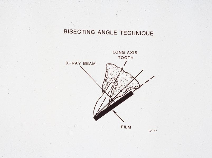
28 Bisecting Angle technique Three (3) imaginary lines help position the tube head for radiographs -Through long axis of tooth -Through film packet -Line dividing in half Film placed against lingual surface Cone directed at 90 degree angle to bisecting line
29
30 Film placement Periapical, general Anterior - long dimension vertical to floor Posterior - long dimension horizontal to floor Embossed dot - place toward incisal occlusal edge

31 Periapical, specific Incisors - centered on midline behind central...

32 Cuspids - centered on cuspid Bicuspids - centered on 2nd bicuspid

33 Molars - centered on 2nd molar
34 Horizontal bitewings Long dimension horizontal to floor Forward edge of film positioned to distal 1/3 of most anterior cuspid Use +8 degrees vertical angulation
35
36 Vertical bitewings - ( 4 periapicals ) Long dimension vertical to floor Bicuspid view - center on 2nd bicupid Molar View - center on contact between 1st & 2nd molar
37 Film processing Darkroom - light proof room with a small 7 1/2 watt filtered light that does not affect x-ray film Processing solutions - Developer, Fixer, Water
38 Film processing Automated processing - enclosed transport system; radiographs available for immediate use; Consistency of results through automated control of temperature and time Portable manual developer (Quick-fix ) - fully self contained developer / fixer with a light safe plastic lid and two light safe hand holes on front

39 Troubleshooting radiographic errors Exposure errors Foreign images - dentures, jewelry, glasses

40 Blurred image - movement of patient, film, & tubehead Double exposure - exposing same film twice

41 Herringbone image - exposing film packet on wrong side


42 Cone cutting - failure of primary beam to expose entire film

43 Overlapping - improper horizontal angulation


44 Elongation - decreased vertical angulation

45 Foreshortening - increased vertical angulation
46 No image - operator /equipment malfunction Thin / light image - underexposure Dense or dark image - overexposure
47 Processing Errors No image Film unexposed Fixing before developing Thin or light image Cold, exhausted /diluted developer Insufficient developing time
48 Dense or dark image -Hot or concentrated developer -Excessive time in developer Partial image -Film not completely immersed in developer (manual) -Overlapping films (automatic)
49 Spots, streaks, or stains -Contaminated/dirty work area,. -Insufficient rinsing between dev./fix. Fogged film -Light leaks; improper safe light -Contaminated solutions -Outdated film -Improper film storage

50 Static marks - static electricity (procedure / patient)
51 Mounting radiographs Mount Standard mount - 14 periapicals, 2 bitewings Patient s name, SSAN, and date radiographs taken recorded on each mount
52 Procedure Films Identified by anatomical landmarks then mounted Embossed dot (convex) - all exposures mounted and viewed from the facial aspect

53 Specific Film Identification Maxillary molars - Three roots, Max sinus, Max tuberosity

54 Maxillary bicuspids - 1st bicuspid has two roots / maxillary sinus

55 Maxillary cuspids- longest root, maxillary sinus
56 Maxillary incisors - Maxillary sinus, Maxillary suture, nasal cavity

57 Mandibular molars - Angle of mandible, two roots, Mandibular canal
58 Mandibular bicuspid - Single root, Mental foramen visible near apex
59 Mandibular cuspids - mental foramen near bicuspids
60 Mandibular incisors - small crowns /roots
61 Horizontal bitewings distal 1/3 of most anterior cuspid to third molars
62 Disposition of radiographs Maintained in patient record Remove when obsolete - determined by Dentist Radiographs retained for silver recovery program
63 DENTAL RADIOLOGY Identify basic facts and terms of radiology, to include fundamentals of chemistry relating to radiology, with 70% accuracy.
64 Radiation Physics Radiation Health and Safety Components of the Dental X-ray Unit Control Devices Image characteristics Radiographic Film Extra-oral radiography Patient positioning guidelines Exposure Techniques
65 Film Placement Film Processing Troubleshooting Radiographic errors Mounting Radiographs Disposition of Radiographs
66 6b. Utilizing the necessary equipment and materials, expose, process, and mount dental radiographs for a Type I examination.
67 6c. After viewing panoramic radiograph procedures demonstrated by an instructor, perform step-by-step procedures of a panoramic radiograph exposure, exercising radiation safety.
ACADEMY FOR DENTAL ASSISTANTS
 ACADEMY FOR DENTAL ASSISTANTS 12 Week Dental Assisting Program Section 2 Quiz Chapter 38 1. Exposure to radiation: a. no matter how small, has the potential to cause harmful biologic changes. b. has a
Radiation Safety for Dental Auxiliaries. Susan W. Grammer, RDH, M.Ed. Course Content. A. Radiation History and the Use of Radiographs
 Radiation Safety for Dental Auxiliaries Susan W. Grammer, RDH, M.Ed. Course Content A. Radiation History and the Use of Radiographs B. Introduction to Physics C. X-ray Machine and Production of X-Rays
Oral and Maxillofacial Radiology
 Majma ah University College of Dental Medicine Oral and Maxillofacial Radiology [Laboratory Manual] Assist. Prof. Kheirallah M. Mohd Malik Afroz Maxillofacial Surgery and Diagnostic Sciences Department
Dentalelle Tutoring - Faulty Radiographs
 Dentalelle Tutoring - Faulty Radiographs Errors in improperly exposing or processing dental films can produce undesirable dental radiographs of nondiagnostic quality. These are known as faulty radiographs.
Intraoral Imaging. Chapter 41. Copyright 2018, Elsevier Inc. All Rights Reserved. 1
 Intraoral Imaging Chapter 41 Copyright 2018, Elsevier Inc. All Rights Reserved. 1 Learning Objectives Lesson 41.1: Projection Types and the Paralleling Technique 1. Pronounce, define, and spell the key
Periapical Radiography
 Periapical Radiography BARBARA E. DIXON B.D.S., M.Sc., D.P.D.S. Main Indications Detection of Apical infection/inflammation Assessment of the periodontal status After trauma Assessment of Unerupted teeth
Dental Radiography
 Western Technical College 10508103 Dental Radiography Course Outcome Summary Course Information Description Career Cluster Instructional Level Total Credits 2.00 Prepares dental auxiliary students to operate
Dental Hygiene Spring 2018 Summer 2014 Fall COURSE OUTLINE DHT 1032 Dental Radiography 2 Credit Hours
 COURSE OUTLINE DHT 1032 Dental Radiography 2 Credit Hours Course Description This course prepares the dental hygiene student to expose, process and critique intra and extraoral radiographs for clinical
COMMON COURSE OUTLINE: Course discipline/number/title: DS 1300: Dental Radiology
 COMMON COURSE OUTLINE: Course discipline/number/title: DS 1300: Dental Radiology A. CATALOG DESCRIPTION 1. Credits: 3 2. Hours/Week: 2 hour lecture, 2 hour lab 3. Prerequisites (Course discipline/number):
Extraoral Imaging. Chapter 42. Copyright 2018, Elsevier Inc. All Rights Reserved. 1
 Extraoral Imaging Chapter 42 Copyright 2018, Elsevier Inc. All Rights Reserved. 1 Learning Objectives Lesson 42.1: Panoramic Imaging 1. Pronounce, define, and spell the key terms. 2. Discuss panoramic
NEW JERSEY RADIOLOGIC TECHNOLOGY BOARD OF EXAMINERS (BOARD) DENTAL RADIOGRAPHY CURRICULUM REQUIREMENTS
 DENTAL RADIOGRAPHY CURRICULUM REQUIREMENTS") CHRIS CHRISTIE DEPARTMENT OF ENVIRONMENTAL PROTECTION BOB MARTIN Governor Division of Environmental Safety and Health Commissioner Bureau of X-Ray Compliance, Technologist Certification Section KIM GUADAGNO
CHRIS CHRISTIE DEPARTMENT OF ENVIRONMENTAL PROTECTION BOB MARTIN Governor Division of Environmental Safety and Health Commissioner Bureau of X-Ray Compliance, Technologist Certification Section KIM GUADAGNO
NEW JERSEY RADIOLOGIC TECHNOLOGY BOARD OF EXAMINERS (BOARD) DENTAL RADIOGRAPHY CURRICULUM REQUIREMENTS GENERAL REQUIREMENTS
 DENTAL RADIOGRAPHY CURRICULUM REQUIREMENTS GENERAL REQUIREMENTS") CHRIS CHRISTIE DEPARTMENT OF ENVIRONMENTAL PROTECTION BOB MARTIN Governor Division of Environmental Safety and Health Commissioner Bureau of X-Ray Compliance, Technologist Certification Section KIM GUADAGNO
CHRIS CHRISTIE DEPARTMENT OF ENVIRONMENTAL PROTECTION BOB MARTIN Governor Division of Environmental Safety and Health Commissioner Bureau of X-Ray Compliance, Technologist Certification Section KIM GUADAGNO
Radiation Safety & Determining Need for Radiographs
 Radiation Safety & Determining Need for Radiographs Guidelines for Radiographic Examination All radiation is harmful! These guidelines have been established to protect the patient and operator from unnecessary
Proceedings of the World Small Animal Veterinary Association Sydney, Australia 2007
 Proceedings of the World Small Animal Veterinary Association Sydney, Australia 2007 Hosted by: Australian Small Animal Veterinary Association (ASAVA) Australian Small Animal Veterinary Association (ASAVA)
CAMOSUN COLLEGE School of Health & Human Services Dental Programs. DHYG 131 Dental Radiology. Winter, 2013 COURSE OUTLINE
 CAMOSUN COLLEGE School of Health & Human Services Dental Programs DHYG 131 Dental Radiology Winter, 2013 COURSE OUTLINE The Approved Course Description is available on the web @ http://camosun.ca/learn/calendar/current/web/dhyg.html
Intraoral radiographic techniques Introduction There are three main types of intraoral radiographs: Periapical radiograph Bitewing radiograph
 Intraoral radiographic techniques Introduction There are three main types of intraoral radiographs: Periapical radiograph Bitewing radiograph Occlusal radiograph The anatomic area of interest and type
DENTAL ASSISTANT SPECIALTY. Clinical Skills-Radiology
 QTP4Y0X1-2 15 November 2018 DENTAL ASSISTANT SPECIALTY Clinical Skills-Radiology Volume 2 381st Training Squadron 2931 Harney Road Fort Sam Houston, TX 78234 QTP 4Y0X1-2 DENTAL ASSISTANT SPECIALTY Volume
Periodontal Disease. Radiology of Periodontal Disease. Periodontal Disease. The Role of Radiology in Assessment of Periodontal Disease
 Radiology of Periodontal Disease Steven R. Singer, DDS srs2@columbia.edu 212.305.5674 Periodontal Disease! Includes several disorders of the periodontium! Gingivitis! Marginal Periodontitis! Localized
Radiology Safety Certification Course
 Radiology Safety Certification Course Laurie Carter, D.D.S., Ph.D. Virginia Commonwealth University School of Dentistry I. Historical Overview A. X-Rays discovered by Wilhelm Roentgen on Nov. 8, 1895 by
Radiographic Policy *ALL RADIOGRAPHS ARE TAKEN FOR DIAGNOSTIC PURPOSES ONLY
 Radiographic Policy *ALL RADIOGRAPHS ARE TAKEN FOR DIAGNOSTIC PURPOSES ONLY Safety Regulations All Patients having dental radiographs must wear a leaded apron and all operators must wear monitoring badges.
HOUSTON COMMUNITY COLLEGE SYSTEM Coleman College for Health Sciences Dental Assisting Program Fall 2014
 HOUSTON COMMUNITY COLLEGE SYSTEM Coleman College for Health Sciences Dental Assisting Program Fall 2014 TITLE: Dental Radiology I, DNTA 1305 (CRN# 26575, 26732, 26730, 26731) CREDIT: 3 semester hours LECTURE
Level 3 Diploma in Dental Nursing
 LearnerName CompanyName AssessorName Date Submitted Level 3 Diploma in Dental Nursing - 5234 Unit 314 Dental Radiography Below contains all areas that make up the 314 dental exam, please be aware of all
Kodak Dental Radiography Series. Radiation Safety in Dental Radiography. Dental
 Kodak Dental Radiography Series Radiation Safety in Dental Radiography Dental Radiation Safety in Dental Radiography The goal of dental radiography is to obtain diagnostic information while keeping the
Big Sandy Community and Technical College. Course Syllabus
 Big Sandy Community and Technical College Course Syllabus PS Number: 54493 Lecture 54494 Lab Semester: Fall Year: 2015 Faculty Name: Kathy J. Miller, DMD Title: Instructor Course Prefix and Number: DAH
Intraoral Radiography: Principles, Techniques and Error Correction
 Intraoral Radiography: Principles, Techniques and Error Correction Gail F. Williamson, RDH, MS Continuing Education Units: 2 hours Online Course: www.dentalcare.com/en-us/dental-education/continuing-education/ce137/ce137.aspx
X-RAY. Intra-oral radiographic techniques:
 X-RAY Lecture No.7 Intra-oral radiographic techniques: Guidelines for ordering radiographs: 1. Make radiographs only after a proper clinical examinations. 2. Order only those radiographs that directly
Course File 243 DDS Physics of Diagnostic Radiology and Oral and Maxillofacial Radiology
 King Saud University College of Dentistry Dept. of Oral Medicine & Diagnostic Sciences Division of Oral & Maxillofacial Radiology Course File 243 DDS Physics of Diagnostic Radiology and Oral and Maxillofacial
European Veterinary Dental College
 European Veterinary Dental College EVDC Training Support Document Preparation of Radiograph Sets (Cat and Dog) Document version : evdc-tsd-radiograph_positioning_(dog_and_cat)-20120121.docx page 1 of 13
COURSE INFORMATION SHEET. HOURS: 3 Credit Hours, 2 Lecture, 3 Laboratory hours a week, 80 Contact Hours
 COURSE INFORMATION SHEET COURSE NUMBER AND TITLE: DNTA 1305 HOURS: 3 Credit Hours, 2 Lecture, 3 Laboratory hours a week, 80 Contact Hours LECTURE: Friday 10:00-12:00 PM, Room U-127 LAB: TBA, Room U-167
The influence of sensor size and orientation on image quality in intra-oral periapical radiography
 Clinical The influence of sensor size and orientation on image quality in intra-oral periapical radiography Tony Druttman 1 The periapical view is one of the standard intra-oral radiographs by which diagnostic
New York Radiation Health and Safety (NYR)
") New York Radiation Health and Safety (NYR) Exam Outline and Suggested References State Regulations Each state s dental board implements regulations and establishes rules for delegating legally allowable
New York Radiation Health and Safety (NYR) Exam Outline and Suggested References State Regulations Each state s dental board implements regulations and establishes rules for delegating legally allowable
Dr.Sepideh Falah-kooshki
 Dr.Sepideh Falah-kooshki MAXILLA Premaxillary/median palatal suture (radiolucent). Incisive fossa and foramen (radiolucent). Nasal passages (radiolucent). Nasal septum (radiopaque). Anterior nasal spine
-SQA-SCOTTISH QUALIFICATIONS AUTHORITY. Hanover House 24 Douglas Street GLASGOW G2 7NG NATIONAL CERTIFICATE MODULE DESCRIPTOR
 -SQA-SCOTTISH QUALIFICATIONS AUTHORITY Hanover House 24 Douglas Street GLASGOW G2 7NG NATIONAL CERTIFICATE MODULE DESCRIPTOR -Module Number- 0069166 -Session-1986-87 -Superclass- PF -Title- DENTAL RADIOGRAPHY
Film processing procedures have a direct effect on the quality of a radiograph.
 . Film processing procedures have a direct effect on the quality of a radiograph. Processing is a series of steps that changes the latent image on the exposed film into a radiograph by producing a visible
Patient Management Image Selection Radiation Biology, Dosimetry & Protection
 Patient Management Image Selection Radiation Biology, Dosimetry & Protection Objectives: Following this course, the participants will have the information necessary to: 1. Identify the techniques used
Dental Morphology and Vocabulary
 Dental Morphology and Vocabulary Palate Palate Palate 1 2 Hard Palate Rugae Hard Palate Palate Palate Soft Palate Palate Palate Soft Palate 4 Palate Hard Palate Soft Palate Maxillary Arch (Maxilla) (Uppers)
Extraoral Radiology October 10th, 2008
 Extraoral Radiology October 10th, 2008 Steven R. Singer, DDS srs2@columbia.edu 212.305.5674 November 8 th, 1895 Extraoral Projections Images can be produced in the dental office X-ray source can be Intraoral
You know you would like to stop swearing at the computer after each shot. Troubleshooting oral radiography
 You know you would like to stop swearing at the computer after each shot Troubleshooting oral radiography Goals of oral radiology Achieve diagnostic images of the teeth and surrounding bone. Images should
Digital Imaging from a new perspective
 TREATMENT CENTRES HANDPIECES HYGIENE SYSTEMS X-RAY SYSTEMS CEREC TREATMENT CENTRES HANDPIECES HYGIENE SYSTEMS X-RAY SYSTEMS CEREC SIRONA CREATING AND MAINTAINING VALUE. You are right to expect a great
The wonderful world of dental radiology
 The wonderful world of dental radiology Dr Christine Hawke BSc(Vet)(Hons) BVSc(Hons) PhD MACVSc (Veterinary Dentistry) PO Box 3001 Willoughby North 2068 Phone: 0408 782 611 Email: christine@sydneypetdentistry.com.au
6610 NE 181st Street, Suite #1, Kenmore, WA
 660 NE 8st Street, Suite #, Kenmore, WA 9808 www.northshoredentalacademy.com.08.900 READ CHAPTER The Professional Dental Assistant (p.-9) No Key Terms Recall Questions:,,,, and 6 CLASS SYLLABUS DAY READ
The wonderful world of dental radiology
 The wonderful world of dental radiology Dr Christine Hawke BSc(Vet)(Hons) BVSc(Hons) PhD MACVSc (Veterinary Dentistry) PO Box 3001 Willoughby North 2068 Phone: 0408 782 611 Email: christine@sydneypetdentistry.com.au
Common Intra Oral Radiographic Errors Made by Dental Students
 GMJ. 2013;2(2):44-48 Common Intra Oral Radiographic Errors Made by Dental Students Abdolaziz Haghnegahdar 1, Pegah Bronoosh 1, Mohamad Mehdi Taheri 2, Amin Farjood 2 1 Department of Oral and Maxillofacial
 2016 course three self-study course The Ohio State University College of Dentistry is a recognized provider for ADA CERP credit. ADA CERP is a service of the American Dental Association to assist dental
EFFECTIVE DATE: Fall 2011
 DAE 106 Dental Assisting Radiography Approved: February 4, 2011 EFFECTIVE DATE: Fall 2011 COURSE PACKAGE FORM Team Leader and Members Tracy Gift, Robbi Baleno Date of proposal to Curriculum Sub-committee:
RECTANGULAR COLLIMATION
 A SPECIAL REPORT RECTANGULAR COLLIMATION No longer a matter of choice! Robert Langlais DDS MS FACD University of Texas San Antonio, Texas This Special Report is made available by Interactive Diagnostic
Selection Criteria. It s Time to Clean up Your Image: Better Radiographic Technique. Changes in Document 2/29/2016. Optimize Your Radiographic Imaging
 KDA 2016 It s Time to Clean up Your Image: Better Radiographic Technique Optimize Your Radiographic Imaging Intraoral Technique Error Recognition and Correction Use of Digital Receptors Panoramic Technique
Prosthetic Options in Implant Dentistry. Hakimeh Siadat, DDS, MSc Associate Professor
 Prosthetic Options in Dentistry Hakimeh Siadat, DDS, MSc Associate Professor Dental Research Center, Department of Prosthodontics & Dental s Faculty of Dentistry, Tehran University of Medical Sciences
Dental Data Checklist. UNIDENTIFIED PERSON FILE Data Collection Entry Guide. City, State, and ZIP. Street Address. FAX Number.
 Investigating Agency Agency Case Number Street Address City, State and Zip Telephone Number FAX Number Medical Examiner/Coroner Medical Examiner/Coroner Case Number Street Address City, State, and ZIP
Techniques of local anesthesia in the mandible
 Techniques of local anesthesia in the mandible The technique of choice for anesthesia of the mandible is the block injection and this is attributed to the absence of the advantages which are present in
Full Mouth Survey. Delta Dental of Massachusetts DeltaDentalMA.com
 X-Rays Having X-rays taken is a painless procedure that uses small amounts of radiation to capture images of your teeth and bones. Because your dentist takes precautions and the amount of radiation used
Higher National Unit specification: general information
 Higher National Unit specification: general information Unit code: H0AH 36 Superclass: PF Publication date: February 2015 Source: Scottish Qualifications Authority Version: 05 Unit purpose This Unit is
tips FoR MaSteRiNG VETERINARY NURSING EDUCATION
 tips FoR MaSteRiNG FuLL MOuTH radiographs Kelly Vearil RVT, VTS (Dentistry) Learning Objective: After reading this article, the veterinary technician will be able to understand the indications of intraoral
Dental Radiography STANDARDS & GUIDELINES TABLE OF CONTENTS. College of Dental Surgeons of BC
 STANDARDS & GUIDELINES Dental Radiography TABLE OF CONTENTS Standards and guidelines inform practitioners and the public of CDSBC s expectations for registrants. This document primarily contains guidelines
Contemporary Implant Dentistry
 Contemporary Implant Dentistry C H A P T ER 1 4 O F C O N T E M P OR A R Y O R A L A N D M A X I L L OFA C IA L S U R G E RY B Y : D R A R A S H K H O J A S T EH Dental implant is suitable for: completely
Test Bank CHAPTER 1. Multiple Choice. 1. Who was awarded the first Nobel Prize for physics in 1901, for his experimental work with. radiation?
 Test Bank CHAPTER 1 Multiple Choice 1. Who was awarded the first Nobel Prize for physics in 1901, for his experimental work with radiation? a. W. J. Morton b. O. Walkhoff c. W. D. Coolidge d. W. C. Roentgen
1B Getting Ready for Instrumentation: Mathematical Principles and Anatomic Descriptors
 MODULE 1B Getting Ready for Instrumentation: Mathematical Principles and Anatomic Descriptors Module Overview This module contains a review of the mathematical principles and anatomic descriptors used
Arrangement of the artificial teeth:
 Lecture Prosthodontic Dr. Osama Arrangement of the artificial teeth: It s the placement of the teeth on a denture with definite objective in mind or it s the setting of teeth on temporary bases. Rules
Introduction. Clinical Periodontal Examination 11/1/16
 Is Radiologic Assessment of Alveolar Crest Height Useful to Monitor Periodontal Disease Activity? Based on DCNA chapter: Hattan A.M. Zaki, Kenneth R. Hoffmann, Ernest Hausmann, Frank A. Scannapieco Periodontitis,
Radiology. & supporting structures. Lec. 14 Common diseases of teeth Dr. Areej
 Radiology Lec. 14 Common diseases of teeth Dr. Areej & supporting structures A radiograph is only one part of the diagnostic process. Usually one does NOT make a diagnosis solely from a radiograph. A diagnosis
Jordan University of Science and Technology Faculty of Applied Medical Sciences Department of Applied Dental Sciences 1 st Semester
 Jordan University of Science and Technology Faculty of Applied Medical Sciences Department of Applied Dental Sciences 1 st Semester Course Title Oral Radiology I Course Number ADS 351 Prerequisites - Course
DENTAL RATE FEE SCHEDULE rates effective 5/1/15 through 6/30/15
 Procedure Code D0120 Description April 2014 Fee Rate cute 16.75% Amount of Reduction May/June 2015 Fee $28.00 $28.00 Periodic Oral Exam Ages 0 thru 18 D0120 Periodic Oral Exam Ages 19 thru 20 and Pregnant
DENTAL ASST: DENTAL SCIENCE II (721)
") DESCRIPTION The second assessment in a series, Dental Assistant II prepares students to assist a dentist or dental hygienist performing the functions of a dental practice. Topics include: chair-side assisting,
DESCRIPTION The second assessment in a series, Dental Assistant II prepares students to assist a dentist or dental hygienist performing the functions of a dental practice. Topics include: chair-side assisting,
2018 Dental Code Set For dates of service from 1/1/ /31/2018
 D0120 PERIODIC ORAL EVALUATION - ESTABLISHED PATIENT D0140 LIMITED ORAL EVALUATION - PROBLEM FOCUSED D0150 COMPREHENSIVE ORAL EVALUATION - NEW OR ESTABLISHED PATIENT D0160 DETAILED AND EXTENSIVE ORAL EVALUATION
2018 Dental Code Set
 D0120 D0140 D0150 D0160 D0180 D0210 D0220 D0230 D0240 D0250 D0251 D0270 D0272 D0273 D0274 D0277 D0290 D0310 D0330 D0340 D0350 D0393 D0470 D0502 PERIODIC ORAL EVALUATION ESTABLISHED PATIENT LIMITED ORAL
Jordan University of Science and Technology Faculty of Applied Medical Sciences Department of Allied Dental Sciences Course Syllabus
 Jordan University of Science and Technology Faculty of Applied Medical Sciences Department of Allied Dental Sciences 2013-2014 Course Syllabus Course Title Course Information ORAL RADIOLOGY I Course Code
Dental Assistant II PRECISION EXAMS
 PRECISION EXAMS Dental Assistant II EXAM INFORMATION Items 31 Points 62 Prerequisites DENTAL ASSISTANT I Grade Level 12 Course Length ONE SEMESTER Career Cluster HEALTH SCIENCE NCHSE HEALTH SCIENCE BUNDLE
Figure (2-6): Labial frenum and labial notch.
: Labial frenum and labial notch.") The anatomy of the edentulous ridge in the maxilla and mandible is very important for the design of a complete denture. The consistency of the mucosa and architecture of the underlying bone is different
The anatomy of the edentulous ridge in the maxilla and mandible is very important for the design of a complete denture. The consistency of the mucosa and architecture of the underlying bone is different
Central Incisor DR.Ahmed Al-Jobory B.D.S.,M.Sc. Conservative Department
 Dental Anatomy Lecture 3 Central Incisor DR.Ahmed Al-Jobory B.D.S.,M.Sc. Conservative Department The permanent maxillary Incisors Maxillary incisor are four in number. The maxillary central incisor is
MA 2030 Radiography Skills for Medical Assistants
 South Central College MA 2030 Radiography Skills for Medical Assistants Common Course Outline Course Information Description This course takes a comprehensive look at the skills and processes needed to
ATTACHMENT AA DentaQuest of Illinois, LLC
 DentaQuest of Illinois, LLC 112 ATTACHMENT AA DentaQuest of Illinois, LLC HFS Dental Program Fee Schedule for and Adult Beneficiaries Rates Effective July 1, 2009 Please note: have limited dental coverage.
PH-04A: Clinical Photography Production Checklist With A Small Camera
 PH-04A: Clinical Photography Production Checklist With A Small Camera Operator Name Total 0-49, Passing 39 Your Score Patient Name Date of Series Instructions: Evaluate your Series of photographs first.
Dental Radiography Series
 Dental Radiography Series Exposure and Processing for dental film radiography. Quality diagnostic radiographs are essential in the practice of dentistry. Equally important is the need to keep exposure
Columbus State Community College Allied Health Department Dental Hygiene Technology
 Columbus State Community College Allied Health Department Dental Hygiene Technology COURSE: DHY 1130 Dental Radiography CREDITS: 3 CLASS HOURS PER WEEK: 2 Lecture 2 Lab PREREQUISITES: BIO 2300, BIO 2232,
Visibility of Maxillary and Mandibular Anatomical Landmarks in Digital Panoramic Radiographs: A Retrospective Study
 Visibility of Maxillary and Mandibular Anatomical Landmarks in Digital Panoramic Radiographs: A Retrospective Study Srisha Basappa, Smitha JD, Nishath Khanum*, Santosh Kanwar, Mahesh MS and Archana Patil
Normal Radiographic Anatomy Maxillary Central Area
 VARIA Normal Radiographic Anatomy Maxillary Central Area Carmen Elena Georgescu 1, Gabriela Tãnase 2, Augustin Mihai 3 Bucharest, Romania Summary The radiopgraphic recognition of disease requires knowledge
Dental Radiography Series
 Dental Radiography Series Guidelines for prescribing dental radiographs. Background Radiological s are used to discover and define the type and extent of disease in many clinical situations. However, public
Table of Contents. Introduction 3. Background 4
 Training manual Table of Contents Introduction 3 Background 4 What are X-rays? 4 How are X-rays Generated? 5 Primary and Scatter Radiation 6 Interactions with Matter 6 Biological Effects of Radiation 7
Radiation Protection in Dental Intraoral Radiography Edition
 Radiation Protection In Intraoral Dental Radiography is one of a series of training CDs designed for staff that use or are exposed to radiation in their daily work. It would be useful to take the Basic
Kois Dento-Facial Analyzer System Instructions
 These instructions apply to the following items: M Panadent Corporation 580 S. Rancho Avenue Colton, California 92324, USA Tel: (909) 783-1841 USA & Canada (800) 368-9777 Kois Dento-Facial Analyzer System
ALABAMA DENTAL HYGIENE PROGRAM 50 QUESTIONS PRE ENTRANCE EXAM
 ALABAMA DENTAL HYGIENE PROGRAM 50 QUESTIONS PRE ENTRANCE EXAM NAME DATE RETURN COMPLETED EXAM WITH APPLICATION You may copy for future reference 1. One cause of decay is the Streptococcus mutans bacteria
Attachment G. Orthodontic Criteria Index Form Comprehensive D8080. ABBREVIATIONS CRITERIA for Permanent Dentition YES NO
 First Review IL HFS Dental Program Models Second Review Ortho cad Attachment G Orthodontic Criteria Index Form Comprehensive D8080 Ceph Film X-Rays Photos Narrative Patient Name: DOB: ABBREVIATIONS CRITERIA
Basic radiation protection & radiobiology
 Basic radiation protection & radiobiology By Dr. Mohsen Dashti Patient care & management 202 Wednesday, October 13, 2010 Ionizing radiation. Discussion issues Protecting the patient. Protecting the radiographer.
The Effect of X Ray Vertical Angulation on Radiographic Assessment of Alveolar Bone Loss
 1 The Effect of X Ray Vertical Angulation on Radiographic Assessment of Alveolar Bone Loss ABSTRACT M. Mehdizadeh MD*, M. Amintavakoli MD**, M. Allahverdi MD*** Introduction: Radiographs provide unique
Lamar Institute of Technology DHYG Course Syllabus & Lab Manual
 Lamar Institute of Technology DHYG 1304 Course Syllabus & Lab Manual Taught by: Lisa Harrell, RDH, BS Office 206 (409) 839-2906 lrharrell@lit.edu SCHEDULE (Lecture) 1 st week 2 nd week 3 rd week Chapter
Fundamental & Preventive Curvatures of Teeth and Tooth Development. Lecture Three Chapter 15 Continued; Chapter 6 (parts) Dr. Margaret L.
 Dr. Margaret L.") Fundamental & Preventive Curvatures of Teeth and Tooth Development Lecture Three Chapter 15 Continued; Chapter 6 (parts) Dr. Margaret L. Dennis Proximal contact areas Contact areas are on the mesial and
Fundamental & Preventive Curvatures of Teeth and Tooth Development Lecture Three Chapter 15 Continued; Chapter 6 (parts) Dr. Margaret L. Dennis Proximal contact areas Contact areas are on the mesial and
Evaluation of a Digital Measurement Tool to Estimate Working Length in Endodontics
 Volume 2 Number 1 Winter Issue, 2001 Evaluation of a Digital Measurement Tool to Estimate Working Length in Endodontics Abstract The purpose of the study was to compare the segmental measurement tool from
A Radiology Portfolio: Today s Solutions for Successful Imaging 1. HISTORY
 A Radiology Portfolio: Today s Solutions for Successful Imaging Course Description: Advances in technology have made a significant impact on the field of dental radiography. For dental practices to make
Archived SECTION 14 - SPECIAL DOCUMENTATION REQUIREMENTS
 SECTION 14 - SPECIAL DOCUMENTATION REQUIREMENTS 14.1 CERTIFICATE OF MEDICAL NECESSITY...2 14.2 OPERATIVE REPORT...2 14.2.A PROCEDURES REQUIRING A REPORT...2 14.3 PRIOR AUTHORIZATION REQUEST...2 14.3.A
INDIANA HEALTH COVERAGE PROGRAMS
 INDIANA HEALTH COVERAGE PROGRAMS PROVIDER CODE TABLES Note: Due to possible changes in Indiana Health Coverage Programs (IHCP) policy or national coding updates, inclusion of a code on the code tables
Educational Objectives:
 Earn 3 CE credits This course was written for dentists, dental hygienists, and assistants. For additional information on this topic, please see Strategies for Optimal Intraoral Digital Imaging, Part 1
Examination of teeth and gingiva
 Examination of teeth and gingiva Siriporn Chattipakorn, DDS, PhD. SUBJECTIVE HISTORY Chief complaint In patient s own words My tooth hurts when I chew hard foods I can t drink cold drink I have bad breath
Upper arch. 1Prosthodontics. Dr.Bassam Ali Al-Turaihi. Basic anatomy & & landmark of denture & mouth
 1Prosthodontics Lecture 2 Dr.Bassam Ali Al-Turaihi Basic anatomy & & landmark of denture & mouth Upper arch Palatine process of maxilla: it form the anterior three quarter of the hard palate. Horizontal
Dental Extraoral X-ray Systems
 Dental Extraoral X-ray Systems PROPOSED REVISIONS TO 4732.XXXX, 1.0 4732.#### DENTAL EXTRAORAL X-RAY SYSTEMS; STATIONARY AND MOBILE. Subpart 1. Applicability. A registrant s x-ray system used for dental
UNDERSTANDING DIGITAL DENTISTRY: CBCT AND INTRA-ORAL 30 SCANNING
 UNDERSTANDING DIGITAL DENTISTRY: CBCT AND INTRA-ORAL 30 SCANNING -=- & UNDERSTANDING DIGITAL DENTISTRY: CBCT AND INTRA-ORAL 30 SCANNING ----CBCTi-------iTERO------ NewTom VGi *Vertical Patient Positioning
Radiation Safety Manual
 King Abdulaziz University Faculty of Dentistry Radiation Safety Manual FOR X-RAY EQUIPMENT OPERATORS October 2009 Radioactivity and Radiation All matter in our environment is made of atoms. Most atoms
Student Course Document
 Dental Hygiene Department Student Course Document COURSE CODE & TITLE: DEN 1218 and DEN 1218L DENTAL RADIOLOGY TERM: PRING 2017 COORDINATOR: CONTACT INFORMATION: OFFICE HOUR : HANTEL CHILD -WILLIAM, RDH
Low Dose Excellent Image Quality Rapid Reconstruction
 Low Dose Excellent Image Quality Rapid Reconstruction Efficient 3 in 1 Dental X-ray System CBCT > Precise 3-D Anatomical structures - Accurate diagnosis for doctors - Safe implant for patients > Significant
Elevators. elevators:- There are three major components of the elevator are:-
 Elevators Elevators:- Are exo-levers, instrument designed to elevate or luxate the teeth or roots from their bony socket in close or surgical method of extraction to force a tooth or root along the line