Avenue L Lateral Lumbar Cage. Surgical Technique
|
|
|
- Caitlin Pearl Underwood
- 6 years ago
- Views:
Transcription

1 Lateral Lumbar Cage Surgical Technique
2 Lateral Lumbar Cage Indication (United States) The Avenue L Lateral Lumbar Cage system is indicated for intervertebral body fusion of the lumbar spine, from L2 to S1, in skeletally mature patients who have had six months of non-operative treatment. The device is intended for use at either one level or two contiguous levels for the treatment of degenerative disc disease (DDD) with up to Grade I spondylolisthesis. DDD is defined as back pain of discogenic origin with degeneration of the disc confirmed by history and radiographic studies. The device system is designed for use with or without integrated fixation and must be used in conjunction with posterior supplemental fixation (e.g. pedicle screws). The device system is intended to be used with autograft and/or allogenic bone graft composed of cancellous and/or corticocancellous bone graft to facilitate fusion. Note: VerteBRIDGE Plating is the integrated fixation designed specifically for the Avenue L cage. Additional supplemental fixation options must be used with the Avenue L cage (with or without VerteBRIDGE Plating) which may include posterior pedicle systems or other posterior fusion systems cleared by the FDA. LDR, a division of Zimmer Biomet Spine, does not practice medicine. Each physician should exercise his or her own independent judgment in the diagnosis and treatment of an individual patient, and this information does not purport to replace the comprehensive training physicians have received.

3 Discectomy & Endplate Preparation Once the desired disc space is reached, a Penfield is used to sweep the residual muscle off of the disc space. To begin the discectomy, incise the annulus using a bayoneted knife. The Cobb Elevator is then used to separate the disc from the upper and lower end plates. It may be necessary to gently mallet the Cobb Elevator in order to fully release the superior and inferior aspects of the contralateral annulus. Note: It is not necessary to remove all the annular disc tissue anterio-posteriorly. It is sufficient to remove the space corresponding to the cage. Maintaining the annular layers and the anterior ligament optimizes cage stability and facilitates arthrodesis. Cobb Elevator


4 Discectomy & Endplate Preparation Fully insert the Paddle Distractors, which are provided in incremental heights, and gradually distract the disc space. The final disc height achieved should be consistent with the disc heights of the adjacent levels. Paddle Distractor T-Handle


5 Discectomy & Endplate Preparation Complete the discectomy and end plate preparation using preferred instruments. CAUTION: Endplate preparation should result in vascularization between the endplate and bone graft without weakening the cortical layer.


6 Implant Trial Selection Proper implant selection is made using the Implant Trials. They are sized by length, width, height and lordosis in order to accommodate variation in patient anatomy. An implant sizing table is also available. Implant Trial T-Handle


7 Implant Trial Selection Insert the appropriate Implant Trial in the intervertebral space using the Slotted Mallet. Confirm position under fluoroscopy: Lateral image: Antero-posterior positioning and position in rotation. A/P image: Centering, lateral coverage and position in rotation. Once the proper size is determined, the Implant Trial is removed from the intervertebral space, and the corresponding cage can be implanted. Note: During fluoroscopy, the T-handle can be removed for better visualization. Note: The through holes in the Implant Trial are used to determine the appropriate implant length. Slotted Mallet Note: Implant Trial can be removed with the Slotted Mallet by guiding it along the trial shaft.


8 Implant Trial Selection Choosing the correct Implant Trial is crucial. The antero-posterior and lateral coverage of the Implant Trial on the vertebral endplate must be optimal in order to provide the best possible position and stability of the cage. In the A/P view, the Implant Trial must sit on the peripheral ring of the dense cortical bone. The Implant Trial (and resulting cage position) can protrude contralaterally per surgeon preference. The lordosis as well as the anterior and posterior heights must be chosen to obtain a height similar to adjacent discs, as well as the correct sagittal balance for the patient. In this example, a 0 Implant Trial does not provide proper vertebral plate contact. INADEQUATE ANTERIOR FIT In this example, the 0 Implant Trial has been replaced by a 6 trial: PROPER ANTERIOR FIT


9 In Line Slap Hammer - Optional Insert the ¼ square shaft connection of the instrument into the distal collet of the Slap Hammer. Insert the ¼ square shaft connection of the instrument into the distal collet of the Slap Hammer. Slotted Mallet 9


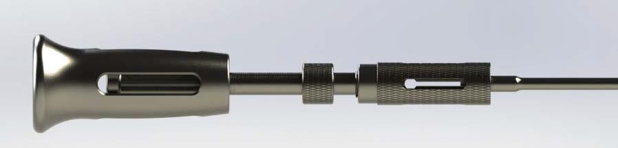
10 In Line Slap Hammer - Optional Slide the In-Line Slap Hammer Handle in a controlled manner along the shaft to remove Outer locking sleeve Slide the In-Line Slap Hammer Handle in a controlled manner along the shaft to remove the instrument.

11 In Line Slap Hammer - Optional In preparation for sterilization the slap hammer lock collet needs to be fully unlocked as shown below. Collet unlocked
12 Implant Sizing Table VerteBRIDGE Plate Height (mm) Plate H08 H10 H12 H14 H16 Short (IR6001T) Medium (IR6002T) Long (IR6003T) Reference Number & Graft Volume (cc) 0 Lordosis 6 Lordosis L W H08 H10 H12 H14 H16 H08 H10 H12 H14 H IR6208P (1.7) 22 IR6808P (2.4) IR6308P (2.0) 22 IR6908P (2.9) IR6408P (2.3) 22 IR7008P (3.4) IR6508P (2.5) 22 IR7108P (3.8) IR6608P (2.6) 22 IR7208P (4.2) IR6210P (2.0) IR6810P (2.8) IR6310P (2.3) IR6910P (3.4) IR6410P (2.6) IR7010P (3.9) IR6510P (2.9) IR7110P (4.4) IR6610P (3.0) IR7210P (4.8) IR6212P (2.3) IR6812P (3.4) IR6312P (2.8) IR6912P (4.1) IR6412P (3.2) IR7012P (4.7) IR6512P (3.4) IR7112P (5.3) IR6612P (3.6) IR7212P (5.8) IR6214P (2.7) IR6814P (3.9) IR6314P (3.2) IR6914P (4.8) IR6414P (3.6) IR7014P (5.5) IR6514P (4.0) IR7114P (6.1) IR6614P (4.2) IR7214P (6.7) IR6216P (3.1) IR6816P (4.5) IR6316P (3.7) IR6916P (5.4) IR6416P (4.1) IR7016P (6.3) IR6516P (4.5) IR7116P (7.0) IR6616P (4.8) IR7216P (7.7) IR6228P (1.6) IR6828P (2.3) IR6328P (1.9) IR6928P (2.8) IR6428P (2.1) IR7028P (3.2) IR6528P (2.3) IR7128P (3.6) IR6628P (2.5) IR7228P (4.0) IR6230P (1.9) IR6830P (2.6) IR6330P (2.2) IR6930P (3.2) IR6430P (2.5) IR7030P (3.7) IR6530P (2.8) IR7130P (4.2) IR6630P (2.9) IR7230P (4.6) IR6232P (2.3) IR6832P (3.2) IR6332P (2.7) IR6932P (3.9) IR6432P (3.0) IR7032P (4.5) IR6532P (3.3) IR7132P (5.0) IR6632P (3.5) IR7232P (5.5) IR6234P (2.7) IR6834P (3.8) IR6334P (3.2) IR6934P (4.5) IR6434P (3.6) IR7034P (5.3) IR6534P (3.9) IR7134P (5.9) IR6634P (4.1) IR7234P (6.4) IR6236P (3.1) IR6836P (4.3) IR6336P (3.6) IR6936P (5.2) IR6436P (4.1) IR7036P (6.0) IR6536P (4.4) IR7136P (6.7) IR6636P (4.7) IR7236P (7.4) 12

13 Cage Holder Assembly and Cage Loading Introduce and screw the threaded rod into the Cage Holder. Cage Holder Threaded Rod Cage Holder Handle 1. Press the button on the Cage Holder Handle 2. Insert the Cage Holder into the Cage Holder Handle 3. Release the Button Note: Low profile cage holder also available SI-AVEL-0001 Cage Holder (Assembled)


14 Implant Trial Selection Hook the Cage Holder in the groove located on the posterior side of the cage. Note: Make sure that one of the grooves in the slotted knob aligns with the groove in the Cage Holder handle, so that the Impactor can be introduced. Screw the slotted knob finger tight to secure the cage onto the Cage Holder. CAUTION: Do not overtighten,, as this may strip the thread in the cage. Slotted knob Groove of the handle
15 Cage Preparation The graft chamber must be filled with bone graft. Place the cage in the Graft Support. Graft Support Compact the bone graft into the chamber using the Graft Compactor. Graft Compactor


16 Cage Insertion Insert the cage in the intervertebral space by successive impactions on the Cage Holder, along the lateral axis. Note the orientation of the Cage Holder. Its posterior face has been labeled. The Adjustable Stop can be added to the Cage Holder to control the positioning of the cage during insertion. The position of the Adjustable Stop can be changed intraoperatively with the Adjustable Stop Screwdriver. Adjustable Stop Note: The Adjustable Stop enables a variable positioning of the implant and maintains it during the insertion of the plating. Note: With the stop set to 0 at the beginning of insertion, the proximal end of the cage will sit approximately 1.25 mm inside the disc space. Adjustable Stop Screwdriver 16


17 Cage Insertion Confirm proper implant placement using fluoroscopy. Lateral View: The posterior face of the Cage Holder corresponds to the posterior face of the cage, allowing visualization of the A/P position of the cage. A/P View: Central markers 1 and 2 indicate the implant center (their alignment proves the absence of rotation). Lateral marker 3 indicates the lateral edge of the cage. Under fluoroscopy, the edge of the cage inserter will indicate the proximal edge of the cage. Note: The Modular Handle may be removed to facilitate fluoroscopic imaging. Note: When placed at the proper depth, the proximal edge of the cage is inset approximately 1.25mm. Note: If a wider cage is desired to protrude contralaterally to obtain full cortical support, the central markers will be o set from the midline in a true A/P view. 17
, it is essential to use fluoroscopy to confirm proper cage placement")
 Figure 5.")
18 Cage Insertion The Cage Holder (IR R) and optional Adjustable Stop (IR R) are available in the standard instrument set so that the surgeon has access to either option. Note: Regardless of which Cage Holder is used, and whether or not the optional modular depth stop is assembled (Figure 4 and 5), it is essential to use fluoroscopy to confirm proper cage placement and that the cage position is maintained during deployment of the VerteBRIDGE plating. Under the fluoroscopy, the distal edge of the Cage Holder will indicate the proximal edge of the cage. Images should be taken during and after cage and plate impactions. Figure 4. Fluoroscopic assessment with the existing Cage Holder ( and Adjustable Stop) Figure 5. Fluoroscopic assessment with the new Low Profile Cage Holder

19 Starter Awl The Avenue L Starter Awl is used after insertion of the Avenue L cage and confirmation of the proper placement of the Avenue L Cage using fluoroscopy.


20 Step 1: Starter Awl Selection & Assembly Select the starter awl depending on the anchoring VerteBRIDGE plate size chosen: Starter Awl S» VerteBRIDGE Anchoring plate S Starter Awl M» VerteBRIDGE Anchoring plate M Starter Awl L» VerteBRIDGE Anchoring plate L One instrument to prepare both trajectories.


 to note the trajectory of the Awl.")
21 Step 1: Starter Awl Selection & Assembly Engage the thinner part of the holder in the groove of the cage holder handle. Area of thinner width. When the instrument is placed blade make sure that the holder is fully engaged. There are markings (1 or 2 ) to note the trajectory of the Awl. These are the same as the numbering system on the PEEK cartridge of the Avenue L VerteBRIDGE Plates Holder 21


22 Step 2: Create the 1st Groove Apply pressure to the Awl Holder until Awl is seated in the Implant Holder housing. Once seated apply thumb pressure to engage the Awl tip with the desired endplate Continue the insertion with a mallet until fully seated. Laser etch lines on the awl holder and implant holder act as a secondary check to ensure awl is fully seated Remove the Starter Awl with a mallet or slotted slap- hammer Insert the 1st Avenue L Anchoring Plate following the Avenue L surgical technique. Stops 22
23 Step 2: Create the 1st Groove Disassemble the Starter Awl and invert such that the 2 is showing. Perform the assembly on the cage holder as in Step 1 Repeat the steps in Step 2 to create the second groove. After completing the second groove remove the Starter Awl.


24 Plate Insertion Once the position of the cage is determined, the plating can be inserted. The impaction of the plates is performed sequentially. Each plate comes pre-assembled on a radiolucent single-use Plate Holder. Insert the first Plate Holder on the Impactor up to the starting mark. Make sure the face marked 1 is aligned with the mark on the Impactor. Insert the thinnest part of the Impactor in the groove of the Cage Holder handle. As the two instruments are joined, the mechanical stop guides the Impactor along the Cage Holder. Mechanical stop Plate Holder Impactor Mark Face number 1

25 Plate Insertion Using your thumb, press the Impactor until the plate bites into the vertebral body. Confirm plate trajectory with fluoroscopy. When trajectory is confirmed the plate may be fully inserted. Mallet the Impactor until it reaches the mechanical stop and the impaction marks are aligned. Check for proper placement of the first anchoring plate using fluoroscopy. Note: The anchoring plate releases automatically from its holder. The empty plate holder is retained on the Impactor. Note: When the Impactor is fully seated the impaction marks will be aligned. Note: For 2-level implantation, L anchoring plates should not be used in the shared vertebra. Before inserting the plates, make sure that the vertebral bodies have s u cient height to avoid contact between the plates of the two implants.


26 Plate Insertion Insert the second anchoring plate holder on the Impactor up to the starting mark. Make sure the face marked 2 is aligned with the mark on the Impactor. Insert the second anchoring plate by impacting on the Impactor until it reaches its mechanical stop and the impaction marks are aligned. Note: The anchoring plate releases automatically from its Plate Holder. The empty Plate Holder is retained on the Impactor. Check for proper placement of the second anchoring plate using fluoroscopy. CAUTION: At this step of the procedure, make sure that numbers 1 and 2 of the two Plate Holders are visible.


27 Final verification and Cage Holder removal Unscrew the slotted knob to release the threaded shaft from the cage. Disengage the Cage Holder s hook from the cage groove with a slight posterior translation and remove it carefully from the intervertebral disc space. A/P and lateral fluoroscopic verification of the plate placement confirms optimal position. 27

28 Final Verification and Cage Holder Removal Dispose of the Plate Holders at the end of the procedure.


29 Supplemental Fixation VerteBRIDGE plate trajectory must be reviewed via fluoroscopy prior to implanting supplemental posterior fixation. Steps should be taken during pedicle screw placement to confirm the screws will not contact the deployed VerteBRIDGE plating. Make the initial path through the pedicle with the awl and/or pedicle probe. Use the tap to create a path for the screw threads.


30 Supplemental Fixation Use a pedicle probe to ensure that there is no interference between the screw trajectory and the VerteBRIDGE plating. If there is interference between the screw path and the VerteBRIDGE plating during Steps 1-3, either create a new screw trajectory or select a shorter screw.

31 Revision: VerteBRIDGE Plate Removal A Revision System is provided in the event the plating must be removed Hook Revision System CAUTION: Plates are single-use only and should not be reinserted once they have been removed.


32 Revision: VerteBRIDGE Plate Removal 1. Insert the Revision System hook parallel to and between the two plates. 1. Rotate the instrument 90, so that the curve of the handle points toward the anchoring plate to be removed. This aligns the pins of the Revision System with the corresponding implant slots (threaded hole and groove). 2. Screw the knob until the head of the revision system contacts the cage.


33 Revision: VerteBRIDGE Plate Removal Once the plate is locked to the Revision System, slightly lever the instrument away from the plate that is being removed. This action unlocks the plate and in conjunction with the Slotted Mallet, eases plate removal. Repeat the previous steps in order to engage and extract the remaining anchoring plate. Revision - cage removal Reattach the Cage Holder to the implant and use the Slotted Mallet to progressively extract the implant.


34 Additional Instruments Lateral Disc Targeting Tool Part # SI-AVEL-0022 Angled Up Cobb Elevators 19mm Part # SI-AVEL-0025 Disc Starters 17mm x 6mm Part # SI-AVEL mm x 6mm Part # SI-AVEL-0005 Cupped Cobb Elevators 13mm Part # SI-AVEL mm Part # SI-AVEL mm Part # SI-AVEL-0017 Straight Cobb Elevator 22mm Part # SI-AVEL-018 Disc Evacuator 17mm x 6mm Part # SI-AVEL mm x 8mm Part # SI-AVEL mm x 10mm Part # SI-AVEL mm x 12mm Part # SI-AVEL mm x 6mm Part # SI-AVEL mm x 8mm Part # SI-AVEL mm x 10mm Part # SI-AVEL mm x 12mm Part # SI-AVEL-0013 Revolving Disc Cutter 8mm Part # SI-AVEL mm Part # SI-AVEL mm Part # SI-AVEL-0021 Angled Down Cobb Elevators 19mm Part # SI-AVEL


35 Additional Instruments Apophyseal Ring Bridge Part # SI-AVEL-0014 Note: Apophyseal Ring Bridges are intended to be used with low-profile cage holder only. Fluoro Modulator Part # SI-AVEL
36 Device description The Avenue L implants are devices whose primary functions are to add a solid structure to a graft so as to enable the stabilization of intervertebral height, after discectomy, during the time of graft setting and achieve a maximum surface of fusion. Various sizes of these implants are available, so that adaptations can be made to take into account the pathology and individual patient. In addition, so as to favor bone growth, the Avenue L lateral cage must be filled with bone graft. The Avenue L implants are manufactured from (radiolucent) PEEK-OPTIMA LT1 with surgical titanium alloy (Ti6Al4V) radiological position markers. The VerteBRIDGE Anchoring Plates are manufactured from surgical titanium alloy (Ti6Al4V). Instrumentation designed for implantation of the Avenue L Lateral Lumbar Cage is manufactured from biocompatible materials such as medical grade stainless steel. Indications for use The Avenue L Lateral Lumbar Cage system is indicated for intervertebral body fusion of the lumbar spine, from L2 to S1, in skeletally mature patients who have had six months of nonoperative treatment. The device is intended for use at either one level or two contiguous levels for the treatment of degenerative disc disease (DDD) with up to Grade I spondylolisthesis. DDD is defined as back pain of discogenic origin with degeneration of the disc confirmed by history and radiographic studies. The device system is designed for use with or without integrated fixation and must be used in conjunction with posterior supplemental fixation (e.g. pedicle screws). The device system is intended to be used with autograft and/or allogenic bone graft composed of cancellous and/or corticocancellous bone graft to facilitate fusion. Contraindications Cardiac problems. Abuse of medicine, drugs, tobacco or alcohol (which change the ossification power). Bony abnormalities preventing safe anchoring plate fixation. Material sensitivity, documented or suspected. Any mental or neuromuscular disorder which would create an unacceptable risk of fixation failure or complications in post-operative care. Bone stock compromised by disease, infection or prior implantation which cannot provide adequate support and or fixation to the implant. Obesity can produce loads on the spinal system which can lead to failure of the fixation of the device or to failure of the device itself. Recent infection, fever or hyper-leukocytosis. Open wounds. Bone absorption, osteopenia and/or osteoporosis. Patients having inadequate tissue coverage over the operative site. Pregnancy. Excessive local inflammation Other medical (for example: anesthetics risks) or surgical conditions which would preclude the potential benefit of spinal implant surgery such as the presence of tumors, congenital abnormalities, elevation of sedimentation rate unexplained by other diseases. Major spinal instabilities. Degenerative spondylolisthesis grade II or more.
37 Warnings Risks associated with general surgery, orthopedic surgery, and the use of general anesthesia should be explained to the patient prior to surgery. It is also recommended that the advantages and disadvantages of surgery, the implants, as well as alternative treatment methods be explained to the patient. Potential risks associated with the use of this system, which may require additional surgery, include device component failure (bending, loosening or fracture), loss of fixation, non-union, fracture of the vertebra, neurological injury, vascular or visceral injury, neurological complications, over-distraction, trauma to nerve root or dura, incorrect implant positioning, implant migration, pseudoarthrosis, disc height loss (impaction of implant into vertebral end plates), allergy or inflammation, general adverse effects related to surgical procedures (e.g. anesthesia, infection), subsidence, and expulsion. The device can break if it is subjected to increased loading associated with delayed union or non-union. If healing is delayed or does not occur, the implant could eventually break due to material fatigue. Factors such as the patient weight, activity level, and compliance to weight bearing or load bearing instructions, have an effect in the stresses to which the implant may be subjected, and may affect the longevity of the implant. Patients with previous spinal surgery at the level(s) to be treated may have different clinical outcomes compared to those without a previous surgery. Discard all damaged or mishandled implants. Under no circumstances may the implants be re-used. Although the device may appear intact on removal, internal modification due to the stress and strains placed on it or small defects may exist which may lead to fracture of the implant. Implants removed from a patient that contact bodily tissues or fluids should never be reused at risk of contamination of the patient. Mixing Metal: Some degree of corrosion occurs on all implanted metal and alloys. Contact of dissimilar metals (e.g. stainless steels and titanium), however, may accelerate this corrosion process. The presence of corrosion may accelerate fatigue fracture of implants and the amount of metal compounds released into the body system may also increase. Manufacturers employ different materials, manufacturing specifications and differing design parameters. Components of the Avenue L Interbody Fusion System should not be used in conjunction with components from any other manufacturer, except for the Zimmer Biomet Spine Timberline retractor. Any decision by a surgeon to remove the device should take into consideration such factors as the risk to the patient of the additional surgical procedure as well as the difficulty of removal. Implant removal should be followed by adequate postoperative management to avoid fracture. Before implanting the Avenue L lateral cage, the vertebral plates must be carefully prepared, being careful not to weaken the cortical bone to avoid implant subsidence. The setting and possible repositioning of the Avenue L lateral cage must be done with the cage holder attached to the cage. In order to ensure its stability in the intervertebral space, the Avenue L implant must be used with a supplemental internal fixation system (plate and/or screws type). Do not attempt to reposition the implant after anchoring plates have been deployed into the vertebral endplates. Avenue L has not been evaluated for safety and compatibility in the MR environment. It has not been tested for heating, migration, or image artifact in the MR environment. The safety of Avenue L in the MR environment is unknown. Scanning a patient who has this implant may result in patient injury. Precautions Being a technically demanding procedure presenting a risk of serious injury to the patient, the implantation of intersomatic systems should be performed only by experienced spine surgeons with specific training in the use of this system and who have knowledge of the present instructions for use. Based on fatigue testing results, when using the Avenue L Implant System, the physician/surgeon should consider the levels of implantation, patient weight, patient activity level, other patient conditions, etc., which may impact on the performance of this system. Patients who smoke have been shown to have an increased incidence of non-unions. Such patients should be advised of this fact and warned of the potential consequences.
38 Precautions (cont.) If the patient is involved in an occupation or activity which applies inordinate stress upon the implant (e.g., running, lifting of significant loads, or muscle strain), resultant forces can cause failure of the device. In some cases, progression of degenerative disease may also be so advanced at the time of implantation that they may substantially decrease the expected useful life to the implant. In such cases, orthopedic devices may be considered only as a delaying technique or to provide temporary relief. Before clinical use, the surgeon should thoroughly understand all aspects of the surgical procedure and limitations of the system. This device is recommended for use only by surgeons familiar with preoperative and surgical techniques, cautions and potential risks associated with spinal surgery. Knowledge ofsurgical techniques, selection and placement of implants, and pre and post-operative patient management are considerations essential to a successful surgical outcome. Patients should be instructed in detail about the limitations of the implants, including but not limited to the impact of excessive loading through patient weight or activity, and should be taught to govern their activities accordingly. Appropriate selection, placement and fixation of the spinal system components are critical factors which affect implant service life. Accordingly, strict adherence to the indications, contraindications, precautions, and warnings for this product is essential to potentially maximize service life. Care must be taken to protect the components from being marred, nicked or notched as a result of contact with metal or abrasive objects. Alterations will produce defects in surface finish and internal stresses which may become the focal point for eventual breakage of the implant. Inspection and trial assembly are recommended prior to surgery to determine if the instruments have been damaged during storage or prior procedures. After any surgery, it is necessary to check the proper position of the implants and to follow the evolution of the fusion using appropriate techniques For the anchoring plates, it is imperative to respect the following points: During multi-level implantations, care should be taken in plate size selection to minimize the possibility of adjacent plate interference. Ensure the cage does not protrude proximally outside the intervertebral disc space to be sure that the anchoring plates are properly positioned in the vertebral body. Prior to implanting the posterior construct, use fluoroscopy to verify the trajectory of the anchoring plates to avoid impingement with pedicle screws. If there is potential for the plates to contact pedicle screws, it may be necessary to change the trajectory of the screws. Sale of this product is restricted to physicians. MR Safety Information Non-clinical testing has demonstrated that the Interbody Cage Systems are MR-Conditional. Patients can be scanned safely immediately after implantation under the following conditions: Static magnetic field of 1.5 Tesla (1.5T) or 3.0-Tesla (3.0T). Maximum spatial gradient field of 3100 G/cm (31 T/m) for 1.5T Systems and 1500 G/cm (15T/m) for 3.0T systems Normal Operating Mode: Maximum whole-body specific absorption rate (SAR) of o 2.0 W/kg for 15 minutes of scanning at 1.5T. 2.0 W/kg for 15 minutes of scanning at 3.0T. When other methods of supplemental fixation are used, also follow the MR conditional labeling for the additional components. Under the scan conditions defined above, the Avenue L Implant System is expected to produce a maximum temperature rise of less than 1 C after 15 minutes of continuous scanning. In non-clinical testing, the image artifact caused by the device extends approximately 1.0cm from the Avenue L Implant when imaged with a gradient echo pulse sequence in either a 1.5T or a 3.0T MRI system.

39 Retractor System 39
 will be exposed at the end of the dilator (see figure 2). The ball probe should never stick out from the distal end of the dilator probe.")

40 Avenue L Retractor System Plug the probe to a suitable electrical source via DIN touch proof connectors. Insert carefully the probe in the smallest dilator tube groove. The detection ball (located at the end of the probe) will be exposed at the end of the dilator (see figure 2). The ball probe should never stick out from the distal end of the dilator probe. Do not overstrain nor press too hard while inserting the probe. Use the dilator holder to handle the dilators (see figure 3). Dilator holder can be connected to the table clamp to avoid surgeon hand exposure during X-rays Slide down carefully the smallest dilator containing the probe into the wound. Remove the probe from the dilator once nerve monitoring is completed and slide carefully the largest dilator on top of the smallest dilator. Nerve monitoring can be performed at this stage using the groove located in the dilator. Once the largest dilator is in position, it will provide the surgeon with the desired diameter where the retractor blades will fit tightly around it for the insertion of the device. Removing advice: First remove the probe, then the dilator. Dispose the probe after use. Fig. 2 Fig. 3


.")
41 Retractor, Blades & Wrench The retractor, blades and wrench are reusable devices. Choose the blades size using the graduation on the dilators (blades length depending on the thickness of the patient, on the selected access for the surgery or on the surgeon's needs). Fix the 3 selected blades on the retractor body: they slide vertically either from the top or from beneath, until a click can be heard: Place a right blade (R marked) on the right arm (R marked) (see figure 4) Place a left blade (L marked) on the left arm (L marked) Place a central blade (C marked) on the middle arm Before inserting the retractor in the wound, check that the retractor is well closed: blades shall make a closed circle (see figure 4). If necessary, use the right and left screws to reduce the angle of the blades (refer to figure 6 for more details). Then slide down the retractor body with its blades in the wound around the largest dilator (see figure 5). Figure 4
42 Retractor, Blades & Wrench Spreading is obtained via 3 different movements (see figure 6) a) Spreading of the lateral blades: Squeezing the handle and screw the knob to maintain spreading as needed (see figure 7) b) Tilting movement of the lateral blades: to enlarge the well on the bottom, use the screws on the left and right arms: screwing opens angularly the blades, unscrewing closes the blades (see figure 8). c) Lateral movement of the selected blades: Screwing the big black knob will enlarge the opening, either the central blade will slide posterior or the 2 lateral blades will slide anterior, depending on where the retractor clamp is fixed to the table (see figure 9). Figure 7 Figure 8 Figure 9
43 Retractor, Blades & Wrench Tip: The wrench can be used to screw and unscrew every knob of the retractor. Removing advice: To remove the blades from the retractor, use the push button on each arm of the retractor and pull the corresponding blade (see figure 10). Figure 10
44 Retractor, Blades & Wrench Below are instructions regarding the use of the Avenue Adjustable Table Clamp. Attach the Adjustable Table Clamp to the bed rail by turning the handle clockwise until the jaws have firmly grasped the rail. Typically the Adjustable Table Clamp is attached on the bed rail opposite the surgeon. Once the Adjustable Table Clamp is securely fastened to the bed rail insert the Avenue Flexible Arm and proceed starting on page 10 of the AVE ST4 Rev A. 44

45 Retractor, Blades & Wrench The table clamp is a reusable device. It's a flexible arm which is used to fasten the retractor to the surgery table To fix the flexible arm to the surgery table, refer to the specific Instruction For Use of the table clamp. Use knob 1 to fasten the table clamp base to the surgical table rail (see figure 11). Use knob 2 to set the height of the arm. Use knob 3 to lock the three articulations. Use knob 4 to fasten the retractor with the arm. Note: Optional table clamp shown. 45
46 Retractor, Blades & Wrench Use the black connector to fix the table clamp to the retractor (see figure 12). To fix the retractor on the table clamp, depending on the surgeon choice, there are 2 different options: Central blade is mobile & lateral blades are fixed: Use the steel lock to lock the retractor body and lateral blades. Only the central blade will slide forwards and backwards when screwing and unscrewing the black knob. Central blade is fixed and lateral blades are mobile: Use the black lock to immobilize the central arm. Screwing and unscrewing the black knob will slide the lateral blades (and the retractor body) forwards or backwards. Figure 12

47 Disposable Light Mats & Extension Cords The light mats are sterile single use products. Check the expiration date and the packaging before use. Dispose after use. The extension cords are reusable. The light mat and its optic fiber cable are employed to light up the bottom of the wound. Plug the extension cord to the light mat. Plug the extension cord to a suitable light source, following the instructions for use of the light mats. Slide carefully the mat through the dedicated slot inside the blade until the appropriate depth (see figure 13). Removing advice: To remove the disposable light mat, slide carefully the light mat out of the blade. Dispose after use. Figure 13 47


 (In that case, the 4th blade support")

48 Light Cable & Light Holder The light cable and the light holder are reusable. The light cable helps to light up the wound. These lights are inserted into the light holder. The light holders are placed in their corresponding holes: Spot 1: on the retractor body (see figure 14) Spot 2: on the optional 4th blade support (see figure 15) (In that case, the 4th blade support uses the same holes as the light cables (spot 1): both plastic connectors of the 4th blade support are inserted in the retractor body as shown in figure 15. Figure 14 Figure 15

.")

 before locking the shim (or broach) by pushing the firing pin")
 into the blade dedicated slot until the")
49 Shim, broach and shim/broach holder Shim, broach and shim/broach holder are reusable products. Clip the shim (or broach) lug in the shim/broach holder dedicated claw on the top end of the holder (see figure 16). The laser marking on the shim (or broach) provides you with the correct orientation. Make sure the lug is well tightened by the holder claw (see figure 17) before locking the shim (or broach) by pushing the firing pin (with the thumb) (see figure 18). Insert the shim (or broach) into the blade dedicated slot until the suitable depth (see figures 19 & 20). Release the shim (or broach) by pushing the trigger (with the forefinger). Figure 16 Figure 17 Figure 18 Figure 19 Figure 20
 is similar as the shim/broach procedure (see precedent chapter 1.3.8 Shim, broach and shim/broach holder).")
50 Optional blade extension The blade extension is reusable. Shim, Broach & Shim/Broach Holder - Handling of the blade extension (see figure 21) is similar as the shim/broach procedure (see precedent chapter Shim, broach and shim/broach holder). Clip the blade extension lug in The blade extension is reusable. Handling of the blade extension (see figure 21) is similar to the shim/broach procedure. holder (see figure 16). Make sure the lug is well tightened by the holder claw (see figure 17) before locking the blade extension by pushing the firing pin (with the thumb) (see figure 18). Insert the blade extension into the blade dedicated slot until the suitable depth (see figure 22). pin (with the thumb) (see figure 18). the shim/broach holder dedicated claw on the top end of the - Make sure the lug is well tightened by the holder claw (see figure 17) before locking the blade extension by pushing the firing Figure 22 Figure 22 Figure 21 - Insert the blade extension into the blade dedicated slot until the suitable depth (see figure 22).
51 Optional 4th Blade & Support The 4th blade and its support are reusable. Clip of the 4th blade support connectors to the retractor spots (see figure 23) (they are the same spots used to plug the light cables in chapter Light cable and light holder). Select the suitable 4th blade (depending on the required length and width). Insert the 4th blade in the dedicated groove inside the support. At this point the 4th blade is still mobile: it can be moved up and down (through the groove support) or laterally (support is sliding), even angularly (see figure 24). Lock the height and the lateral position of the 4th blade screwing the knob with the wrench (see figure 25). Figure 23 Figure 24 Figure 25
52 Cleaning, Sterilization & Maintenance Recommendations for the handling of these surgical instruments are as follows: Do not let blood or tissue dry on the instrument Rinse the instrument immediately after use and before decontamination. As much as possible, manipulate instruments made from different metals separately. Check the functionality and cleanliness of each instrument before use. Time dwell between use and cleaning of the devices should be minimal. Devised should be carried into wet wraps from the surgery room to the cleaning room. Some devised should be disassembled prior to cleaning, some should be handled specifically prior to cleaning, as described below Disassembling of the retractor Hold the tips away from each other screwing the lateral nut and turn the black knob until the central arm is totally out. Both handles have to be removed from the retractor body. Push the left and right buttons to release both handles (see figure 27). Figure 27
53 Shim/Broach Holder Disassembling Disassemble the shim/broach holder for cleaning: take off the pin (see figure 28), then pull the firing pin and hold up the rod (see figure 29). Figure 28 Figure th blade support special handling care The support must be completely opened (maximum wingspan) to maximize the access for cleaning (see figure 30). Screw must be totally loosened. Figure 30
54 Maintenance Use surgery lubricant before each use. Apply only where it is recommended in the following pictures (red circles). WARNING: DO NOT lubricate any part which will be in contact with the patient Retractor Table clamp connector
55 Maintenance Shim holder

56 Disclaimer: This document is intended exclusively for physicians and is not intended for laypersons. Information on the products and procedures contained in this document is of a general nature and does not represent and does not constitute medical advice or recommendations. Because this information does not purport to constitute any diagnostic or therapeutic statement with regard to any individual medical case, each patient must be examined and advised individually, and this document does not replace the need for such examination and/or advice in whole or in part. Caution: Federal (USA) law restricts this device to sale by or on the order of a physician. Rx Only. Please refer to the package inserts for important product information, including, but not limited to, indications, contraindications, warnings, precautions, adverse effects, and patient counseling information. ldr.com 2017 LDR, a division of Zimmer Biomet Spine, Inc. All rights reserved. All content herein is protected by copyright, trademarks and other intellectual property rights owned by or licensed to LDR, a division of Zimmer Biomet Spine, Inc. or one of its affiliates unless otherwise indicated, and must not be redistributed, duplicated or disclosed, in whole or in part, without the express written consent of Zimmer Biomet Spine. This material is intended for health care professionals and the Zimmer Biomet Spine sales force. Distribution to any other recipient is prohibited. AVE ST 9 Rev C
nva Anterior Lumbar Interbody Fusion System
 nva Anterior Lumbar Interbody Fusion System 1 IMPORTANT INFORMATION FOR PHYSICIANS, SURGEONS, AND/OR STAFF The nv a, nv p, and nv t are an intervertebral body fusion device used in the lumbar spine following
nvt Transforaminal Lumbar Interbody Fusion System
 nvt Transforaminal Lumbar Interbody Fusion System 1 IMPORTANT INFORMATION FOR PHYSICIANS, SURGEONS, AND/OR STAFF The nv a, nv p, and nv t are an intervertebral body fusion device used in the lumbar spine
nvp Posterior Lumbar Interbody Fusion System
 nvp Posterior Lumbar Interbody Fusion System 1 IMPORTANT INFORMATION FOR PHYSICIANS, SURGEONS, AND/OR STAFF The nv a, nv p, and nv t are an intervertebral body fusion device used in the lumbar spine following
EXACTECH SPINE. Operative Technique. Cervical Spacer System. Surgeon focused. Patient driven. TM
 EXACTECH SPINE Operative Technique Cervical Spacer System Surgeon focused. Patient driven. TM ACAPELLA ONE Acapella One Cervical Spacer System is an anterior cervical discectomy and fusion device with
Thoracolumbar Solutions. Avenue T. TLIF Cage. with ertebridge PLATING TECHNOLOGY. Surgical Technique Guide
 Thoracolumbar Solutions Avenue T TLIF Cage with ertebridge PLATING TECHNOLOGY Surgical Technique Guide 2 Avenue T TLIF Cage Surgical Technique VerteBRIDGE Plating is the integrated fixation designed specifically
Alamo T Transforaminal Lumbar Interbody System Surgical Technique
 Transforaminal Lumbar Interbody System Surgical Technique Table of Contents Indications and Device Description.............. 1 Alamo T Implant Features and Instruments...........2 Surgical Technique......................
Cervical Spacer System surgical technique
 Blackhawk TM Cervical Spacer System surgical technique Blackhawk TM The BLACKHAWK Cervical Spacer System is designed to provide biomechanical stabilization as an adjunct to fusion. Spinal fixation should
Alamo C. Cervical Interbody System Surgical Technique. An Alliance Partners Company
 Cervical Interbody System Surgical Technique Table of Contents Indications for Use................................1 Device Description............................... 1 Alamo C Instruments..............................
TM TM Surgical Technique
 TM TM Surgical Technique TABLE OF CONTENTS Reli SP Spinous Plating System Overview Device Description Implant Features Indications Instruments Access Instruments Preparation Instruments Insertion Instruments
OPERATIVE TECHNIQUE. CONSTRUX Mini PTC. Mini PTC Spacer System
 OPERATIVE TECHNIQUE CONSTRUX Mini PTC Mini PTC Spacer System TABLE OF CONTENTS Introduction 1 Operative Technique 2 Part Numbers 6 Indications For Use 7 INTRODUCTION 1 INTRODUCTION The CONSTRUX Mini PTC
Imola Lateral IBF System Surgical Technique
 Imola Lateral IBF System Surgical Technique IMOLA CIRCUIT TABLE OF CONTENTS Design Rationale Instructions for Use Surgical Technique 1. Table Mounting 2. Surgical Planning & Targeting 3. Access and Preparation
HawkeyeTM Peek. surgical technique
 HawkeyeTM Peek surgical technique Introduction The ChoiceSpine HAWKEYE Vertebral Body Replacement (VBR) System is intended for use in the thoracolumbar spine (T1 - L5) to replace a collapsed, damaged,
Veyron -C Anterior Cervical System Surgical Technique
 Veyron -C Anterior Cervical System Surgical Technique 2 Veyron-C Anterior Cervical System Surgical Technique Veyron-C Anterior Cervical System Surgical Technique Description, Indications & Contraindications...3
Zimmer Anterior Buttress Plate System. Surgical Technique
 Zimmer Anterior Buttress Plate System Surgical Technique 2 Zimmer Anterior Buttress Plate System Surgical Technique Zimmer Anterior Buttress Plate System Surgical Technique Description, Indications & Contraindications...
ACP1 CERVICAL PLATE SPINAL SYSTEM SURGICAL TECHNIQUE GUIDE II.
 I. ACP1 CERVICAL PLATE II. SPINAL SYSTEM SURGICAL TECHNIQUE GUIDE I. Introduction The Gold Standard Orthopaedics, LLC ACP1 Spinal System was designed with surgeons to incorporate strength, functionality,
SURGICAL TECHNIQUE GUIDE TRESTLE. Anterior Cervical Plating System
 SURGICAL TECHNIQUE GUIDE TRESTLE Anterior Cervical Plating System 2 SURGICAL TECHNIQUE GUIDE SURGICAL TECHNIQUE GUIDE System Features Large window enables visualization of graft site and end plates Screw
Surgical Technique. Apache Anterior Lumbar Interbody Fusion
 Surgical Technique Apache Anterior Lumbar Interbody Fusion 2 Table of Contents Page Preoperative Planning 4 Patient Positioning 4 Disc and Endplate Preparation 4 Distraction/Size Selection 5 Implantation
A U X I L I A R Y C O N N E C T O R S Surgical Technique
 A U X I L I A R Y C O N N E C T O R S Surgical Technique AUXILIARY CONNECTORS ISSYS LP Auxiliary Connectors The ISSYS LP auxiliary connectors were designed to provide medial-lateral variability for the
Apache Cervical Interbody Fusion Device. Surgical Technique. Page of 13. LC-005 Rev F
 LC-005 Rev F Apache Cervical Interbody Fusion Device Page of 13 Surgical Technique INDICATIONS: When used as an intervertebral body fusion device, the Genesys Spine Interbody Fusion System is indicated
Table of Contents.
 surgical technique The Ambassador TM Anterior Cervical Plate System is a versatile system of implants and instruments with a variety of sizes to provide optimal anatomic compatibility. The integrated cam
Advantage ALIF. Keith Shevlin Managing Director
 Advantage ALIF Unit 10, 9-11 Myrtle Street, Crows Nest NSW 2065 Keith Shevlin Managing Director keithshevlin@precisionsurgical.com.au Advantage ALIF Introduction & Indications for Use 1 Surgical Technique
Royal Oak IBFD System Surgical Technique Posterior Lumbar Interbody Fusion (PLIF)
") Royal Oak IBFD System Surgical Technique Posterior Lumbar Interbody Fusion (PLIF) Preoperative Planning Preoperative planning is necessary for the correct selection of lumbar interbody fusion devices.
Royal Oak IBFD System Surgical Technique Posterior Lumbar Interbody Fusion (PLIF) Preoperative Planning Preoperative planning is necessary for the correct selection of lumbar interbody fusion devices.
Zimmer Facet Screw System Surgical Technique
 Zimmer Facet Screw System Surgical Technique 2 Zimmer Facet Screw System Surgical Technique Zimmer Facet Screw System Surgical Technique Description, Indications & Contraindications...3 Surgical Technique...4
TABLE OF CONTENTS. ShurFit Anterior Cervical Interbody Fusion (ACIF) System Overview 2. Implant Specifications 3. Instrument Features 4
 System Overview 2. Implant Specifications 3. Instrument Features 4") Surgical Technique TABLE OF CONTENTS ShurFit Anterior Cervical Interbody Fusion (ACIF) System Overview 2 Product Highlights 2 Indications 2 Implant Specifications 3 Instrument Features 4 Surgical Technique
Surgical Technique TABLE OF CONTENTS ShurFit Anterior Cervical Interbody Fusion (ACIF) System Overview 2 Product Highlights 2 Indications 2 Implant Specifications 3 Instrument Features 4 Surgical Technique
Posterior Lumbar Interbody Fusion System
 Px Posterior Lumbar Interbody Fusion System Px PEEK INTERBODY FUSION SYSTEM INDICATIONS FOR USE The Innovasis Px PEEK IBF System is an intervertebral body fusion device for use in patients with degenerative
SURGICAL TECHNIQUE MANUAL. InterFuse T
 1 CONTENTS InterFuse T Product Description 3 Indications for Use 3 X-Ray Marker Locations 4 Product Specifications 4 Instrument Set 5 Step 1 Preoperative Planning 8 Patient Positioning 8 Step 2 Disc Removal
InFix. Anterior Lumbar Device. Surgical Technique Guide
 InFix Anterior Lumbar Device Surgical Technique Guide 2 InFix Anterior Lumbar Device Surgical Technique Guide InFix Anterior Lumbar System s modular design is intended to restore lordosis, disc height
C-THRU Anterior Spinal System
 C-THRU Anterior Spinal System Surgical Technique Manufactured From Contents Introduction... Page 1 Design Features... Page 2 Instruments... Page 3 Surgical Technique... Page 4 Product Information... Page
Solitaire Anterior Spinal System
 Surgical Technique Solitaire Anterior Spinal System Independent Stabilization for the Anterior Column Available in Titanium and Contents Introduction... Page 1 Design Features... Page 2 Instruments...
INTELLIGENT SPINAL SYSTEM
 INTELLIGENT SPINAL SYSTEM I. Introduction II. Product Specification III. Surgical Technique IV. Ordering Information V. IFU for Lospa IS SPINAL SYSTEM The LOSPA IS spinal system consists of
MODULAR DESIGN OFFERS FREEDOM OF CHOICE. Surgical Technique
 MODULAR DESIGN OFFERS FREEDOM OF CHOICE Surgical Technique Joint Spine Sports Med MectaLIF Anterior Surgical Technique 2 INDEX 1. INTRODUCTION 4 1.1 Material & Marker 5 2. INDICATIONS 5 3. CONTRAINDICATIONS
Y o u r Id e a s En g i n e e r e d t o Li f e
 ISSYS LP Spinal Fixation System Surgical Guide Y o u r Id e a s En g i n e e r e d t o Li f e In t r o d u c t i o n ISSYS LP Sp i n a l Fixation System The foundation of the ISSYS LP Spinal Fixation System
Threshold Pedicular Fixation System Surgical Technique
 Threshold Pedicular Fixation System Surgical Technique Table of Contents Patient Preparation and Positioning... 2 Determining Incision Location... 3 Assembling the Cannulated Awl... 4 Guide Wire Placement...
EIT TLIF Cage. For Natural Bone Ingrowth with EIT Cellular Titanium
 EIT TLIF Cage For Natural Bone Ingrowth with EIT Cellular Titanium EIT TLIF Cage Surgical Technique EIT Cellular Titanium provides active fusion area» ~ 80% porosity» ~ 650 µm diamond pore size» open interconnected
VTI INTERFUSE T SURGICAL TECHNIQUE FORWARD THINKING FOR THE BACK. 1/20
 VTI INTERFUSE T SURGICAL TECHNIQUE FORWARD THINKING FOR THE BACK. 1/20 CONTENTS InterFuse T Product Description Indications for Use X-Ray Marker Locations Product Specifications Instrument Set 3 4 5 STEP
VTI INTERFUSE S SURGICAL TECHNIQUE FORWARD THINKING FOR THE BACK.
 VTI INTERFUSE S SURGICAL TECHNIQUE FORWARD THINKING FOR THE BACK. CONTENTS InterFuse S Product Description Indications for Use X-Ray Marker Locations and Product Specifications Instrument Set 3 4 5-7 STEP
ACP. Anterior Cervical Plate System SURGICAL TECHNIQUE
 ACP Anterior Cervical Plate System SURGICAL TECHNIQUE ACP TABLE OF CONTENTS INTRODUCTION 4 INDICATIONS AND CONTRAINDICATIONS 5 WARNINGS AND PRECAUTIONS 6 IMPLANT DESCRIPTION 7 INSTRUMENTS 10 SURGICAL
Surgical Technique Manual
 InterFuse S Interbody Fusion System Surgical Technique Manual VERTEBRAL TECHNOLOGIES MS 4043-02 Rev. O Product Overview Introduction The VTI InterFuse S implant is an interbody fusion device that combines
Thoracolumbar Solutions ROI-A. ALIF Cage. Surgical Technique Guide
 Thoracolumbar Solutions ROI-A ALIF Cage Surgical Technique Guide 2 ROI-A ALIF Cage Surgical Technique Featuring VerteBRIDGE Plating Technology ROI-A ALIF Cage Surgical Technique Guide 3 TABLE OF CONTENTS
Thoracolumbar Solutions. Zyston Curve. Interbody Spacer System. Surgical Technique Guide
 Thoracolumbar Solutions Zyston Curve Interbody Spacer System Surgical Technique Guide 2 Zyston Curve Interbody Spacer System Surgical Technique Guide The Zyston Curve Interbody System is designed to optimize
CROSS -FUSE P E E K V B R / I B F SYST E M
 S U R G I C A L T E C H N I Q U E CROSS -FUSE P E E K V B R / I B F SYST E M S U R G I C A L S Y S T E M O V E R V I E W 2 CROSS-FUSE P E E K V B R / I B F S Y S T E M S U R G I C A L T E C H N I Q U E
Aesculap CeSpace TM PEEK and CeSpace TM XP Spinal Implant System Instructions for Use
 Aesculap CeSpace TM PEEK and CeSpace TM XP Spinal Implant System Instructions for Use Page 1 of 5 Indications for use: When used as a Vertebral Body Replacement Device: The CeSpace PEEK and CeSpace XP
PILLAR AL. Anterior Lumbar Interbody Fusion (ALIF) and Partial Vertebral Body Replacement (pvbr) PEEK Spacer System OPERATIVE TECHNIQUE
 and Partial Vertebral Body Replacement (pvbr) PEEK Spacer System OPERATIVE TECHNIQUE") PILLAR AL PEEK Spacer System Anterior Lumbar Interbody Fusion (ALIF) and Partial Vertebral Body Replacement (pvbr) OPERATIVE TECHNIQUE Table of Contents 1 INTRODUCTION 2 PRE-OPERATIVE TECHNIQUE 3 OPERATIVE
PILLAR AL PEEK Spacer System Anterior Lumbar Interbody Fusion (ALIF) and Partial Vertebral Body Replacement (pvbr) OPERATIVE TECHNIQUE Table of Contents 1 INTRODUCTION 2 PRE-OPERATIVE TECHNIQUE 3 OPERATIVE
L-VARLOCK. Posterior Lumbar Cage with adjustable lordosis. S urgical T echnique
 L-VARLOCK Posterior Lumbar Cage with adjustable lordosis S urgical T echnique Introduction Designed and manufactured by KISCO International, L-VARLOCK cages are made of titanium alloy Ti 6AI 4V (standards
Thunderbolt. surgical technique. MIS Pedicle Screw System. Where Nimble and Secure Intersect
 Thunderbolt TM MIS Pedicle Screw System Where Nimble and Secure Intersect surgical technique i www.choicespine.com System Features Dovetail set screw: Minimizes head splay and cross-threading Secure connection
TABLE OF CONTENTS. Vault C Anterior Cervical Discectomy 2 and Fusion (ACDF) System Overview. Implants 3. Instruments 5. Surgical Technique 10
 System Overview. Implants 3. Instruments 5. Surgical Technique 10") Surgical Technique TABLE OF CONTENTS Vault C Anterior Cervical Discectomy 2 and Fusion (ACDF) System Overview Indications 2 Implants 3 Instruments 5 Surgical Technique 10 1. Preoperative planning 10 2.
Surgical Technique TABLE OF CONTENTS Vault C Anterior Cervical Discectomy 2 and Fusion (ACDF) System Overview Indications 2 Implants 3 Instruments 5 Surgical Technique 10 1. Preoperative planning 10 2.
Royal Oak Cervical Plate System
 Royal Oak Cervical Plate System Manufactured by Nexxt Spine, Inc. Royal Oak Cervical Plate System INTRODUCTION FEATURES AND BENEFITS Table of Contents SURGICAL TECHNIQUE Step 1. Patient Positioning Step
BAK/C Cervical Anterior Interbody Fusion System
 Surgical Technique BAK/C Cervical Anterior Interbody Fusion System The Comfortable Choice for Cervical Fusion BAK/C Cervical Surgical Technique 1 The BAK/C Cervical Fusion System is an alternative to conventional
Cervical Solutions ROI-C. Cervical Cage. with ertebridge PLATING TECHNOLOGY. Surgical Technique Guide
 Cervical Solutions ROI-C Cervical Cage with ertebridge PLATING TECHNOLOGY Surgical Technique Guide 2 ROI-C Cervical Cage Surgical Technique Guide ROI-C featuring VerteBRIDGE plating is a zero-profile,
product overview Implant heights range from 8mm-20mm in 2mm increments, with two lordocic angle options of 6 and 12.
 ETHOS A-Spacer PEEK System Surgical Technique Guide Synchronizing Medical Innovation with Global Markets product overview The SyncMedical Ethos PEEK IBF System is an intervertebral body fusion device for
100 Interpace Parkway Parsippany, NJ
 100 Interpace Parkway Parsippany, NJ 07054 www.biometspine.com 800-526-2579 All trademarks are the property of Biomet, Inc. or one of its subsidiaries, unless otherwise indicated. Rx Only. 2009 EBI, LLC.
Valencia Pedicle Screw Surgical Technique
 Valencia Pedicle Screw Surgical Technique VALENCIA CIRCUIT TABLE OF CONTENTS Design Rationale Indications for Use Surgical Technique 1. Pedicle Preparation 2. Screw Insertion 3. Rod Placement 4. Locking
Cervical Solutions. Trinnect. Hydrated Anterior Cervical Spacer System. Surgical Technique Guide
 Cervical Solutions Trinnect Hydrated Anterior Cervical Spacer System Surgical Technique Guide 2 Trinnect Hydrated Anterior Cervical Spacer System Surgical Technique Guide Trinnect Cervical Allograft Spacers
surgical technique ROI-T
 surgical technique ROI-T TLIF CAGE Surgical Technique ROI-T TLIF cage Table of Contents Approach and facet/pedicle preparation... 3 Controlled distraction... 4 Discectomy and endplate preparation... 6
EasyStep. Operative technique
 Operative technique EasyStep - Step staple This publication sets forth detailed recommended procedures for using Stryker Osteosynthesis devices and instruments. It offers guidance that you should heed,
Cervical Solutions. Optio-C Anterior Cervical Plate. with Allograft/Autograft. Surgical Technique Guide
 Cervical Solutions Optio-C Anterior Cervical Plate with Allograft/Autograft Surgical Technique Guide 2 Optio-C Anterior Cervical Plate with Allograft/Autograft Surgical Technique Guide The Optio-C System
EFSPINE CERVICAL COMBINED SET DISC PROTHESIS ORGANIZER BOX
 EFSPINE CERVICAL COMBINED SET INSTRUMENTS CERVICAL CAGE & DISC PROTHESIS ORGANIZER BOX Cervical Thoracic Thoraco - Lumbar Sacral EFSPINE CERVICAL COMBINED SET CERVICAL IMPLANTS INTRODUCTION Cervical Disc
Pinit Plate Small Bone Fusion System Bone Plate & Screw System
 Pinit Plate Small Bone Fusion System Bone Plate & Screw System Description The Pinit Plate Small Bone Fusion System consists of 2-hole bone plates made available in three length options and two thickness
JuggerLoc Bone-to-Bone System for Ankle Syndesmosis Fixation. Surgical Technique
 JuggerLoc Bone-to-Bone System for Ankle Syndesmosis Fixation Surgical Technique Table of Contents Position and Preparation... 2 Incision... 2 Fracture Reduction... 2 Drill Fibula and Tibia... 3 JuggerLoc
Zimmer Trabecular Metal Ankle Interpositional Spacer and Trabecular Metal Ankle Fusion Spacer
 Zimmer Trabecular Metal Ankle Interpositional Spacer and Trabecular Metal Ankle Fusion Spacer Surgical Technique 2 Zimmer Trabecular Metal Ankle Interpositional Spacer and Trabecular Metal Ankle Fusion
O PE RATIV E TE C HN IQ U E. ProView. Expandable Retractor System U.S. EDITION
 O PE RATIV E TE C HN IQ U E ProView M I N I M A L A C C E S S P O R TA L ( M A P ) S Y S T E M Expandable Retractor System U.S. EDITION Table of Contents 1 INTRODUCTION 2 OPERATIVE TECHNIQUE 9 PART NUMBERS
VTI INTERLINK PEDICLE SCREW SYSTEM
 VTI INTERLINK PEDICLE SCREW SYSTEM SURGICAL TECHNIQUE FORWARD THINKING FOR THE BACK. DEVICE DESCRIPTION The VTI InterLink Pedicle Screw System is comprised of polyaxial pedicle screws in various diameters
OPERATIVE TECHNIQUE PTC PEEK FORZA. spacer system
 OPERATIVE TECHNIQUE PTC PEEK FORZA spacer system TABLE OF CONTENTS Introduction 1 Operative Technique 2 Instruments 12 FORZA PEEK Part Numbers 20 FORZA PTC Part Numbers 22 Modular Implant Inserter 24 Disassembly
SURGICAL TECHNIQUE ROI-T TM TLIF INTERSOMATIC IMPLANT TRANS-FORAMINAL APPROACH
 TLIF INTERSOMATIC IMPLANT TRANS-FORAMINAL APPROACH Table of Contents page Step 1 - Articular resection.................................................................................. 3 Step 2 - Pedicle
Technique Guide. T-PAL. Transforaminal posterior atraumatic lumbar spacer system.
 Technique Guide T-PAL. Transforaminal posterior atraumatic lumbar spacer system. Table of Contents Introduction T-PAL 2 AO Principles 4 Indications and Contraindications 5 Surgical Technique Preparation
M.I.S. MAKE IT SMART IN ONE SYSTEM. Surgical Technique. Hip Knee Spine Navigation
 M.I.S. MAKE IT SMART IN ONE SYSTEM Surgical Technique Hip Knee Spine Navigation M.U.S.T. Mini Open Surgical Technique Hip Knee Spine Navigation 2 C O N T E N T S 1 INTRODUCTION 4 2 SURGICAL TECHNIQUE 5
Surgical Technique INTERSOMATIC CERVICAL CAGE
 R INTERSOMATIC CERVICAL CAGE NEOCIF IMPLANTS NEOCIF is an implant designed to make anterior cervical interbody fusion (ACIF) easier and to remove the need for structural autologous graft. The cage is made
LUMBAR POSTERIOR MINIMALLY INVASIVE SYSTEM. Surgical Technique
 LUMBAR POSTERIOR MINIMALLY INVASIVE SYSTEM Surgical Technique Joint Spine Sports Med M.U.S.T. Mini Open Surgical Technique Joint Spine Sports Med CAUTION Federal law (USA) restricts this device to sale
TiLock XT Minimally Invasive Surgery (MIS) Pedicle Screw System
 Pedicle Screw System") TiLock XT Minimally Invasive Surgery (MIS) Pedicle Screw System The Genesys Spine TiLock XT Minimally Invasive Surgery (MIS) Pedicle Screw System consists of rods (straight and curved), lock screws, and
TiLock XT Minimally Invasive Surgery (MIS) Pedicle Screw System The Genesys Spine TiLock XT Minimally Invasive Surgery (MIS) Pedicle Screw System consists of rods (straight and curved), lock screws, and
OPERATIVE TECHNIQUE COVER IMAGE OPTIONAL (DETAIL) IMAGE SKYHAWK. lateral interbody fusion system lateral plate system
 IMAGE SKYHAWK. lateral interbody fusion system lateral plate system") OPERATIVE TECHNIQUE COVER IMAGE OPTIONAL (DETAIL) IMAGE SKYHAWK lateral interbody fusion system lateral plate system TABLE OF CONTENTS Introduction 1 Pre-Operative Technique 2 Operative Technique 3 Part
OPERATIVE TECHNIQUE COVER IMAGE OPTIONAL (DETAIL) IMAGE SKYHAWK lateral interbody fusion system lateral plate system TABLE OF CONTENTS Introduction 1 Pre-Operative Technique 2 Operative Technique 3 Part
HydraLok. Operative Technique. Polyaxial Pedicle Screw System
 HydraLok Operative Technique Polyaxial Pedicle Screw System Table of Contents Introduction...1 OPERATIVE TECHNIQUE OVERVIEW...2 DETAILED OPERATIVE TECHNIQUE...4 LOCATE AND PREPARE THE PEDICLE...4 PROBE
ROI-A Oblique. ertebridge PLATING TECHNOLOGY
 surgical technique ROI-A Oblique ALIF CAGE with ertebridge PLATING TECHNOLOGY Surgical Technique ROI-A Oblique ALIF CAGE Table of Contents Patient positioning and oblique surgical approach... Discectomy
Surgical Technique. Apache Posterior Lumbar Interbody Fusion Apache Transforaminal Lumbar Interbody Fusion
 Surgical Technique Apache Posterior Lumbar Interbody Fusion Apache Transforaminal Lumbar Interbody Fusion 2 Table of Contents Page Preoperative Planning 4 Patient Positioning 5 Disc Exposure 5 Disc and
Lapidus Arthrodesis System Instructions for Use
 Lapidus Arthrodesis System Instructions for Use Description The AlignMATE Lapidus Arthrodesis System consists of bone plates and bone screws (locking, non-locking and interfragmentary), which are intended
Luminary ALIF. Disc preparation and implant insertion instruments.
 Luminary ALIF. Disc preparation and implant insertion instruments. Technique Guide Instruments and implants approved by the AO Foundation Table of Contents Introduction Luminary ALIF 2 AO Principles 4
FastFrame External Fixation System. Damage Control Surgical Technique
 FastFrame External Fixation System Damage Control Surgical Technique 1 FastFrame External Fixation System Damage Control Surgical Technique Table of Contents Introduction... 2 Indications and Contradictions...
Cervical Solutions. Alta. ACDF System. Surgical Technique Guide
 Cervical Solutions Alta ACDF System Surgical Technique Guide 2 Alta ACDF System Surgical Technique Guide A comprehensive system to address a continuum of fixation requirements and anatomic demands. Alta
OPERATIVE TECHNIQUE. anterior cervical plating system
 OPERATIVE TECHNIQUE 3º anterior cervical plating system Introduction 1 Pre-Operative Technique 2 Oerative Technique 3 Instructions for Use 12 Part Numbers 16 The surgical technique shown is for illustrative
XRL A modular expandable radiolucent vertebral body replacement system
 XRL A modular expandable radiolucent vertebral body replacement system This publication is not intended for distribution in the USA. SURGICAL TECHNIQUE Table of Contents Introduction XRL 2 AO Spine Principles
Technique Guide Small Bone Fusion System
 Technique Guide Small Bone Fusion System The Pinit Plate Small Bone Fusion System is a super low profile, modular bone plate and screw system designed to stabilize a bunionectomy with a medial to lateral
TiLock 2 Spinal System. Surgical Technique
 TiLock 2 Spinal System Surgical Technique Table of Contents Page Preoperative Planning 4 Pedicle Preparation 5 Probe 5 Tap Pedicle 6 Screw Options 7 Screw Insertion 8 Aligning the Windows 9 Rod Insertion
SYNFIX EVOLUTION SECURED SPACER SYSTEM
 SYNFIX EVOLUTION SECURED SPACER SYSTEM Instruments and implants for stand-alone anterior lumbar interbody fusion Instruments and implants approved by the AO Foundation. This publication is not intended
SURGICAL TECHNIQUE ROI-C TM ANTERIOR CERVICAL CAGE
 SURGICAL TECHNIQUE ROI-C TM ANTERIOR CERVICAL CAGE SURGICAL TECHNIQUE ROI-C TM Table of Contents page 1 - Disc location............................................................................................
SynCage. Surgical Technique. This publication is not intended for distribution in the USA. Instruments and implants approved by the AO Foundation.
 SynCage Surgical Technique This publication is not intended for distribution in the USA. Instruments and implants approved by the AO Foundation. Image intensifier control Warning This description alone
X-spine Surgical Technique
 X-spine Surgical Technique The X90 Pedicle Screw System Revolutionary Design and Function This document is intended exclusively for experts in the field, particularly physicians, and is not intended for
Transforaminal Lumbar Interbody Fusion Cage (TLIF)
") Transforaminal Lumbar Interbody Fusion age (TLIF) 990100010 DOULE ENGINE MEDIL MTERIL O., LTD. No. 218 Houxiang Road, Haicang District, Xiamen 361022, P.R.hina Tel: +86 592 6087101 Fax: +86 592 6587078
Transforaminal Lumbar Interbody Fusion age (TLIF) 990100010 DOULE ENGINE MEDIL MTERIL O., LTD. No. 218 Houxiang Road, Haicang District, Xiamen 361022, P.R.hina Tel: +86 592 6087101 Fax: +86 592 6587078
operative technique Universal Application
 operative technique Universal Application Introduction Introduction Building upon the design rationale of the Xia Spinal System, the new Xia Spinal System represents the latest advancement in spinal implant
VECTRA-T SURGICAL TECHNIQUE. The Translational Anterior Cervical Palate System. This publication is not intended for distribution in the USA.
 VECTRA-T The Translational Anterior Cervical Palate System This publication is not intended for distribution in the USA. SURGICAL TECHNIQUE Image intensifier control This description alone does not provide
TABLE OF CONTENTS. 2 (8144 Rev 2)
") 1 (8144 Rev 2) TABLE OF CONTENTS Introduction Conventus CAGE TM - Proximal Humerus...3 Indications and Contraindications...4 Surgical Summary...5 Patient Positioning & Approach...6 Surgical Technique Plate
1 (8144 Rev 2) TABLE OF CONTENTS Introduction Conventus CAGE TM - Proximal Humerus...3 Indications and Contraindications...4 Surgical Summary...5 Patient Positioning & Approach...6 Surgical Technique Plate
TiLock XT Minimally Invasive Surgery (MIS) Pedicle Screw System
 Pedicle Screw System") Minimally Invasive Surgery (MIS) Pedicle Screw System Surgical Technique Guide 2 Minimally Invasive Surgery (MIS) Pedicle Screw System The Genesys Spine Minimally Invasive Surgery (MIS) Pedicle Screw System
Minimally Invasive Surgery (MIS) Pedicle Screw System Surgical Technique Guide 2 Minimally Invasive Surgery (MIS) Pedicle Screw System The Genesys Spine Minimally Invasive Surgery (MIS) Pedicle Screw System
VLIFT System Overview. Vertebral Body Replacement System
 VLIFT System Overview Vertebral Body Replacement System VLIFT System System Description The VLIFT Vertebral Body Replacement System consists of a Distractible In Situ (DIS) implant, which enables the surgeon
GIZA Surgical Technique
 GIZA Surgical Technique Vertebral Body Replacement System Manufactured by Titanium alloy material provides mechanical integrity during insertion and distraction, x-ray visibility, and biocompatibility*
EXCELLA ll. Spinal System
 EXCELLA ll Spinal System Excella II Spinal System INDICATIONS FOR USE The Innovasis Excella II Spinal System is intended for use in the non-cervical area of the spine. WARNING: The safety and effectiveness
MEDIAN AND OBLIQUE SURGICAL TECHNIQUE. ROI-A ALIF CAGE with. ertebridge PLATING TECHNOLOGY
 MEDIAN AND OBLIQUE SURGICAL TECHNIQUE ROI-A ALIF CAGE with ertebridge PLATING TECHNOLOGY SURGICAL TECHNIQUE ROI-A ALIF CAGE Table of Contents Access... Discectomy... Trial... Implant Cage... Insert First
ROI-C CERVICAL CAGE with
 SURGICAL TECHNIQUE ROI-C CERVICAL CAGE with ertebridge PLATING TECHNOLOGY SURGICAL TECHNIQUE ROI-C CERVICAL CAGE Table of Contents Distraction and Discectomy... 3 Trialing... 4 Implant Insertion Depth
3.5 mm Locking Attachment Plate
 For Treatment of Periprosthetic Fractures 3.5 mm Locking Attachment Plate Surgical Technique Table of Contents Introduction 3.5 mm Locking Attachment Plate 2 Indications 4 Surgical Technique Preparation
MIS Cemented Tibial Component
 MIS Cemented Tibial Component NexGen Complete Knee Solution Surgical Technique Table of Contents Surgical Exposure... 2 Finish the Tibia... 2 Position Based on Anatomic Landmarks... 3 Lateral Posterior
Headless Compession Screw 2.5 / 3.0
 SURGICAL TECHNIQUE Headless Compession Screw 2.5 / 3.0 Titanium or Stainless Steel Cannulated Headless Design Multiple Thread Options Torx Driver Sterile and Non-Sterile Options Simple Instrumentation