Dual-Energy Imaging of Bone Marrow Edema on a Dedicated Multi-Source Cone-Beam CT System for the Extremities
|
|
|
- Noreen Pitts
- 6 years ago
- Views:
Transcription

1 Dual-Energy Imaging of Bone Edema on a Dedicated Multi-Source Cone-Beam CT System for the Extremities W Zbijewski, 1 A Sisniega, 1 JW Stayman, 1 N Packard, 2 J Yorkston, 2 G Thawait, 3 S Demehri, 3 J Fritz, 3 JH Siewerdsen 1,3 1 Dept.of Biomedical Engineering, 2 Carestream Health 3 Russel H. Morgan Dept. of Radiology, The I-STAR Laboratory Imaging for Surgery, Therapy, and Radiology Collaborators C Bingham, S Ghazarian (JHU Rheumatology) M Mahesh (JHU Radiology) Funding Support NIH 2R01-CA NIH 1R21-AR NIH 1R01-EB
 Soft-tissue contrast resolution Flexible platform Three-Source Configuration Custom fixed-anode unit Three x-ray tubes arranged axially Increased Field-of-View Reduced cone beam")
2 Dedicated Extremities CBCT Extremities CBCT Standing and sitting scans Dose ~10 mgy High spatial resolution (~0.5 mm) Soft-tissue contrast resolution Flexible platform Three-Source Configuration Custom fixed-anode unit Three x-ray tubes arranged axially Increased Field-of-View Reduced cone beam artifacts New method for Dual-Energy CBCT Obviates need for double scan Dedicated Extremities CBCT Extremities CBCT Standing and sitting scans Dose ~10 mgy High spatial resolution (~0.5 mm) Soft-tissue contrast resolution Flexible platform Three-Source Configuration Custom fixed-anode unit Three x-ray tubes arranged axially Increased Field-of-View Reduced cone beam artifacts New method for Dual-Energy CBCT Obviates need for double scan 1 source 3 source


 DE Imaging of Bone Edema Bone Edema")


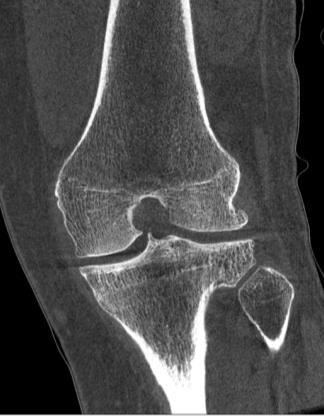
3 Dedicated Extremities CBCT Extremities CBCT Standing and sitting scans Dose ~10 mgy High spatial resolution (~0.5 mm) Soft-tissue contrast resolution Flexible platform Dual-Energy (DE) Imaging Arthrography Iodine / Bone / Soft Tissue Gout Uric Acid / Bone / Soft Tissue Bone Mineral Density (BMD) Bone Edema (BME) Fat / Bone / Soft Tissue Zbijewski et al, Med Phys (2014) DE Imaging of Bone Edema Bone Edema (BME) Increased fluid (water) content Decreased fat (yellow marrow) content Arthritis, trauma, metastases Detectable in T2-weigthed MRI CBCT T2 MRI BME DE Imaging of BME Challenging in conventional CT Partial volume effect from trabeculae Dual-Energy Virtual calcium subtraction Water + marrow maps CT DE CT BME T2 MRI Pache et al, Radiology (2010)
4 DE with Three-Source CBCT Imaging Setup Emulates extremities CBCT 10:1 antiscatter grid LE: 60 kvp HE: 105 kvp (with or w/o high-z filter) 3-Source DE Firing Pattern Central source (2): LE 360 projections Axial sources (1 + 3): HE 180 projections each Only oblique rays at central slice Image-based DE Compared to double scan DE at equivalent dose SDD ~56 cm SAD ~43 cm HE 3 6 cm LE 2 HE 1 DE with Three-Source CBCT Imaging Setup Emulates extremities CBCT 10:1 antiscatter grid LE: 60 kvp HE: 105 kvp (with or w/o high-z filter) 3-Source DE Firing Pattern Central source (2): LE 360 projections Axial sources (1 + 3): HE 180 projections each Only oblique rays at central slice Image-based DE Compared to double scan DE at equivalent dose =1 o =1 0 SDD ~56 cm SAD ~43 cm HE 3 6 cm LE 2 HE 1
 involves LE 2 projections High Energy")
 HE 3 ˆ arg max L ; y R Forward model")
 combines HE 1 and HE 3 projections HE3 reconstruction HE1 reconstruction")
5 Image Reconstruction in 3-Source DE CBCT Penalized Likelihood (PL) ˆ arg max L ; y R Forward model L(µ; y) Edge-preserving Huber penalty R Low Energy reconstruction L(µ; y) involves LE 2 projections High Energy Reconstruction L(µ; y) combines HE 1 and HE 3 projections LE 2 central slice Image Reconstruction in 3-Source DE CBCT Penalized Likelihood (PL) HE 3 ˆ arg max L ; y R Forward model L(µ; y) Edge-preserving Huber penalty R Low Energy reconstruction L(µ; y) involves LE 2 projections HE 1 High Energy Reconstruction L(µ; y) combines HE 1 and HE 3 projections HE3 reconstruction HE1 reconstruction Combined reconstruction

6 Image-based DE Three-material decomposition Water + Fat/ + Cortical Bone Volume preservation constraint Narrow separation between Fat and Water Artifact Correction Fast GPU Monte Carlo scatter correction ~5 min correction per scan DE Decomposition Calibration of LE and HE Water and : ROIs of pure material Bone: Measurements over a range of BMDs + linear fit μ LE μ HE Sisniega et al (PMB 2015) Vol. Frac Vol. Frac Vol. Frac. Image-based DE Three-material decomposition Water + Fat/ + Cortical Bone Volume preservation constraint Narrow separation between Fat and Water Artifact Correction Fast GPU Monte Carlo scatter correction ~5 min correction per scan DE Decomposition Calibration of LE and HE Water and : ROIs of pure material Bone: Measurements over a range of BMDs + linear fit μ LE μ HE (µ ) HE LE: 60 kvp HE: 105 kvp fat (µ ) LE water bone Sisniega et al (PMB 2015) Vol. Frac Vol. Frac Vol. Frac.

7 Base Materials Edema Inserts Base Materials Edema Inserts Tissue-Mimicking Materials Cortical Bone=Dipotassium Phosphate K 2 HPO 4 /Fat=Ethanol Both soluble in water Stable mixtures up to ~ fat Edema: Decreasing fraction of ethanol Imaging Experiments ~10 cm diameter phantom HE: 105 kvp, 0.1 mas/frame, ~7 mgy CTDI LE: 60 kvp, 0.8 mas/frame, ~7 mgy CTDI Experimental Study Water Volume Composition 100% H 2 O 100% C 2 H 6 O K 2 HPO 4 Cortical Bone 100% K 2 HPO4 75 mg/ml K 2 HPO 4 in H 2 O Ethanol 100 mg/ml NIST K 2 HPO Bone 4 in H 2 0 Water NIST Fat 90% 75% 60% Goodsitt et al, Investig. Radiol (1987) Tissue-Mimicking Materials Cortical Bone=Dipotassium Phosphate K 2 HPO 4 /Fat=Ethanol Both soluble in water Stable mixtures up to ~ fat Edema: Decreasing fraction of ethanol Imaging Experiments ~10 cm diameter phantom HE: 105 kvp, 0.1 mas/frame, ~7 mgy CTDI LE: 60 kvp, 0.8 mas/frame, ~7 mgy CTDI Experimental Study Water Volume Composition 100% H 2 O 100% C 2 H 6 O Cortical Bone 100% K 2 HPO4 75 mg/ml K 2 HPO 4 in H 2 O 100 mg/ml K 2 HPO 4 in H % 75% 60% Goodsitt et al, Investig. Radiol (1987)


8 Water Vol. Fraction Vol. Fraction Cortical Bone Vol. Fraction FBP reconstruction Three-Material DE Decomposition Narrow beam double scan DE Water Bone Three-Material DE Decomposition Narrow beam double scan DE Nominal Nominal Nominal


9 Water Vol. Fraction Vol. Fraction Cortical Bone Vol. Fraction W. Zbijewski SPIE Medical Imaging 2015 FBP reconstruction Three-Material DE Decomposition Narrow beam double scan DE Water Bone Three-Material DE Decomposition Narrow beam double scan DE


10 Water Vol. Fraction Vol. Fraction Cortical Bone Vol. Fraction FBP reconstruction Full Beam Three-Material DE Decomposition Full beam + scatter correction Water Bone Full Beam Scatter Correction Three-Material DE Decomposition Full beam + scatter correction
11 Accuracy of Bone Volume Fraction Relative difference with narrow beam DE Full Beam Scatter Correction Rel. diff.=0.04 Rel. diff. =0.06 Rel. diff. =0.01 Rel. diff.=0.03 Rel. diff.=0.01 Rel. diff. =0.01 Rel. diff.=0.12 Rel. diff.=0.03 Rel. diff.=0.01 Rel. diff.= Bone vol. frac Bone vol. frac. Bone and Fat Fractions in Cadaveric Knee LE: 60 kvp, 7 mgy HE: 105 kvp, 7 mgy PL reconstruction 105 kvp Three-Source DE Fat vol. frac. Bone vol. frac.


12 Bone and Fat Fractions in Cadaveric Knee LE: 60 kvp, 7 mgy HE: 105 kvp, 7 mgy PL reconstruction 105 kvp Three-Source DE frac.=0.74±0.13 Bone frac.=0.059±0.008 frac.=0.88±0.08 Bone frac.=0.064±0.005 frac.=0.65±0.16 Bone frac.=0.034±0.004 frac.=0.48±0.18 Bone frac.=0.027± Fat vol. frac. Bone vol. frac. Bone and Fat Fractions in Cadaveric Knee LE: 60 kvp, 7 mgy HE: 105 kvp, 7 mgy PL reconstruction frac.=0.95±0.03 Bone frac. =0.032±0.005 Three-Source DE Fat vol. frac Bone vol. frac frac.=0.85±0.09 Bone frac.=0.029±0.006
13 Conclusions Fat/ Three-Source CBCT CBCT: adaptable platform for new imaging configurations Improved image quality (artifact and field of view) Detection of Edema in DE CBCT volume fraction changes 20% Requires high accuracy in HU Scatter correction (even with grid) (fast MC-GPU) 3-source DE performance comparable to double scan Reduced imaging time compared to double scan Bone Quantitative CBCT Bone Imaging DE for BMD measurement <15% of value High spatial resolution - bone microarchitecture Comprehensive assessment of bone health
Extremities CBCT. Advances in Cone-Beam CT and Emerging Applications: Clinical Motivation 7/14/2015
 JHU Radiology 50 100 150 200 250 300 350 400 450 500 50 100 150 200 250 300 350 400 450 JHU Radiology Advances in Cone-Beam CT and Emerging Applications: Extremities CBCT W Zbijewski Department of Biomedical
Cone-Beam CT for MSK Extremities
 8/6/0 Diagnostic Image Quality Evaluation of an Extremity Cone-Beam CT Scanner: Pre-Clinical and First Clinical Results Abdullah Muhit Wojciech Zbijewski, J Webster Stayman John Yorkston, Nathan Packard,
Peripheral Quantitative CT (pqct) Using a Dedicated Extremity Cone-Beam CT Scanner
 Using a Dedicated Extremity Cone-Beam CT Scanner") SPIE Medical Imaging 23 Peripheral Quantitative CT (pqct) Using a Dedicated Extremity Cone-Beam CT Scanner A. A. Muhit, a S. Arora, a M. Ogawa, a Y. Ding, a W. Zbijewski, a J. W. Stayman, a G. Thawait,
SPIE Medical Imaging 23 Peripheral Quantitative CT (pqct) Using a Dedicated Extremity Cone-Beam CT Scanner A. A. Muhit, a S. Arora, a M. Ogawa, a Y. Ding, a W. Zbijewski, a J. W. Stayman, a G. Thawait,
Dual-Energy CT: The Technological Approaches
 Dual-Energy CT: The Technological Approaches Dushyant Sahani, M.D Director of CT Associate Professor of Radiology Massachusetts General Hospital Harvard Medical School Email-dsahani@partners.org Disclosure
Acknowledgments. A Specific Diagnostic Task: Lung Nodule Detection. A Specific Diagnostic Task: Chest CT Protocols. Chest CT Protocols
 Personalization of Pediatric Imaging in Terms of Needed Indication-Based Quality Per Dose Acknowledgments Duke University Medical Center Ehsan Samei, PhD Donald Frush, MD Xiang Li PhD DABR Cleveland Clinic
Computed tomography Acceptance testing and dose measurements
 Computed tomography Acceptance testing and dose measurements Jonas Andersson Medical Physicist, Ph.D. Department of Radiation Sciences University Hospital of Norrland, Umeå Sweden Contents The Computed
Toshiba Aquillion 64 CT Scanner. Phantom Center Periphery Center Periphery Center Periphery
 Comparison of radiation dose and imaging performance for the standard Varian x-ray tube and the Richardson Healthcare ALTA750 replacement tube for the Toshiba Aquillion CT scanners. by Robert L. Dixon,
True Dual Energy. Dr. Stefan Ulzheimer, Siemens Healthcare GmbH. DEfinitely Siemens
 DEfinitely Siemens True Dual Energy Dr. Stefan Ulzheimer, Siemens Healthcare GmbH International version. Not for distribution in the US. Unrestricted Siemens AG 2015 All rights reserved. The products/features
ESTABLISHING DRLs in PEDIATRIC CT. Keith Strauss, MSc, FAAPM, FACR Cincinnati Children s Hospital University of Cincinnati College of Medicine
 ESTABLISHING DRLs in PEDIATRIC CT Keith Strauss, MSc, FAAPM, FACR Cincinnati Children s Hospital University of Cincinnati College of Medicine CT Dose Indices CTDI INTRODUCTION CTDI 100, CTDI w, CTDI vol
Estimating Iodine Concentration from CT Number Enhancement
 Estimating Iodine Concentration from CT Number Enhancement Rosemary Eaton, Andrew Shah, Jane Shekhdar Medical Physics, Mount Vernon Hospital CT Users Group 4 th October 212, Edinburgh Summary Background
CURRENT CT DOSE METRICS: MAKING CTDI SIZE-SPECIFIC
 CURRENT CT DOSE METRICS: MAKING CTDI SIZE-SPECIFIC Keith Strauss, MSc, FAAPM, FACR Cincinnati Children s Hospital University of Cincinnati College of Medicine Acknowledgments John Boone, PhD Michael McNitt-Grey,
CT NUMBER ACCURACY ANALYSIS FOR RADIOTHERAPY TREATMENT PLANNING IMAGING
 CT NUMBER ACCURACY ANALYSIS FOR RADIOTHERAPY TREATMENT PLANNING IMAGING Julian Liu a, Keisha Robinson a, DhanaJayan Kothandan a and Joshua Luis b (a) Cancer Centre London (b) University College London
Breast CT and Dosimetry
 2013 ICTP/IAEA Training Course on Radiation Protection of Patients Trieste Breast CT and Dosimetry John M. Boone, Ph.D., FAAPM, FSBI, FACR Professor and Vice Chair (Research) of Radiology Professor of
Implementation of the 2012 ACR CT QC Manual in a Community Hospital Setting BRUCE E. HASSELQUIST, PH.D., DABR, DABSNM ASPIRUS WAUSAU HOSPITAL
 Implementation of the 2012 ACR CT QC Manual in a Community Hospital Setting BRUCE E. HASSELQUIST, PH.D., DABR, DABSNM ASPIRUS WAUSAU HOSPITAL Conflict of Interest Disclaimer Employee of Aspirus Wausau
CT Scanning Protocol For V2R Guided Surgery Solutions
 CT Scanning Protocol For V2R Guided Surgery Solutions 2 V2R CT Scanning Protocol \\ Contents Contents General requirements... 3 V2R Dual Scan Protocol... 5 V2R Single Scan Protocol... 8 Overview... 10
SPECIFIC PRINCIPLES FOR DOSE REDUCTION IN HEAD CT IMAGING. Rajiv Gupta, MD, PhD Neuroradiology, Massachusetts General Hospital Harvard Medical School
 SPECIFIC PRINCIPLES FOR DOSE REDUCTION IN HEAD CT IMAGING Rajiv Gupta, MD, PhD Neuroradiology, Massachusetts General Hospital Harvard Medical School OUTLINE 1 st Presentation: Dose optimization strategies
Ultralow Dose Chest CT with MBIR
 Ultralow Dose Chest CT with MBIR Ella A. Kazerooni, M.D. Professor & Director Cardiothoracic Radiology Associate Chair for Clinical Affairs University of Michigan Disclosures Consultant: GE Healthcare
GATE MONTE CARLO DOSIMETRY SIMULATION OF MARS SPECTRAL CT
 GATE MONTE CARLO DOSIMETRY SIMULATION OF MARS SPECTRAL CT R Aamir, C Lowe, J Damet, P Carbonez, A P H Butler, N Schleich, N G Anderson MARFO, Emmanuel Geant4 User Workshop 2017, Wollongong Overview MARS
National Cancer Institute
 National Cancer Institute A Dosimetry Summary of CT Participants in the National Lung Screening Trial (NLST) U.S. DEPARTMENT OF HEALTH AND HUMAN SERVICES National Institutes of Health AAPM 2015 Anaheim,
An Update of VirtualDose Software Used for Assessing Patient Organ Doses from CT Examinations
 An Update of VirtualDose Software Used for Assessing Patient Organ Doses from CT Examinations Aiping Ding, X. George Xu Rensselaer Polytechnic Institute Troy, NY USA http://rrmdg.rpi.edu 1 Acknowledgements
Patient Dosimetry in Mammography and Tomosynthesis:
 2013 ICTP/IAEA Training Course on Radiation Protection of Patients Trieste Patient Dosimetry in Mammography and Tomosynthesis: What to measure, why and how John M. Boone, Ph.D., FAAPM, FSBI, FACR Professor
Cone Beam CT Protocol Optimisation for Prostate Imaging with the Varian Radiotherapy OBI imaging system. Dr Craig Moore & Dr Tim Wood
 Cone Beam CT Protocol Optimisation for Prostate Imaging with the Varian Radiotherapy OBI imaging system Dr Craig Moore & Dr Tim Wood Background With the increasing use of CBCT imaging alongside complex
CT SCAN PROTOCOL. Shoulder
 CT SCAN PROTOCOL Shoulder Purpose and Summary CT images made with this protocol are used to provide the orthopedic surgeon with a detailed 3D anatomical reconstruction of the patient s scapula and proximal
DIAGNOSTIC IMAGING. OPTIMIZED.
 ABOUT LED DENTAL SEE THE DIFFERENCE Using our years of business insight and clinical experience as a foundation, LED Dental takes the uncertainty out of your imaging purchase decision. We offer our clients
Detection of clinically suspected scaphoid bone fractures using a dedicated cone-beam CT (CBCT). A retrospective study of 139 patients.
. A retrospective study of 139 patients.") Detection of clinically suspected scaphoid bone fractures using a dedicated cone-beam CT (CBCT). A retrospective study of 139 patients. Award: Winner Poster No.: P-0043 Congress: ESSR 2014 Type: Scientific
Detection of clinically suspected scaphoid bone fractures using a dedicated cone-beam CT (CBCT). A retrospective study of 139 patients. Award: Winner Poster No.: P-0043 Congress: ESSR 2014 Type: Scientific
Dual-Energy CT Applications in Radiation Therapy
 THE UNIVERSITY OF WISCONSIN MADISON Dual-Energy CT Applications in Radiation Therapy - Jessica Miller 1 Disclosures Funding provided by Siemens Medical 2 Learning objectives General principles of dual
Utility of Dual-Energy CT to Evaluate Patients with Hip and Pelvis Pain in the ER Setting
 Utility of Dual-Energy CT to Evaluate Patients with Hip and Pelvis Pain in the ER Setting Johnson, T., Moran, E., Glazebrook, K., Leng, S., Fletcher, J., and McCollough, C. An educational review ER011
Typical PET Image. Elevated uptake of FDG (related to metabolism) Lung cancer example: But where exactly is it located?
 Lung cancer example: But where exactly is it located?") Typical PET Image Elevated uptake of FDG (related to metabolism) Lung cancer example: But where exactly is it located? PET/CT Oncology Imaging Anatometabolic fusion images are useful in the management
Typical PET Image Elevated uptake of FDG (related to metabolism) Lung cancer example: But where exactly is it located? PET/CT Oncology Imaging Anatometabolic fusion images are useful in the management
Measurement of organ dose in abdomen-pelvis CT exam as a function of ma, KV and scanner type by Monte Carlo method
 Iran. J. Radiat. Res., 2004; 1(4): 187-194 Measurement of organ dose in abdomen-pelvis CT exam as a function of ma, KV and scanner type by Monte Carlo method M.R. Ay 1, M. Shahriari 2, S. Sarkar 3, P.
PACS: ERGONOMIC CONSIDERATIONS 1
 RADIOLOGY RESEARCH Radiographic Tomosynthesis: Acquisition Parameters Michael J. Flynn, PhD Henry Ford Health System Detroit, MI Learning Objectives Learn.. 1. Appreciate the importance of scan direction,
Gender differences in CT calcium scoring: A phantom study
 Gender differences in CT calcium scoring: A phantom study Nicholas Petrick, Qin Li, Benjamin Berman, Marios A Gavrielides, Rongping Zeng, Berkman Sahiner CDRH/OSEL/DIDSR U.S. Food and Drug Administration
Doses from Cervical Spine Computed Tomography (CT) examinations in the UK. John Holroyd and Sue Edyvean
 examinations in the UK. John Holroyd and Sue Edyvean") Doses from Cervical Spine Computed Tomography (CT) examinations in the UK John Holroyd and Sue Edyvean Why a new dose survey? Number of enquires received concerning the current NDRL Concern that could
Doses from Cervical Spine Computed Tomography (CT) examinations in the UK John Holroyd and Sue Edyvean Why a new dose survey? Number of enquires received concerning the current NDRL Concern that could
Dual Energy CT: a new tool in evaluation of the urinary tract stones composition in clinical practice - initial study
 Dual Energy CT: a new tool in evaluation of the urinary tract stones composition in clinical practice - initial study Poster No.: C-2279 Congress: ECR 2013 Type: Scientific Exhibit Authors: M. Guzi#ski,
Fetal Dose Calculations and Impact on Patient Care
 Fetal Dose Calculations and Impact on Patient Care Matt Hough, MS, DABR, DABMP Florida Hospital Diagnostic Medical Physics and Radiation Safety Resource ACR-SPR Practice Parameter for Imaging Pregnant
CT Head Dose Reduction Using Spiral Scanning Protocol
 CT Head Dose Reduction Using Spiral Scanning Protocol Reed, William J MD; Broderick, Daniel F, MD; Weindling, Steven M, MD; Czervionke, Leo F MD; and Morin, Richard L; PhD; Mayo Clinic, Department of Radiology;
To Shield or Not to Shield? Lincoln L. Berland, M.D.
 To Shield or Not to Shield? Lincoln L. Berland, M.D. Disclosures Consultant to: Nuance, Inc. Page 2 Breast Radiation on CT Use of chest CT has increased in women vulnerable to cancer induction by radiation.
Dual-Energy 101: Principles, Methods and Dose
 Dual-Energy 101: Principles, Methods and Dose Juan Carlos Ramirez-Giraldo, Ph.D Staff Scien2st, Collabora2ons Manager SE Region ISCT San Francisco, 2017 Siemens Medical Solu2ons USA, Inc., 2017 Page 1
Radiation Dose Reduction: Should You Use a Bismuth Breast Shield?
 Radiation Dose Reduction: Should You Use a Bismuth Breast Shield? Lincoln L. Berland, M.D., F.A.C.R. Michael V. Yester, Ph.D. University of Alabama at Birmingham Breast Radiation on CT Use of chest CT
Alessandro Albonico Philips
 Alessandro Albonico Philips Alessandro.albonico@philips.com Noise (Standard Deviation in HU) Virtually noise-free Characteristic of a true knowledge-based IR 80 70 Standard Recon idose4 Level6 1 mm Slice
Mineral Density Of Subchondral Bone May Be Quantitatively Evaluated Using A Clinical Cone Beam Computed Tomography Scanner
 Mineral Density Of Subchondral Bone May Be Quantitatively Evaluated Using A Clinical Cone Beam Computed Tomography Scanner Mikael J. Turunen, PhD 1, Juha Töyräs, PhD 1, Harri Kokkonen, PhD 2, Jukka S.
PET in Radiation Therapy. Outline. Tumor Segmentation in PET and in Multimodality Images for Radiation Therapy. 1. Tumor segmentation in PET
 Tumor Segmentation in PET and in Multimodality Images for Radiation Therapy Wei Lu, Ph.D. Department of Radiation Oncology Mallinckrodt Institute of Radiology Washington University in St. Louis Outline
AAPM Task Group 180 Image Guidance Doses Delivered During Radiotherapy: Quantification, Management, and Reduction
 AAPM Task Group 180 Image Guidance Doses Delivered During Radiotherapy: Quantification, Management, and Reduction Parham Alaei, Ph.D. Department of Radiation Oncology University of Minnesota NCCAAPM Fall
Feasibility of improving cone-beam CT number consistency using a scatter correction algorithm.
 Thomas Jefferson University Jefferson Digital Commons Department of Medicine Faculty Papers Department of Medicine 11-4-2013 Feasibility of improving cone-beam CT number consistency using a scatter correction
STRUCTURED EDUCATION REQUIREMENTS EFFECTIVE: JANUARY 1, 2016
 Computed Tomography The purpose of structured education is to provide the opportunity for individuals to develop mastery of discipline-specific knowledge that, when coupled with selected clinical experiences,
Patient Dose Estimates. from CT Examinations. Patient Dose Estimates
 Patient Dose Estimates from CT Examinations Patient Dose Estimates from CT Examinations John M. Boone, Ph.D. Professor and Vice Chairman of Radiology Professor of Biomedical Engineering University of California,
Accounting for Imaging Dose
 Accounting for Imaging Dose High Profile Over-exposures Lead to Growing Concern FDA issues warning in October 2009-209 patients exposed to 8 times typical dose for CT brain perfusion scan (3-4 Gy) - Some
Introduction. Modalities used in imaging guidance. Flat panel detector. X-ray Imaging Dose to Patients in the Era of Image-Guided Radiation Therapy
 X-ray Imaging Dose to Patients in the Era of Image-Guided Radiation Therapy George Ding, Ron Price, Charles Coffey Vanderbilt-Ingram Cancer Center Vanderbilt University Medical Center, Nashville, TN Introduction
Physics (CT VI-Cone Beam CT)
") SSK15 Physics (CT VI-Cone Beam CT) W ednesday, Dec. 2 10:30AM - 12:00PM Location: S403B CT PH AMA PRA Category 1 Credits : 1.50 ARRT Category A+ Credits: 1.50 Stephen J. Glick, PhD, Silver Spring, MD (Moderator)
SSK15 Physics (CT VI-Cone Beam CT) W ednesday, Dec. 2 10:30AM - 12:00PM Location: S403B CT PH AMA PRA Category 1 Credits : 1.50 ARRT Category A+ Credits: 1.50 Stephen J. Glick, PhD, Silver Spring, MD (Moderator)
Doses from pediatric CT examinations in Norway Are pediatric scan protocols developed and in daily use?
 Doses from pediatric CT examinations in Norway Are pediatric scan protocols developed and in daily use? Eva Godske Friberg * Norwegian Radiation Protection Authority, P.O. Box, Østerås, Norway Abstract.
8/3/2016. Consultant for / research support from: Astellas Bayer Bracco GE Healthcare Guerbet Medrad Siemens Healthcare. Single Energy.
 U. Joseph Schoepf, MD Prof. (h.c.), FAHA, FSCBT-MR, FNASCI, FSCCT Professor of Radiology, Medicine, and Pediatrics Director, Division of Cardiovascular Imaging Consultant for / research support from: Astellas
BioMedical quantitative X-Ray Imaging. Emmanuel Brun Researcher Inserm Université Grenoble Alpes
 BioMedical quantitative X-Ray Imaging Emmanuel Brun Researcher Inserm Université Grenoble Alpes 1 Outline Introduction K-Edge Imaging Patient imaging at the European synchrotron Medical Phase Contrast
Low Dose Era in Cardiac CT
 Low Dose Era in Cardiac CT DIANA E. LITMANOVICH, MD Department of Radiology Beth Israel Deaconess Medical Center Harvard Medical School Disclosures Neither I nor my immediate family members have a financial
Original Research THE USE OF REFORMATTED CONE BEAM CT IMAGES IN ASSESSING MID-FACE TRAUMA, WITH A FOCUS ON THE ORBITAL FLOOR FRACTURES
 DOI: 10.15386/cjmed-601 Original Research THE USE OF REFORMATTED CONE BEAM CT IMAGES IN ASSESSING MID-FACE TRAUMA, WITH A FOCUS ON THE ORBITAL FLOOR FRACTURES RALUCA ROMAN 1, MIHAELA HEDEȘIU 1, FLOAREA
Implementation & optimization of a lung cancer screening CT program. Presented by Izabella Barreto at the 2016 Florida AAPM Chapter Meeting
 Implementation & optimization of a lung cancer screening CT program Presented by Izabella Barreto at the 2016 Florida AAPM Chapter Meeting Izabella Barreto, Nathan Quails, Catherine Carranza, Nathalie
8/3/2016. DBT Physics Basic to Advanced: Primer On Tomosynthesis. Tomosynthesis Pedigree
 DBT Physics Basic to Advanced: Primer On Tomosynthesis Andrew D. A. Maidment, Ph.D. University of Pennsylvania Department of Radiology Acknowledgements of Support Research support from the Komen Foundation,
Experimental measurement with an anthropomorphic phantom of the proton dose distribution in the presence of metal implants
 E13195: Slightly better series/treatment with PRONE combination with vertical field. - This field is only possible in case the tumor is not too caudal (problem with energy). - It will increase the integral
CT Dose Estimation. John M. Boone, Ph.D., FAAPM, FSBI, FACR Professor and Vice Chair of Radiology. University of California Davis Medical Center
 CT Dose Estimation John M. Boone, Ph.D., FAAPM, FSBI, FACR Professor and Vice Chair of Radiology 1 University of California Davis Medical Center CT Dose Estimation Introduction The CTDI Family of Metrics
Cardiac CT - Coronary Calcium Basics Workshop II (Basic)
") Cardiac CT - Coronary Calcium Basics Workshop II (Basic) J. Jeffrey Carr, MD, MSCE Dept. of Radiology & Public Health Sciences Wake Forest University School of Medicine Winston-Salem, NC USA No significant
Cardiac CT - Coronary Calcium Basics Workshop II (Basic) J. Jeffrey Carr, MD, MSCE Dept. of Radiology & Public Health Sciences Wake Forest University School of Medicine Winston-Salem, NC USA No significant
Optimizing radiation dose by varying age at pediatric temporal bone CT
 JOURNAL OF APPLIED CLINICAL MEDICAL PHYSICS, VOLUME 16, NUMBER 1, 2015 Optimizing radiation dose by varying age at pediatric temporal bone CT Daichi Noto, 1 Yoshinori Funama, 2a Mika Kitajima, 3 Daisuke
Calculation of Effective Doses for Radiotherapy Cone-Beam CT and Nuclear Medicine Hawkeye CT Laura Sawyer
 Calculation of Effective Doses for Radiotherapy Cone-Beam CT and Nuclear Medicine Hawkeye CT Laura Sawyer Department of Medical Physics and Bioengineering, Royal United Hospital, Bath Overview Varian Acuity
Norland Densitometry A Tradition of Excellence
 Norland Densitometry A Tradition of Excellence Norland DXA Bone Density Measurement Osteoporosis is a disease marked by reduced bone strength leading to an increased risk of fractures. About 54 million
Managing the imaging dose during Image-guided Radiotherapy. Martin J Murphy PhD Department of Radiation Oncology Virginia Commonwealth University
 Managing the imaging dose during Image-guided Radiotherapy Martin J Murphy PhD Department of Radiation Oncology Virginia Commonwealth University Radiographic image guidance has emerged as the new paradigm
Outcomes in the NLST. Health system infrastructure needs to implement screening
 Outcomes in the NLST Health system infrastructure needs to implement screening Denise R. Aberle, MD Professor of Radiology and Bioengineering David Geffen School of Medicine at UCLA 1 Disclosures I have
Image Fusion, Contouring, and Margins in SRS
 Image Fusion, Contouring, and Margins in SRS Sarah Geneser, Ph.D. Department of Radiation Oncology University of California, San Francisco Overview Review SRS uncertainties due to: image registration contouring
8/18/2011. Acknowledgements. Managing Pediatric CT Patient Doses INTRODUCTION
 Managing Pediatric CT Patient Doses Keith J. Strauss, MSc, FAAPM, FACR President X-Ray Computations, Inc. Boston, Massachusetts Acknowledgements Marilyn Goske, MD John Boone, PhD Cynthia McCollough, PhD
Dental ConeBeam Computed Tomography (CBCT) X-ray Systems
 X-ray Systems") Dental ConeBeam Computed Tomography (CBCT) X-ray Systems PROPOSED REVISIONS TO 4732.XXXX, 1.0 4732.#### DENTAL CONEBEAM COMPUTED TOMOGRAPHY (CBCT) X-RAY SYSTEMS; STATIONARY AND MOBILE. Subpart 1. Applicability.
Dental ConeBeam Computed Tomography (CBCT) X-ray Systems PROPOSED REVISIONS TO 4732.XXXX, 1.0 4732.#### DENTAL CONEBEAM COMPUTED TOMOGRAPHY (CBCT) X-RAY SYSTEMS; STATIONARY AND MOBILE. Subpart 1. Applicability.
The Computed Tomography Examination
 CONTENT SPECIFICATIONS The Computed Tomography Examination The purpose of The American Registry of Radiologic Technologists (ARRT ) Computed Tomography Examination is to assess the knowledge and cognitive
Andrew Karellas, PhD
 Advanced Imaging for Breast Cancer: Screening, Diagnosis, and Assessing Response to Therapy The Role of Tomosynthesis Andrew Karellas, PhD Department of Radiology University of Massachusetts Medical School
A Snapshot on Nuclear Cardiac Imaging
 Editorial A Snapshot on Nuclear Cardiac Imaging Khalil, M. Department of Physics, Faculty of Science, Helwan University. There is no doubt that nuclear medicine scanning devices are essential tool in the
354 Korean J Radiol 9(4), August 2008
, August 2008") Review of Failed CT Phantom Image Evaluations in 2005 and 2006 by the CT Accreditation Program of the Korean Institute for Accreditation of Medical Image Hye Jung Park, MD 1 Seung Eun Jung, MD 1, 2 Young
Review of Failed CT Phantom Image Evaluations in 2005 and 2006 by the CT Accreditation Program of the Korean Institute for Accreditation of Medical Image Hye Jung Park, MD 1 Seung Eun Jung, MD 1, 2 Young
Hampton University Proton Therapy Institute
 Hampton University Proton Therapy Institute Brief introduction to proton therapy technology, its advances and Hampton University Proton Therapy Institute Vahagn Nazaryan, Ph.D. Executive Director, HUPTI
Powered by. Dedicated MRI
 Powered by Dedicated MRI Provides the latest software and hardware upgrade configuration powered by exp technology: boosting productivity, increasing image quality, and adding new acquisition techniques.
Radiology Update 2017
 Radiology Update 2017 John K. Phillips, MD Affiliated Assistant Professor of Radiology University of Tennessee Health Sciences Center Chief, Radiology and Nuclear Medicine VA Memphis Disclosures Financial:
RADIATION PROTECTION IN DIAGNOSTIC AND INTERVENTIONAL RADIOLOGY. L19: Optimization of Protection in Mammography
 IAEA Training Material on Radiation Protection in Diagnostic and Interventional Radiology RADIATION PROTECTION IN DIAGNOSTIC AND INTERVENTIONAL RADIOLOGY L19: Optimization of Protection in Mammography
True Low Dose. Exact time to display image on screen may vary upon computer and network configuration.
 RAYSCAN ALPHA PLUS True Low Dose Cone Beam CT Industry Leading Resolution High resolution images provide all the clinical information needed while keeping radiation exposure low. Endodontics - Smallest
IMAGE GENTLY HOW CAN YOU HELP?
 IMAGE GENTLY HOW CAN YOU HELP? Keith J. Strauss, MSc, FAAPM, FACR Director, Radiology Physics & Engineering Children s s Hospital Boston Harvard Medical School Acknowledgment Marilyn J. Goske,, MD Robert
Translating Protocols Across Patient Size: Babies to Bariatric
 Translating Protocols Across Patient Size: Babies to Bariatric Cynthia H. McCollough, PhD, FACR, FAAPM Professor of Radiologic Physics Director, CT Clinical Innovation Center Department of Radiology Mayo
Fundamentals, Techniques, Pitfalls, and Limitations of MDCT Interpretation and Measurement
 Fundamentals, Techniques, Pitfalls, and Limitations of MDCT Interpretation and Measurement 3 rd Annual Imaging & Physiology Summit November 20-21, 21, 2009 Seoul, Korea Wm. Guy Weigold, MD, FACC Cardiovascular
Quantitative and Qualitative Assessment of Thorax Cone Beam CT Image Quality across Multiple Imaging Systems
 Quantitative and Qualitative Assessment of Thorax Cone Beam CT Image Quality across Multiple Imaging Systems Matthew Williams Pre-registration Clinical Scientist Velindre NHS Trust, Cardiff Computed Tomography
CT Dosimetry in the Clinical Environment: Methods and Analysis
 CT Dosimetry in the Clinical Environment: Methods and Analysis Manuel Arreola, Ph.D. DABR Associate Chair of Radiology Director, Medical Physics Graduate Program Department of Radiology University of Florida
CT Optimisation for Paediatric SPECT/CT Examinations. Sarah Bell
 CT Optimisation for Paediatric SPECT/CT Examinations Sarah Bell Sarah.bell14@nhs.net Outline 1. Introduction 2. Aims and Objectives 3. Methods 4. Results 5. Discussion 6. Conclusions 7. References Introduction
Contrast-Enhanced Digital Mammography
 2015 ARRS Breast Symposium Contrast-Enhanced Digital Mammography John Lewin, M.D. Diversified Radiology of Colorado CEDM - Outline History Technique Literature Review / Cases Clinical Status Inexpensive,
4 Essentials of CK Physics 8/2/2012. SRS using the CyberKnife. Disclaimer/Conflict of Interest
 SRS using the CyberKnife Sonja Dieterich, PhD, DABR Associate Professor University of California Davis Disclaimer/Conflict of Interest Consulting agreements with Broncus Medical and CyberHeart, Inc. Scientific
Multislice CT versus cone beam CT: dosimetric and image quality comparision.
 Multislice CT versus cone beam CT: dosimetric and image quality comparision. Poster No.: C-2589 Congress: ECR 2015 Type: Authors: Scientific Exhibit C. Dionisi 1, A. Crispo 2, E. Stefani 3, A. Lovaglio
3D Cone beam CT & Digital Radiography Dedicated to Otorhinolaryngology
 3D Cone beam CT & Digital Radiography Dedicated to Otorhinolaryngology Multi-functional imaging solution3 RAYSCAN m is an unique 2-in-1 imaging solution, combining Cone Beam CT and Digital Radiography,
Advancing critical clinical decisions
 Radiation Oncology Computed Tomography Big Bore RT Advancing critical clinical decisions Pursuit of the successful outcome Powerful technology delivers Radiation therapy can be effective in helping your
Image Guided Proton Therapy and Treatment Adaptation
 Image Guided Proton Therapy and Treatment Adaptation www.hollandptc.nl d.r.schaart@tudelft.nl Cancer in The Netherlands About 1 in 3 people get cancer in some stage of their life 86.800 new cancer patients
Validation of in vivo cancellous bone assessment using high resolution imaging and histologic examination
 Validation of in vivo cancellous bone assessment using high resolution imaging and histologic examination Luke Arentsen, PhD Medical Physics Resident University of Minnesota Cancellous bone has a higher
Measurement of computed tomography dose profile with pitch variation using Gafchromic XR-QA2 and thermoluminescence dosimeter (TLD)
") Journal of Physics: Conference Series PAPER OPEN ACCESS Measurement of computed tomography dose profile with pitch variation using Gafchromic XR-QA2 and thermoluminescence dosimeter (TLD) To cite this
Journal of Physics: Conference Series PAPER OPEN ACCESS Measurement of computed tomography dose profile with pitch variation using Gafchromic XR-QA2 and thermoluminescence dosimeter (TLD) To cite this
THE TUFFEST STUFF CT REGISTRY REVIEW Live Lecture Seminar SATURDAY CURRICULUM
 1. The CT Imaging Chain-10 major components & their functions a. The x-ray tube b. Generator c. Filter d. Pre-patient collimator e. Pre-detector collimator f. Detector system g. Analog to digital converter
COMPUTED TOMOGRAPHY COURSE
 CT Radiography RAD 421 4 th year semester 2 Course Lecture Tutorial Practical Credit hours CT Radiography 2-1 2 Course Description The course explores the basic physical and technical principles of CT
Learning objective. Outline. Acknowledgements. KV CBCT Imaging Part I. R Hammoud AAPM 2008 CE-Therapy (SAM) 1
 1") 1 2 KV CBCT Imaging Part I Rabih Hammoud, MS, DABR Henry Ford Health System Detroit, Michigan Acknowledgements Indrin Chetty, PhD Teamour Nurushev, PhD Harrison Guan, PhD Jinkoo Kim, PhD JianYue Jin, PhD
1 2 KV CBCT Imaging Part I Rabih Hammoud, MS, DABR Henry Ford Health System Detroit, Michigan Acknowledgements Indrin Chetty, PhD Teamour Nurushev, PhD Harrison Guan, PhD Jinkoo Kim, PhD JianYue Jin, PhD
ACR MRI Accreditation Program. ACR MRI Accreditation Program Update. Educational Objectives. ACR accreditation. History. New Modular Program
 ACR MRI Accreditation Program Update Donna M. Reeve, MS, DABR, DABMP Department of Imaging Physics University of Texas M.D. Anderson Cancer Center Educational Objectives Present requirements of the new
A feasibility study of multislice X-ray CT imaging of gel dosimeters using the zero scan method
 JOURNAL OF APPLIED CLINICAL MEDICAL PHYSICS, VOLUME 15, NUMBER 4, 2014 A feasibility study of multislice X-ray CT imaging of gel dosimeters using the zero scan method Muhammad B. Kakakhel, 1 Tanya Kairn,
Imaging of Scattered Radiation for Real Time Tracking of Tumor Motion During Lung SBRT
 Imaging of Scattered Radiation for Real Time Tracking of Tumor Motion During Lung SBRT April 25 nd, 2015 Lung Cancer Lung cancer is the most lethal cancer: Over 224,000 new diagnoses in the U.S. predicted
SOMATOM Drive System Owner Manual Dosimetry and imaging performance report
 www.siemens.com/healthcare SOMATOM Drive System Owner Manual Dosimetry and imaging performance report Table of contents 1 Dosimetry and imaging performance report 5 1.1 Dose information 5 1.1.1 General
Metal Artefact Reduction in CT
 Metal Artefact Reduction in CT DANIEL MARRINER Metal Artefact Reduction in CT Metal Artefact Clinical Indications for MAR SEMAR and How It Works Technical Considerations Case Studies utilising SEMAR Metal
CS 9300 Family. The power of flexibility
 CS 9300 Family The power of flexibility The new CS 9300 digital imaging system from Carestream Dental take the guesswork out of examinations The all-in-one CS 9300 is the most versatile multimodality imaging
Quanitative and qualitative analysis of Image Quality and Imaging Dose of KV CBCT from XVI Elekta Linac.
 IOSR Journal of Applied Physics (IOSR-JAP) e-issn: 2278-4861.Volume 9, Issue 5 Ver. IV (Sep. - Oct. 2017), PP 46-52 www.iosrjournals.org Quanitative and qualitative analysis of Image Quality and Imaging
Radiation Dose Reduction Strategies in Coronary CT Angiography
 Radiation Dose Reduction Strategies in Coronary CT Angiography Noor Diyana Osman, PhD noordiyana@usm.my Contents: Introduction Radiation dosimetry in CT Radiation risk associated with coronary CT angiography