Pattern Formation and Morphogenesis of Fungiform Papillae during Mouse Development
|
|
|
- Lydia Ellen Clarke
- 6 years ago
- Views:
Transcription
1 Pattern Formation and Morphogenesis of Fungiform Papillae during Mouse Development Jae-Young Kim Department of Medical Science The Graduate School, Yonsei University
2 Pattern Formation and Morphogenesis of Fungiform Papillae during Mouse Development Directed by Professor Han-Sung Jung The Doctoral Dissertation submitted to the Department of Medical Science, the Graduate School of Yonsei University in partial fulfillment of the requirements for the degree of Doctor of Philosophy Jae-Young Kim June 2004
3 This certifies that the Doctoral Dissertation of Jae-Young Kim is approved Han-Sung Jung Kwang-Kyun Park Yun-Jung Yoo Myounghee Kim Young-Ho Kim The Graduate School Yonsei University June 2004
4 ACKNOWLEDGEMENTS I would like to thank Professor Han-Sung Jung for invaluable advice and guidance. My thanks also go to Professor Hee-Jin Kim, Professor Kyoung-Nam Kim, Dr. Keiichi Akita at Tokyo Medical Dental University, Dr. Hitoshi Yamamoto at Nihon University and Professor Yi-Ping Chen at Tulane University for help me on several experiments. It is great pleasure to thank Sung-Won Cho, Soo-Jin Song, Heui-Jung Hwang, Jong-Min Lee, Jinglei Cai, Kyoung-Won Cho, Hyun-Do Park, Hyun-Ho Kwak, Kwan-Hyun Youn, and Min-Jung Lee for letting me collaborate on several experiments. I am greatful to many people from the department of Oral Biology, College of Dentistry, Yonsei University, past and present, who made my Ph.D. course enjoyable. Special thanks to Professor Syng-Il Lee, Professor Kwang-Kyun Park, and Professor Soon-Chul Park for encouragement. Finally, special thanks go to my family for supporting me always. Jae-Young Kim
5 TABLE OF CONTENTS ABSTRACT... 1 I. INTRODUCTION Tongue and its appendage papillae Outline of tongue and gustatory papillae development in vertebrate Morphogenesis and pattern formation of fungiform papillae development Pattern formation and molecular studies in gustatory papillae development II. MATERIALS AND METHODS Animals Histology Immunohistochemistry SEM (scanning electron microscope) In situ hybridization In vitro organ culture Di.I. application and fate mapping Recombination assay Whole mount immunostaining Beads implantation Antisense oligodeoxynucleotides (AS-ODN) Image processing and counting the Shh expression spots... 20
6 III. RESULTS Morphological findings Expression pattern of signalling molecules Recombination assay Whole mount immunohistostaining Gene expression pattern after in vitro organ culture Alteration of gene expression pattern after the treatment of antisense Cx43 and implantation of NOGGIN bead to in vitro organ culture.. 39 IV. DISCUSSION Morphogenesis of fungiform papillae in the mouse development Expression of signalling molecules prior to morphological changes E13.5 epithelium determines the position of fungiform papillae Developmental role of gap junction during mouse tongue development Expression patterns of signalling molecules after in vitro organ culture Gene expression pattern in fungiform papillae following Cx43 antisense treatment Gene expression pattern in fungiform papillae following NOGGIN bead treatment V. CONCLUSION REFERENCES ABSTRACT (IN KOREAN)... 77
7 LIST OF FIGURES Figure 1. Schematic diagram of mouse tongue and gustatory papillae. 5 Figure 2. Ultrastructural study of the fungiform papillae. 23 Figure 3. Expression of signalling molecules. 26 Figure 4. Recombination assay. 30 Figure 5. Whole mount immunostaining using Cx Figure 6. Signalling molecules expression pattern after in vitro organ culture for 48 hours at E Figure 7. Alteration of gene expression pattern after the treatment of antisense Cx43 ODN. 42 Figure 8. Alteration of gene expression pattern after the NOGGIN bead implantation. 44 Figure 9. The number of Shh expression spots of tongue dorsal surface. 46 Figure 10. Schematic diagram of the regulation of signalling molecules during fungiform papillae development. 61 Figure 11. Schematic diagram showing the epithelial mesenchymal interactions at early morphogenesis of fungiform papillae. 63
8 LIST OF TABLE Table 1. The oligodeoxynucleotide sequences used in this study. 41
9 ABSTRACT The tongue is a part of organ that plays important roles in swallowing, taste the food, and speech in human. Anatomically, the tongue is divided into an oral part and a pharyngeal part by the terminal sulcus. The following four different types of tongue papillae, which are small organs, can be found on the dorsal surface of the tongue: the fungiform, circumvallate, foliate and filiform papillae. Interestingly, development of fungiform papillae showed the stereotyped pattern on the dorsal surface of the tongue in mice from embryonic day 12 (E12) to E17. The patterned distribution and developmental processes of the fungiform papillae suggest possible similarities between the fungiform papillae and other epithelial appendages, including teeth, feathers, and hairs, which develop via epithelial mesenchymal interaction. The histological results and ultrastructural observations showed the development of specific structures in the epithelium into fungiform papillae. Many signalling factors, including signalling molecules and ECM (Extracellular matrix) molecules such as Shh, Bmps, Fgfs, laminin, CAM (Cell adhesion molecule) and cytokeratins, might be involved in the morphogenesis of fungiform papillae during the early development. In order to obtain direct evidence of the
10 epithelium and mesenchyme interaction during fungiform papillae morphogenesis, the formation of fungiform papillae like structure was examined after a recombination assay. It was conducted after the recombination assay at E12.5 and E13.5 for 2 days using an in vitro organ culture. From the recombination assay results, the E13.5 epithelial portion of the fungiform papillae could determine the position of the newly formed fungiform papillae with the epithelial signalling molecules. E13.5 was a critical stage for fungiform papillae morphogenesis. To understand the signalling regulations of fungiform papillae, expression patterns of Shh, Bmp-2 and connexin 43 were examined. The results showed that the spatio-temporal expression pattern of signalling molecules is of evidence for proper formation of fungiform papillae. Furthermore, antisense oligodeoxynucleotide (AS-ODN) connexin 43 was treated to analyze the developmental functions of gap junction and NOGGIN protein, which is an antagonist of Bmps, was treated during in vitro organ culture. The expression patterns of signalling molecules, Shh and Bmp-2 were altered by inhibition of gap junctions. Shh, which is known as a key molecule to determine spacing pattern, expression patterns were disturbed after the inhibition of Bmp-2 as well. These results revealed that pathway of the signalling molecules during fungiform papillae development. Fungiform papillae can be considered to be small epithelial appendages, which are formed with the specific patterned array via the epithelial mesenchymal interactions.
11 I. INTRODUCTION 1. Tongue and its appendage papillae During mouse embryogenesis, the terminal sulcus divides the developing tongue into an oral part and a pharyngeal part. Four different types of tongue papillae can be found on the dorsal surface: fungiform, circumvallate, foliate and filiform papillae. These papillae are distributed in a specific pattern. 1 The fungiform, foliate, and circumvallate papillae are known as the gustatory papillae, which contain taste buds and work as sensory organs. 2 The gustatory papillae are comprised of an epithelial covering over a broad core of connective tissue. Taste buds, which are discrete collections of approximately 40 to 60 cells within the papilla epithelium, are an oval sensory end organ that is involved in the perception of chemical stimuli and in taste transduction. 3,4,5 The fungiform papillae are located on the oral part of the tongue with a specific patterned array. One circumvallate papilla is located in the middle part of the terminal sulcus in rodents. In humans, about 7 to 9 circumvallate papillae are located along the terminal sulcus (Fig. 1). Foliate papillae are
12 found on the sides of the posterior one-third of the human tongue and are well developed at birth, but revert to a rudimentary structure in adults. The filiform papillae, which contain no taste buds, cover the entire anterior part of the dorsal surface of the tongue and consist of cone-shaped structures, each with a core of connective tissue that is covered by an epithelium expressing hair-related keratins. 6 The information on the tongue papillae development presented in this review was obtained from previous experiments in sheep, rodents, and salamanders as well as from studies in human development. 1,7,8 The initial study using a vertebrate tongue was aimed at examining the three-dimensional structure of the embryonic and fetal stages of the tongue and its appendages, the tongue papillae, using light microscopy and scanning electron microscopy. 1,7 It was reported that during the development of the tongue papillae many factors might be involved in morphogenesis, including the ECM (extracellular matrix) molecules such as laminin, CAM (Cell adhesion molecule) and cytokeratins Recently, the circumvallate and fungiform papillae were examined using a variety of experimental approaches by protein expressions, signalling network and tissue interactions In particular, the development of fungiform papillae, which are one of the gustatory papillae, was examined as an epithelial appendage. 15 In this study, the formation of the fungiform papillae is discussedas a model system for pattern formation because they have a specific patterned array on the dorsal surface of the tongue. 4,5,16
13 Filiform papillae Circumvallate papilla Median sulcus Foliate papilla Fungiform papilla
14 Figure 1. Schematic diagram of mouse tongue and gustatory papillae. Diagram of the mouse tongue shows general boundaries of oral part, pharyngeal part, and intermolar eminence of tongue. Mouse has a single circumvallate papilla in the middle of the posterior part. Mouse has stereotyped array of fungiform papillae on the anterior part of the tongue. Three types of papillae including fungiform, foliate, and circumvallate papillae have taste bud.
15 2. Outline of tongue and gustatory papillae development in vertebrate During the initial stages of organogenesis, the induction and formation of the epithelial appendages, including feathers, hairs, mammary glands, and teeth require specific and complex epithelial-mesenchymal interactions throughout their development Although the vertebrate epithelial appendages have specific structures and diverse functions, most epithelial appendages have similar developmental stages, including induction, morphogenesis, and differentiation. 20,21 In feathers or in scale forming regions of the skin of chick embryos, the epidermal placode is the first morphological event for the formation of an epithelial appendage. It provides a signal to the dermis, which results in the formation of dermal condensations immediately beneath the placode. 22 The formation of the epidermal placodes is determined by the mesodermal signals, which was revealed by the heterospecific tissue recombinations of the ectodermal and mesodermal components of the embryonic skin. 23 Furthermore, the mesenchyme determines the number, size, location as well as the structural identity of the appendages, while the epithelium determines the zoological class type (feathers or hair) and the competence state. 24 Therefore, the molecular mechanisms are conserved during the evolution of the skin as well as another developmental model system such as the limbs. Moreover, similar molecules appear to be essential for its proper development. 25 The development of the gustatory papillae has similar pattern of formation with the other epithelial appendages. The fungiform papillae also form a repeating unit with a highly regular spatial pattern through the epithelial mesenchymal interactions over the dorsal surface of the tongue during embryonic development. A recent study reported that a reciprocal
16 interaction between the tongue epithelium and the underlying mesenchyme is of fundamental importance in determining the individual position of the fungiform papilla as well as its stage-dependent competency. 14 In mice, the fungiform papillae begin to develop as an epithelial thickening at E13. As the tongue develops, the mesenchymal cells become more rounded and closely apposed to form a condensate. The connective tissue is overlaid by an epithelium, which is two cells thick. The periderm cells, which form a superficial thin sheet of cells, are formed over the epithelium in early stage. During fungiform papillae development, the epithelial cells elongate in the primordium and thicken. 14,26,35 The tongue epithelium and mesenchyme undergo subsequent morphogenesis with a stereotyped pattern until E17. Previous studies have suggested that the morphogenesis of the gustatory papillae requires a series of invaginations and evaginations, which are coordinated interactions among the lingual epithelium and mesenchyme as well as in the nerve innervation of the tongue. 5,27 The development of the fungiform papillae can be divided into two phases. The first phase is the morphogenesis phase, in which the papillae structures are produced. The second phase is the innervation phase, in which the taste buds develop. In the innervation phase, the fungiform papillae display a different developmental procedure for complete development than the other epithelial appendages. In the case of the fungiform papilla, the nerve innervation is required to develop into sensory organs with taste buds, as shown during the development of mice. 4,27,28 Therefore, the innervation phase represents a maturation phase or functional phase. 14 Jitpukdeebodintra et al., after examining the cell adhesion molecules, reported that the formation of the circumvallate papilla initially depends on cell migration, which likely to play a major role during circumvallate placode formation. 12 During the
17 initial tongue innervation, the nerves follow a spatiotemporary restricted pathway, suggesting that neural guidance is governed by signalling molecules from environmental factors such as BDNF (Brain derived neurotrophic factor), NT4 (Neurotrophin-4), E-cadherin, and laminin. 2,5,8 Indeed, as a special sensory organ, the gustatory papillae are closely associated with nerve innervation, which has been examined in detail studies on taste buds. 29
18 3. Morphogenesis and pattern formation of fungiform papillae development Previous studies have suggested that the morphogenesis of the tongue papillae requires coordinated interactions among the lingual epithelium and mesenchyme as well as nerve innervation of the tongue. 2,30 The development of fungiform papillae can be divided into two stages. One is the morphogenesis, which produces the papillae structures. The other is nerve innervation, which develops the taste buds. 4,27,28,31-34 Recent studies of the tongue in developing mouse embryos emphasized that nerve innervations are needed to develop the taste bud. 2,30 Other studies showed that the Shh, Ptc, Gli-1, Bmp-2, Bmp-4, and Fgf-8 transcripts are expressed within the epithelia of the primordial tongue. 27,35 Especially, Shh plays important role in establishing papillary boundaries. 13 It was suggested that the expression of the signalling molecules is of fundamental importance during papillae morphogenesis, and occurs prior to the morphological changes. 13,15,27,35,36 However, there is no detailed description of the series of epithelial and mesenchymal changes during the early development of the fungiform papillae. Moreover, there is no direct evidence of the epithelium and mesenchyme interactions during the early development of fungiform papillae. This study focused on the morphogenesis of the fungiform papillae structure and attempted to identify the critical stage in the position of the fungiform papillae structure. The scanning electron microscopy (SEM), histology, and gene expression results were examined collectively to comprehend the precise development mechanism of the fungiform papillae. Both an in vitro organ culture system and an in vitro recombination system were used to obtain direct evidence of the epithelium and mesenchyme interactions, which build up the fungiform papillae structure in early development, and these systems
19 preclude the lingual nerve innervations. The recombination assay in this study suggests that the epithelium and mesenchyme interaction plays an important role during the early development of the tongue papillae, and the E13.5 epithelium has the potential to develop newly formed fungiform papillae structures prior to nerve innervations.
20 4. Pattern formation and molecular studies in gustatory papillae development BMP-2 is a member of the transforming growth factor superfamily, which is expressed in many regions of the developing embryo including the developing fungiform papillae. 35,78,85 They have been shown to be involved in many developmental processes including epithelial mesenchymal interactions during tooth and limb development. 79,81,82 BMP-2 also is a part of a signalling cascade that controls development of the facial primordia. 75 The SHH protein is involved in morphogenesis and pattern formation of the tongue papillae as a morphogen, which directs the cells toward a nonpapillary fate, and as a mitogen, which causes the expansion of the interplacodal epithelium. 13 Disruption of Shh signalling during rat tongue development revealed that the development and patterning of the fungiform papillae depends on this signalling pathway. 16 Gap junctions are specialized areas of the cell membranes that allow direct exchange of signalling molecules and metabolites between the cytoplasm of neighboring cells. 42 They are organized collections of protein channels that allow ions and small molecules to enable communicate between adjacent cells. These protein channels that make up the gap junctions consist of two "hemi-channels" or connexons, each connexon representing an oligomeric arrangement of six polypeptides. 43 Each connexon is composed of six transmembrane proteins called connexins, which are localized symmetrically around a central pore. The connexin family of proteins is encoded by at least 19 genes in rodents with many homologues cloned from other species. 44,45 They are named according to their molecular weight. Connexin 43 (Cx43), for example, is a 43 kda protein and is widely expressed in embryo and adult tissues. 46,47 Signals such as hormones and growth and differentiation factors are thought to perform their function through affecting levels and activities of connexins. 39 Gap
21 junctions are observed in many developing tissues including cardiac muscle, and have been suggested to participate in a variety of roles including the regulation of signal transmission, cell proliferation, differentiation, apoptosis, and tissue homeostasis. 43 One of the major gap junction proteins in chick and mouse embryonic limb is alpha-1 connexin or connexin-43 (Cx43). Cx43 transcript is detected both in the apical ectodermal ridge (AER) and the posterior mesenchyme by stage 20 in the chick and the AER by embryonic day 10 in the mouse A major role of connexins in the early mesenchyme may be to enable the mesenchymal cells in the polarizing region to communicate with the anterior mesenchyme. 49,52 Expression of Cx43 in the developing limb bud is complex, and some variations exist between the mouse 48,51,53 and the chick. 50 Expression levels of Cx43 54 and the patterning genes of Shh 55 and Bmp-2 56 are higher in the ZPA. It has been suggested that interactions between signalling molecules and junctional communication would play an important role in controlling fungiform papillae development. 54 I show here that Cx43 protein expression significantly correlates with fungiform papillae development. In order to define the relationship between Shh and Bmp-2, the expression patterns of these genes were compared by using in vitro organ culture. Antisense oligodeoxynucleotide (AS-ODN) Cx43 was treated to analyze the developmental functions of Cx The monomers are deoxynucleotides like those in DNA. There are usually only of them, hence "oligo". This sequence (3' to 5') is antisense, that is, complementary to the sense sequence of a molecule of mrna. Physically block the ability of ribosomes to move along the messenger RNA or simply hasten the rate at which the mrna is degraded within the cytosol. 58 I investigated the function of Bmp-2 during tongue development by ectopically expressing NOGGIN, a potent antagonist of the BMPs. 57 The expression patterns of signalling molecules, Shh and Bmp-2 were altered by inhibition of gap junctions. Expression patterns of
22 Shh, which is known as a key molecule to determine spacing pattern, were disturbed after the inhibition of Bmp-2 as well. These results suggest that the signalling cascade through the gap junction is sufficient for initiating papillary pattern and morphogenesis.
23 II. MATERIALS AND METHODS 1. Animals Adult ICR mice were housed in a temperature-controlled room (22±1 C) under artificial illumination (lights on from 05:00 h to 17:00 h) and at 55% relative humidity, with free access to food and water. mice embryos were obtained from time-mated pregnant mice. The day that the presence of the vaginal plug was confirmed was designated embryonic day 0 (E0). Embryos at development stages (E12-17, daily intervals) were used in the present study. 2. Histology The mouse embryos were fixed in 4% paraformaldehyde (PFA) in PBS overnight at 4, and embedded in paraffin wax. Specimens were cut with a 5 thickness, and the sections were stained with both hematoxylin and eosin (H&E). 3. Immunohistochemistry For immunohistochemistry, the specimens were fixed with 4% paraformaldehyde at 4. They were equilibrated in a 30% sucrose solution for cryoprotection and embedded in an OCT compound (Sakura Finetek, USA). Expression pattern of Cx43 was examined by 7 cryosection after whole mount immunohistochemistry. Subsequent to the whole mount in situ hybridization as well as the Di.I. microinjections, 20
24 serial sagittal sections were cut using a cryostat and mounted on a silane-coated slide glass slide (Muto Pure Chemicals. Co., Japan). 4. SEM (scanning electron microscope) The tongues, which were removed from the mandible, were prefixed with 2.5% glutaraldehyde in PBS for 24 h at 4, rinsed with PBS for 8 hours at 4, and postfixed with 1% osmium tetroxide in PBS for 2 h. The samples were then dehydrated in a graded series of ethanol, followed by critical point drying using a HCP-2 apparatus (Hitachi, Tokyo, Japan) with CO 2 as the transitional fluid. The specimens, which were mounted on stubs, were coated with platinum and examined by scanning electron microscopy (SEM, S-4500; Hitachi, Tokyo, Japan). 5. In situ hybridization Whole cultured tissue was washed in PBST and was treated for minutes with proteinase K (10 /ml in PBST). It was washed twice in PBST and refixed for 1 hour in 4% PFA. Embryos were then prehybridized for 2 hours at 60 in hybridization buffer containing 50% deionized formamide. Hybridization was performed overnight at 65 in hybridization buffer containing /ml riboprobe. Excess probe was removed by sequential washes in 2X standard saline citrate (SSC; three times at 60 ), 0.2 X SSC (three times at 60 ), 1:1 of 0.2 X SSC:0.1 M phosphate buffer (PB), and PB (twice). Nonspecific binding in the tissue was blocked for 1-2 hours in 15% goat serum, 2 mg/ml bovine serum albumin, and 0.3% Triton X-100 in 0.1 M PB. After this treatment, the specimen
25 was incubated overnight with anti-digoxigenin antibody conjugated to alkaline phosphatase diluted 1:2,000 in blocking solution. Excess antibody was removed by washes in 0.1 M PB, and the tissue was equilibrated with color buffer containing 100 mm Tris, ph 9.5; 50 mm MgCl 2 ; 100 mm NaCl; and 0.1% Tween 20. The Bmp-2, Bmp-4, and Shh plasmids were kindly provided by Dr. Y. -P. Chen. 6. In vitro organ culture To examine the regulation of signalling molecules after micro dissecting the tongues from a E12.5 mouse mandible, the tongue was separated into the aboral and oral parts. It was then cultured in medium containing 10% fetal bovine serum for 2 days by applying a slight modification of Trowel's culture method. 7. Di.I. application and Fate Mapping Di.I. (1,19-dioctadecyl-3,3,39,39-tetramethyl indocarbocyanine perchlorate; Molecular Probes, Eugene, OR) was used as a cell tracer to observe cell migration during tongue development. A 0.3% w/v Di.I. in DMSO was used for microinjection. The injection of Di.I. was performed using 10 cm borosilicate capillary pipettes (Sutter Instruments, BF ), pulled using a Sutter Instrument Flaming Brown micro pipette puller, and that were filled by capillary action. Using a device of electricity. The lipophilic carbocyanine dye inserts into the membrane of the cells adjacent to the injection site. The exact position of the dye can be determined by using fluorescent microscope. Injections were done onto anterior part of
26 tongue dorsal surface. 8. Recombination assay Recombination of the tongue using the intact epithelium and mesenchyme was done at E12.5 and E13.5. The tongue was dissected at E12.5 and E13.5, incubated in 2.4 unit dispase II for 15 to 20 min at 3 7, and washed in medium containing 10% fetal bovine serum. The epithelium and mesenchyme were separated using a dissection microscope. The epithelia were trimmed to equal sizes beneath a transparent dish using graph paper as a guide. A lipophilic dye, Di.I. (Molecular Probes, UK; 2 mg/ml in dimethyl formamide), was injected as a marker of the epithelial thickening regions at E13.5. The epithelia remained intact in the media. The epithelium was placed on top of the mesenchyme with a 90-degree rotation and the recombinants were cultured. After 24 and 48 hours recombination, the recombinants were examined using epifluorescence microscopy (LEICA, MZ FL III). Shh expression was observed using whole mount in situ hybridization after the recombination assay to identify the fungiform papillae forming area. 9. Whole mount immunostaining Collect embryos in ice cold PBS. The embryos were fixed in freshly prepared 4% paraformaldehyde (PFA) in PBS overnight at 4. Wash the embryos in PBS, three times for 15 min. The embryos were washed in PBS with 1% Triton X-100 and 10% fetal calf serum (TS-PBS) three times for l hour. The embryos
27 were incubated with primary antibody, mouse monoclonal antibody against the Connexin43, (Zymed, Cat No ) for 24 hours. Then, the embryos were washed with TS-PBS, 6 times for 30 min. The embryos were incubated with peroxidase-conjugated goat anti-mouse immunoglobulins (diluted 1/200 in TS-PBS) overnight. After washing the embryos with PBT 6 times for 30 minutes, the antibody was localized with DAB staining kit. 10. Beads implantation Affigel Blue beads, (BioRad) 150 mm in diameter, were dried and then soaked in 0.5 mg/ml of human recombinant NOGGIN (Regeneron). For examination of gene regulation of fungiform papillae development, a NOGGIN soaked bead was implanted in the anterior part of the tongue at E The tongues were used for the whole mount in situ hybridization, after the bead implantation. PBS soaked beads were implanted as the control experiment. 11. Antisense oligodeoxynucleotides (AS-ODN) Cx43 specific antisense ODN was designed and shown to be effective in knocking down Cx43 protein levels (sequence ; GenBank accession no. X61576). 58 Control ODN sequence was used as a sense control for the mouse Cx43 sequence ODN (Table 1). ODN were purchased from GENOTECH (KOREA).
28 12. Image processing and counting the Shh expression spots Whole tongues were photographed and digitized by using a Dimage Scan Multi film scanner (Minolta, Osaka, Japan). Bright field and cultured tongue images were collected with digital camera (LEICA, DC-300F). Digital images of cultured tongues, which were analyzed by using whole mount in situ hybridization with Shh mrna, were collected. Tongues were scored double blind for the presence of both large and small (<10 diameter) Shh-positive papillary spots. The mean numbers of papillae per tongue were compared for different treatment groups by ANOVA.
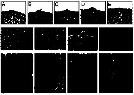
29 III. RESULTS 1. Morphological findings In order to understand the precise development of the fungiform papillae, transverse sections of the E13 to E17 mouse embryo tongue were stained with H&E. The morphological changes of the tongue epithelium surface were examined by SEM. Epithelial thickening was detected at E13, and the epithelial cells at the basal epithelium on the anterior part of the tongue increased in height (Fig. 2A). Epithelial thickening was detected at E13.5 on the dorsal surface of the tongue with a specific pattern from the anterior to posterior (Fig. 2F and J). At E14, epithelial thickening, under the surface region, became concave as the condensed mesenchymal cells migrated upward (Fig. 2B). At E14.5, archlike structures arose on the dorsal surface of the tongue with a stereotyped pattern, and the number of these structures increased (Fig. 2G and K). The number of these regions had grown by this stage. At E15, the epithelial cells continued to elongate, but the epithelial cells in the center of the archlike structure became thinner. In addition, the underlying mesenchymal cells of the epithelial thickening region were condensed (Fig. 2C). At E16, the epithelial cells continued to increase in height, and filiform papillae began to develop. The epithelial cells in the center of the archlike structure became thinner, and the underlying mesenchyme migrated upward (Fig. 2D). At E16.5, archlike structures rose on the dorsal surface with the highest length, and the tongue grew into the shape of an adult one. The distribution pattern and number of fungiform papillae were observed using SEM (Fig. 2H and L). At E17, fungiform papillae were observed on the anterior part of the tongue. The heights between the elevation of the
30 underlying mesenchyme and the stratified cells became similar. The surface of the stratified regions developed into the filiform papillae (Fig. 2E). In adults, the fungiform papillae were surrounded by filiform papillae (Fig. 2I and M).
31
32 Fig. 2. Ultrastructural study of the fungiform papillae. (A-E) Histology of fungiform papillae during morphogenesis. (F-M) SEM study of mouse tongue development. (A) E13, the epithelium thickening. (B) E14, the epithelium developed into an archlike structure and the underlying mesenchymal cells were condensed. (C) E15, the epithelium of the fungiform papillae area, and their neighbor cells grew together. (D) E16, the filiform papillae began to develop. (E) E17, the development of the fungiform papillae into adult fungiform papillae (scale bar: (A-E) 25 ). (F, J) E13.5, the fungiform papillae structures were observed on the dorsal surface, median sulcus (arrowhead), the fungiform papilla structures (arrow). (G, K) E14.5 the fungiform papillae structures raised on the dorsal surface. (H, L) E16.5 the fungiform papilla structures on the anterior part of the tongue. (I, M) PN 10 weeks, adult fungiform papillae were detected in the middle of the filiform papillae (scale bar: (F-I) 250 ; (J-M) 50 ).
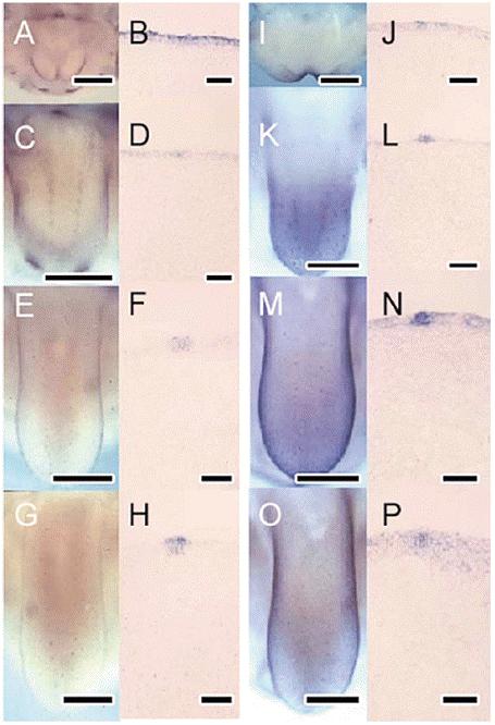
33 2. Expression patterns of signalling molecules The Bmp-4 and Shh expression patterns during the formation of the fungiform papillae in the mouse embryos from E12.5 to E14 were examined using whole mount in situ hybridization in order to determine the relationship between the morphological changes and the expression pattern of the signalling molecules. Transverse sections were observed after the whole mount in situ hybridization to confirm the regions where the signalling molecules were expressed. Shh expression was detected in the dorsal surface of the tongue at E12.5. This expression was broadly detected in the lateral swelling (Fig. 3A and B). Bmp-4 had a similar expression pattern to Shh. However, Bmp-4 expression was limited to a restricted area and was stronger there than in the other epithelium layers (Fig. 3I and J). At E13, the Bmp-4 and Shh expression patterns were more localized and specific than at E12.5 (Fig. 3C and K). The distribution patterns of gene expression on the dorsal surface of the tongue resembled the SEM observations (Fig. 2E and J and Fig. 3C and K), and these expression patterns were restricted to the epithelium (Fig. 4D and L). At E13.5, both Bmp-4 and Shh were strongly expressed in a localized spot, which appeared histologically identical to the neighboring epithelial cells (Fig. 2F and N). These gene expression patterns coincided with the distribution patterns of the elevated regions (Fig. 2E and M). At E14, the Bmp-4 expression level was lower and broadened, but Shh expression was even more restricted and localized in the epithelial thickening regions (Fig. 3G and O). Both Bmp-4 and Shh expression were detected in the archlike structures (Fig. 3H and P).
34
35 Fig. 3. Expression of signalling molecules. (A-H) Shh, (I-P) Bmp-4 expression. (A, B) E12.5, Shh expression was detected in the epithelium. (C, D) E13, along the median sulcus, Shh expression with a stereotyped expression pattern and the anterior part of the mandible incisors was detected. (E, F) E13.5, Shh expression was restricted to certain areas of the epithelium. (G, H) E14, from the anterior to the posterior of the tongue, a stereotyped pattern expression was found on the dorsal surface of the tongue (scale bar: (A, C, E, G) 250 ; (B, D, F, H) 50 ). (I, J) E12.5, Bmp-4 was expressed broadly in the epithelium. (K, L) E13, Bmp-4 expression was detected with a stereotyped pattern, which was restricted to the epithelium. (M, N) E13.5, the strong Bmp-4 expression level was restricted to the archlike structures. The dorsal surface of the tongue expression spots was specified to produce a stereotyped pattern. (O, P) E14, Bmp-4 expression changed to be broad on the dorsal surface (scale bar: (I, K, M, O) 250 ; (J, L, N, P) 50 ).
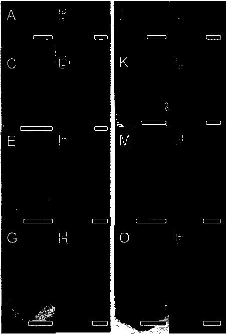
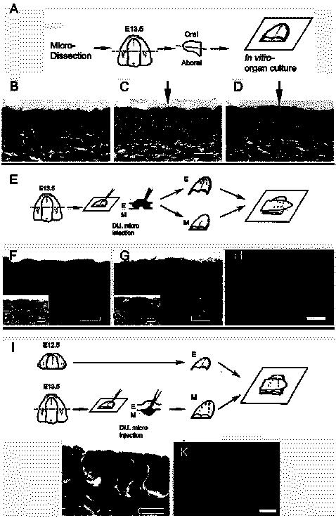
36 3. Recombination assay An in vitro organ culture was used as a control. For the in vitro organ culture, the E13.5 tongues were microdissected using a tungsten needle. The tongue was separated into an aboral and oral part. These microdissections allowed the fungiform papillae-like structures to develop (Fig. 4A). After the in vitro organ culture, the fungiform papillae-like structures were observed using the paraffin sections and H&E staining. Twenty-four hours later, the in vitro organ cultured tongue showed fungiform papillae-like structures. Archlike structures could be observed over the epithelium area, and there was a small amount of condensation in the underlying mesenchyme area (Fig. 4B). After 48 hours, the epithelial cells grew to make an archlike structure. Those structures were similar to the E14 fungiform papillae structure (Fig. 4C). After 72 hours, the fungiform papillae-like structures, which developed as E15 fungiform papillae, were examined. However, these structures were different from those observed in vivo at E15. There was no elevation above the epithelium (Fig. 4D). A recombination assay was performed to determine the role of the E13.5 epithelium and mesenchyme during early fungiform papillae development. Recombination of the tongue using intact epithelium and mesenchyme was observed using an E13.5 tongue. A Di.I. microinjection was made in the epithelium of the fungiform papillae at E13.5 in order to determine the properties of the E13.5 epithelium (Fig. 4E). All the morphological structures were disrupted after the recombination assay (data not shown). No morphological changes were observed in the recombinants of the E13.5 tongues 24 hours later (Fig. 4F). However, Shh expression was detected in the epithelium of the recombinants (Fig. 4F*). Cross sections of the newly formed fungiform
37 papillae-like structures appeared 48 hours after the recombination (Fig. 4G). Frozen sections after the whole mount in situ hybridization exhibited epithelial expression of Shh 48 hours after the recombination (Fig. 4G*). All the Di.I. microinjected epithelium of the fungiform papillae was observed to have archlike structure using fluorescence microscopy. This suggested that the E13.5 epithelium can induce newly formed fungiform papillae-like structures in the epithelium with the epithelial signalling (Fig. 4H). An additional recombination assay was done to understand the role of the E13.5 mesenchyme. Di.I. was microinjected into the underlying mesenchyme of the fungiform papillae at E13.5 (Fig. 4I). Another recombination using the epithelium of the E12.5 tongue and mesenchyme from an E13.5 tongue showed newly formed fungiform papillae-like structures 48 hours later (Fig. 4J). Newly formed fungiform papillae-like structures could not be found in Di.I. labeled mesenchyme area using fluorescent microscopy (Fig. 4K).
38
39 Fig 4. Recombination assay. In vitro organ culture of fungiform papillae at E13.5 (A, B, C, D), recombination assay of fungiform papillae at E13.5 (E, F, G, H), expression of signalling molecule after the recombination assay (F*, G*), and the recombination assays E12.5 and E13.5 (I, J, K). (A) Schematic diagram of the in vitro organ culture. After microdissecting the tongue from an E13.5 mouse mandible, the tongue was separated into the aboral and oral parts. It was then cultured using a slight modification of Trowell's culture method. (B) 24 hours later, it changed little from E13.5. (C) 48 hours later, the arrow indicates an archlike structure and the underlying mesenchymal cells were condensed. (D) 72 hours later, an archlike structure was detected in the epithelium and a mesenchymal core rose to the epithelium (scale bar: 50 ). (E) Schematic diagram of the recombination assay. After microdissecting the tongue from an E13.5 mouse mandible, the tongue was separated into an aboral and oral part. The aboral part was treated with dispase II (2.4 unit for 15 min) at 37 and the epithelium was removed with a fine needle. The Di.I. microinjection accommodating to the epithelium, which showed a specific fungiform papillae structure. The epithelium rotated 90 degree and was then reattached to the mesenchyme (to determine the origin of that structure). It was then cultured using a slight modification of Trowell's culture method (E, epithelium; M, mesenchyme). (F) 24 hours after the recombination, most morphological structures were disrupted. (F*) Shh expression was restricted to certain area of the epithelium. (G) 48 hours later, it formed archlike structures in the epithelium. (G*) A strong Shh expression level was detected only in the archlike structure. (H) The Di.I. labeled epithelium induced a specific structure of the fungiform papillae (blue line shows basement membrane). (I) Schematic diagram of the recombination assay with E12.5 and E13.5. The Di.I. microinjection was accommodated to the mesenchyme of the E13.5 tongue (E, epithelium; M, mesenchyme). (J) 48 hours later, it formed archlike structures in the epithelium. (K) The fungiform papilla structure was not observed on the
40 Di.I. injected mesenchymal area (scale bar: (B, C, D, F, G, H) 50 ; (F*, G*) 10 ; (J, K) 25 ).
41 4. Whole mount immunohistostaining In order to determine the relationship between the morphological changes and the expression pattern of Cx43. The Cx43 expression patterns during the formation of the fungiform papillae in the mouse embryos from E11 to E14 were examined using whole mount immunostaining and transverse sections were examined after the whole mount immunostaining to confirm the regions where the Cx43 was expressed. Cx43 expression was broadly detected in the lateral swelling at E11.5 (Fig. 5A). This expression was detected in epithelium and underlying mesenchyme of the tongue at E11.5 (Fig. 5D). At E12.5, Cx43 expression patterns were more localized and specific than at E11.5 (Fig. 5B), and Cx43 expression was restricted to the epithelium (Fig. 5E). At E13.5, Cx43 was strongly expressed in a localized spot, which appeared histologically identical to the neighboring epithelial cells (Fig. 5C, F).
42 A B C D E F
43 Figure 5. Whole mount immunostaining using Cx43. (A-C) Whole mount immunostaining of mouse tongue. (A) E11.5, there was a broad expression pattern of Cx43. (B) E12.5, Cx43 expression spots were observed on the dorsal surface of the tongue. (C) E13.5, Cx43 expressions were localized with specific patterned array. (D-F) Frozen sections after the whole mount immunostaining. Dotted lines indicate basement membrane. (D) E11.5, Cx43 expressions were localized in both epithelium and mesenchyme. (E) E12.5, Cx43 expressions were localized in epithelium. (F) E13.5, Cx43 expressed in the fungiform papillae forming epithelium (arrow heads indicate the strongest expression spots). (scale bar: (A, B) 100 ; (C) 200 ; (D-F) 40 ).
44 5. Gene expression pattern after in vitro organ culture To examine the expression patterns of signalling molecules, E12.5 tongue was used for in vitro organ culture. After the micro dissecting the tongues at E12.5 mouse mandible, the tongue was separated into the aboral and oral parts. It was then cultured for 2 days in a medium containing 10% fetal bovine serum using a slight modification of Trowell's culture method (Fig. 6G). Shh expression was detected in the dorsal surface of the tongue after 2 days of culture at E12.5 (Fig. 6A). Bmp-2 showed similar expression pattern to Shh. However, The intensity of Bmp-2 expression was weaker than that of Shh expression (Fig. 6C). After whole mount in situ hybridization, transverse sections were performed to examine the precise expression pattern of Shh and Bmp-2. Both Shh and Bmp-2 were strongly expressed in a localized spot in epithelium, which appeared histologically identical to the neighboring epithelial cells (Fig. 6B, C, E, and F).
45 G
46 Figure 6. Signalling molecules expression pattern after in vitro organ culture for 48 hours at E12.5. (A-C) Shh and (D-F) Bmp-2 expression patterns were observed after the whole mount in situ hybridization. These expression patterns are similar to the in vivo expressions at E14. (A) Shh expressed on the dorsal surface of the tongue with specific array, which were observed at E14 mouse tongue. (B, C) Transverse sections showed Shh expression in the fungiform papillae like structure in epithelium. (D) Bmp-2 expressions were examined after in vitro organ culture. Expression patterns were similar to Shh expression pattern. The intensity of expressions were less than that of Shh. (E, F) Bmp-2 expressions were also localized in epithelium. (G) Schematic diagram of in vitro culture at E12.5. (scale bar: (A, D) 250 ; (B, C, E, F) 50 ). Dotted lines indicate the section level.
47 6. Alteration of gene expression pattern after the treatment of antisense Cx-43 and implantation of NOGGIN bead to in vitro organ culture. Cx43 specific AS-ODN was designed and shown to be effective in knocking down Cx43 protein levels (sequence ; GenBank accession no. X61576). 58 Control ODN sequences were used as a sense control for the mouse Cx43 sequence ODN (Table 1). Both Bmp-2 and Shh expressions were altered after treatment of Cx43 AS-ODN. Shh expression was denser than the control expression on the dorsal surface of the tongue (Fig. 7A). Otherwise, Bmp-2 expression was decreased and restricted in the anterior part of the tongue (Fig. 7D). Expressed cell population densities were disrupted by Cx43 AS-ODN (Fig. 9). Shh and Bmp-2 expression patterns were altered by Cx43 AS-ODN and confirmed with transverse section after whole mount in situ hybridization. Intensity of Shh expression was stronger than that of the control (Fig. 3B), while Bmp-2 expression was weaker (Fig. 7E). Moreover, it is shown that antisense Cx43 AS-ODN altered expression pattern of signalling molecules (Fig. 7C, F and Fig. 9). For examination of the gene regulation of fungiform papillae development, a NOGGIN soaked bead was implanted in the anterior part of the right side of the tongue at E12.5. PBS bead implantation was examined as the control. After the bead implantation, signalling molecules were examined using whole mount in situ hybridization. After NOGGIN bead implantation, Shh expression was altered near the bead-implanted area (Fig. 8A). Transverse sections showed that the size of fungiform papilla structure was larger than that of the control culture, and The intensity of Shh expression was more intense than in the control culture (Fig. 8B, C). After the PBS bead implantation, there were no critical changes from control culture (Fig. 8D, E). Bmp-2 expressions were altered after the NOGGIN bead implantation (Fig. 8F). Bmp-2 expressions were moved to the mesenchyme near
48 to NOGGIN bead (Fig. 8G). After the NOGGIN bead implantation, the size of fungiform papillae was larger than that of the control culture (Fig. 8H). The number of Shh expression spots was examined after the whole mount in situ hybridization as a marker for spacing pattern. Since Shh is well known as a major signalling molecule to set up of pattern formation and morphogenesis during fungiform papillae development (Fig. 9). Control sense Cx43 ODN treated and PBS bead implanted tongues showed similar number of Shh expression spots of control culture, it was 1.8 spots/100 2 in the anterior part of the dorsal tongue area. However, after Cx43 AS-ODN treatment and NOGGIN bead implantation, the number of Shh expression spots was increased as 7.4 and 3.6 spots/100 2 in the anterior part of the dorsal tongue area (Fig. 9).
49 Table 1. The oligodeoxynucleotide (ODN) sequences used in this study. ODN Sequence Mouse Cx43 specific GTA ATT GCG GCA GGA GGA ATT GTT TCT GTC Sense control GAC AGA AAC AAT TCC TCC TGC CGC AAT TAC Cx43, Connexin43 (Law et al., 2002).
50
51 Figure 7. Alteration of gene expression pattern after the treatment of antisense Cx43 ODN. AS Cx43 ODN (Antisense Cx43 Oligodeoxynucleotide) was designed as GTA ATT GCG GCA GGA GGA ATT GTT TCT GTC. Pattern formation of fungiform papillae was disrupted following Cx43 AS-ODN treatment. (A) Shh expressions were altered following Cx43 AS-ODN. Shh expression spots were increased on the dorsal surface of the tongue. (B, C) Shh expressed in the fungiform papillae structures in epithelium. (D) The number of Bmp-2 expression spots were decreased on the dorsal surface of the tongue after Cx43 AS-ODN. (E, F) Although Bmp-2 expressed in the fungiform papillae forming epithelium, the intensity of expressions was decreased. (scale bar: (A, D) 250 ; (B, C, E, F) 50 ). Dotted lines indicate the section level.
52
53 Figure 8. Alteration of gene expression pattern following the NOGGIN bead implantation (concentration of NOGGIN protein was 0.5 mg/ml). Red circles indicate beads. (A) The NOGGIN bead was implanted at right side of the dorsal surface of the tongue. The size of Shh expression spot was larger than that of the control. (B) Transverse section showed that Shh expressions were localized in epithelial cells. (C) The size of fungiform papillae like structures were increased. (D) PBS bead was implanted as a control experiment. There was no critical change from the control. (E, F) Transverse sections were also examined. (G) Afte the NOGGIN bead implantation, Bmp-2 expressed near to the NOGGIN bead with broad pattern. (H, I) Transverse section showed that Bmp-2 expressed in mesenchyme, where is the beneath to the fungiform papillae like structure. (scale bar: (A, E, G) 250 ; (B, C, F, H, I) 50 ). Dotted lines indicate the section level.
54 Spots/100 μm The number of Shh expression spots Control +48hrs AS Cx43+48hrs CS Cx43+48hrs NOGGIN+48hrs PBS+48hrs Figure 9. The number of Shh expression spots of tongue dorsal surface. Alteration of Shh expression pattern was examined by counting the expression spots after the Cx43 AS-ODN treatment and the NOGGIN bead implantation (spots/100 2 ).
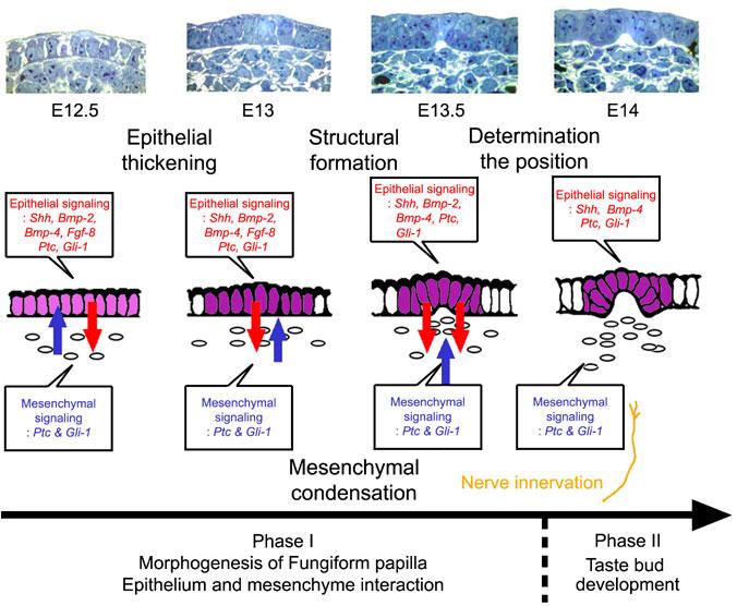
55 IV. DISCUSSION 1. Morphogenesis of fungiform papillae in the mouse development Tongue papillae are types of epithelial appendages, which share a similar process to other organs, such as the hairs, kidneys, teeth, feathers, and mammary glands. 15 However, fungiform papillae have different developmental procedures from other epithelial appendages for complete development. Fungiform papillae require nerve innervations to develop into sensory organs. 4,28,30-34 Fungiform papillae begin to develop with epithelial thickening. 7,59-61 At E12.5, cells of the epithelial layer become elongated and the periderm thickens in some restricted areas of the epithelium. From E13 to E16, the epithelium and mesenchyme undergo subsequent morphogenesis. There is a series of invaginations and evaginations, as the epithelium and mesenchyme interact to form tongue papillae with an epithelial covering over a mesenchymal core. The early stages of fungiform papillae formation resemble those of other specialized epithelial appendages, particularly the hairs, teeth, and feather buds. 21,62-64 In those developing systems, cascades of intercellular signalling interactions specify the organ position and induce morphogenesis. 64,65 This study showed the precise morphogenesis of the mouse tongue development. These results confirmed the previous results of mouse tongue development using SEM. 7 At E13.5, fungiform papillae are raised on the dorsal surface of the tongue, which form two rows from the anterior to the posterior, along the median sulcus. From this stage, the fungiform papillae could be observed using a dissecting microscope. These rows had a pattern, called a "spacing pattern", on the dorsal surface. 37 The density of the fungiform papillae was higher in the anterior part of the tongue. At E14, fungiform papillae
56 developed anterior to the lateral side. The distribution pattern and density of the fungiform papillae developed to the adult one. At E16, the filiform papillae began to grow on the dorsal surface of the tongue. Therefore, the adult fungiform papillae could be observed, which were surrounded by filiform papillae. The histological results showed a similar morphogenesis pattern as that shown by the ultrastructural findings. It appears that the morphogenesis of the fungiform papillae is similar to that observed in the formation of feathers, hair, and teeth, which results from the intimate collaboration of the epithelium and mesenchyme. 21,62-64
57 2. Expression of signalling molecules prior to morphological changes Recent studies of the tongue in developing mouse embryos have shown that Shh, Ptc, Gli-1, Bmp-2, Bmp-4, and Fgf-8 transcripts are expressed within the epithelia of the tongue primordia, because these signalling molecules are of fundamental importance in papillae morphogenesis. 27,35 In addition, this study confirmed the previous results on gene expression during tongue papillae development. These signalling molecules might regulate the early stages of organogenesis and produce signalling networks, which have been reported in many organs, including tooth buds, 66 limb buds, 67,68 and feather buds. 69 Gene expression was explained as a morphological marker of the tongue papillae. Based on these results, the expression of these signalling molecules can only be detected in the epithelium prior to the morphological changes. Initially, Bmp-4 and Shh expressions were detected in restricted area, which changed to a specific structure at E13. These signalling molecules share a similar expression pattern. However, there were differences in the intensity and the width of the expression in the epithelium. Secondly, the signalling molecules were detected as dotted patterns on the dorsal surface of the tongue. Furthermore, the specific stereotyped spacing pattern of the fungiform papillae could be observed. Overall, these results suggest that the signalling molecules, which have been implicated in the morphogenetic regulation of many other epithelial appendages, are also associated with the initial stages of tongue papillae formation. 13 From these gene expression results, these signalling molecules are expressed in restricted area in the epithelium, which changes morphologically. It was assumed that the tongue papillae share a similar formation process as observed with other epithelial appendages.
58 3. E13.5 epithelium determines the position of fungiform papillae The formation of skin appendages requires an interaction between the epithelium and mesenchyme. 21,62-64 The mesenchyme determines the number, size, location, and structural identity of the appendage, while the epithelium determines the orientation and competence state. From the morphological results in this study, most of the signalling molecules were expressed in the epithelium. However, morphological changes were observed in the epithelium and mesenchyme. It is thought that the epithelium might play an important role during early development, as observed in hair, feather, and tooth development. In addition, the results suggest that these epithelial signalling molecules regulate the morphogenesis of the mesenchyme at E13.5. Compared to the other epithelial appendages, tongue papillae have technical disadvantages. This study could not observe the fungiform papillae on the tongue dorsal surface by a dissecting microscope prior to E13.5. Therefore, a Di.I. microinjection was available from E13.5. A previous study reported that prior to E14, the nerve does not reach the basement membrane of the fungiform papillae forming area. 27 Consequently, E13.5 was selected to examine the role of the epithelium and mesenchyme interaction for the development of fungiform papillae with the Di.I. microinjection. A recombination assay using an in vitro organ culture after the recombination assay with the intact epithelium and mesenchyme was made at E13.5 for 2 days in order to confirm the properties of the epithelium during mouse tongue development. After 24 hours, an archlike structure was observed in the epithelium. It had similar figures to the E14 fungiform papillae in vivo. After 48 hours, the labeled epithelium induced fungiform papillae-like structures, which were observed over the newly formed fungiform papillae
59 structures. These findings suggest that the epithelium can induce the fungiform papillae-like structures, and the epithelium has properties that can localize the epithelial appendages at E13.5. The signalling molecule, Shh, was chosen as a morphological marker of the fungiform papillae after the recombination assay to confirm the identity of newly formed fungiform papillae. 13 Shh expression was also detected in the epithelium of the newly formed fungiform papillae. This suggested that the E13.5 epithelium can induce newly formed fungiform papillae-like structures in the epithelium with the epithelial signalling. This study also examined the reversal recombination using the epithelium of the E13.5 tongue and the mesenchyme of the E12.5 tongue. These observations showed the Di.I. labeled epithelium formed fungiform papillae structures, which were similar to the results of the E13.5 recombination (data not shown). However, recombination using the epithelium of the E12.5 tongue and the mesenchyme of the E13.5 tongue produced the fungiform papillae structure regardless of Di.I. microinjected mesenchyme. It suggests that E13.5 is a crucial stage for determining the position of the fungiform papillae structure and that there is mutual interaction between tongue epithelium and mesenchyme which work together for papillae development at early mouse embryogenesis.
60 4. Developmental role of gap junction during mouse tongue development. Gap junctions are intercellular channels that are involved in cell-to-cell communication. 83 Small molecules can pass through the gap junction channels, such as ions and second messengers with molecular weights less than 1 kda. 84 Gap junctions are considered to be important for maintaining tissue homeostasis and for communicating certain intracellular signals. Gap junctions are also observed in many developing tissues including cardiac muscle and nerve,and have been suggested in variety of roles including the regulation of signal transmission, cell proliferation, differentiation, apoptosis, and tissue homeostasis. 43 One of the major gap junction proteins in chick and mouse embryonic limb is alpha-1 connexin, also known as Cx A major role of connexins in the early mesenchyme may be to enable mesenchymal cells in the polarizing region to communicate with the anterior mesenchyme. 49,52 I have demonstrated changing patterns of Cx43 expression and gap junction formation during mice fungiform papillae development. The patterns of Cx43 expression were examined by using whole mount immunostaining method. The Cx43 was expressed in the dorsal surface of the mice tongue from E11.5. At E12.5, expression of Cx43 was localized in the epithelium. In particular, At E13.5, expressions of Cx43 were examined in fungiform papillae forming area. These expression patterns are similar to those of the signalling molecules such as Shh and Bmps. These findings implicate that there is the relationship between gap junction and signalling molecules as it has been known in limb development
61 5. Expression patterns of signalling molecules after in vitro organ culture. Recent studies of the fungiform papillae in developing mouse embryos have shown that Shh, Ptc, Gli-1, Bmp-2, Bmp-4 and Fgf-8 transcripts are expressed in the fungiform papillae forming area prior to morphogenesis, indicating that the signalling molecules are of fundamental importance in papillae morphogenesis. 27,35 And, Shh is required during initiation, determination, and differentiation of fungiform papillae. 13 In mice tongue development ,27,35 BMP-2 has been shown to be involved in many developmental processes including epithelial mesenchymal interactions during tooth and limb development. 81,82 BMP-2 is also a part of a signalling cascade that controls development of the facial primordial. 75 Interestingly, in vitro organ culture of mouse tongue was allowed to examine the regulation of signalling molecules. So I could examine the expression pattern of signalling molecules, Bmp-2 and Shh, after in vitro organ culture at E12.5. After 2 days of culture, Shh expression was observed on the tongue dorsal surface with stereotyped pattern using whole mount in situ hybridization. Expression pattern of Bmp-2 was also examined at fungiform papillae forming area of dorsal surface. Both signalling molecules were expressed in fungiform papillae forming area in the epithelium. These expression patterns are similar to those of the E14, in vivo control. This in vitro organ culture method would be helpful to understand the signalling pathway during early development of fungiform papillae. In particular, the regulations between activation and inhibition of signalling molecules can be examined by using various methods, such as AS-ODN and protein beads implantation.
62 6. Gene expression pattern in fungiform papillae following Cx43 antisense treatment. Previously, it is suggested that interactions between signalling molecules and junctional communication would play an important role in controlling development Here, in an attempt to understand more clearly how communication through Cx43 channels influences the cascade of gene expression involved in the patterning of the fungiform papillae. I examined that Cx43 protein expression significantly correlates with fungiform papillae development by using Cx43 AS-ODN treatment, Cx43 specific AS-ODN was designed and shown to be effective in knocking down Cx43 protein levels. 58 Control sense ODN (CS-ODN) a sequence was used as a sense control for the mouse Cx43 sequence ODN. Shh expression pattern was altered and disrupted following Cx43 AS-ODN treatment. The number of Shh expression spots increased from the control. The number of Shh expression spots in the control specimen was 1.8 spots/100 2 in the anterior part of the dorsal tongue area. While the numbers of Shh expression spots of Cx43 CS-ODN treated specimen were similar to that of the control, the Cx43 AS-ODN treated specimen showed much higher density of 7.4 spots/ These results demonstrate that the Cx43 mediated regulation of fungiform papillae development is important in fungiform papillae patterning and that transient knockdown of Cx43 expression leads to disruption of fungiform papillae patterning. Bmp-2 expression pattern was altered and decreased following Cx43 AS-ODN treatment. Histological sections, after whole mount in situ hybridization, showed that the intensity of Bmp-2 expression was decreased from the control. In addition, Bmp-2 expressions were decreased while Shh expressions were increased following Cx43 AS-ODN treatment. These results suggest that Shh and Bmp-2 are downstream of Cx43 mediated signalling in the
63 particular cascade of fungiform papillae development. This signalling cascade in fungiform papillae development is similar to that of the limb development. 50,52
64 7. Gene expression pattern in fungiform papillae following NOGGIN bead treatment. In order to understand the signalling regulation during early fungiform papillae development, ectopic-expressing NOGGIN, which is a potent antagonist of the BMPs, was treated to in vitro organ culture by using the bead implantation. Bmp-2 expression in epithelium was blocked by BMP-inhibitor NOGGIN protein. Interestingly, localization of Bmp-2 expressions was moved to mesenchyme. Shh expression was slightly altered by NOGGIN protein. The density of Shh expression spot was increased to 3.6 spots/100 2 in the anterior part of the dorsal tongue surface. After the NOGGIN bead implantation, a morphological disturbance in fungiform papilla was observed by transverse section during fungiform papillae development. The size of fungiform papilla was larger than that of the control. This change could be due to the effect of the regulation of the signalling molecules, such as Bmp-2, Shh and Cx43, during early development of fungiform papillae prior to nerve innervations. These altered expression patterns suggest that there would be a relationship between Bmp-2 expression and nerve innervations, which were excluded by this in vitro organ culture system. These alteration could be due to the inhibitory regualation between Bmp-2 and nerve factors. 86 To sum it up, Cx43 plays an important role during fungiform papillaedevelopment. Antisense Cx43 ODN treatment leads to a down-regulation of Bmp-2 expression and an up-regulation of Shh expression. NOGGIN bead implantation leads to the alteration of size of a fungiform papilla and up-regulation of Shh expression. These treatments resulted in the development of abnormal fungiform papillae morphology and disrupted pattern formation of signalling molecules. It is suggested that there are regulations among Shh,
65 Bmp-2, and Cx43 mediated signalling can regulate pattern formation and morphogenesis. Transient knockdown of Cx43 leads to up-regulation of Shh, which in turn leads to disruption of pattern formation of fungiform papillae. I note that Cx43 is also expressed in the facial primordia and the limb during development. These regions are very similar to fungiform papillae in developmental terms. Therefore, it would be interesting to look at the role of Cx43 in these regions to determine whether it has a function similar to the one proposed here for the fungiform papillae.
66 V. CONCLUSION The development of the epithelial appendages has been examined intensively over the last 30 years, mainly using a number of model systems such as feathers, hair, teeth, as well as the mammary and salivary glands ,70-74 The fungiform papillae might be another new model system for examining epithelial appendage development. All these model systems share similar developmental processes in the early embryonic stages. In particular, fungiform papillae development shows some similarities with the development of other epithelial appendages. First, prior to the morphological changes, the signalling molecules are expressed in the fungiform papillae forming area with a specific patterned array. Second, the morphogenesis of the fungiform papillae has a specific structure such as epithelial thickening and mesenchymal condensation, which are observed in the morphogenesis of the other epithelial appendages. Third, the stereotyped pattern, which is governed by signalling molecules such as Shh, Bmp-2, and Cx43, is present during fungiform papillae development (Fig. 10). 61 Fourth, the fungiform papillae develop through reciprocal interactions between epithelial and mesenchymal tissue. It would be interesting to know how, in the case of the gustatory papillae, individual position, size and the spacing pattern are determined (Fig. 11). Moreover, fungiform papillae have different developmental procedures from the other epithelial appendages. Nerve innervation is required for the development of mouse embryo taste buds, 16,27 whereas the fungiform papillae can develop independently of nerve innervation in salamanders. 4,28 This result suggested that nerve innervation is not essential for the initiation of fungiform papillae, 14 but rather for the complete development of the functional and matured fungiform papillae as sensory organs in mammals. Therefore, an
67 investigation of the primary induction system for the individual papilla should be performed. In addition, the finding the mesenchymal factors to complete understand epithelial mesenchymal interactions, just as it revealed in limb development, early Fgfs signallings could be good candidates for mesenchymal signalling molecules. And the induction of the ectopic papillae as new study fields would be another challenge. A culture system of the mouse tongue has allowed in our laboratory an investigation into the participation of three tissue types, the epithelium, mesenchyme and neural tissue during the initiation, morphogenesis, growth, and maintenance of the fungiform papillae. 14 Concerning evolution, in order to understand the diversity of the fungiform papillae and how it is conserved, a comparative study using other molecular markers and available approaches is suggested. The development of fungiform papillae represents an ideal model system for examining the epithelial mesenchymal interactions, nerve innervation and spacing pattern formation during embryonic development. 1. Prior to the morphological changes, the signalling molecules are expressed with the stereotyped pattern in the fungiform papillae forming area. 2. The morphogenesis of the fungiform papillae showed specific structures such as epithelial thickening and mesenchymal condensation. 3. Fungiform papillae develop through the reciprocal interactions between epithelium and mesenchymal tissue. 4. Development of fungiform papillae was governed by signalling molecules, such as Shh, Bmp-2 and connexin Fungiform papillae can be considered to be small epithelial
68 appendages, which are formed with the specific patterned array via the epithelial mesenchymal interactions
69 As Cx43 Cx-43 AS-ODN Cx-43 Cx43 (Gap junction) Bmp-2 Shh Hall, 2003 NOGGIN Patterning & Morphogenesis
70 Figure 10. Schematic diagram of the regulation of signalling molecules during fungiform papillae development (Red bar; inhibition, Blue arrow; activation). Previous report showed that Shh is a key molecule for fungiform papillae patterning. 13 Cx43 regulates Bmp-2 and Shh with different ways. NOGGIN also would play an important role for patterning and morphogenesis of fungiform papillae.
71
Cytological and Histological Study of Adult and Neonate Epidermis in Thick and Thin Skin of Various Anatomical Sites
 Available online on www.ijpqa.com International Journal of Pharmaceutical Quality Assurance 218; 9(2); 174-179 doi: 1.25258/ijpqa.v9i2.13642 ISSN 975 956 Research Article Cytological and Histological Study
Dorsum of the tongue. Oral Part exhibit lingual papillae of the 4 types. Oral Part of Tongue divided into Left & right halves by shallow median groove
 Histology of TONGUE Figure 22.13 Dorsum of the tongue Oral Part of Tongue divided into Left & right halves by shallow median groove Oral Part exhibit lingual papillae of the 4 types a. filiform papillae,
Intravital Microscopic Interrogation of Peripheral Taste Sensation
 Supplementary Information Intravital Microscopic Interrogation of Peripheral Taste Sensation Myunghwan Choi 1, Woei Ming Lee 1,2, and Seok-Hyun Yun 1 * 1 Harvard Medical School and Wellman Center for Photomedicine,
Yara Saddam. Amr Alkhatib. Ihsan
 1 Yara Saddam Amr Alkhatib Ihsan NOTE: Yellow highlighting=correction/addition to the previous version of the sheet. Histology (micro anatomy) :- the study of tissues and how they are arranged into organs.
Ahtiainen et al., http :// /cgi /content /full /jcb /DC1
 Supplemental material JCB Ahtiainen et al., http ://www.jcb.org /cgi /content /full /jcb.201512074 /DC1 THE JOURNAL OF CELL BIOLOGY Figure S1. Distinct distribution of different cell cycle phases in the
The Epithelial-Mesenchymal Interaction Plays a Role in the Maintenance of the Stem Cell Niche of Mouse Incisors via Fgf10 and Fgf9 Signaling
 The Open Biotechnology Journal, 2008, 2, 111-115 111 The Epithelial-Mesenchymal Interaction Plays a Role in the Maintenance of the Stem Cell Niche of Mouse Incisors via Fgf10 and Fgf9 Signaling Tamaki
Supplementary Figure 1: Signaling centers contain few proliferating cells, express p21, and
 Supplementary Figure 1: Signaling centers contain few proliferating cells, express p21, and exclude YAP from the nucleus. (a) Schematic diagram of an E10.5 mouse embryo. (b,c) Sections at B and C in (a)
Taste buds, each of which consists of about 80 specialized
 TISSUE ENGINEERING: Part C Volume 00, Number 00, 2016 ª Mary Ann Liebert, Inc. DOI: 10.1089/ten.tec.2015.0377 METHODS ARTICLE Taste Bud Labeling in Whole Tongue Epithelial Sheet in Adult Mice Nandakumar
Histology = the study of tissues. Tissue = a complex of cells that have a common function
 { EPITHELIAL TISSUE Histology = the study of tissues Tissue = a complex of cells that have a common function The Four Primary Tissue Types: Epithelium (epithelial tissue) covers body surfaces, lines body
Tongue In the buccal cavity of the digestive system
 Tongue In the buccal cavity of the digestive system same layers as those of tubular organs Mucosa, submucosa, and muscularis muscularis = the muscularis externa no muscularis mucosa 1 Tongue ling = tongue
Spacing patterns on tongue surface-gustatory papilla
 Int. J. Dev. iol. 48: 157-161 (2004) Spacing patterns on tongue surface-gustatory papilla HN-SUNG JUNG*, KEIICHI KIT 1 and JE-YOUNG KIM Division of natomy and Developmental iology, Department of Oral iology,
Determination of the Distribution of Cilia on the Surface of the Mantle of Cypraea caputserpentis utilizing Scanning Electron Microscopy
 Determination of the Distribution of Cilia on the Surface of the Mantle of Cypraea caputserpentis utilizing Scanning Electron Microscopy DURATION September 10, 1990- May 7, 1991 Tracie A. Yokoi Advisor
ODONTOGENESIS- A HIGHLY COMPLEX CELL-CELL INTERACTION PROCESS
 ODONTOGENESIS- A HIGHLY COMPLEX CELL-CELL INTERACTION PROCESS AMBRISH KAUSHAL, MALA KAMBOJ Department of Oral and Maxillofacial Pathology Career Post Graduate Institute of Dental Sciences and Hospital
Brain Development III
 Brain Development III Neural Development In the developing nervous system there must be: 1. The formation of different regions of the brain. 2. The ability of a neuron to differentiate. 3. The ability
Tetrapod Limb Development
 Biology 4361 Developmental Biology Tetrapod Limb Development July 29, 2009 Tetrapod Limbs Merlin D. Tuttle Vicki Lockard and Paul Barry Father Alejandro Sanchez Anne Fischer Limb Development - Overview
The role of neurotrophin receptors in taste development.
 University of Louisville ThinkIR: The University of Louisville's Institutional Repository Electronic Theses and Dissertations 8-2013 The role of neurotrophin receptors in taste development. Da Fei University
Whole Mount Drosophila Embryo In Situ Hybridization with RNA probes 2/5/2001 Leslie Vosshall
 Whole Mount Drosophila Embryo In Situ Hybridization with RNA probes 2/5/2001 Leslie Vosshall DAY ONE All incubations are done at room temperature unless otherwise noted. All solutions and all containers
LIST OF ORGANS FOR HISTOPATHOLOGICAL ANALYSIS:!! Neural!!!!!!Respiratory:! Brain : Cerebrum,!!! Lungs and trachea! Olfactory, Cerebellum!!!!Other:!
 LIST OF ORGANS FOR HISTOPATHOLOGICAL ANALYSIS:!! Neural!!!!!!Respiratory:! Brain : Cerebrum,!!! Lungs and trachea! Olfactory, Cerebellum!!!!Other:! Spinal cord and peripheral nerves! Eyes, Inner ear, nasal
Vertebrate Limb Patterning
 Vertebrate Limb Patterning What makes limb patterning an interesting/useful developmental system How limbs develop Key events in limb development positioning and specification initiation of outgrowth establishment
Tetrapod Limb Development
 Biology 4361 Developmental Biology Tetrapod Limb Development July 29, 2009 Tetrapod Limbs Merlin D. Tuttle Vicki Lockard and Paul Barry Father Alejandro Sanchez Anne Fischer Limb Development - Overview
HUMAN ANATOMY II STUDY NOTES. At the end of this chapter the student should be able to answer the following questions:
 HUMAN ANATOMY II STUDY NOTES CHAPTER ONE The Special Senses Learning objectives At the end of this chapter the student should be able to answer the following questions: 1. What is the gross and histological
Supporting Information
 Supporting Information Lu et al. 10.1073/pnas.1719109115 SI Materials and Methods Mice. For tamoxifen-induced recombination, mice were given tamoxifenpreparedincornoil(20mg/ml;sigma)at5mgper25gof body
(A) PCR primers (arrows) designed to distinguish wild type (P1+P2), targeted (P1+P2) and excised (P1+P3)14-
 PCR primers (arrows) designed to distinguish wild type (P1+P2), targeted (P1+P2) and excised (P1+P3)14-") 1 Supplemental Figure Legends Figure S1. Mammary tumors of ErbB2 KI mice with 14-3-3σ ablation have elevated ErbB2 transcript levels and cell proliferation (A) PCR primers (arrows) designed to distinguish
1 Supplemental Figure Legends Figure S1. Mammary tumors of ErbB2 KI mice with 14-3-3σ ablation have elevated ErbB2 transcript levels and cell proliferation (A) PCR primers (arrows) designed to distinguish
Development of teeth. 5.DM - Pedo
 Development of teeth 5.DM - Pedo Tooth development process of continuous changes in predetermined order starts from dental lamina A band of ectodermal cells growing from the epithelium of the embryonic
Cells and viruses. Human isolates (A/Kawasaki/173/01 [H1N1], A/Yokohama/2057/03 [H3N2],
![Cells and viruses. Human isolates (A/Kawasaki/173/01 [H1N1], A/Yokohama/2057/03 [H3N2],](/thumbs/86/93801604.jpg "Cells and viruses. Human isolates (A/Kawasaki/173/01 [H1N1], A/Yokohama/2057/03 [H3N2],") Supplementary information Methods Cells and viruses. Human isolates (A/Kawasaki/173/01 [H1N1], A/Yokohama/2057/03 [H3N2], and A/Hong Kong/213/03 [H5N1]) were grown in Madin-Darby canine kidney (MDCK) cells
Supplementary information Methods Cells and viruses. Human isolates (A/Kawasaki/173/01 [H1N1], A/Yokohama/2057/03 [H3N2], and A/Hong Kong/213/03 [H5N1]) were grown in Madin-Darby canine kidney (MDCK) cells
Neutrophils contribute to fracture healing by synthesizing fibronectin+ extracellular matrix rapidly after injury
 Neutrophils contribute to fracture healing by synthesizing fibronectin+ extracellular matrix rapidly after injury Bastian OW, Koenderman L, Alblas J, Leenen LPH, Blokhuis TJ. Neutrophils contribute to
How we study the brain: a survey of methods used in neuroscience
 How we study the brain: a survey of methods used in neuroscience Preparing living neurons for recording Large identifiable neurons in a leech Rohon-Beard neurons in a frog spinal cord Living slice of a
Dr. Heba Kalbouneh. Dr. Heba Kalbouneh. Dr. Heba Kalbouneh
 Dr. Heba Kalbouneh Dr. Heba Kalbouneh Dr. Heba Kalbouneh Tissue: is a group of cells that serve the same function, they are surrounded by extra cellular matrix. The 4 basic types of tissue: 1. epithelial
Tetrapod Limb Development
 IBS 8102 Cell, Molecular and Developmental Biology Tetrapod Limb Development February 11, 2008 Tetrapod Limbs Merlin D. Tuttle Vicki Lockard and Paul Barry Father Alejandro Sanchez Anne Fischer Limb Patterning
BCL11B Regulates Epithelial Proliferation and Asymmetric Development of the Mouse Mandibular Incisor
 BCL11B Regulates Epithelial Proliferation and Asymmetric Development of the Mouse Mandibular Incisor Kateryna Kyrylkova 1, Sergiy Kyryachenko 1, Brian Biehs 2 *, Ophir Klein 2, Chrissa Kioussi 1 *, Mark
Tissues. tissue = many cells w/ same structure and function. cell shape aids its function tissue shape aids its function
 Tissues tissue = many cells w/ same structure and function cell shape aids its function tissue shape aids its function Histology = study of tissues 4 types of tissues Epithelial coverings contact openings
Prepared By Student. Dania Abed Al-majeed. Rahma Raad Hanna. Balqees Mohammed Aasim. Dania Hisham. Rasha Rafiee
 Prepared By Student Rahma Raad Hanna Balqees Mohammed Aasim Dania Hisham Dania Abed Al-majeed Rasha Rafiee Epithelia Epithelia can be derived from ectoderm, mesoderm or endoderm -ectoderm gives rise to
Acute myocardial infarction (MI) on initial presentation was diagnosed if there was 20 minutes
 on initial presentation was diagnosed if there was 20 minutes") SUPPLEMENTAL MATERIAL Supplemental Methods Diagnosis for acute myocardial infarction Acute myocardial infarction (MI) on initial presentation was diagnosed if there was 20 minutes or more of chest pain
SUPPLEMENTAL MATERIAL Supplemental Methods Diagnosis for acute myocardial infarction Acute myocardial infarction (MI) on initial presentation was diagnosed if there was 20 minutes or more of chest pain
 Drawings illustrating the human pharyngeal apparatus. Drawings illustrating the human pharyngeal apparatus. Drawings illustrating the human pharyngeal apparatus. Drawings illustrating the human pharyngeal
Skin. Kristine Krafts, M.D.
 Skin Kristine Krafts, M.D. Skin Lecture Objectives Describe the functions of skin. Describe the structure, location and function of the cell types found in epidermis: keratinocytes, melanocytes, Langerhans
VEGFR2-Mediated Vascular Dilation as a Mechanism of VEGF-Induced Anemia and Bone Marrow Cell Mobilization
 Cell Reports, Volume 9 Supplemental Information VEGFR2-Mediated Vascular Dilation as a Mechanism of VEGF-Induced Anemia and Bone Marrow Cell Mobilization Sharon Lim, Yin Zhang, Danfang Zhang, Fang Chen,
MULTIPLE CHOICE. Choose the one alternative that best completes the statement or answers the question.
 Exam Name MULTIPLE CHOICE. Choose the one alternative that best completes the statement or answers the question. 1) All of the following are synthesized along various sites of the endoplasmic reticulum
Skin (Integumentary System) Wheater, Chap. 9
 Wheater, Chap. 9") Skin (Integumentary System) Wheater, Chap. 9 Skin (Integument) Consists of skin and associated derivatives Largest organ of body (21 ft 2 ; 9 lbs.; has 11 miles of blood vessels) Functions: Protection
Skin (Integumentary System) Wheater, Chap. 9 Skin (Integument) Consists of skin and associated derivatives Largest organ of body (21 ft 2 ; 9 lbs.; has 11 miles of blood vessels) Functions: Protection
Biology. Dr. Khalida Ibrahim
 Dr. Khalida Ibrahim Biology Histology: Histology: is the study of the tissues of the body. Tissue: group of similar cells combined to perform a common function. The human body is composed of only 4 basic
口腔生理學 ( 含顎咬合 ) Oral physiology (occlusion included) 學習目標. Oral moucosa 參考資料. Classification. Mucosa
 Oral physiology (occlusion included) 學習目標. Oral moucosa 參考資料. Classification. Mucosa") 口腔生理學 ( 含顎咬合 ) Oral physiology (occlusion included) Mucosa 臺北醫學大學牙醫學系張維仁老師 E-mail cweijen1@tmu.edu.tw 學習目標 1. let the student to understand the base knolwedge of oral physisology. 2.The student can firsther
口腔生理學 ( 含顎咬合 ) Oral physiology (occlusion included) Mucosa 臺北醫學大學牙醫學系張維仁老師 E-mail cweijen1@tmu.edu.tw 學習目標 1. let the student to understand the base knolwedge of oral physisology. 2.The student can firsther
Urogenital Development
 2-5-03 Urogenital Development Greg Dressler Assoc. Professor Dept. of Pathology x46490 Dressler@umich.edu The Origin of the Kidney In the vertebrate embryo, the first stage of kidney development occurs
The Formation of Endoderm-Derived Taste Sensory Organs Requires a Pax9-Dependent Expansion of Embryonic Taste Bud Progenitor Cells
 The Formation of Endoderm-Derived Taste Sensory Organs Requires a Pax9-Dependent Expansion of Embryonic Taste Bud Progenitor Cells Ralf Kist 1,2, Michelle Watson 1, Moira Crosier 1, Max Robinson 2, Jennifer
The Beauty of the Skin
 The Beauty of the Skin Rose-Anne Romano, Ph.D Assistant Professor Department of Oral Biology School of Dental Medicine State University of New York at Buffalo The Big Question How do approximately 50 trillion
Construction of Nephron by Fusion of Adult Glomeruli to Ureteric Buds with Type V Collagen. Yusuke Murasawa, Pi-chao Wang
 Construction of Nephron by Fusion of Adult Glomeruli to Ureteric Buds with Type V Collagen Yusuke Murasawa, Pi-chao Wang Abstract Although tissue engineering of artificial organs such as skin or cartilage
Human Pluripotent Stem Cell Cardiomyocyte Differentiation Kit (PSCCDK) Introduction Kit Components Cat. # # of vials Reagent Quantity Storage
 Introduction Kit Components Cat. # # of vials Reagent Quantity Storage") Human Pluripotent Stem Cell Cardiomyocyte Differentiation Kit (PSCCDK) Catalog #5901 Introduction Human pluripotent stem cells (hpsc), including embryonic stem cells (ESC) and induced pluripotent stem
Human Pluripotent Stem Cell Cardiomyocyte Differentiation Kit (PSCCDK) Catalog #5901 Introduction Human pluripotent stem cells (hpsc), including embryonic stem cells (ESC) and induced pluripotent stem
Expression of acid base transporters in the kidney collecting duct in Slc2a7 -/-
 Supplemental Material Results. Expression of acid base transporters in the kidney collecting duct in Slc2a7 -/- and Slc2a7 -/- mice. The expression of AE1 in the kidney was examined in Slc26a7 KO mice.
UNIVERSITY OF NAIROBI
 UNIVERSITY OF NAIROBI UNIVERSITY EXAMINATIONS 2013/2014 LEVEL I MID-SEMESTER II EXAMINATION FOR THE DEGREE OF BACHELOR OF SCIENCE IN NURSING (BScN) AND BACHELOR OF PHARMACY (B.PHARM) MARKING SCHEME HNS101/UPC106:
Neurodevelopment II Structure Formation. Reading: BCP Chapter 23
 Neurodevelopment II Structure Formation Reading: BCP Chapter 23 Phases of Development Ovum + Sperm = Zygote Cell division (multiplication) Neurogenesis Induction of the neural plate Neural proliferation
Epithelial Tissue. Functions include: 1. Protection 4. Absorption 2. Secretion 5. Filtration 3. Sensory reception
 Tissues There are 4 primary tissue types in the human body: 1. Epithelial (covering/lining) 2. Connective (support) 3. Muscle (movement) 4. Nervous (control) Epithelium Epithelial Tissue Covers the surface
Investigating the role of EphAl ephrin-a signalling during trigeminal ganglion axon guidance
 Investigating the role of EphAl ephrin-a signalling during trigeminal ganglion axon guidance A thesis submitted for the degree of Doctor of Philosophy Molecular and Biomedical Science (Discipline of Genetics),
Histology Notes -Part 1: Epithelial Tissues
 Introduction Group of cells w/ similar structure & function = TISSUE Four Basic Tissue Types 1. Epithelial-covers 2. Connective-supports 3. Muscular*-produces movement (will discuss in the muscular system
Tissues Review 4 type
 Tissues Review 4 type Tissues Definition: a group of closely associated cells that perform related functions and are similar in structure Between cells: nonliving extracellular material Four basic types
20 2 Stomach Fig. 2.1 An illustration showing different patterns of the myenteric plexus peculiar to the regions in the guinea-pig stomach stained wit
 Stomach 2 The stomach is unique in that ICC have a different distribution in proximal and distal regions of the same organ. ICC-CM and ICC-LM are densely distributed throughout the thick circular and longitudinal
Epidermis. Integumentary system
 Epidermis the doctor mentioned at the begging of the lecture that the slides is from different sources and has information and details that is enough for us so we don t have to go back and read from the
Hepatogenesis I Liver development
 Hepatogenesis I Liver development HB 308 George Yeoh Room 2.59 MCS Building yeoh@cyllene.uwa.edu.au Topics Early liver development Tissue interaction - role of morphogens and cytokines Liver enriched transcription
Supplementary Figure 1 Expression of Crb3 in mouse sciatic nerve: biochemical analysis (a) Schematic of Crb3 isoforms, ERLI and CLPI, indicating the
 Schematic of Crb3 isoforms, ERLI and CLPI, indicating the") Supplementary Figure 1 Expression of Crb3 in mouse sciatic nerve: biochemical analysis (a) Schematic of Crb3 isoforms, ERLI and CLPI, indicating the location of the transmembrane (TM), FRM binding (FB)
Supplementary Figure 1 Expression of Crb3 in mouse sciatic nerve: biochemical analysis (a) Schematic of Crb3 isoforms, ERLI and CLPI, indicating the location of the transmembrane (TM), FRM binding (FB)
Supplemental Information. Otic Mesenchyme Cells Regulate. Spiral Ganglion Axon Fasciculation. through a Pou3f4/EphA4 Signaling Pathway
 Neuron, Volume 73 Supplemental Information Otic Mesenchyme Cells Regulate Spiral Ganglion Axon Fasciculation through a Pou3f4/EphA4 Signaling Pathway Thomas M. Coate, Steven Raft, Xiumei Zhao, Aimee K.
Standard Operating Procedure
 1.0 Purpose: 1.1 Relaxation, dissection, weighing and fixation of heart for histological analysis. Changes in heart weight and wall thickness are linked to cardiovascular phenotypes. This protocol describes
Inner ear development Nervous system development
 Upcoming Sessions April 22: Nervous System Development Lecture April 24: Reviews of Axonal Pathfinding in Sensory Systems April 29: Inner Ear Development Lecture May 1: May 6: May 8: Auditory System Pathfinding
SUPPLEMENTARY MATERIAL. Sample preparation for light microscopy
 SUPPLEMENTARY MATERIAL Sample preparation for light microscopy To characterize the granulocytes and melanomacrophage centers, cross sections were prepared for light microscopy, as described in Material
Functional Development of Neuronal Networks in Culture -An in vitro Assay System of Developing Brain for Endocrine Disruptors
 Functional Development of Neuronal Networks in Culture -An in vitro Assay System of Developing Brain for Endocrine Disruptors Masahiro Kawahara and Yoichiro Kuroda Tokyo Metropolitan Institute for Neuroscience
Medical Biology. Dr. Khalida Ibrahim
 Dr. Khalida Ibrahim Medical Biology MUSCLE TISSUE 1. Muscle tissue is characterized by its well-developed properties of contraction. 2. Muscle is responsible for the movements of the body and the various
Ch 4. Skin and Body Membranes
 Ch 4 Skin and Body Membranes TITLE HISTOLOGY SLIDES & NOTES ESSENTIAL QUESTION What tissues compose the integumentary system? Stratified Squamous Epithelium Stratified = several layers; Squamous = shape
Abstract Introduction
 Decreased colocalization of synapsin I and Munc13 within presynaptic axon terminals of the earthworm neuromuscular junction when stimulated could help determine how the two proteins interact during neurotransmitter
Supporting Information
 Supporting Information Creuzet 10.1073/pnas.0906072106 Fig. S1. Migrating CNC cells express Bmp inhibitors. (A) HNK1 immunolabeling of migrating CNC cells at 7 ss. (B) At this stage, Fgf8 is strongly expressed
Tissues. Definition. A group of similar cells and their intercellular substances specialized to perform a specific function.
 Chapter 4 - Tissues Tissues Definition A group of similar cells and their intercellular substances specialized to perform a specific function. Tissues Epithelial covers exposed surfaces, lines internal
Instructions for Use. APO-AB Annexin V-Biotin Apoptosis Detection Kit 100 tests
 3URGXFW,QIRUPDWLRQ Sigma TACS Annexin V Apoptosis Detection Kits Instructions for Use APO-AB Annexin V-Biotin Apoptosis Detection Kit 100 tests For Research Use Only. Not for use in diagnostic procedures.
Immature organoids appear after hours.
 THE ESSENTIALS OF LIFE SCIENCE RESEARCH GLOBALLY DELIVERED Allison Ruchinskas, B.S., and James Clinton, Ph.D. ATCC Cell Systems, Gaithersburg, MD INTRODUCTION Figure 1. Mouse small intestinal organoid
Further studies on the melanophores of periodic albino mutant of Xenopus laevis
 J. Embryol. exp. Morph. 91, 65-78 (1986) 65 Printed in Great Britain The Company of Biologists Limited 1986 Further studies on the melanophores of periodic albino mutant of Xenopus laevis T. FUKUZAWA AND
Supplementary Materials and Methods
 Supplementary Materials and Methods Whole Mount X-Gal Staining Whole tissues were collected, rinsed with PBS and fixed with 4% PFA. Tissues were then rinsed in rinse buffer (100 mm Sodium Phosphate ph
SUPPLEMENTARY INFORMATION
 Supplementary Figure 1. Experimental strategy to generate sensory hair cells in the mammalian cochlea and evaluate their morphological, molecular, and biophysical properties. A) Expression plasmid encoding
 Many Forms of Cell-Cell Communication Regulate Tissue Function and Phenotype Physiological Functions of Gap Junctions Homeostasis buffering/sharing of ions, nutrients, and water Metabolic support nutrient
evolution and development of primate teeth
 evolution and development of primate teeth diversity of mammalian teeth upper left molars buccal mesial distal lingual Jernvall & Salazar-Ciudad 07 trends in dental evolution many similar single-cusped
Tissues. Definition. A group of similar cells and their intercellular substances specialized to perform a specific function.
 Chapter 4 - Tissues Tissues Definition A group of similar cells and their intercellular substances specialized to perform a specific function. Tissues Epithelial covers exposed surfaces, lines internal
Supplemental Figure 1. Quantification of proliferation in thyroid of WT, Ctns -/- and grafted
 Supplemental Figure 1. Quantification of proliferation in thyroid of WT, Ctns -/- and grafted Ctns -/- mice. Cells immunolabeled for the proliferation marker (Ki-67) were counted in sections (n=3 WT, n=4
CYTOMORPHOLOGY MODULE 28.1 INTRODUCTION OBJECTIVES 28.2 GENERAL GUIDELINES. Notes
 28 CYTOMORPHOLOGY 28.1 INTRODUCTION Light microscopic examination of stained cells in smears is the method of choice of diagnostic cytology. It allows classification of most normal cells as to type and
2. Epithelial Tissues Dr. Manal Othman
 Biology-232 GENERAL HISTOLOGY 2. Epithelial Tissues Dr. Manal Othman Anatomy Department CMMS, AGU HISTOLOGY: w Study of the structure and function of tissues and organs at the microscopic levels. w Tissues
Nature Neuroscience: doi: /nn Supplementary Figure 1
 Supplementary Figure 1 Subcellular segregation of VGluT2-IR and TH-IR within the same VGluT2-TH axon (wild type rats). (a-e) Serial sections of a dual VGluT2-TH labeled axon. This axon (blue outline) has
Supplemental Data. Wnt/β-Catenin Signaling in Mesenchymal Progenitors. Controls Osteoblast and Chondrocyte
 Supplemental Data Wnt/β-Catenin Signaling in Mesenchymal Progenitors Controls Osteoblast and Chondrocyte Differentiation during Vertebrate Skeletogenesis Timothy F. Day, Xizhi Guo, Lisa Garrett-Beal, and
Supplemental Information. Tissue Myeloid Progenitors Differentiate. into Pericytes through TGF-b Signaling. in Developing Skin Vasculature
 Cell Reports, Volume 18 Supplemental Information Tissue Myeloid Progenitors Differentiate into Pericytes through TGF-b Signaling in Developing Skin Vasculature Tomoko Yamazaki, Ani Nalbandian, Yutaka Uchida,
Essentials in Head and Neck Embryology. Part 3 Development of the head, face, and oral cavity
 Essentials in Head and Neck Embryology Part 3 Development of the head, face, and oral cavity Outline General overview of prenatal development Embryonic period phase 1 Formation of bilaminar disk Formation
SUPPLEMENTARY FIG. S2. Teratoma. Portion of a teratoma composed of neural tissue. The large cells in the central part correspond to ganglion cells.
 Supplementary Data SUPPLEMENTARY FIG. S1. Teratoma. The tumor is composed predominantly of keratinizing squamous epithelium (Sq), which forms cysts filled with keratin (arrows). The tumor also contains
In vitro scratch assay: method for analysis of cell migration in vitro labeled fluorodeoxyglucose (FDG)
") In vitro scratch assay: method for analysis of cell migration in vitro labeled fluorodeoxyglucose (FDG) 1 Dr Saeb Aliwaini 13/11/2015 Migration in vivo Primary tumors are responsible for only about 10%
In vitro scratch assay: method for analysis of cell migration in vitro labeled fluorodeoxyglucose (FDG) 1 Dr Saeb Aliwaini 13/11/2015 Migration in vivo Primary tumors are responsible for only about 10%
of an untreated HS-stained BAEC monolayer viewed using a laser confocal microscope; Bar = 10 µm.
 Supplemental Figure 1: EC monolayer heparan sulfate fluorescence intensity. (A) Enface perspective of an untreated HS-stained BAEC monolayer viewed using a laser confocal microscope; Bar = 10 µm. (B) Enface
SHORT COMMUNICATION. Human Papillomavirus Type 11 E1 Ú E4 and L1 Proteins Colocalize in the Mouse Xenograft System at Multiple Time Points
 VIROLOGY 214, 259 263 (1995) SHORT COMMUNICATION Human Papillomavirus Type 11 E1 Ú E4 and L1 Proteins Colocalize in the Mouse Xenograft System at Multiple Time Points DARRON R. BROWN,*,,1 JANINE T. BRYAN,
BI 121 LAB. WEEK 2: Tissues (continued); Integumentary System
; Integumentary System") BI 121 LAB 2-1 WEEK 2: Tissues (continued); Integumentary System This week you will 1) Review the four major tissue types 2) Review the characteristics of epithelial tissues. 3) Learn the major characteristics
BI 121 LAB 2-1 WEEK 2: Tissues (continued); Integumentary System This week you will 1) Review the four major tissue types 2) Review the characteristics of epithelial tissues. 3) Learn the major characteristics
Integumentary System. Integumentary System
 1. General aspects a. The integumentary system consists of several organs major organ of the system is the skin other organs are relatively small and they can be considered as specialized structures of
o A cushion of fat surrounds most of the eye
 Name Period SPECIAL SENSES The Senses of touch o Temperature o Pressure o Pain o Smell o Taste o Sight o Hearing o Equilibrium The Eye and Vision are in the eyes has over a o Most of the eye is enclosed
Reformation of Taste Buds by Crossed Sensory Nerves in the Rat s Tongue
 Acta physiol. scand. 1970. 79. 88-94 From the Department of Physiology, Veterinarhogskolan, Stockholm, Sweden and the Department of Zoology, University of Michigan, Ann Arbor, Michigan, U.S.A. Reformation
CELL BIOLOGY - CLUTCH CH CELL JUNCTIONS AND TISSUES.
 !! www.clutchprep.com CONCEPT: CELL-CELL ADHESION Cells must be able to bind and interact with nearby cells in order to have functional and strong tissues Cells can in two main ways - Homophilic interactions
Fish Skin Grafts Promote Superior Cell Ingrowth Compared to Amnion Allografts, Human Cadaver Skin and Mammalian Extracellular Matrix (ECM)
") Fish Skin Grafts Promote Superior Cell Ingrowth Compared to Amnion Allografts, Human Cadaver Skin and Mammalian Extracellular Matrix (ECM) Christopher L. Winters, DPM American Health Network Indianapolis,
Fish Skin Grafts Promote Superior Cell Ingrowth Compared to Amnion Allografts, Human Cadaver Skin and Mammalian Extracellular Matrix (ECM) Christopher L. Winters, DPM American Health Network Indianapolis,
Toluidin-Staining of mast cells Ear tissue was fixed with Carnoy (60% ethanol, 30% chloroform, 10% acetic acid) overnight at 4 C, afterwards
 overnight at 4 C, afterwards") Toluidin-Staining of mast cells Ear tissue was fixed with Carnoy (60% ethanol, 30% chloroform, 10% acetic acid) overnight at 4 C, afterwards incubated in 100 % ethanol overnight at 4 C and embedded in
Toluidin-Staining of mast cells Ear tissue was fixed with Carnoy (60% ethanol, 30% chloroform, 10% acetic acid) overnight at 4 C, afterwards incubated in 100 % ethanol overnight at 4 C and embedded in
Mesenchymal control over elongating and branching morphogenesis in salivary gland development
 J. Embryol. exp. Morph. Vol. 66, pp. 9-, 98 9 Printed in Great Britain Company of Biologists Limited 98 Mesenchymal control over elongating and branching morphogenesis in salivary gland development ByHIROYUKI
Lecture Series 4 Cellular Membranes. Reading Assignments. Selective and Semi-permeable Barriers
 Lecture Series 4 Cellular Membranes Reading Assignments Read Chapter 11 Membrane Structure Review Chapter 12 Membrane Transport Review Chapter 15 regarding Endocytosis and Exocytosis Read Chapter 20 (Cell
****************************************************************************************************** INTEGUMENTARY SYSTEM
 BIOLOGY 211: HUMAN ANATOMY & PHYSIOLOGY ****************************************************************************************************** INTEGUMENTARY SYSTEM ******************************************************************************************************
STIRLING, M.D., SC.D., F.R.S.R, Regius Professor of the Institutes
 ON THE EPITHELIUM OF THE CORNEA. By WM. STIRLING, M.D., SC.D., F.R.S.R, Regius Professor of the Institutes of Medicine, and D AVID SKINNER, M.A., Student of Medicine in the Universty of Aberdeen. (PI.
04 Development of the Face and Neck. Development of the Face Development of the neck
 04 Development of the Face and Neck Development of the Face Development of the neck Development of the face Overview of facial development The fourth week ~ the twelfth week of prenatal development Between
Fig. S1. Upregulation of K18 and K14 mrna levels during ectoderm specification of hescs. Quantitative real-time PCR analysis of mrna levels of OCT4
 Fig. S1. Upregulation of K18 and K14 mrna levels during ectoderm specification of hescs. Quantitative real-time PCR analysis of mrna levels of OCT4 (n=3 independent differentiation experiments for each
Bmp4 regulates the fate of epithelial seam for lip development
 Bmp4 regulates the fate of epithelial seam for lip development Piao Zheng Guo The Graduate School Yonsei University Department of Dental Science Bmp4 regulates the fate of epithelial seam for lip development
Elastic Skeleton of Intracranial Cerebral Aneurysms in Rats
 1722 Elastic Skeleton of Intracranial Cerebral Aneurysms in Rats Naohiro Yamazoe, MD, Nobuo Hashimoto, MD, Haruhiko Kikuchi, MD, and Fumitada Hazama, MD In an attempt to clarify the developmental mechanism