Case Report. International Journal of Scientific Study. Abstract
|
|
|
- Horatio Wells
- 6 years ago
- Views:
Transcription

1 Maxillary Central Incisor with two Root Canals and two Separated Roots: A Marcelo Santos Coelho 1, Claudia Fernandes de Magalhães Silveira 2, Marco Túlio Oréfice 3, Augusto Shoji Kato 4, Carlos Eduardo Fontana 5, Carlos Eduardo da Silveira Bueno 6 1 DDS, Centro de Pesquisas Odontológicas São Leopoldo Mandic. 2 MSc, Professor, Centro de Pesquisas Odontológicas São Leopoldo Mandic. 3 DDS, Professor, Centro de Pesquisas Odontológicas São Leopoldo Mandic. 4 MSc, Professor, Centro de Pesquisas Odontológicas São Leopoldo Mandic. 5 MSc, Professor, Centro de Pesquisas Odontológicas São Leopoldo Mandic. 6 PhD, Professor, Centro de Pesquisas Odontológicas São Leopoldo Mandic. Abstract Maxillary central incisors have been reported as presenting with only 1 root canal and a single root in 100% of cases. Variations in the number of roots or canals in the upper central incisors are rare. Therefore, to achieve a technically satisfactory endodontic outcome, the clinician must have adequate knowledge of the internal canal morphology and be aware of the possible variations. The purpose of this study was to report a clinical case with a varying number of roots in a right maxillary central incisor. After the appropriate cleaning and shaping of the missed root canal, it was filled using the Tagger s technique. Cone beam computed tomography follow up showed complete healing of the lesion after 36 months. Keywords: Cone Beam Computed Tomography, Dental Operating Microscope, Internal Anatomy, Maxillary Central Incisor, Root Canal Treatment Introduction: The success of root canal treatment is highly dependent on the cleanliness and shaping of the root canal system. The aim is the removal of pulp tissue and bacteria and their by-products, while the canal is shaped in preparation to receive the filling material.¹ To achieve cleanliness and decontamination of the canal, adequate knowledge of the internal anatomy of the teeth and possible variations is essential. The use of a dental operating microscope with adequate instruments that permit visualization and negotiation of the root canal system is also important. 2 as Vertucci 5 in 1984 have also evaluated the internal anatomy of the teeth and reported the same findings. Despite these findings of 1 root canal in a single root being presented in the vast majority of cases, some variations have been reported. Reid 6 et al. reported 2 cases of maxillary permanent incisors with 2 root canals in a single root. In 2003, Genovese 7 reported a maxillary central incisor with 2 separated roots. In addition, Sponchiado 8 et al. reported a case with this variation to the anatomy in a tooth with coronal macrodontia. In 2009, Gondin 9 reported an upper incisor presenting 3 root canals. : A 42-year-old Caucasian woman was referred for endodontic treatment of the maxillary right central incisor due to apical radiolucency and prosthetic indications. The tooth was asymptomatic, with an absence of sinus tract or swelling. A provisional acrylic crown was in position. The tooth had been Since the first report by Hess 3 in 1925, the maxillary central incisor has been reported as presenting with 1 root canal and a single root in 100% of cases. In 1975, De Deus 4 studied the internal dental anatomy of 1137 teeth. Among them were 37 maxillary central incisors and all of them had 1 root canal in a single root. Further studies such previously treated by an endodontic specialist, but 77 July-September 2013 Volume 01 Issue 02
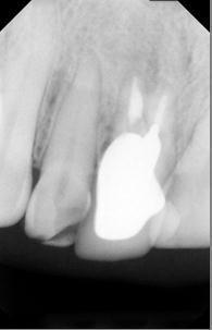
2 without the use of a microscope or any other kind of magnification. Radiographic examination (fig. 1) revealed that the tooth had 2 separated roots. The buccal root exhibited radiopaque material, and the palatal root showed a narrow canal and an apical radiolucent area. After local anesthetization with 3% prilocaine and 0.03 IU/mL felipressine (DFL, Rio de Janeiro, Brasil), the tooth was isolated with a rubber dam, all the provisional cement was removed and the pulp chamber was irrigated with 2.5% sodium hypochlorite (Biodinâmica, São Paulo, Brazil). Using an 8 magnification on a dental operating microscope (DFVasconcellos, Rio de Janeiro, Brazil), the gutapercha present in the buccal canal was assessed. The entrance of the palatal root was obliterated. Using TRA 01 and TRA 24D ultrasonic tips (Dental Trinks, São Paulo, Brazil) and with the illumination and magnification provided by the microscope, the palatal root was located and negotiated. The working length was established with an apex locator (Sybron Endo, California, USA) and a radiograph was taken to confirm the patency of the canal. No treatment was performed on the buccal canal. Chemical and mechanical instrumentation was performed with Gates Glidden burs (DentsplyMaillefer, Ballaigues, Switzerland), manual files (Flexofile, DentsplyMaillefer, Ballaigues, Switzerland), and rotary Ni-Ti files (MTwo, VDW, Munich, Germany). At every change of instrument, the canal was thoroughly irrigated with sodium hypochlorite; after preparation, it was flooded with 17% EDTA (Biodinâmia, São Paulo, Brazil) for 3 min. Passive ultrasonic irrigation (PUI) was used for 20 sec to activate the hypochlorite; this procedure was repeated 2 more times. After final hypochlorite irrigation, the root canal was dried with paper points. A dressing of calcium hydroxide with saline solution was left inside the canal for 14 days. At the following appointment, the patient was asymptomatic with the provisional crown not showing any clinical sign of leakage. After anesthesia and placement of the rubber dam, the root canal was again accessed. The calcium hydroxide dressing was removed, the canal was irrigated with sodium hypochlorite then EDTA, and passive ultrasonic irrigation was performed using the same protocol as for the first visit, and finally the root canal was dried with paper points. Filling was performed with standard #60 gutta percha points and accessory M, FM, and F (Dentsply, Petropolis, Brasil). Endofill (Dentsply, Petropolis, Brasil) cement and Tagger s technique were used to complete the root canal filling. Final radiographs were taken from the orto, mesial, and distal aspects. A prosthesis was cemented and the patient was referred for general dentistry. Due to the initial attempt at canal location, a deviation was observed in the palatal root. (fig. 2) Thirty-six months after treatment, the patient was asymptomatic; the probing test was normal and a permanent crown was complete. Radiographic examination (fig. 3) revealed that the radiolucent area had become normal with characteristics of a healing area. With the aid of cone beam computed tomography the independent roots could be visualized (fig 4,5). The original lesion was completely healed, and there was no perforation at the palatal root. 78 July-September 2013 Volume 01 Issue 02


3 Figure No. 1 Figure No. 2 Figure No. 3 Figure No. 4 Figure No July-September 2013 Volume 01 Issue 02
4 Discussion: Conventional endodontic treatment, particularly in cases of anatomic variation, must be performed efficiently to ensure functionality of the tooth. Substantial coronal destruction may jeopardize prosthetic rehabilitation and encourage patients to reconsider prosthesis on implant. Teeth with a small coronal remnant and apical radiolucency may have a favorable outcome in cases where the root canal system is properly negotiated and filled. Root canal retreatment is usually more cost-effective than an implant-supported restoration. 10 Considering that the palatal root had no canal obturation, performing apical surgery in this case would have been unlikely to be successful. 11 Variations in the anatomy of the root canal may be associated with coronal aberrations such as dens invaginatus, 12,13 talon cusp fusion, 14 or germination, even with a clinically normal crown. 15 In this case, the patient had no natural crown, precluding the assessment of the original morphology. The case reported herein exhibited a rare situation of a maxillary central incisor with 2 independent root canals, classified as a class IV as described by Vertucci. 5 The success in this case was largely dependent on the localization, negotiation, and proper treatment of the palatal root. In spite of having had previous appointments with an endodontic specialist, the complete domain of the internal anatomy was not achieved. It is believed that the use of magnification and illumination may increase the success of accessing calcified canals or those with an 16, 17 uncommon morphology. It is important to use ultrasonic tips with different shapes when removing calcifications, pulp nodules, or materials that obliterate the canal entrance. The utilization of microsonics 18 is a safe way to deal with difficult anatomies by minimizing the risk of perforation or other adverse events. Modern endodontic practice must involve not only knowledge of the internal anatomy, but also the technology necessary to adequately negotiate the entire root canal system. Conclusion: The use of dental operating microscope and the appropriate ultrasonic tips can be considered an important armamentarium to locate root canals. The root canal treatment of the tooth reported in this study was effective, less invasive and cost effective in comparison with an implant-supported single crown. Referances: 1. Schilder H. Cleaning and shaping the root canal. Dent Clin North Am Apr;18(2): Wu D, Shi W, Wu J, Wu Y, Liu W, Zhu Q. The clinical treatment of complicated root canal therapy with the aid of a dental operating microscope. Int Dent J Oct;6(5): Hess W. The anatomy of the root-canals of the teeth of the permanent dentition, part 1. New York: William Wood and Co., De Deus QD. Endodontia, 5th ed. Medsi: Rio de Janeiro, Vertucci F. Root canal anatomy of the human teeth. Oral Surg Oral Med Oral Pathol.1984 Nov;58(5): Reid JS, WP Sauders, DG Macdonald. Maxillary permanent incisors with two root canals: a report of two cases. Int Endod J Jul;26(4): Genovese FR, Marsico EM. Maxillary central incisor with two roots: a case report. JEndod Mar;29(3): Sponchiado Jr. EC, Ismail HA, Braga MR, de Carvalho FK, Simoes CA. Maxillary central incisor with two root canals: a case report. JEndod Oct;32(10): Gondim Jr. E, Setzer F, Zingg P, Karabucak B. A Maxillary Central Incisor with Three Root Canals: A. JEndod Oct;35(10): Kim S.G, Solomon C Cost-effectiveness of endodontic Molar Retreatment Compared with Fixed Partial Dentures and Single-Tooth Implant Alternatives. JEndod Mar;37(3): July-September 2013 Volume 01 Issue 02
5 11. Von Arx T, Peñarrocha M, Jensen S. Prognostic Factors in Apical Surgery with Root-end Filling: A Meta-analysis. J Endod Jun;36(6): Beltes P. Endodontic Treatment in Three Cases of Dens Invaginatus. J Endod Jun:23(6): Alani A, Bishop K. Dens Invaginatus: part 1- Classification, Prevalence and actiology. Int Endod J Dec;41(12): Ozcelik B, Atila B. Bilateral Palatal Talon Cusps on Permanent Maxillary Lateral Incisors: A. Eur J Dent Jan;5(1): Lambruschini MG, Camps J. A Two-rooted Maxillary Central Incisor with a Normal Clinical Crown. JEndod Feb;19(2): Görduysus MÖ, Gordysus M, Friedman S. Operating Microscope Improves Negotiation of Second Mesiobuccal Canals in Maxillary Molars. JEndod Nov;27(11): Selden HS. The role of a dental operating microscope in improved nonsurgical treatment of "calcified" canals..oral Surg Oral Med Oral Pathol Jul;68(1): Cunha RS, Davini F, Fontana CE, Miguita KB, Bueno CES. O conceito microsonics: primeiro molar superior com cinco canais relato de caso. RSBO 2011 Apr-Jun;8(2): Corresponding Author Marcelo Santos Coelho Rua Emilio Ribas 776/13, Campinas SP Brazil. coelho_marcelo@yahoo.com.br 81 July-September 2013 Volume 01 Issue 02
ENDODONTIC MANAGEMENT OF A MANDIBULAR FIRST MOLAR WITH SIX CANALS : A CASE REPORT
 ENDODONTIC MANAGEMENT OF A MANDIBULAR FIRST MOLAR WITH SIX CANALS : A CASE REPORT Author Name: Sreenath Narayanan INTRODUCTION Accurate diagnosis and successful endodontic therapy is always a challenge
Case Report Endodontic Treatment of Maxillary Premolar with Three Root Canals Using Optical Microscope and NiTi Rotatory Files System
 Case Reports in Dentistry Volume 2013, Article ID 710408, 4 pages http://dx.doi.org/10.1155/2013/710408 Case Report Endodontic Treatment of Maxillary Premolar with Three Root Canals Using Optical Microscope
Case Report Endodontic Management of Maxillary Second Molar with Two Palatal Roots: A Report of Two Cases
 Volume 2012, Article ID 590406, 4 pages doi:10.1155/2012/590406 Case Report Endodontic Management of Maxillary Second Molar with Two Palatal Roots: A Report of Two Cases Surbhi Patel 1 and Pawan Patel
Management of a Type III Dens Invaginatus using a Combination Surgical and Non-surgical Endodontic Therapy: A Case Report
 Management of a Type III Dens Invaginatus using a Combination Surgical and Non-surgical Endodontic Therapy: A Case Report Mithra N. Hegde, BDS, MDS, FPFA; Aditya Shetty, BDS, MDS; Rekha Sagar, BDS, MDS
Endodontic Treatment After Autotransplantation of Tooth with Complete Root Formation
 Endodontic Treatment After Autotransplantation of Tooth with Complete Root Formation Caio Cesar Souza 1, Carlos Eduardo da Silveria Bueno 1, Augusto Shogi Kato 1, Rina Andrea Pelegrine 1 Ana Paula Simezo
Gorduysus MO et al. Operating Microscope improves ability to locate and negotiate second canal
 ORIGINAL RESEARCH I.J.C.M.R Operating microscope improves ability to locate and negotiate second canal in mandibular ıncisors Mehmet Omer Gorduysus, Melahat Gorduysus, Zeliha Yilmaz * Department of Endodontics,
Case Report Three Independent Mesial Canals in a Mandibular Molar: Four-Year Followup of a Case Using Cone Beam Computed Tomography
 Case Reports in Dentistry Volume 2013, Article ID 891849, 4 pages http://dx.doi.org/10.1155/2013/891849 Case Report Three Independent Mesial Canals in a Mandibular Molar: Four-Year Followup of a Case Using
WaveOne Gold reciprocating instruments: clinical application in the private practice: Part 2
 C L I N I C A L WaveOne Gold reciprocating instruments: clinical application in the private practice: Part 2 Peet van der Vyver 1 and Martin Vorster 2 1 Department of Odontology, School of Dentistry, University
e74 Department of Conservative Dentistry and Endodontics, Meenakshi Ammal Dental College a Reader.
 Endodontic management of a maxillary first molar with two palatal roots and a single fused buccal root diagnosed with spiral computed tomography - a case report Velayutham Gopikrishna, MDS, a Joseph Reuben,
Case Report Endodontic Treatment of Bilateral Maxillary First Premolars with Three Roots Using CBCT: A Case Report
 Case Reports in Dentistry, Article ID 505676, 4 pages http://dx.doi.org/10.1155/2014/505676 Case Report Endodontic Treatment of Bilateral Maxillary First Premolars with Three Roots Using CBCT: A Case Report
Case Report Root Canal Treatment of Mandibular Second Premolar with Three Separate Roots and Canals Using Spiral Computed Tomographic
 Case Reports in Dentistry, Article ID 816576, 4 pages http://dx.doi.org/10.1155/2014/816576 Case Report Root Canal Treatment of Mandibular Second Premolar with Three Separate Roots and Canals Using Spiral
Limited To Endodontics Newsletter. Limited To Endodontics A Practice Of Endodontic Specialists July Volume 2
 Limited To Endodontics Newsletter LTE Limited To Endodontics A Practice Of Endodontic Specialists July 1 2009 Volume 2 Endodontic Treatment For The Compromised Tooth The goal of endodontic therapy is to
THE GENERAL DENTIST AND TIMELY REFERRAL TO THE ENDODONTIST
 CLINICAL THE GENERAL DENTIST AND TIMELY REFERRAL TO THE ENDODONTIST Andrei Berdichewsky, DDS 1 The use of endodontic treatment to solve problems related to pulpal and periapical pathologies is extremely
36 year-old Caucasian male presented for evaluation and treatment of tooth #3.
 Case Report William Hu Non-Surgical Retreatment #3 36 year-old Caucasian male presented for evaluation and treatment of tooth #3. Subjective Chief complaint: I was referred to see if you can do a new root
Endodontic Treatment of a Mandibular First Premolar With Three Root Canals: A Case Report
 J Res Dentomaxillofac Sci http://www.jrdms.dentaiau.ac.ir e(issn): 2383-2754 p(issn):2588-4166 Journal of Research in Dental and Maxillofacial Sciences Endodontic Treatment of a Mandibular First Premolar
Evidence-based decision-making in endodontics
 Clin Dent Rev (2017) 1:6 https://doi.org/10.1007/s41894-017-0006-0 TREATMENT Evidence-based decision-making in endodontics Eyal Rosen 1 Igor Tsesis 1 Received: 15 June 2017 / Accepted: 9 July 2017 / Published
Treatment of perforating internal root resorption with MTA: a case report
 127 Journal of Oral Science, Vol. 54, No. 1, 127-131, 2012 Case Report Treatment of perforating internal root resorption with MTA: a case report Eduardo Nunes 1), Frank F. Silveira 1,2), Janir A. Soares
Large periapical lesion: Healing without knife and incision
 Large periapical lesion: Healing without knife and incision Ridhima Suneja College of Dentistry, Gulf Medical University, Ajman, UAE ABSTRACT Three dimensional obturation of root space has always yielded
RSBO Revista Sul-Brasileira de Odontologia ISSN: Universidade da Região de Joinville Brasil
 RSBO Revista Sul-Brasileira de Odontologia ISSN: 1806-7727 fbaratto@uol.com.br Universidade da Região de Joinville Brasil Salles Gonçalves Pais, Andressa; Fontana, Carlos Eduardo; Sigrist De Martin, Alexandre;
The Graduate School Yonsei University Department of Dentistry Myoungah Seo
 The Graduate School Yonsei University Department of Dentistry Myoungah Seo A Masters Thesis Submitted to the Department of Dentistry and the Graduate School of Yonsei University in partial fulfillment
Success in root canal treatment (RCT) is dependent. Second Mesiobuccal Canal Treatment in a Predoctoral Dental Clinic: A Retrospective Clinical Study
 is dependent. Second Mesiobuccal Canal Treatment in a Predoctoral Dental Clinic: A Retrospective Clinical Study") Second Mesiobuccal Canal Treatment in a Predoctoral Dental Clinic: A Retrospective Clinical Study Marcelo Santos Coelho, DDS, MSc; Jeffrey M. Parker, DMD; Peter Z. Tawil, DMD, MS, FRCD(C), Dip ABE Abstract:
Second Mesiobuccal Canal Treatment in a Predoctoral Dental Clinic: A Retrospective Clinical Study Marcelo Santos Coelho, DDS, MSc; Jeffrey M. Parker, DMD; Peter Z. Tawil, DMD, MS, FRCD(C), Dip ABE Abstract:
Radix entomolaris A case report
 ISSN: Printed version: 1806-7727 Electronic version: 1984-5685 RSBO. 2012 Jul-Sep;9(3):340-4 Case Report Article Radix entomolaris A case report Felipe Davini 1 Rodrigo Sanches Cunha 2 Carlos Eduardo Fontana
Non-surgical endodontic treatment f invaginatus type III using cone bea Title tomography and dental operating mic report.
 Non-surgical endodontic treatment f invaginatus type III using cone bea Title tomography and dental operating mic report. Author(s) Kato, H. Journal Bulletin of Tokyo Dental College, 5 URL http://hdl.handle.net/10130/3722
Field Guide to the Ultrasonic Revolution
 Helsē Ultrasonic Field Guide to the Ultrasonic Revolution 20 Endo Tasks... Simplified. Sparking an Ultrasonic Revolution At Helse Ultrasonic, our unwavering mission is to turn your ultrasonic unit into
Limited To Endodontics Newsletter. Limited To Endodontics A Practice Of Endodontic Specialists June Volume 4
 Limited To Endodontics Newsletter LTE Limited To Endodontics A Practice Of Endodontic Specialists June 1 2011 Volume 4 Endodontic Treatment Utilizing 3D Imaging Recent advances in technology, such as rotary
Complex endodontic treatment of an immature type III dens invaginatus. A case report
 doi:10.1111/j.1365-2591.2008.01414.x CASE REPORT Complex endodontic treatment of an immature type III dens invaginatus. A case report E. R. Fregnani 1,2, L. F. B. Spinola 2,J.R.O.Sônego 2, C. E. S. Bueno
Creating a glide path for rotary NiTi instruments: part two
 Clinical Creating a glide path for rotary NiTi instruments: part two Peet van der Vyver 1 Introduction In part one of this series the author discussed the rationale for the preparation of a glide path
PORTFOLIO 01. Assigning a Level of Difficulty to Your Endodontic Cases
 PORTFOLIO 01 Assigning a Level of Difficulty to Your Endodontic Cases Root Canal Specialty Associates provides care in all phases of surgical and nonsurgical endodontics. With decades of combined experience,
Mandibular first molar with four canals in the mesial root
 C L I N I C A L Mandibular first molar with four canals in the mesial root Hamed Karkehabadi 1, Ricardo Machado 2 and Lucas da Fonseca Roberti Garcia 3 Abstract A 35-year-old female patient with intermittent
Case Report Endodontic Management of a Maxillary First Molar with Two Palatal Canals and a Single Buccal Canal: A Case Report
 Case Reports in Dentistry Volume 2012, Article ID 389387, 4 pages doi:10.1155/2012/389387 Case Report Endodontic Management of a Maxillary First Molar with Two Palatal Canals and a Single Buccal Canal:
Case Report Management of Complex Root Canal Curvature of Bilateral Radix Entomolaris: Three-Dimensional Analysis with Cone Beam Computed Tomography
 Case Reports in Dentistry Volume 2013, Article ID 697323, 4 pages http://dx.doi.org/10.1155/2013/697323 Case Report Management of Complex Root Canal Curvature of Bilateral Radix Entomolaris: Three-Dimensional
Staining Potential of Calcium Hydroxide and Monochlorophenol Following Removal of AH26 Root Canal Sealer
 Staining Potential of Calcium Hydroxide and Monochlorophenol Following Removal of AH26 Root Canal Sealer Abstract Aim: The focus of this study was to examine the staining potential of calcium hydroxide
Diagnosis and Treatment of Three-Rooted Maxillary Premolars
 Diagnosis and Treatment of Three-Rooted Maxillary Premolars Hacer Deniz Arisu a Tayfun Alacam b Abstract Anatomical variations must be considered in clinical and radiographical evaluations during endodontic
Treatment Options for the Compromised Tooth: A Decision Guide
 Treatment Options for the Compromised Tooth: A Decision Guide www.aae.org/treatmentoptions ROOT AMPUTATION, HEMISECTION, BICUSPIDIZATION Case One Hemisection of the distal root of tooth #19. 13 mo. Recall
Endodontic treatment of mandibular incisors with two root canals: Report of two cases
 Blackwell Publishing IncMalden, USAAEJAustralian Endodontic Journal1329-1947 2007 The Authors;? 2007 2731Case ReportMandibular Incisors with Two CanalsY. S. Kabak and P. V. Abbott Aust Endod J 2007; 33:
Case Report. July 2015; Vol. 12, No. 7. Vineet Agrawal 1, Sonali Kapoor 2, Mukesh Patel 3
 Case Report Ultrasonic Technique to Retrieve a Rotary Nickel-Titanium File Broken Beyond the Apex and a Stainless Steel File from the Root Canal of a Mandibular Molar: A Case Report Vineet Agrawal 1, Sonali
Shah. Management of a maxillary second premolar with an S-shaped root canal - An endodontic. Management of a maxillary second premolar
 Case Report Management of a maxillary second premolar with an S-shaped root canal - An endodontic challenge Nabi Shahnaz 1, Amin Khalid 2, Hussain Aijaz 3, Baba Irfan Ashraf 4*, Aasim Farooq Shah 5 1 PG
Root Canal Treatment. with a mechanical treatment system. Clinical case
 Endo motor with MANI Silk File Root Canal Treatment with a mechanical treatment system Case file by Markus Ludolph, Dortmund/Germany. Focus of activities: Endodontics. Clinical case In January of 2016,
Treatment Options for the Compromised Tooth
 New Edition Treatment Options for the Compromised Tooth A Decision Guide American Association of Endodontists www.aae.org/treatmentoptions TREATMENT PLANNING CONSIDERATIONS The Treatment Options for the
Endodontic management of radix entomolaris: Case reports
 2015; 2(1): 15-19 ISSN Print: 2394-7489 ISSN Online: 2394-7497 IJADS 2015; 2(1): 15-19 2015 IJADS www.oraljournal.com Received: 24-02-2015 Accepted: 08-04-2015 Dr. Uday Kamath Dentistry and Endodontics,
KING SAUD UNIVERSITY College of Dentistry. Department of Restorative Dental Sciences DIVISION OF ENDODONTICS COURSE OUTLINE 323 RDS
 KING SAUD UNIVERSITY College of Dentistry Department of Restorative Dental Sciences DIVISION OF ENDODONTICS COURSE OUTLINE 323 RDS Pre-Clinical Endodontics Three (3) Credit Hours Third Year 2014-2015 Prepared
Nestor Cohenca Professor Department of Endodontics Department of Pediatric Dentistry Diplomate, ABE
 Clinical Application of High-Resolution CBCT in Endodontics Time to Change Strategy! Nestor Cohenca Professor Department of Endodontics Department of Pediatric Dentistry Diplomate, ABE cohenca@uw.edu Clinical
In vitro evaluation of root canal preparation with plastic endodontic rotary finishing file - A SEM study
 Original Research In vitro evaluation of root canal preparation with plastic endodontic rotary finishing file - A SEM study ASHUTOSH * ASEEM P. TIKKU ** ANIL CHANDRA ** PROMILA VERMA *** RAKESH K. YADAV
Maxillary First Molar with Three Mesiobuccal Root Canals: A Case Report
 JKAU: Med. Sci., Vol. 19 No. 2, pp: 99-108 (2012 A.D. / 1433 A.H.) DOI: 10.4197/Med. 19-2.8 Maxillary First Molar with Three Mesiobuccal Root Canals: A Case Report Laila A. Bahammam, BDS, MSC, CERT ENDOD
Fundamentals of Endodontics Peter Briggs, Ahmed Farooq and Tracy Watford, Trish Moore and QED
 Fundamentals of Endodontics Peter Briggs, Ahmed Farooq and Tracy Watford, Trish Moore and QED Practical Hands-on course St George s Hospital, SW17 0QT St George s Dental Simulation Today Take the opportunity
Endodontic treatment of a maxillary first molar having three mesiobuccal canals with the aid of spiral computed tomography: a case report
 495 Journal of Oral Science, Vol. 52, No. 3, 495-499, 2010 Case Report Endodontic treatment of a maxillary first molar having three mesiobuccal canals with the aid of spiral computed tomography: a case
MAXILLARY FIRST MOLAR : AN ENIGMA FOR ENDODONTISTS
 ISSN: 0976-3104 CASE REPORT OPEN ACCESS MAXILLARY FIRST MOLAR : AN ENIGMA FOR ENDODONTISTS Sowmya B*, Sarika Chandra, Abha Telang, Sylvia Mathew, John V George Department of Conservative Dentistry and
Journal of Craniomaxillofacial Research. Vol. 3, No. 4 Autumn 2016
 Journal of Craniomaxillofacial Research Vol. 3, No. 4 Autumn 2016 The use of cone beam computed tomography in diagnosis and surgical management of a case of internal root resorption: A case report Samane
Douglas D. Lancaster, COL, DC
 Douglas D. Lancaster, COL, DC 3 August 2016 Army Post Graduate Dental School Prevalence of the middle mesial canal in non-surgical root canal treated mandibular first and second molars in a local military
Surgical Retreatment of an Invaginated Maxillary Central Incisor Following Overfilled Endodontic Treatment: A Case Report
 Surgical Retreatment of an Invaginated Maxillary Central Incisor Following Overfilled Endodontic Treatment: A Case Report Hakan Ozbas a Rustem Kemal Subay b Melike Ordulu c ABSTRACT This case report presents
Bypassing Separated Instruments in the Root Canal Two Case Reports
 IOSR Journal of Dental and Medical Sciences (IOSR-JDMS) e-issn: 2279-0853, p-issn: 2279-0861.Volume 15, Issue 6 Ver. XI (June. 2016), PP 08-13 www.iosrjournals.org Bypassing Separated Instruments in the
Chronic pulp canal obliteration after dental trauma and orthodontic treatment: a case report
 original article Chronic pulp canal obliteration after dental trauma and orthodontic treatment: a case report Laís C. PACHECO 1 Fabiola ORMIGA 2 Heloisa GUSMAN 3 Patrícia de Andrade RISSO 3 DOI: http://dx.doi.org/10.14436/2358-2545.6.2.041-046.oar
MANAGEMENT OF ROOT RESORPTION- A REBIRTH CASE REPORTS DEPARTMENT OF CONSERVATIVE DENTISTRY AND ENDODONTICS
 MANAGEMENT OF ROOT RESORPTION- A REBIRTH CASE REPORTS AUTHORS Dr. SHALINI.H, PG Student Dr. B. RAMAPRABHA, MDS Professor Dr. M. KAVITHA, MDS Professor and HOD DEPARTMENT OF CONSERVATIVE DENTISTRY AND ENDODONTICS
Jim Ruckman. 65 year-old Caucasian female presented for evaluation and treatment of tooth #19.
 Case Report Jim Ruckman Non-Surgical Root Canal Therapy #19 65 year-old Caucasian female presented for evaluation and treatment of tooth #19. Subjective Chief complaint: I was seen in the dental school
An In Vivo Evaluation of Two Types of Files used to Accurately Determine the Diameter of the Apical Constriction of a Root Canal: An In Vivo Study
 An In Vivo Evaluation of Two Types of Files used to Accurately Determine the Diameter of the Apical Constriction of a Root Canal: An In Vivo Study Sumeet Darda, BDS, MDS; Narendra Manwar, BDS, MDS; Manoj
Virtuosity is in your hands. Endodontic and Microsurgical Ultrasonic Instruments
 Virtuosity is in your hands Endodontic and Microsurgical Ultrasonic Instruments The advantages of ProUltra tips Patented contra-angled feature enhances access to all teeth Abrasive coating increases efficiency
Diagnosis and endodontic management of two rooted mandibular second premolar: A case report
 International Journal of Sciences & Applied Research www.ijsar.in Diagnosis and endodontic management of two rooted mandibular second premolar: A case report Saima Jamal*, Swathi, Rajaram Naik Department
Case Report Endodontic Treatment and Esthetic Management of a Geminated Central Incisor Bearing a Talon Cusp
 Case Reports in Dentistry, Article ID 123681, 4 pages http://dx.doi.org/10.1155/2014/123681 Case Report Endodontic Treatment and Esthetic Management of a Geminated Central Incisor Bearing a Talon Cusp
Endodontic treatment of maxillary lateral incisors with anatomical variations
 Case report ISSN 2234-7658 (print) / ISSN 2234-7666 (online) Endodontic treatment of maxillary lateral incisors with anatomical variations Moon-Hwan Lee, Jung- Hong Ha, Myoung-Uk Jin, Young-Kyung Kim,
Journal of American Science 2014;10(12)
") Morphological variations in the root canal system of mandibular Premolars : A case series Youssef A. Algarni, BDS Specialist of Endodontics, Jeddah, KSA dr_yousseef@hotmail.com Abstract: The mandibular
Morphological variations in the root canal system of mandibular Premolars : A case series Youssef A. Algarni, BDS Specialist of Endodontics, Jeddah, KSA dr_yousseef@hotmail.com Abstract: The mandibular
Failure of root canal therapy often
 Anatomy Microcomputed tomographic evaluation of mandibular molars with single distal canals Arthur F. Lamia, DDS, MS n N.J. McDonald, BDS, MS The aim of this study was to evaluate, using microcomputed
ENDODONTIC MANAGEMENT OF C-SHAPED CANAL IN MANDIBULAR SECOND MOLAR: A CASE REPORT
 Case Report International Journal of Dental and Health Sciences Volume 03, Issue 04 ENDODONTIC MANAGEMENT OF C-SHAPED CANAL IN MANDIBULAR SECOND MOLAR: A CASE REPORT Thakur Savita 1,Rani Nidhi 2,Dosanjh
Diagnosis and treatment of teeth with primary endodontic lesions mimicking periodontal disease: three cases with long-term follow ups
 Case report ISSN 2234-7658 (print) / ISSN 2234-7666 (online) http://dx.doi.org/10.5395/rde.2014.39.1.56 Diagnosis and treatment of teeth with primary endodontic lesions mimicking periodontal disease: three
Unusual root canal anatomy in a maxillary second molar
 www.edoriumjournals.com CLINICAL IMAGES PEER REVIEWED OPEN ACCESS Unusual root canal anatomy in a maxillary second molar Toshiko Inoue, Makoto Saito, Fumio Nishimura, Takashi Miyazaki ABSTRACT Abstract
Mid Mesial Canal in Mandibular Molars: Two Case Report and A Review of Literature
 ISSN 2455-4499; Vol.10, Issue 03 (March 2018) Pg. no. 27-32. Institute of Research Advances https://research-advances.org/index.php/irajas Mid Mesial Canal in Mandibular Molars: Two Case Report and A Review
Challenges and Current Concepts in the Preparation of Root Canal System in Radix Entomolaris Case Reports
 IOSR Journal of Dental and Medical Sciences (IOSR-JDMS) e-issn: 2279-0853, p-issn: 2279-0861.Volume 15, Issue 2 Ver. VI (Feb. 2016), PP 77-81 www.iosrjournals.org Challenges and Current Concepts in the
Restoration of a Grossly Carious tooth using Canal Projection: A Comparative Analysis of different materials for use as Canal Projectors
 Restoration of a Grossly Carious tooth using Canal Projection: A Comparative Analysis of different materials for use as Canal Projectors Saurabh Ahuja 1, RPS Bedi 2, Ankur Garg 3, Himantika Singh 4, Nidhi
Complicated untreated apical periodontitis causing paraesthesia: A case report
 Aust Endod J 2017 CASE REPORT Complicated untreated apical periodontitis causing paraesthesia: A case report Domenico Ricucci, MD, DDS 1, * ; Simona Loghin, DDS 1 ; and Jose F. Siqueira Jr, DDS, MSc, PhD
An unusual maxillary canine with two separated root canals
 pissn 225-0864 / eissn 2093-879 http://dx.doi.org/0.5395/jkacd.20.36.5.43 Case report A maxillary canine with two separated root canals: a case report Dong-Ryul Shin, Jin-Man Kim 2, Duck-Su Kim 3, Sun-Young
Endodontic Management of Radix Entomolaris in First and Third Mandibular Molars with Hybrid Rotary Systems A Case Series.
 Annals of Dental Research (2011) Vol 1 (1): 73-79 Mind Reader Publications: All Rights Reserved Annals of Dental Research www.adres.yolasite.com www.journalshub.com Case Series Endodontic Management of
Index. shapes, sizes, and locations, 282 small bone defects, through-and-through bone defect, treatment techniques, 285, 287
 Index A Accessory canal, 18 20 Accidental pulp exposure, 100, 103 104 Adhesive dentistry, 174 175 Ankylosis, 69 70 Anterior superior alveolar (ASA), 81 Anxiety, 36 Apical periodontitis asymptomatic, 214
Influence of cervical preflaring on apical file size determination - An in vitro study
 Original Research Influence of cervical preflaring on apical file size determination - An in vitro study VASUNDHARA SHIVANNA * DEEPALI AGARWAL ** ABSTRACT Aim: To investigate the influence of cervical
Journal of Dental & Oro-facial Research Vol. 14 Issue 01 Jan. 2018
 Journal of Dental & Oro-facial Research Vol. 14 Issue 01 Jan. 2018 Management of Non-Vital Teeth with Open Apices using MTA: Two Case Reports *Karan Narang 1, Mohini Nayak 2, Abdul Wahed, 3 John V. George
LOCATING CANALS. Strategies, Armamentarium and Techniques. by Clifford J. Ruddle, DDS. Advanced Endodontics DENTISTRY TODAY February 2017
 Advanced Endodontics DENTISTRY TODAY February 2017 LOCATING CANALS Strategies, Armamentarium and Techniques by Clifford J. Ruddle, DDS Over the past decades, many technologies have come to market to help
Computed TomographyEvaluationof Root Canal Desobturation of Lower Human Incisors
 IOSR Journal of Dental and Medical Sciences (IOSR-JDMS) e-issn: 2279-0853, p-issn: 2279-0861.Volume 17, Issue 8 Ver. 2 (August. 2018), PP 60-64 www.iosrjournals.org Computed TomographyEvaluationof Root
Advanced Endodontics Course
 Advanced Endodontics Course 2017 London Course Information Booklet Hello and Welcome! Welcome to one of UK s most hands on Advanced Endodontics course where you will be receiving the best hands on teaching
Comparison of Spreader Penetration during Lateral Compaction of 0.04 and 0.02 Tapered Gutta-Percha Master Cones
 Original Article Comparison of Spreader Penetration during Lateral Compaction of 0.04 and 0.02 Tapered Gutta-Percha Master Cones M. Saatchi 1, L. Etesami 2 1 Assistant Professor, Department of Endodontics,
EFFICACY OF NON-SURGICAL RETREATMENT WITH AND WITHOUT USING ULTRASONICS AND DENTAL MICROSCOPE
 Original Article International Journal of Dental and Health Sciences Volume 03,Issue 0 EFFICACY OF NON-SURGICAL RETREATMENT WITH AND WITHOUT USING ULTRASONICS AND DENTAL MICROSCOPE Liana Kinjrawi,Aziz
CONTENTS. Endodontic therapy Permanent open apex teeth Intracanal Medication. A. Introduction I. Problems II. III. IV. B. Research C.
 CONTENTS A. Introduction I. Problems II. III. IV. Endodontic therapy Permanent open apex teeth Intracanal Medication B. Research C. Conclusion INTRODUCTION A. Problems 1. In permanent teeth with open apex
Case Note Retrieval of a separated file using Masserann technique: A case report
 Kathmandu University Medical Journal (2006), Vol. 4, No. 2, Issue 14, 238-242 Case Note Retrieval of a separated file using Masserann technique: A case report Pai ARV 1, Kamath MP 2, Basnet P 3 1 Associate
Case Report: Maxillary second molar with two palatal canals diagnosed with cone beam computed tomography: a case report
 Case Report: Maxillary second molar with two palatal canals diagnosed with cone beam computed tomography: a case report Dr Dhara Shukla 1,Dr Girish Parmar 2, Dr Abhishek Parmar 3 1 PG Part II Student,
Case Report Independent and Confluent Middle Mesial Root Canals in Mandibular First Molars: A Report of Four Cases
 Case Reports in Dentistry Volume 2012, Article ID 103125, 6 pages doi:10.1155/2012/103125 Case Report Independent and Confluent Middle Mesial Root Canals in Mandibular First Molars: A Report of Four Cases
Advanced Endodontics Course
 Advanced Endodontics Course 2017 London Course Information Booklet Hello and Welcome! Welcome to one of UK s most hands on Advanced Endodontics course where you will be receiving the best hands on teaching
Calcium Hydroxide And Zinc Oxide Eugenol As Root Canal Filling Materials In Primary Molars: A Comparative Study
 Clinical Update By S. G. Damle and U. M. Nadkarni, Department of Pedodontics and Preventive Dentistry, Nair Hospital Dental College, Mumbai, India. Address for correspondence: Dr S. G. Damle, Professor
5 Days Comprehensive Endodontic Course Topics
 5 Days Comprehensive Endodontic Course Topics 1. Pulp Dentin Complex/Retrogressive Changes: The significance of structural elements and its physiological, pathological and age related changes on the diagnosis
Lodha Ena* M. Kala** INTRODUCTION
 Indian Journal of Dental Education 23 Volume 2 Number 4 October-December 2010 A clinical study on the incidence of C shaped root canals in mandibular second molars in patients visiting the department of
A maxillary lateral incisor with four root canals
 doi:10.1111/j.1365-2591.2011.01984.x A maxillary lateral incisor with four root canals J. Kottoor 1, R. Murugesan 2 & D. V. Albuquerque 3 1 Department of Conservative Dentistry and Endodontics, Mar Baselios
Maxillary first molar with 7 root canals diagnosed using cone-beam computed tomography
 Case report ISSN 2234-7658 (print) / ISSN 2234-7666 (online) Maxillary first molar with 7 root canals diagnosed using cone-beam computed tomography Evaldo Rodrigues 1, Antônio Henrique Braitt 2, Bruno
P. Variant Anatomy of Mandibular First Permanent Molar:
 Case Report Variant Anatomy of Mandibular 10.5005/jp-journals-10031-1119 First Permanent Molar: A Case Series Variant Anatomy of Mandibular First Permanent Molar: A Case Series 1 Pratima R Shenoi, 2 Rajesh
Case Report Endodontic Management of a Maxillary Molar with Three Mesiobuccal Canals
 Case Reports in Dentistry, Article ID 320196, 4 pages http://dx.doi.org/10.1155/2014/320196 Case Report Endodontic Management of a Maxillary Molar with Three Mesiobuccal Canals Sirisha Gundam, 1 Radhika
ENDODONTIC TREATMENT OF PREMOLAR WITH UNUSUAL ANATOMY AND HYPERCEMENTOSIS CASE REPORT
 ENDODONTIC TREATMENT OF PREMOLAR WITH UNUSUAL ANATOMY AND HYPERCEMENTOSIS CASE REPORT ABSTRACT NOUMAN NOOR 2 MANZOOR AHMED 3 NUSRAT JABEEN 4 SADAF HUMAYOUN The purpose of this article was to report the
Primary molars with extra root canals - a case series
 Case Series Primary molars with extra root canals - a case series Sagar Chawla, 1 Mousami Goswami, 2 Priyanka Sachdeva, 3 Vidhi Walia. 4 1,3,4 Postgraduate student, 2 Professor and Head, Department of
Nonsurgical retreatment of type III DENS invaginatus
 Case Report: Nonsurgical retreatment of type III DENS invaginatus Dr Nupur Dhanak, Dr Sunita Garg, Dr Abhishek Parmar, Dr Mansi Dave Dept. of Conservative Dentistry and Endodontics, Govt Dental College
Survey on Knowledge about Access Cavity Preparation
 Survey on Knowledge about Access Cavity Preparation 1 Sanjay Madhavan, 2 Dr.Chandana 1 II BDS student, Saveetha Dental College and Hospital, Chennai 2 Department of Conservative Endodontics, Saveetha Dental
International Journal of Basic and Clinical Studies (IJBCS) 2014;3(1): Uysal I and Oztekin F
 2014;3(1): Uysal I and Oztekin F") Endodontic Treatment of Mandibular Incisors with Periapical Lesion by Using Single Instrument: Cone Beam Computed Tomography Imaging Ibrahim Uysal 1 Faruk Oztekin 2 1 PhD, Dicle University, Faculty of
Endodontic Treatment of Mandibular Incisors with Periapical Lesion by Using Single Instrument: Cone Beam Computed Tomography Imaging Ibrahim Uysal 1 Faruk Oztekin 2 1 PhD, Dicle University, Faculty of
Original article: A Comparative evaluation of three different Ni-Ti rotary files using Crown Down technique. An Exvivo study using C T scan.
 Bangladesh Journal of Medical Science Vol. 16 No. 02 April 17 Original article: A Comparative evaluation of three different Ni-Ti rotary files using Crown Down technique. An Exvivo study using C T scan.
CLINICAL AND RADIOGRAPHIC EVALUATION OF DIRECT PULP CAPPING PROCEDURES PERFORMED BY POSTGRADUATE STUDENTS
 CLINICAL AND RADIOGRAPHIC EVALUATION OF DIRECT PULP CAPPING PROCEDURES PERFORMED BY POSTGRADUATE STUDENTS Monica Monea Alexandru Sitaru Tudor Hantoiu Department of Odontology and Oral Pathology, Faculty
Endo Easy Efficient. one file endo. The RECIPROC Technique Steps to Success.
 Endo Easy Efficient one file endo The RECIPROC Technique Steps to Success www.vdw-dental.com 1. Selecting the Correct Instrument Pre-Operative Radiograph DECISION canal is completely visible wide or medium