Anatomy of Oral Cavity DR. MAAN AL-ABBASI
|
|
|
- Diana Sherman
- 6 years ago
- Views:
Transcription


1 Anatomy of Oral Cavity DR. MAAN AL-ABBASI
2 By the end of this lecture you should be able to: 1. Differentiate different parts of the oral cavity 2. Describe the blood and nerve supply of mucosa and muscles of palate and tongue 3. Identify the effect of nerve paralysis on those muscles and their related function 4. Summarise the lymphatic drainage of oral cavity

3 Parts of Oral Cavity

4 II. MOUTH PROPER Oral Cavity Proper Oral Vestibule

5 Floor of mouth Each sublingual compartment contains submandibular gland and ducts, lingual and hypoglossal nerve and the sublingual vessels.
6 o Sensory Nerve Supply Roof: by greater palatine and nasopalatine nerves (branches of maxillary nerve) Floor: by lingual nerve (branch of mandibular nerve) Cheek: by buccal nerve (branch of mandibular nerve) o Motor Muscle in the cheek (buccinator) and the lip (orbicularis oris) are supplied by the branches of the facial nerve

7 HARD PALATE o COVERED BY MUCOUS MEMBRANE AND FORMS A PARTITION BETWEEN THE ORAL AND THE NASAL CAVITY Median raphe o THE MUCOUS MEMBRANE AND THE PERIOSTEUM CANNOT BE SEPARATED (MUCOPERIOSTEUM)

8 SOFT PALATE MOVABLE PORTION AND IS ATTACHED TO THE HARD PALATE PALATINE TONSIL IS ALSO CALLED ISTHMUS OF FAUCES OR THE TONSILLAR SINUS

9 Muscles of Palate raises the uvula brings soft palate in contact with posterior pharyngeal wall, preventing food from going upward into the nasopharynx tenses the soft palate raises the tongue
10 Sensory Nerve Supply Mostly by the maxillary nerve through its branches: Greater palatine nerve Lesser palatine nerve Nasopalatine nerve Glossopharyngeal nerve supplies the region of the soft palate
11 Motor Nerve Supply All the muscles are supplied by the: Pharyngeal plexus Except tensor veli palatine that supplied by: Nerve to medial pterygoid, a branch of the mandibular division of the trigeminal nerve

12 Blood Supply Branches of the maxillary artery Greater palatine Lesser palatine Sphenopalatine Ascending palatine, branch of the facial artery Ascending pharyngeal, branch of the external carotid artery

13 Lymphatic drainage

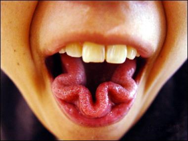
14 The Tongue

15 Intrinsic tongue muscles Inferior Longitudinal: moves tip up & down Superior Longitudinal: moves tip up & down Transverse: narrows & lengthens tongue Vertical: flattens & depresses tongue

16 Extrinsic tongue muscles Styloglossus: Pulls tongue up & back Palatoglossus: Pulls tongue back

17 Coronal section of tongue Genioglossus: Prevents tongue from falling back Hyoglossus: Depresses tongue
18 Movements Protrusion: Genioglossus on both sides acting together Retraction: Styloglossus and hyoglossus on both sides acting together Depression: Hyoglossus and genioglossus on both sides acting together Elevation: Styloglossus and palatoglossus on both sides acting together

19
20 Nerve Supply of Tongue Anterior 2/3 Posterior 1/3 Sensory Lingual Glossopharyngeal Motor Hypoglossal *** Taste Chorda tympani Glossopharyngeal *** except palatoglossus which is supplied by pharyngeal plexus

21 Nerve Supply of Tongue

22 Papillae in tongue

23 Papillae in tongue Fungiform: at tip & sides of tongue Circumvallate: just in front of terminal sulcus Foliate: at posterior lateral margins of tongue Filiform: centre of tongue, have no taste buds

24 Tongue Map? Sweet = Sucrose Salty = NaCl Sour = HCl Bitter = Quinine Umami = Glutamate

25 Taste Pathway

26 Blood Supply Arteries: Lingual artery Tonsillar branch of facial artery Ascending pharyngeal artery Veins: Lingual vein, ultimately drains into the internal jugular vein Lingual artery & vein Hypoglossal nerve Dorsal lingual artery & vein Deep lingual vein

27 Lymphatic Drainage Tip: Submental nodes bilaterally & then deep cervical nodes Anterior two third: Submandibular unilaterally & then deep cervical nodes Posterior third: Deep cervical nodes (jugulodigastric mainly)
 (due to large frenulum) Lesion of the")

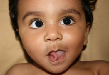
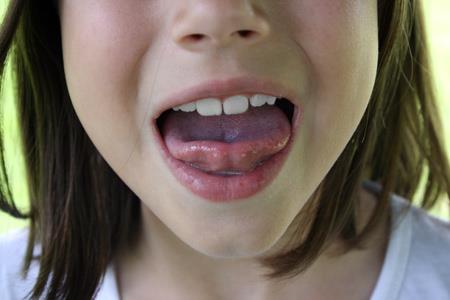
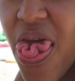
28 Lacerations of the tongue Clinical Notes Tongue-Tie (ankyloglossia) (due to large frenulum) Lesion of the hypoglossal nerve The protruded tongue deviates toward the side of the lesion Tongue is atrophied & wrinkled

29 Thank You
Subdivided into Vestibule & Oral cavity proper
 Extends from the lips to the oropharyngeal isthmus The oropharyngeal isthmus: Is the junction of mouth and pharynx. Is bounded: Above by the soft palate and the palatoglossal folds Below by the dorsum
Basic Anatomy and Physiology of the Lips and Oral Cavity. Dr. Faghih
 Basic Anatomy and Physiology of the Lips and Oral Cavity Dr. Faghih It is divided into seven specific subsites : 1. Lips 2. dentoalveolar ridges 3. oral tongue 4. retromolar trigone 5. floor of mouth 6.
-Ibrahim Al-Naser. -Dr Al- Muhtaseb. 1 P a g e
 -1 -Ibrahim Al-Naser - -Dr Al- Muhtaseb 1 P a g e The Digestive System The doctor started the lecture by talking about the class rules. The GI system is an organ system, it is divided into: The Alimentary
Oral cavity : consist of two parts: the oral vestibule and the oral cavity proper. Oral vestibule : is slit like space between.
 Oral cavity Oral cavity : consist of two parts: the oral vestibule and the oral cavity proper Oral vestibule : is slit like space between the teeth, buccal gingiva, lips, and cheeks 1 Oral cavity Oral
Nose & Mouth OUTLINE. Nose. - Nasal Cavity & Its Walls. - Paranasal Sinuses. - Neurovascular Structures. Mouth. - Oral Cavity & Its Contents
 Dept. of Human Anatomy, Si Chuan University Zhou hongying eaglezhyxzy@163.com Nose & Mouth OUTLINE Nose - Nasal Cavity & Its Walls - Paranasal Sinuses - Neurovascular Structures Mouth - Oral Cavity & Its
Dr.Ban I.S. head & neck anatomy 2 nd y. جامعة تكريت كلية طب االسنان املرحلة الثانية
 جامعة تكريت كلية طب االسنان التشريح مادة املرحلة الثانية أ.م.د. بان امساعيل صديق 6102-6102 1 The Palate The palate forms the roof of the mouth and the floor of the nasal cavity. It is divided into two
بسم اهلل الرحمن الرحيم
 بسم اهلل الرحمن الرحيم Today we will talk about digestive system in the head & neck We have the mouth, teeth, tongue, palate & salivary glands all of these are included in this lecture *First we will start
ORAL CAVITY, ESOPHAGUS AND STOMACH
 ORAL CAVITY, ESOPHAGUS AND STOMACH 1 OBJECTIVES By the end of the lecture you should be able to: Describe the anatomy the oral cavity, (boundaries, parts, nerve supply). Describe the anatomy of the palate,
The Digestive System in the Head and Neck
 The Digestive System in the Head and Neck The Mouth The Lips The lips are two fleshy folds that surround the oral orifice They are covered on the outside by skin and are lined on the inside by mucous membrane
Today's lecture discuss : 1- the mouth. 5-the salivary glands
 Today's lecture discuss : 1- the mouth 3-the tongue 2-the teeth 4-the palates 5-the salivary glands ( u dnt have to refer to the slides, I've included everything in slides ( 1-27 ) except some figures.
Veins of the Face and the Neck
 Veins of the Face and the Neck Facial Vein The facial vein is formed at the medial angle of the eye by the union of the supraorbital and supratrochlear veins. connected through the ophthalmic veins with
Lec [8]: Mandibular nerve:
![Lec [8]: Mandibular nerve:](/thumbs/94/121295776.jpg "Lec [8]: Mandibular nerve:") Lec [8]: Mandibular nerve: The mandibular branch from the trigeminal ganglion lies in the middle cranial fossa lateral to the cavernous sinus. With the motor root of the trigeminal nerve [motor roots lies
Lec [8]: Mandibular nerve: The mandibular branch from the trigeminal ganglion lies in the middle cranial fossa lateral to the cavernous sinus. With the motor root of the trigeminal nerve [motor roots lies
SCHOOL OF ANATOMICAL SCIENCES Mock Run Questions. 4 May 2012
 SCHOOL OF ANATOMICAL SCIENCES Mock Run Questions 4 May 2012 1. With regard to the muscles of the neck: a. the platysma muscle is supplied by the accessory nerve. b. the stylohyoid muscle is supplied by
Temporal region. temporal & infratemporal fossae. Zhou Hong Ying Dept. of Anatomy
 Temporal region temporal & infratemporal fossae Zhou Hong Ying Dept. of Anatomy Temporal region is divided by zygomatic arch into temporal & infratemporal fossae. Temporal Fossa Infratemporal fossa Temporal
PTERYGOPALATINE FOSSA
 PTERYGOPALATINE FOSSA Outline Anatomical Structure and Boundaries Foramina and Communications with other spaces and cavities Contents Pterygopalatine Ganglion Especial emphasis on certain arteries and
Bisection of Head & Nasal Cavity 頭部對切以及鼻腔. 解剖學科馮琮涵副教授 分機
 Bisection of Head & Nasal Cavity 頭部對切以及鼻腔 解剖學科馮琮涵副教授 分機 3250 E-mail: thfong@tmu.edu.tw Outline: The structure of nose The concha and meatus in nasal cavity The openings of paranasal sinuses Canals, foramens
Face. Definition: The area between the two ears and from the chin to the eye brows. The muscles of the face
 Face Definition: The area between the two ears and from the chin to the eye brows. The muscles of the face The muscle of facial expression (include the muscle of the face and the scalp). All are derived
HUMAN ANATOMY II STUDY NOTES. At the end of this chapter the student should be able to answer the following questions:
 HUMAN ANATOMY II STUDY NOTES CHAPTER ONE The Special Senses Learning objectives At the end of this chapter the student should be able to answer the following questions: 1. What is the gross and histological
Temporal fossa Infratemporal fossa Pterygopalatine fossa Terminal branches of external carotid artery Pterygoid venous plexus
 Outline of content Temporal fossa Infratemporal fossa Pterygopalatine fossa Terminal branches of external carotid artery Pterygoid venous plexus Boundary Content Communication Mandibular division of trigeminal
Infratemporal fossa: Tikrit University college of Dentistry Dr.Ban I.S. head & neck Anatomy 2 nd y.
 Infratemporal fossa: This is a space lying beneath the base of the skull between the lateral wall of the pharynx and the ramus of the mandible. It is also referred to as the parapharyngeal or lateral pharyngeal
Lecture 07. Lymphatic's of Head & Neck. By: Dr Farooq Amanullah Khan PMC
 Lecture 07 Lymphatic's of Head & Neck By: Dr Farooq Amanullah Khan PMC Dated: 28.11.2017 Lymphatic Vessels Of the 800 lymph nodes in the human body, 300 are in the Head & neck region. The lymphatic vessels
Cranial Nerve VII - Facial Nerve. The facial nerve has 3 main components with distinct functions
 Cranial Nerve VII - Facial Nerve The facial nerve has 3 main components with distinct functions Somatic motor efferent Supplies the muscles of facial expression; posterior belly of digastric muscle; stylohyoid,
Lips and labial mucosa
 Lips and labial mucosa External portion of the lips: the vermilion border and the skin Vermilion border : the exposed red portion of the lip, covered by mucous membrane, no mucous glands Boundary: the
Prevertebral Region, Pharynx and Soft Palate
 Unit 20: Prevertebral Region, Pharynx and Soft Palate Dissection Instructions: Step1 Step 2 Step 1: Insert your fingers posterior to the sternocleidomastoid muscle, vagus nerve, internal jugular vein,
Oral Cavity and Pharynx. The Oral Cavity. The oral cavity is divided into two major portions: the vestibule and the cavum oris.
 11 Oral Cavity and Pharynx Persons who specialize in the care and treatment of the oral cavity have a great responsibility. The oral cavity participates actively in respiration, nutrition, and excretion
ANTERIOR CERVICAL TRIANGLE (Fig. 2.1 )
") 2 Neck Anatomy ANTERIOR CERVICAL TRIANGLE (Fig. 2.1 ) The boundaries are: Lateral: sternocleidomastoid muscle Superior: inferior border of the mandible Medial: anterior midline of the neck This large triangle
2 Neck Anatomy ANTERIOR CERVICAL TRIANGLE (Fig. 2.1 ) The boundaries are: Lateral: sternocleidomastoid muscle Superior: inferior border of the mandible Medial: anterior midline of the neck This large triangle
Trigeminal Nerve Anatomy. Dr. Mohamed Rahil Ali
 Trigeminal Nerve Anatomy Dr. Mohamed Rahil Ali Trigeminal nerve Largest cranial nerve Mixed nerve Small motor root and large sensory root Motor root Nucleus of motor root present in the pons and medulla
APRIL
 APRIL - 2003 OCTOBER - 2003 February 2009 [KU 652] Sub. Code : 4131 FIRST B.D.S DEGREE EXAMINATION (Modified Regulations III) Paper I HUMAN ANATOMY, HISTOLOGY AND EMBRYOLOGY Time : Three hours
The Pharynx. Dr. Nabil Khouri MD. MSc, Ph.D
 The Pharynx Dr. Nabil Khouri MD. MSc, Ph.D Introduction The pharynx is the Musculo-fascial halfcylinder that links the oral and nasal cavities in the head to the larynx and esophagus in the neck Common
Trigeminal Nerve Worksheets, Distributions Page 1
 Trigeminal Nerve Worksheet #1 Distribution by Nerve Dr. Darren Hoffmann Dental Gross Anatomy, Spring 2013 We have drawn out each of the branches of CN V in lecture and you have an idea now for their basic
Dr.Ban I.S. head & neck anatomy 2 nd y جامعة تكريت كلية طب االسنان مادة التشريح املرحلة الثانية أ.م.د. بان امساعيل صديق 6102/6102
 جامعة تكريت كلية طب االسنان مادة التشريح املرحلة الثانية أ.م.د. بان امساعيل صديق 6102/6102 Pterygopalatine fossa: The pterygopalatine fossa is a cone-shaped depression, It is located between the maxilla,
3-Deep fascia: is absent (except over the parotid gland & buccopharngeal fascia covering the buccinator muscle)
") The Face 1-Skin of the Face The skin of the face is: Elastic Vascular (bleed profusely however heal rapidly) Rich in sweat and sebaceous glands (can cause acne in adults) It is connected to the underlying
The Face 1-Skin of the Face The skin of the face is: Elastic Vascular (bleed profusely however heal rapidly) Rich in sweat and sebaceous glands (can cause acne in adults) It is connected to the underlying
Anatomy Sheet: Oral cavity Done by: rasha Rakan edited by: khansaa Mahmoud
 Anatomy Sheet: Oral cavity Done by: rasha Rakan edited by: khansaa Mahmoud The oral cavity has 2 parts: 1. Oral vestibule: outer part that consists of outside the teeth, between the teeth, the cheeks and
The Neck the lower margin of the mandible above the suprasternal notch and the upper border of the clavicle
 The Neck is the region of the body that lies between the lower margin of the mandible above and the suprasternal notch and the upper border of the clavicle below Nerves of the neck Cervical Plexus Is formed
Oral cavity landmarks
 By: Dr. Ahmed Rabah Oral cavity landmarks The knowledge of oral anatomy and physiology will help the operator and provides enough landmarks to act as positive guide during denture construction. This subject
Dr.Ban I.S. head & neck anatomy 2 nd y. جامعة تكريت كلية طب االسنان املرحلة الثانية أ.م.د. بان امساعيل صديق 6102/6102
 جامعة تكريت كلية طب االسنان التشريح مادة املرحلة الثانية أ.م.د. بان امساعيل صديق 6102/6102 Parotid region The part of the face in front of the ear and below the zygomatic arch is the parotid region. The
Mohammad Mohtaseb. Nour Hussein. Faisal Nimri
 2 Mohammad Mohtaseb Nour Hussein Faisal Nimri Muscles of the tongue The tongue is a muscular organ and contains intrinsic and extrinsic muscles. The intrinsic muscle contains vertical, oblique, and transverse
Respiratory System. Cambridge University Press Concise Anatomy for Anaesthesia Andreas G. Erdmann Excerpt More information
 Respiratory System 1 The mouth DESCRIPTION The mouth extends from the lips (anterior) to the isthmus of the fauces (posterior). There are two sections: Vestibule slit-like cavity between the cheeks/lips
ORAL & PHARYNGEAL STRUCTURES
 ORAL & PHARYNGEAL STRUCTURES Pedro Amarante Andrade, PhD LCSC06 BIOSCIENCES FOR SPEECH AND LANGUAGE THERAPY 09/10/2017 1 Oral & pharyngeal structures Dentition THIS SESSION Tongue Taste & Sensation Tonsillar
Parotid Gland. Parotid Gland. Largest of 3 paired salivary glands (submandibular; sublingual) Ramus of Mandible. Medial pterygoid.
 Ramus of Mandible. Medial pterygoid.") Parotid region Parotid Gland Largest of 3 paired salivary glands (submandibular; sublingual) Ramus of Mandible Medial pterygoid Cross section of mandible Masseter D S SCM Parotid Gland Mastoid Process
Parotid region Parotid Gland Largest of 3 paired salivary glands (submandibular; sublingual) Ramus of Mandible Medial pterygoid Cross section of mandible Masseter D S SCM Parotid Gland Mastoid Process
Introduction. Key terms. Key terms. Key terms 10/12/2016 EQUINE UPPER DIGESTIVE SYSTEM. Learning Objectives for My Lectures. What are the functions?
 Learning Objectives for My Lectures Recognize the importance of the head. List the functions of the digestive system. EQUINE UPPER DIGESTIVE SYSTEM Dr. Fawzy Elnady Prof. of Anatomy and Embryology Cairo
Structure and Nerve Supply of The Larynx
 Kingdom of Bahrain Arabian Gulf University College of Medicine and Medical sciences Structure and Nerve Supply of The Larynx This presentation was originally prepared by: Dr. Kumar Notes were added by:
Neck of Condylar. Process. Anterior Border of Ramus. Mandibular. Foramen. Posterior Border of Ramus Incisive Fossa.
 Learning Outcomes The Mandible Surface Anatomy Muscle Attachments The (FOM) Muscles of the FOM The Tongue Muscles of the Tongue The Submandibular Region Submandibular Gland Sublingual Gland Lingual The
- Reem Akiely. -Wardeh Al-Swalmeh. - Mohammad Al-Muhtaseb. 1 P a g e
 -2 - Reem Akiely -Wardeh Al-Swalmeh - Mohammad Al-Muhtaseb 1 P a g e The palate: * Hard palate * Soft palate the Uvula: is a muscular structure present In the midline of the soft palate (اللهاة) The Hard
University of Palestine. Final Exam 1 st Semester 2014/2015 Total Grade: 60
 Question One: MCQ: 1- The coronal suture joins the a) frontal and parietal bones. b) left and right parietal bones. c) parietal and occipital bones. d) parietal, squamous temporal and greater wing of the
THE INTERIOR OF THE PHARYNX. By Dr. Muhammad Imran Qureshi
 THE INTERIOR OF THE PHARYNX By Dr. Muhammad Imran Qureshi The Cavity The cavity of the pharynx is divided into: 1. The Nasal part (called Nasopharynx) 2. The Oral part (called the Oropharynx), 3. And the
Anatomy 2. Parotid bed (V.imp): meaning that gland is sleeping on structures and they are:
: meaning that gland is sleeping on structures and they are:") Anatomy 2 Parotid Gland: "refer to previous sheet for extra details." Its pyramidal in shape, apex is toward pharynx. Its Medial surface is divided into Anterio-medial and posterio-medial and its posterio-medial
Anatomy 2 Parotid Gland: "refer to previous sheet for extra details." Its pyramidal in shape, apex is toward pharynx. Its Medial surface is divided into Anterio-medial and posterio-medial and its posterio-medial
Dental Anatomy and Physiology for Clinical Dental Technicians. with Marnie Hayward
 Dental Anatomy and Physiology for Clinical Dental Technicians with Marnie Hayward Salivary glands Parotid Submandibular Sublingual Salivary glands position Parotid glands Lie below ear and behind angle
Anterior triangle of neck
 Anterior triangle of neck Dept. of Anatomy Zhou Hong Ying Outline boundary and subdivisions of ant. triangle contents of the triangle Muscles: suprahyoid muscles, infrahyoid muscles Nerves: CNⅩ, CNⅪ, CNⅫ,
The Digestive System. Chapter 23 Anatomy of the Digestive System Part 1
 The Digestive System Chapter 23 Anatomy of the Digestive System Part 1 Overview Organs: Mouth, pharynx, esophagus, stomach, small intestine, and large intestine Overview Accessory Organs Teeth, tongue,
Tikrit University collage of dentistry Dr.Ban I.S. head & neck anatomy 2 nd y. Lec [5] / Temporal fossa :
![Tikrit University collage of dentistry Dr.Ban I.S. head & neck anatomy 2 nd y. Lec [5] / Temporal fossa :](/thumbs/88/115294566.jpg "Tikrit University collage of dentistry Dr.Ban I.S. head & neck anatomy 2 nd y. Lec [5] / Temporal fossa :") Lec [5] / Temporal fossa : Borders of the Temporal Fossa: Superior: Superior temporal line. Inferior: gap between zygomatic arch and infratemporal crest of sphenoid bone. Anterior: Frontal process of the
Lec [5] / Temporal fossa : Borders of the Temporal Fossa: Superior: Superior temporal line. Inferior: gap between zygomatic arch and infratemporal crest of sphenoid bone. Anterior: Frontal process of the
OPEN ACCESS ATLAS OF OTOLARYNGOLOGY, HEAD & NECK OPERATIVE SURGERY
 OPEN ACCESS ATLAS OF OTOLARYNGOLOGY, HEAD & NECK OPERATIVE SURGERY BUCCINATOR MYOMUCOSAL FLAP The Buccinator Myomucosal Flap is an axial flap, based on the facial and/or buccal arteries. It is a flexible
Upper arch. 1Prosthodontics. Dr.Bassam Ali Al-Turaihi. Basic anatomy & & landmark of denture & mouth
 1Prosthodontics Lecture 2 Dr.Bassam Ali Al-Turaihi Basic anatomy & & landmark of denture & mouth Upper arch Palatine process of maxilla: it form the anterior three quarter of the hard palate. Horizontal
Mohammad Hisham Al-Mohtaseb. Lina Mansour. Reyad Jabiri. 0 P a g e
 2 Mohammad Hisham Al-Mohtaseb Lina Mansour Reyad Jabiri 0 P a g e This is only correction for the last year sheet according to our record. If you already studied this sheet just read the yellow notes which
(loco-regional disease)
") (loco-regional disease) (oral cavity) (circumvillae papillae) (subsite) A (upper & lower lips) B (buccal membrane) C (mouth floor) D (upper & lower gingiva) E (hard palate) F (tongue -- anterior 2/3 rds
(loco-regional disease) (oral cavity) (circumvillae papillae) (subsite) A (upper & lower lips) B (buccal membrane) C (mouth floor) D (upper & lower gingiva) E (hard palate) F (tongue -- anterior 2/3 rds
Cranial nerves.
 Cranial nerves eaglezhyxzy@163.com Key Points of Learning Name Components Passing through Peripheral distribution Central connection Function Cranial nerves Ⅰ olfactory Ⅱ optic Ⅲ occulomotor Ⅳ trochlear
Anatomy of the Trigeminal Nerve
 19 Anatomy of the Trigeminal Nerve.1 Introduction 0. The Central Part of the Trigeminal Nerve 1..1 Origin 1.. Trigeminal Nuclei.3 The Peripheral Part of the Trigeminal Nerve 4.3.1 Ophthalmic Nerve 4.3.
function - sensory & postganglionic sympathetic [communication from the internal carotid plexus in the cavernous sinus] innervation of the mucosa of
![function - sensory & postganglionic sympathetic [communication from the internal carotid plexus in the cavernous sinus] innervation of the mucosa of](/thumbs/74/71276096.jpg "function - sensory & postganglionic sympathetic [communication from the internal carotid plexus in the cavernous sinus] innervation of the mucosa of") Nerves I. Cranial nerves A. Olfactory (CN I) 1. Olfactory bulb 2. Olfactory tract B. Optic n. (CNII) function - carries visual sensory information from the neural retina to the diencephalon & midbrain
Nerves I. Cranial nerves A. Olfactory (CN I) 1. Olfactory bulb 2. Olfactory tract B. Optic n. (CNII) function - carries visual sensory information from the neural retina to the diencephalon & midbrain
Maxilla, ORBIT and infratemporal fossa. Neophytos C Demetriades MD, DDS, MSc Associate professor European University of Cyprus School of Medicine
 Maxilla, ORBIT and infratemporal fossa Neophytos C Demetriades MD, DDS, MSc Associate professor European University of Cyprus School of Medicine MAXILLA Superior, middle, and inferior meatus Frontal sinus
ARTICLE. Imaging of tongue carcinoma
 Cancer Imaging (2006) 6, 186 193 DOI: 10.1102/1470-7330.2006.0029 CI ARTICLE Imaging of tongue carcinoma Cheng K Ong and Vincent F H Chong Department of Diagnostic Radiology, National University Hospital,
Trigeminal Nerve (V)
") Trigeminal Nerve (V) Lecture Objectives Discuss briefly how the face is developed. Follow up the course of trigeminal nerve from its point of central connections, exit and down to its target areas. Describe
Trigeminal Nerve (V) Lecture Objectives Discuss briefly how the face is developed. Follow up the course of trigeminal nerve from its point of central connections, exit and down to its target areas. Describe
For the following questions, indicate the letter that corresponds to the SINGLE MOST APPROPRIATE ANSWER
 GROSS ANATOMY EXAMINATION May 15, 2000 For the following questions, indicate the letter that corresponds to the SINGLE MOST APPROPRIATE ANSWER 1. Pain associated with an infection limited to the middle
Anatomy and Physiology. Bones, Sutures, Teeth, Processes and Foramina of the Human Skull
 Anatomy and Physiology Chapter 6 DRO Bones, Sutures, Teeth, Processes and Foramina of the Human Skull Name: Period: Bones of the Human Skull Bones of the Cranium: Frontal bone: forms the forehead and the
By : Prof Saeed Abuel Makarem & Dr.Sanaa Alshaarawi
 By : Prof Saeed Abuel Makarem & Dr.Sanaa Alshaarawi OBJECTIVES By the end of the lecture, students shouldbe able to: List the nuclei of the deep origin of the trigeminal and facial nerves in the brain
Oral cavity neoplasms imaging staging
 Oral cavity neoplasms imaging staging Poster No.: C-1751 Congress: ECR 2010 Type: Educational Exhibit Topic: Head and Neck Authors: L. Oleaga, M. Squarcia, S. Capurro, J. Berenguer, T. Pujol, M. Olondo,
Head and Face Anatomy
 Head and Face Anatomy Epicranial region The Scalp The soft tissue that covers the vault of skull. Extends from supraorbital margin to superior nuchal line. Layers of the scalp S C A L P = skin = connective
Tongue In the buccal cavity of the digestive system
 Tongue In the buccal cavity of the digestive system same layers as those of tubular organs Mucosa, submucosa, and muscularis muscularis = the muscularis externa no muscularis mucosa 1 Tongue ling = tongue
Omran Saeed. Luma Taweel. Mohammad Almohtaseb. 1 P a g e
 2 Omran Saeed Luma Taweel Mohammad Almohtaseb 1 P a g e I didn t include all the photos in this sheet in order to keep it as small as possible so if you need more clarification please refer to slides In
Dr. Sami Zaqout Faculty of Medicine IUG
 Auricle External Ear External auditory meatus The Ear Middle Ear (Tympanic Cavity) Auditory ossicles Internal Ear (Labyrinth) Bony labyrinth Membranous labyrinth External Ear Auricle External auditory
Anatomic Relations Summary. Done by: Sohayyla Yasin Dababseh
 Anatomic Relations Summary Done by: Sohayyla Yasin Dababseh Anatomic Relations Lecture 1 Part-1 - The medial wall of the nose is the septum. - The vestibule lies directly inside the nostrils (Nares). -
Figure (2-6): Labial frenum and labial notch.
: Labial frenum and labial notch.") The anatomy of the edentulous ridge in the maxilla and mandible is very important for the design of a complete denture. The consistency of the mucosa and architecture of the underlying bone is different
The anatomy of the edentulous ridge in the maxilla and mandible is very important for the design of a complete denture. The consistency of the mucosa and architecture of the underlying bone is different
CRANIAL NERVES. Dr. Amani A. Elfaki Associate Professor Department of Anatomy
 CRANIAL NERVES Dr. Amani A. Elfaki Associate Professor Department of Anatomy LEARNING OBJECTIVES Named the cranial nerves Identify the funcunal component of each cranial nerve Identify the effect of each
Head and Neck I. PHARYNGEAL APPARATUS (FIGURE 12.1; TABLE 12.1)
") chapter 12 Head and Neck I. PHARYNGEAL APPARATUS (FIGURE 12.1; TABLE 12.1) The pharyngeal apparatus consists of the pharyngeal arches, pharyngeal pouches, pharyngeal grooves, and pharyngeal membranes,
chapter 12 Head and Neck I. PHARYNGEAL APPARATUS (FIGURE 12.1; TABLE 12.1) The pharyngeal apparatus consists of the pharyngeal arches, pharyngeal pouches, pharyngeal grooves, and pharyngeal membranes,
Tracing the Cranial Nerves Osteologically
 CN I II III IV V 1 Supra-orbital ethmoidal nn. Ext. nasal V 2 Tracing the Cranial Nerves Osteologically Nucleus of Origin Olfactory tracts of frontal lobe of cerebrum Optic tracts from optic chiasma and
CN I Olfactory. CN II Optic. CN III Oculomotor. Special Sensory Efferent fibers to Olfactory Bulb. Cribiform Plate of Ethmoid
 CN I Olfactory Efferent fibers to Olfactory Bulb Olfactory Tract Olfactory Bulb Cribiform Plate of Ethmoid Anosmia Loss of sense of smell Uncinate Fits olfactory hallucinations To Olfactory Epithelium
*in general the blood supply of the nose comes from branches of the internal and external carotid arteries.
 In the previous lecture we talked about the anatomy of the nasal cavity, today we will talk about its blood supply, venous drainage, innervations, and finally about the paranasal sinuses. When we describe
REVIEW/PREVIEW OF HEAD AND NECK ANATOMY FOR ENT EXAM
 REVIEW/PREVIEW OF HEAD AND NECK ANATOMY FOR ENT EXAM - 2017 PALPATE CAROTID ARTERY: AT LEVEL OF CAROTID BIFURCATION VERTEBRAL LEVEL C4 Sternocleidomastoid Muscle INTERNAL CAROTID EXTERNAL CAROTID COMMON
Structure Location Function
 Frontal Bone Cranium forms the forehead and roof of the orbits Occipital Bone Cranium forms posterior and inferior portions of the cranium Temporal Bone Cranium inferior to the parietal bone forms the
Brain and spinal nerve. By: shirin Kashfi
 Brain and spinal nerve By: shirin Kashfi Nervous system: central nervous system (CNS) peripheral nervous system (PNS) Brain (cranial) nerves Spinal nerves Ganglions (dorsal root ganglions, sympathetic
HEAD & NECK BY NUMBERS THIRD EDITION Copyright 2013, Anatomy Numbers (Phoenix, AZ) All rights reserved
 All rights reserved") HEAD & NECK BY NUMBERS THIRD EDITION Copyright 2013, Anatomy Numbers (Phoenix, AZ) All rights reserved No part of this publication may be reproduced or transmitted in any form or by any means, electronic
HEAD & NECK BY NUMBERS THIRD EDITION Copyright 2013, Anatomy Numbers (Phoenix, AZ) All rights reserved No part of this publication may be reproduced or transmitted in any form or by any means, electronic
The Ear The ear consists of : 1-THE EXTERNAL EAR 2-THE MIDDLE EAR, OR TYMPANIC CAVITY 3-THE INTERNAL EAR, OR LABYRINTH 1-THE EXTERNAL EAR.
 The Ear The ear consists of : 1-THE EXTERNAL EAR 2-THE MIDDLE EAR, OR TYMPANIC CAVITY 3-THE INTERNAL EAR, OR LABYRINTH 1-THE EXTERNAL EAR Made of A-AURICLE B-EXTERNAL AUDITORY MEATUS A-AURICLE It consists
A. The supraclavicular nerves supply sensory fibers to the skin of the clavicular area
 YR 1 GROSS ANATOMY WRITTEN EXAM 2 -- October 10, 1997. CHOOSE THE SINGLE BEST ANSWER FOR QUESTIONS 1-42. 1. Each of the following statements is CORRECT EXCEPT: A. The supraclavicular nerves supply sensory
Gross Anatomy of the. TEMPORAL BONE, EXTERNAL EAR, and MIDDLE EAR
 Gross Anatomy of the TEMPORAL BONE, EXTERNAL EAR, and MIDDLE EAR M1 Gross and Developmental Anatomy 9:00 AM, December 11, 2008 Dr. Milton M. Sholley Professor of Anatomy and Neurobiology Assignment: Head
Nasal region. cartilages: septal cartilage (l); lateral nasal cartilage (2); greater alar cartilages (2); lesser alar cartilages (?
; lateral nasal cartilage (2); greater alar cartilages (2); lesser alar cartilages (?") Nasal region skull bones: nasal and frontal processes of maxilla cartilages: septal cartilage (l); lateral nasal cartilage (2); greater alar cartilages (2); lesser alar cartilages (?) 1 Nasal cavity Roof
Nasal region skull bones: nasal and frontal processes of maxilla cartilages: septal cartilage (l); lateral nasal cartilage (2); greater alar cartilages (2); lesser alar cartilages (?) 1 Nasal cavity Roof
Parotid Gland, Temporomandibular Joint and Infratemporal Fossa
 M1 - Anatomy Parotid Gland, Temporomandibular Joint and Infratemporal Fossa Jeff Dupree Sanger 9-057 jldupree@vcu.edu Parotid gland: wraps around the mandible positioned between the mandible and the sphenoid
Upper Respiratory Tract
 Upper Respiratory Tract Lectures Objectives Describe the structure of nasal cavity including nasal septum. Describe the structure of lateral wall of nasal cavity including conchae and meatuses. Locate
Unit VIII Problem 9 Anatomy of The Ear
 Unit VIII Problem 9 Anatomy of The Ear - The ear is an organ with 2 functions: Hearing. Maintenance of equilibrium/balance. - The ear is divided into 3 parts: External ear. Middle ear (which is also known
Tikrit University College of Dentistry Dr.Ban I.S. head & neck anatomy 2 nd y.
 Lec [3]/The scalp The scalp extends from the supraorbital margins anteriorly to the nuchal lines at the back of the skull and down to the temporal lines at the sides. The forehead, from eyebrows to hairline,
Digestive Anatomy Lab
 Digestive Anatomy Lab In-Lab Exercises I have included the word list in this document. Any descrepencies between this document and the wordlist, you should default to this document. There is a lot of repetition
Lungs a. d. b. c. e.
 Lungs d. e. Lungs Right superior lobe Right middle lobe Right inferior lobe d. Left superior lobe e. Left inferior lobe Sinuses d. Nasal Cavity & Sinuses g. g. i. Nasal Cavity & Sinuses g. h. d. f. e.
Neck-2. Dr. Heba Kalbouneh Associate Professor of Anatomy and Histology
 Neck-2 ` Dr. Heba Kalbouneh Associate Professor of Anatomy and Histology Triangles of the neck Side of the neck Midline Lower border of mandible Line between angle of mandible and mastoid Superior nuchal
Cranial Nerves IX-X (Glossopharyngeal & Vagus Nerves)
") Cranial Nerves IX-X (Glossopharyngeal & Vagus Nerves) Please view our Editing File before studying this lecture to check for any changes. Color Code Important Doctors Notes Notes/Extra explanation Objectives
Cranial Nerves IX-X (Glossopharyngeal & Vagus Nerves) Please view our Editing File before studying this lecture to check for any changes. Color Code Important Doctors Notes Notes/Extra explanation Objectives
Salivary Glands and Teeth. Dr. Nabil Khouri MD, Ph.D
 Salivary Glands and Teeth Dr. Nabil Khouri MD, Ph.D Anatomy and Histology of salivary glands Functions of Saliva Keeps the mouth moist Aids in swallowing Aids in speech Keeps the mouth and teeth clean
Anatomy: head and Neck (6 questions) 1. Prevertebral Flexor Musculature (lying in front of the vertebrae) include all, EXCEPT: Longus Colli.
 1. Prevertebral Flexor Musculature (lying in front of the vertebrae) include all, EXCEPT: Longus Colli.") Anatomy: head and Neck (6 questions) 1. Prevertebral Flexor Musculature (lying in front of the vertebrae) include all, EXCEPT: Longus Colli. Rectus Capitis Anterior. Rectus Capitis Lateralis. Rectus Capitis
Anatomy: head and Neck (6 questions) 1. Prevertebral Flexor Musculature (lying in front of the vertebrae) include all, EXCEPT: Longus Colli. Rectus Capitis Anterior. Rectus Capitis Lateralis. Rectus Capitis