For a new dimension of a practice s success: The Sirona 3D family.
|
|
|
- Simon Bryant
- 6 years ago
- Views:
Transcription


 and laboratories (inlab), Instruments and")


1 CAD / CAM SYSTEMS INSTRUMENTS HYGIENE SYSTEMS TREATMENT CENTERS IMAGING SYSTEMS CAD/ CAM SYSTEMS INSTRUMENTS HYGIENE SYSTEMS TREATMENT CENTERS IMAGING SYSTEMS Subject to technical changes and errors in the text, Order No. A91100-M47-B , Printed in Germany, Dispo-Nr , JP WS 1111X.VX Sirona unique worldwide systems expertise in dental equipment products Sirona develops and manufactures a comprehensive range of dental equipment, including CAD/CAM Systems for dental practices (CEREC) and laboratories (inlab), Instruments and Hygiene Systems, Treatment Centers and Imaging Systems. Sirona manufactures high technology products that guarantee ease of use and a high return on investment for the good of your practice and for the benefit of your patients. In this way, you can approach every challenge you face with confidence. Enjoy every day. With Sirona. Digital volume tomography For a new dimension of a practice s success: The Sirona 3D family. Sirona Dental Systems contact@sirona.com


2 Good reasons for 3D Communicate with your patients with more insight! Today s patients demand the most advanced dental care. Three-dimensional images guide your patients to a better understanding of your diagnoses, increasing their trust in you and increasing treatment plan acceptance. Together with the integrated FaceScanner, the idea of the virtual patient is coming closer to reality. With 3D from Sirona, you will also increase the clinical safety in the cases of difficult diagnoses and you will encounter new possibilities in implantology. Enjoy every day. With Sirona


3 Software Save time with a perfect workflow! Integrated solutions for the entire practice SIDEXIS XG software smoothly integrates with practice management applications, DICOM environments, allowing access to your 3D images from any computer on your network. GALAXIS then guides the user effectively through the 3D volume to expedite diagnosis even in difficult cases. GALILEOS Implant allows you to achieve improved safety during implantation. Partner Galileos with CEREC, and the unique Integrated Implantology procedure reduces appointments required, saving you and your patients money and time. Our strategically designed software solutions provide an efficient workflow and optimal support for all tasks associated with your daily practice routine. They also facilitate cooperation within the practice, as well as between radiological centers and the referring parties. Work findings-oriented. From the GALAXIS 1.7 software version you can mark findings directly in the x-ray volume, save the screen contents with all settings and, if needed, call it up again. A very time and cost-effective solution for your practice! Reporting without loss of time. With the REPORTER software you can quickly and easily create radiological reports that you can then print on film or paper, send as a digital viewer with findings or export in PDF or DICOM format. Referrer concept. Do you want to build a dental radiology center with tools such as findings-oriented reports, DICOM volumes for various 3D software programs or the simple Wrap&Go Viewer, you will be able to meet different referrer requirements


4 Referral network Benefit from a referral network! With a referral network you can make your practice even more profitable and ensure the use of your 3D x-ray unit is maximized and pays for itself sooner. Your options range from providing 3D x-ray images to complete diagnostic services. The demands are different depending on whether your referrals have software into which they can import the 3D images for the diagnosis or whether they want to hire your services for the interpretation. The software solutions from Sirona makes data exchange simple and convenient: The Wrap&Go Viewer lets you easily send the x-ray data set to your colleagues. The complete diagnostic information and an implant treatment plan, if applicable, are included. Using DICOM Export, you can make the data set available for use in 3rd party software solutions. The REPORTER software lets you generate diagnostic-oriented reports, which you can save as PDFs, or print as desired. Sirona s software tools make data exchange with colleagues simple and convenient


5 Integrated FaceScanner Welcome to the practice of the future - Now! With the integrated FaceScanner. With the FaceScanner integrated in GALILEOS x-ray units, the vision of the virtual patient is coming within reach: while taking the x-ray, a virtual facial scan of the patient is captured, which is then automatically superimposed to the x-ray image with extreme precision. The face scan supports you in your therapy communication and makes treatment easier for your patients to understand. Looking towards the future, further development could include the ability to simulate surgical or orthodontic changes to the facial surface in advance and thus depict possible results of therapy in a way that is easier for the patient to understand. Greater integration The included viewer software shows the face scan together with the x-ray volume ideal for patient consultations. Built-in export functions are included so that the data can be used in programs like Dolphin Imaging. The FaceScanner can be added to any GALILEOS unit without creating space restrictions. The FaceScanner integrates itself perfectly into your practice! Greater trust Unlike conventional face scanners, the GALILEOS FaceScanner is laser-free. After capturing the facial surface, texture and coloring in the software help produce a precise virtual image of all soft tissue. The patient can identify much better with a facial image generated in this way than with an x-ray image. This promotes even greater trust! Large Field of View True 180 : GALILEOS face scan covers all relevant structures


6 3D unit family Sirona delivers excellence with every solution. ORTHOPHOS XG 3D: VOL 1: Cylindrical imaging volume of 8 cm diameter x 8 cm height VOL 2: Cylindrical imaging volume of 5 cm diameter x 5.5 cm height GALILEOS Compact: ellipsoid imaging volume with dia. 15 cm x 12 cm height GALILEOS Comfort: spherical imaging volume with dia. 15 cm Whether you are a specialist or a practice-based dentist: the Sirona 3D family offers a solution to meet your unique needs. Each option delivers the best image quality with the lowest dose, and an optimized workflow. As a premium provider with more than 100 years of x-ray experience, Sirona guarantees high-quality, long-lasting and reliable products. Digital volume tomography. In a single scan, our 3D units capture 200 single exposures; our 3D software then reconstructs an x-ray volume in the best-possible ratio of dose and image quality. With the ORTHOPHOS XG 3D you have the option to take a scan in high-definition mode with all available Field of Views. You can also take a smaller volume of 5 cm x 5.5 cm in this HD mode, at 100 μm, that is suitable for endo applications. 1 Best image quality From the positioning of the patient to the completed image on screen: All phases of image capture are carefully synchronized to return the best possible result. Resolution and noise suppression ensure every detail is captured; and the metal artifact reduction captures interference-free images. 2 with the lowest dose Your patients can be assured they are receiving the lowest possible radiation dose, while still obtaining the best possible image for their dental health review and planning. Using state-of-the-art technology, Sirona s image intensifier achieves this low dose even in large scan volumes. Dose can be reduced even further by using special programs in 2D, or reduction to a smaller scan volume in 3D. 3 and an optimized workflow. The GALAXIS imaging software is specifically tailored to enhance the 4D dental workflow. Time required to access information is reduced because findings are saved along with each panorama image (along with corresponding lateral, transversal and axial slices). Implant planning with GALILEOS Implant is so easy that you can perform the planning and consultation of the patient simultaneously. In connection with CEREC you can plan prosthetically and surgically at the same time. Incorporate SICAT drilling templates, and your implant workflow is optimized, as well

7 Indications Many of your patients are candidates for 3D. More insight. More possibilities. 3D imaging reveals vital information such as bone structure and nerve canal location enhancing clinical safety and enabling you to perform Implants in your own practice. When does 3D pay off? Greater reliability with 3D in Implantology Endodontics Oral and maxillofacial surgery Orthodontics General dentistry e.g. evaluate need for augmentation, localize nerve canal e.g. recognizing the finest structures when detecting root fractures and traumas to the dentoalveolar complex, depicting internal and external root resorption, preoperative diagnostics in the case of periapical osseous lesions, preoperative endodontic planning (before apicoectomy) e.g. displaced teeth, fracture diagnostics, sinus diagnostics, cysts, retained roots, orthognathic surgical procedures e.g. displaced, impacted teeth, cephalometric analysis, root resorptions, and cleft lips, jaws and palates e.g. contradictory findings, as well as those that are difficult or impossible to view in the OPG, apical lucency, patient consultations, implantology, smaller oral surgery procedures. Which unit is right for you? Unit ORTHOPHOS XG 3D GALILEOS Compact GALILEOS Comfort Universal hybrid unit for the general practice: with broad therapeutic approach, with the objective of implanting or broadening your implantological offer, and with focus on endodontic treatment. VOL1: Cylinder volume 8 cm diameter x 8 cm height (can be collimated to UJ/LJ) for the toothed jaw area or VOL2: Cylinder volume 5 cm diameter x 5.5 cm height for a single tooth or a quadrant. Pure DVT unit for the practice with a focus on implantology Ellipsoid volume with dia. 15 cm x 12 cm height Volume can be overlaid on UJ or LJ 3D slice navigation Upgradeable to GALILEOS Comfort Integrated Facescanner optional Pure DVT unit for radiology, practices specialized in surgery, practices focused on 3D-oriented orthodontic problems, ENT medicine Spherical volume with dia. 15 cm Volume can be overlaid on UJ or LJ Easypad user panel Increased diagnostic possibilities due to cephalometric view, detail reconstruction in high resolution, integrated Facescanner optional Cylinder volume with dia. 8 cm x 8 cm height for the toothed jaw area volume can be overlaid on UJ or LJ Numerous 2D programs 3D module and ceph arm upgradeable General dental practice n Orthodontic practice n Implantological practice n Implantological clinic n Surgery n Radiology center n ENT practice n n highly recommended. well suited


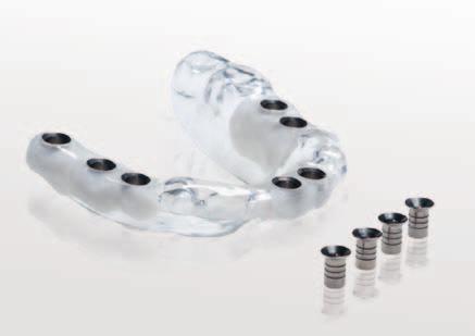
8 Implantology Safe implants: a good diagnosis for your practice s future! For beginners and specialists. With 3D from Sirona, implanting has become very easy: The GALILEOS Implant software guides even beginners efficiently through the planning process within minutes. Thanks to the colored visualization of the nerve canal and the depiction of the bones in all dimensions, you can adapt the implant in an ideal manner to fit the patient s anatomy. This ensures a high degree of safety and longevity of the implants, because unfavorable stress can be avoided by precise planning and implementation. For beginners and specialists. With 3D from Sirona, implanting has become very easy: The GALILEOS Implant software guides even beginners efficiently through the planning process within minutes. Thanks to the colored visualization of the nerve canal and the depiction of the bones in all dimensions, you can adapt the implant in an ideal manner to fit the patient s anatomy. This ensures a high degree of safety and longevity of the implants, because unfavorable stress can be avoided by precise planning and implementation. Safe implementation. Easily obtain Sirona SICAT* drilling templates by sending the planning data, the scan guide and a model to SICAT*. You ll receive an inexpensive and highly precise drilling template. * SICAT GmbH & Co. KG, Brunnenallee 6, Bonn, Tel.:


9 Integrated Implantology Maximum convenience: Only 3 sessions necessary for a perfect implant! With the help of the software imaging, your patients will understand the need for and the anticipated results of their proposed treatment, increasing the likelihood of treatment plan acceptance. Sirona unites 3D imaging and CAD/CAM. Integrated Implantology with CEREC reduces the number of sessions necessary for the comlete implant process, saving you and your patient time and money. When using CEREC in conjunction with GALILEOS or ORTHOPHOS XG 3D, the entire process, starting with the planning and ending with the manufacture of highly precise abutments and crowns, remains in your practice, ensuring highly precise, esthetic and affordable results, quickly. Your patients will feel good about your practice and will recommend you to others. In the future you will even be able to design and mill drilling templates with CEREC. Simultaneous prosthetic and surgical planning. The GALILEOS Implant software unites the prosthetic suggestion from the CEREC software with the 3D x-ray data. This way you will be able to take into consideration both, function as well as esthetics. 1 st Session n Using CEREC Bluecam, take a digital impression of the jaw and plan the prosthetics in the CEREC software. n Using a GALILEOS or ORTHOPHOS XG 3D scan, integrate the prosthetic proposal into the x-ray volume and simultaneously plan the implant based on the surgical and prosthetic conditions. n Load the optical impression and your plan onto the SICAT FTP server and order a surgical guide (SICAT OptiGuide process). 2 nd session n During the second session, have already prepared and have readily available: the drilling template (by SICAT) the implant if possible, the individual abutment created with CEREC and the provisional restoration made with CEREC n The patient will come in for the implant procedure. With the help of the surgical guide (in this example), insert an implant that is immediately loadable, screw in the abutment and cement the provisional restoration. 3 rd session n The patient comes in for treatment. Create the final prosthesis


10 Orthodontics A point of view: What orthodontists need. Displaced or impacted teeth? Root resorptions? In the field of orthodontics, these and many more are cases in which you can see more and diagnose more quickly and precisely with 3D, and thus be more successful. As an orthodontist, you also need special 2D views, which two of our 3D units provide: Whether you decide on ORTHOPHOS XG 3D or GALILEOS Comfort is therefore not a question of opinion, but a question of your requirements. Either way, you will make a good choice when choosing one of those two units. ORTHOPHOS XG 3D: Diversity in 2D. More reliability with 3D. The traditional cephalometric radiography function of the ORTHOPHOS XG 3D gives you lateral, symmetric (p. a. or a. p.) and carpus exposures for diagnosis. Expand these possibilities with the advantages of 3D x-ray imaging, in order to determine the exact spacial locations in cases, such as displaced teeth. GALILEOS Comfort: New diagnostic dimensions with 3D. For high-end orthodontics, GALILEOS Comfort is the unit of choice. The GALAXIS software displays various views simultaneously: classic panorama,cephalometric views and diverse radiological slices, greatly expanding your diagnostic capabilities. Programs for the optional ceph arm for ORTHOPHOS XG 3D n Symmetric a.p., symmetric p.a., lateral, carpus exposures n Asymmetric, image size adjustable 23 cm H x 18 cm W or 23 cm H x 28 cm W n Special projections, e.g. half-axial, Clementschitsch n Quickshot mode for shortened acquisition time n Adjustable crown collimation for dose reduction n Anjustable image size n Orthodontic image series with reduced cooling periods SIDEXIS XG is compatible with common orthodontic analysis programs. With its optional ceph arm, ORTHOPHOS XG 3D provides all of the views important to an orthodontist

, ORTHOPHOS XG 3D is perfectly adapted to fit typical practice needs: One scan captures the entire jaw, with a sufficient")


 VOL 2 (5 cm Ø x 5,5 cm height) Standard-mode HD-mode n 200 images n pulsed scan n")
11 ORTHOPHOS XG 3D ORTHOPHOS XG 3D: The most popular x-ray unit in the world. Now with 3D! ORTHOPHOS XG 3D: VOL 1: Cylindrical imaging volume of 8 cm diameter x 8 cm height VOL 2: Cylindrical imaging volume of 5 cm diameter x 5.5 cm height Optimized for everyday tasks in the dental practice. With a perfectly adapted cyndrical 3D volume (8 cm diameter, 8 cm height), ORTHOPHOS XG 3D is perfectly adapted to fit typical practice needs: One scan captures the entire jaw, with a sufficient field-of-view, avoiding the increased radiation exposure caused by stitching several 3D x-ray images together. The captured volume is also small enough to diagnose time-efficiently. If an even smaller volume is sufficient, simply collimate the height and diameter to 5 cm*. Whenever you decide that you need to see more, the 3D module provides increased security, select VOL 2 with a Field of View of 5 cm diameter x 5.5 cm height. In all standard cases, the extensive panoramic and cephalometric x-ray programs are guaranteed to deliver the right x-ray image in standard mode as well. 2D programs n For an exact summary of the 2D programs, please see the end of this brochure 3D views n Tiltable 2D slices n Custom 3D slicing n PAN, TSA, LSA, axial, sagittal, coronal, 3D model n Implant-oriented view, 1-Click OP reports Standard-mode and HD-mode in comparison Mode VOL 1 (8 cm Ø x 8 cm height) VOL 2 (5 cm Ø x 5,5 cm height) Standard-mode HD-mode n 200 images n pulsed scan n voxel size 160 µm n 500 images n continuous scan n voxel size 160 µm n 200 images n pulsed scan n voxel size 160 µm n 500 images n continuous scan n voxel size 100 µm Automatic sensor change Don t risk dropping a 3D sensor during a manual change. Switch from 2D to 3D at the press of a button, thus saving time, preventing mistakes and eliminating repeat imaging. More reliable diagnosis with MARS The MARS software reduces artifacts caused by metal fillings thus reducing misdiagnoses. Selectable high-definition mode When it comes down to it, the HD mode delivers extremely detailed, accurate images (160 μm voxel size with VOL 1, 100 μm with VOL 2 ideal for endodontics!). MARS 20 21
 includes the complete dentition, as well as the ascending rami and the sinus area.")


12 GALILEOS Compact GALILEOS Compact: A tailored solution with more possibilities. Ellipsoid imaging volume (12 x 15 x 15) cm 3 Not only suited for implantologists. GALILEOS Compact s ellipsoid field of view (15 cm diameter by 12 cm height) includes the complete dentition, as well as the ascending rami and the sinus area. The unit is made to meet the requirements in the area of implantology. In addition, GALILEOS Compact is suited for general practices that already own a modern OPG unit, but would still like to benefit from the advantages of 3D diagnostics with low dose at great volume. GALILEOS Compact can be upgraded to a GALILEOS Comfort at any time. If the large volume is not needed in some cases, simply collimate the field of view to the upper jaw or the lower jaw, a standard feature on all our 3D units. Cropped volumes save time during the diagnostic process and reduce the patient s exposure to radiation. Programs n High contrast option for optimized hard tissueimaging n VO4 3D views n Tiltable 2D slices n Custom 3D slicing n TSA, LSA, AXIAL, SAGITTAL, CORONAL, 3D MODEL n Implant-oriented view, 1-Click OP reports Field-of-view collimation on the upper jaw or lower jaw. Field-of-view collimation on the upper jaw or lower jaw. Perfect patient positioning with bite block, forehead support and laser light localizer


13 GALILEOS Comfort GALILEOS Comfort: High-end imaging, down to the smallest detail. Spherical imaging volume (15 x 15 x 15) cm 3 Superior diagnostics for all indications. With just one scan. With its large, 15 cm spherical volume, GALILEOS Comfort not only captures the entire jaw, including the temporomandibular joints, but also the entire craniofacial anatomy. GALILEOS Comfort is therefore perfectly suited for the practice specialized in oral surgery, practices with 3D-oriented orthodontic specialization, ENT practices, as well as for radiologists. Compared to GALILEOS Compact, expand your imaging options with cephalometric programs, as well as high resolution volume reconstruction. The optionally integrable FaceScanner also allows for precise analysis of the soft-tissue facial surface and provides new possibilities in consultation and patient-education. Programs n VO1 (with option to select high-contrast mode for optimal bone imaging) n VO2 (with option to select high-contrast mode for optimal bone imaging) 3D views n Partly navigable 2D layers n Freely selectable 3D slices n Pan, TSA, LSA, axial, sagittal, coronal, 3D model n 1-click implant reporting n Detail volume with high resolution n CEPH lat., CEPH p. a. / a. p. Integrated FaceScanner for soft-tissue analysis. Ergonimic operation with the Easypad touch panel. Precise positioning with patient immobilization

14 Investment security 3D now or later: Great future prospects! ORTHOPHOS XG 3D ready 3D x-ray doesn t yet fit your current plans for your practice? Keep the option open for the future: Sirona also offers a 3D module, allowing the ORTHOPHOS XG 3D ready to be upgraded to an XG 3D at any time. Benefit from the advantages of the tried-and-tested ORTHOPHOS XG family now with perfect image quality and practice-adjusted workflow. Invest on the safe side, able to take the step into the 3 rd dimension at any time. Sirona cares for its user base, with constant improvements to the software and hardware. We offer new updates in rough annual rhythms, so that our customers are always able to work with the newest technologies, comfortably and economically. If desired, you can also ensure your investment by taking advantage of an extended warranty. The investment in 3D will pay off faster than you may think: thanks to 3D, your patients will have a better understanding of your suggested therapy. Increasing case acceptance by one patient per month pays for the 3D module. Flexibility for tomorrow. Unit firmware can be updated at any time n Customizable touchscreen interface n Periodical software updates n 3D module and ceph arm can be retrofitted at any time n High-end technical specifications to accommodate future applications Your future with 3D. n More security with difficult diagnoses n Innovative, time-efficient workflow n Increased patient trust n Low-risk implant planning and surgery n State-of-the-art Integrated Implantology An investment that will pay off. n Reliable and durable technology provided by Sirona n Tested in numerous clinical studies n Extended warranty possible n Fast amortization through expanded practice spectrum and increased case acceptance n Integrated solutions for the entire practice 26 27
 5 cm diameter x 5.")

 1,6 m x 1,6 m x 2,25 m 1,6 m x 1,6 m x 2,25 m 1,5 m x 1,1 m x 2,25 m (PAN), Recommended room dimensions (depth x width x height) 1,8 m x 1,8 m x 2,5 m 1,8")


")

15 Numbers and facts Technical characteristics at a glance. Technical Overview GALILEOS Comfort GALILEOS Compact ORTHOPHOS XG 3D Field of view 15 cm x 15 cm x 15 cm 12 cm x 15 cm x 15 cm 8 cm x 8 cm (Ø x height) 5 cm diameter x 5.5 cm height Resolution in 3D: isotropic voxel edge size 0,3/0,15 mm 0,3 mm 0.16 mm; 0.1 mm in HD mode Scan time/exposure time 14 s /2 6 s 14 s/2 6 s 2 5 s, 14 s in HD mode ORTHOPHOS XG3D ORTHOPHOS XG3D CEPH GALILEOS Reconstruction time 2,5 4,5 min 4,5 min 4,5 min X-ray generator kv ma Effective dosage (ICRP 2007) μsv (Ludlow) Standard: 75 μsv μsv (Ludlow) (Standard: 37 μsv) μsv (Ludlow) Standard: 75 μsv Min. space requirements (depth x width x height) 1,6 m x 1,6 m x 2,25 m 1,6 m x 1,6 m x 2,25 m 1,5 m x 1,1 m x 2,25 m (PAN), Recommended room dimensions (depth x width x height) 1,8 m x 1,8 m x 2,5 m 1,8 m x 1,8 m x 2,5 m 1,7 m x 1,3 m x 2,5 m (PAN) Radiation shielding Identical to panoremic units: see DIN 6812: June 2002 Identical to panoremic units: see DIN 6812: June 2002 Identical to panoremic units: see DIN 6812: June 2002 Min. door width 66 cm 66 cm 66 cm Weight approx. 120 kg approx. 120 kg approx. 110 kg Technical specifications User interface EasyPad MultiPad EasyPad Patient positioning Standing/seated, chin rest/bite block head fixing system Standing/seated, chin rest/bite block head fixing system Standing/seated, chin rest/bite block, occlusal bite block for automatic patient positioning with 2D PAN images Software optional Views n SIDEXIS Image processing and management software n GALAXIS Diagnosis-based work, clarification of diagnostic tasks n GALILEOS Implant Implant planning software n CEREC meets GALILEOS Simultaneous prosthetic and surgical planning n REPORTER (optional) n FaceScanner Viewer Software* Partly navigable 2D layers, freely selectable 3D slices, Pan, TSA, LSA, axial, sagittal, coronal, 3D model, 1-click implant reporting, Detail volume with high resolution, CEPH lat., CEPH p. a. / a. p. n SIDEXIS Image processing and management software n GALAXIS Diagnosis-based work, clarification of diagnostic tasks n GALILEOS Implant Implant planning software n CEREC meets GALILEOS Simultaneous prosthetic and surgical planning n REPORTER (optional) Tiltable 2D slices, custom 3D slicing, TSA, LSA, axial, sagittal, coronal, 3D model, implant-oriented view, 1-Click OP reports n SIDEXIS Image processing and management software n GALAXIS Diagnosis-based work, clarification of diagnostic tasks n GALILEOS Implant Implant planning software n CEREC meets GALILEOS Simultaneous prosthetic and surgical planning n REPORTER (optional) Tiltable 2D slices, custom 3D slicing, TSA, LSA, axial, sagittal, coronal, 3D model, implant-oriented view, 1-Click OP reports Individually adjustable for seated patients and wheelchairs. Stable base for freestanding mounting. Remote control with exposure parameter display. Retrofit options Facescanner* (optional) Facescanner* (optional), to Comfort version Ceph (optional), also available as 2D unit with 3D upgrade option * from delivery start on


 P10 pediatric")


16 Overview ORTHOPHOS XG 3D: 2D programs at a glance. Temporomandibular joint Sinus Panorama Adjustable radiation angle with open and closed occlusion TM1 lateral with a slice position S1 maxillary sinuses in one image P1 orthoradial radiation Standard exposure Optional panning UJ with a constant magnification of 1.25 LJ right TM2 axial S2 maxillary sinuses in two images P2 without ascending rami with artifact reduction left Individual quadrants TM3 S3 maxillary sinuses in one image (linear) P10 pediatric panorama, beam field reduced in height and length Optional panning Bite wing TM4 UJ LJ Optional panning right S4 maxillary sinuses in two images (linear) P12 thick slice in anterior tooth region left TM5 Multislice in posterior tooth region Quickshot option for all PAN programs Automatic adjustment of the rotation curve to the jaw width Automatic positioning with occlusal bite block BW1 BW2 anterior tooth region TM6 MS
Easy operation. Numerous diagnostic options. X-rays you can rely on: the ORTHOPHOS XG device family. All ORTHOPHOS XG 3Dready programs at a glance.
 CAD /CAM Systems Instruments Hygiene Systems Treatment Centers Imaging Systems Subject to technical changes and errors in the text, Order No. A91100-M47-B346-01-7600, Printed in Germany, Dispo-Nr. 04602,
T h e D e n t a l C o m p a n y FROM DIAGNOSTIC SCAN TO SURGERY, WE SHAPE THE FUTURE OF DENTISTRY.
 T h e D e n t a l C o m p a n y FROM DIAGNOSTIC SCAN TO SURGERY, WE SHAPE THE FUTURE OF DENTISTRY. SIDEXIS SOFTWARE ORTHOPHOS SL D/D SEAMLESS THE NEW STANDARD IN CLINICAL DIAGNOSIS AND PATIENT COMMUNICATION
The 2D x-ray family. dentsplysirona.com
 The 2D x-ray family dentsplysirona.com 02 I 03 As versatile as practice life The Orthophos 2D X-ray family offers the right solution for every practice. From entry into digital radiography to the perfect
A new dimension of success in your practice
 3D Imaging Family A new dimension of success in your practice dentsplysirona.com 2/3 Good reasons for 3D With 3D imaging, you have the ideal basis for a new dimension of success in your practice. BETTER
Digital Imaging from a new perspective
 TREATMENT CENTRES HANDPIECES HYGIENE SYSTEMS X-RAY SYSTEMS CEREC TREATMENT CENTRES HANDPIECES HYGIENE SYSTEMS X-RAY SYSTEMS CEREC SIRONA CREATING AND MAINTAINING VALUE. You are right to expect a great
The 3D x-ray family. dentsplysirona.com
 The 3D x-ray family dentsplysirona.com 02 I 03 As versatile as practice life. The Dentsply Sirona 3D x-ray family offers 3 units, Galileos Comfort Plus, Orthophos SL 3D and Orthophos XG 3D, whose visual
clinical articles management advice practice profiles technology reviews Implant PRACTICE US Volume 3 No 2
 clinical articles management advice practice profiles technology reviews Implant PRACTICE US Volume 3 No 2 Sirona Sirona tailors solutions to the needs of the different markets within dentistry to ensure
A new dimension of success in your practice
 3D Imaging Family A new dimension of success in your practice dentsplysirona.com CEREC Integration Diagnosis Treatment Plan Guided Implantology Endodontics Airway Analysis Functional Occlusal & TMD Orthodontics
ORTHOPHOS XG 3 DS. X-ray systems. ORTHOPHOS XG 3 Digital panoramic X-ray for practical diagnostics.
 ORTHOPHOS XG 3 DS X-ray systems ORTHOPHOS XG 3 Digital panoramic X-ray for practical diagnostics. ORTHOPHOS XG 3 Standard panoramic X-ray with proven technology. competent successful Many years of competence
Head to new heights with your imaging SCANORA 3D
 SCANORA 3D Head to new heights with your imaging Benefits at a glance The solution for dentomaxillofacial and ENT imaging Easy Patient seated for added stability during exposure. Clear, self-explinatory
Straight to the point with your diagnosis.
 CAD/CAM SYSTEMS INSTRUMENTS HYGIENE SYSTEMS TREATMENT CENTERS IMAGING SYSTEMS HELIODENT DS INTRAORAL X-RAY WITH EXCELLENT IMAGE QUALITY Straight to the point with your diagnosis. T h e D e n t a l C o
Imagine the possibilities with Sirona 3D.
 CAD/CAM SYSTEMS I INSTRUMENTS I HYGIENE SYSTEMS I TREATMENT CENTERS I IMAGING SYSTEMS WELCOME TO THE NEW DIMENSION OF DIAGNOSTIC IMAGING Imagine the possibilities with Sirona 3D. T h e D e n t a l C o
X X X. GXS-700 Direct USB Digital Intraoral Sensors. Buy a Sensor Combo and a Digital Pan Unit, Receive $600 Off! GO.BENCO benco.
 8 0 0. G O. B E N C O b e n c o. c o m GS-700 Direct USB Digital Intraoral Sensors Designed to make migrating from film, or upgrading an existing digital system, easier than ever High quality image capture
You design, we create.
 CAD/CAM SYSTEMS INSTRUMENTS HYGIENE SYSTEMS TREATMENT CENTRES IMAGING SYSTEMS infinident SIRONA S CENTRAL PRODUCTION You design, we create. www.infinident.gb.com T h e D e n t a l C o m p a n y infinident:
XPan 3D Plus. FONA Every dental solution you need. Advanced dental technology. Headquarters THE ULTIMATE DIAGNOSTIC SOLUTION DIGITAL DENTISTRY
 FONA Every dental solution you need Through decades of experience and deep understanding of the dental profession, we deliver complete, reliable and accessible solutions.regardless of country or specialisation,
CS 9300 Family. The power of flexibility
 CS 9300 Family The power of flexibility The new CS 9300 digital imaging system from Carestream Dental take the guesswork out of examinations The all-in-one CS 9300 is the most versatile multimodality imaging
VistaVox S 3D from Dürr Dental
 VistaVox S 3D from Dürr Dental 3D and 2D X-ray images with exceptional image quality COMPRESSED AIR SUCTION IMAGING DENTAL CARE HYGIENE Taking diagnostics to the next level VistaVox S combines diagnostic
Laser treatment doesn t have to be a big deal any more.
 CAD/CAM SYSTEMS I INSTRUMENTS I HYGIENE SYSTEMS I TREATMENT CENTERS I IMAGING SYSTEMS SIROLaser THE COMPACT DIODE LASER FOR YOUR PRACTICE Laser treatment doesn t have to be a big deal any more. T h e D
IMPLANT MORE SUCCESSFULLY WITH SICAT SURGICAL GUIDES
 IMPLANT MORE SUCCESSFULLY WITH SICAT SURGICAL GUIDES MAKE EVERY CASE COUNT DIAGNOSTICS AND PLANNING IN 3D. In the field of implantology the use of surgical guides based on 3D X-ray data gives your practice
CS 8100 FAMILY / CS D FAMILY / CS 9300 FAMILY EXTRAORAL SOLUTIONS EXTRAORDINARY POSSIBILITIES
 CS 8100 FAMILY / CS 8100 3D FAMILY / CS 9300 FAMILY EXTRAORAL SOLUTIONS EXTRAORDINARY POSSIBILITIES A SOLUTION FOR EVERY CLINIC AND ANY CONSULTATION The CS 8100 does more than my old machine and is half
ZIRCONIA, SIMPLY FASTER. FULL CONTOUR ZIRCONIA RESTORATIONS IN A SINGLE VISIT ONLY WITH CEREC.
 ZIRCONIA, SIMPLY FASTER. FULL CONTOUR ZIRCONIA RESTORATIONS IN A SINGLE VISIT ONLY WITH CEREC. CEREC MAKES IT HAPPEN. What would you think about providing your patients full contour zirconia crowns in
STL. inside. ineos Blue: Precision, Speed and Control. THE SCANNER WITH Bluecam TECHNOLOGY
 CAD/CAM SYSTEMS INSTRUMENTS HYGIENE SYSTEMS TREATMENT CENTERS IMAGING SYSTEMS THE SCANNER WITH Bluecam TECHNOLOGY ineos Blue: Precision, Speed and Control. STL inside T h e D e n t a l C o m p a n y ineos
Fear turns into satisfaction. PerioScan the gentle prophylaxis competence
 CAD/CAM SYSTEMS INSTRUMENTS HYGIENE SYSTEMS TREATMENT CENTERS IMAGING SYSTEMS PerioScan the gentle prophylaxis competence Fear turns into satisfaction. Gentle prophylaxis Prophylaxis without pain? That
Where technology meets dentistry INNOVATION. at a GLANCE
 Where technology meets dentistry INNOVATION at a GLANCE ABOUT PALTOP Paltop is a premium manufacturer of dental implants that strives to provide highest quality products in the dental arena. Leveraging
ENGLISH. Cefla s.c. - Via Selice Provinciale 23/a, Imola - Italy Tel
 09-2017 NVGEGB161S00 According to the regulations in force, some products and/or features may have different availability and characteristics in areas outside of the European Union. Please contact your
OP 3D Vision The upgradable 3D X-ray system for the strictest demands.
 OP 3D Vision The upgradable 3D X-ray system for the strictest demands. The solution for every task: KaVo OP 3D Vision. Regardless of which dental query you may have, the KaVo ORTHOPANTOMOGRAPH OP 3D Vision
For technical assistance: Tel Working time: 8.30 am pm
 I For technical assistance: Tel. +39 059 392827 Working time: 8.30 am - 5.30 pm Index JD Digital solutions for Intraoral Scanning JD Implant Libraries JD Guided Surgery Kit JD Surgical Guide and 3D Model
SICAT FUNCTION TRACK REAL MOTION IN MOTION. Real 3D jaw motion for the first time
 SICAT FUNCTION TRACK REAL MOTION IN MOTION Real 3D jaw motion for the first time REAL MOTION TIME FOR FACTS THE DIGITALIZATION of treatment procedures based on 3D X-ray data is progressing rapidly. What
THE WAIT IS OVER CS D. 3D imaging is now available for everyone
 THE WAIT IS OVER CS 8100 3D 3D imaging is now available for everyone COMPLEXITY IS NO LONGER THE STANDARD NOW THERE ARE MANY REASONS TO MOVE TO 2D/3D IMAGING Now it s possible to experience nothing but
Versatility And Expandability In One Panoramic.
 Orthoralix 9200 / 9200 DDE Versatility And Expandability In One Panoramic. Panoramic X-ray Systems Intraoral X-ray Systems Digital Intraoral Sensors Digital X-ray Phosphor Plates Intraoral Cameras Imaging
3Shape X1 Scanning redefined
 3Shape X1 Scanning redefined Why choose the X1 Give your patients a great experience No head fixation and sleek design create a comfortable scanning experience for your patient High image quality low dose
PAN CEPH 3D CONE BEAM
 PAN CEPH 3D CONE BEAM 2D - 3D panoramic units PANORAMIC CEPHALOMETRIC 3D CONE BEAM IMAGING I-MAX TOUCH Tactile & naturally intuitive panoramic imaging Discover the simplicity and efficiency this unit can
I AM DEMANDING Type CMOS Flat Panel CMOS CMOS ø 40 x 40 mm, ø 60 x 60 mm, ø 80 x 80 mm, ø 110 x 80 mm
 TECHNICAL SPECIFICATIONS 1168 1501 1978 1237 1551-2351 Ø 1090 PANORAMIC CBCT CEPHALOMETRIC X-RAY SOURCE Tube type High frequency DC generator 2.8 mmal / 85 kv 7.0 mmal / 90 kv 2.8 mmal / 85 kv Operation
DIAGNOSTIC IMAGING. OPTIMIZED.
 ABOUT LED DENTAL SEE THE DIFFERENCE Using our years of business insight and clinical experience as a foundation, LED Dental takes the uncertainty out of your imaging purchase decision. We offer our clients
The Quality Leader in 3D Cone Beam CT
 The Quality Leader in 3D Cone Beam CT The Complete 2-in-1 or 3-in-1 Multi-modality Solution PreXion, with over 15 years of innovation in the medical and dental fields, introduces the PreXion3D Eclipse.
Your stepping stones to success.
 CAD/CAM SYSTEMS INSTRUMENTS HYGIENE SYSTEMS TREATMENT CENTERS IMAGING SYSTEMS THE inlab SYSTEM AND ITS COMPONENT PARTS Your stepping stones to success. T h e D e n t a l C o m p a n y inlab YOUR FUTURE-ORIENTED
Everything. Supplying everything dental for your practice needs
 Everything Connect Dental Supplying everything dental for your practice needs henryschein.co.uk Highly accurate, durable and 3D Conebeam digital image ORTHOPHOS XG3D/SL Practice Management Software Secure
THE POWER OF THE PERFECT FIT
 THE POWER OF THE PERFECT FIT SCAN, DESIGN, MILL The PLANMECA FIT CAD/CAM system allows you to perform highly advanced, powder-free digital restorations, including same-day crowns, inlays, onlays, bridges
3Shape X1 Scanning redefined
 3Shape X1 Scanning redefined Why choose the X1 Give your patients a great experience No head fixation and sleek design create a comfortable scanning experience for your patient High image quality low dose
sironausa.com CAD/CAM for Sirona Dental, Inc Sirona Drive Charlotte, NC 28273
 sironausa.com CAD/CAM for Sirona Dental, Inc. 4835 Sirona Drive Charlotte, NC 28273 Everyone SIRONAUSA.COM WHY CAD/CAM? WHY NOW? Traditional dental procedures, like physical impressions and temporary bridges,
The Optimum Choice for Implantologist
 The Optimum Choice for Implantologist What is essential for your practice? What s the best way to choose a 3D X-ray machine for implant treatment planning? 02 Doctor says.. There are diagnostic limitations
INTRAORAL SCANNER TRANSFORMING CREATIVITY
 INTRAORAL SCANNER TRANSFORMING CREATIVITY COMING SOON! POWDER-FREE SCANNING* Increased efficiency, accuracy, and comfort for your patient. Gesture control module for infection control INTRAORAL SCANNER
STELLARIS 3D 4 IN 1 CBCT SOLUTION FOR ADVANCED DIAGNOSTICS
 STELLARIS 3D 4 IN CBCT SOLUTION FOR ADVANCED DIAGNOSTICS 3 STELLARIS 3D 4 IN CBCT SOLUTION FOR ADVANCED DIAGNOSTICS Stellaris 3D is a complete and compact, fully upgradeable 3D CBCT for a patient, Panoramic
English. Perfect Vision
 English Perfect Vision Everything becomes clearer and simpler with a big F.O.V. The complete dentomaxillofacial volume ready for your diagnosis One scan provides you with an incredible amount of information
VGi - R EN ENGLISH. QR srl - Via Silvestrini, Verona Italy Tel
 VGi - R15.1 - EN ENGLISH QR srl - Via Silvestrini, 20-37135 Verona Italy Tel. +39 045 8202727-045 583500 info@newtom.it www.newtom.it FIRST IN CONE BEAM, ACCURATE IN RESULTS. 360 degree imaging, reduced
Profound understanding of anatomy
 ENGLISH Profound understanding of anatomy The unique Planmeca ProMax 3D product family offers equipment for all maxillofacial imaging. All volumes sizes from the smallest special cases to whole head images
INTRAORAL IMAGING ACCORDING TO YOUR NEEDS Intraoral X-rays
 INTRAORAL IMAGING ACCORDING TO YOUR NEEDS Intraoral X-rays FONA Intraoral X-rays INTRAORAL IMAGING ACCORDING TO YOUR NEEDS FONA intraoral X-ray devices are designed for ease of use, precision, reliability
Cone Beam 3D Imaging
 Cone Beam 3D Imaging NewTom Sets the Standard in 3D Maxillofacial Imaging Cone Beam 3D Imaging The Global Market Leader The Inventors n of Cone Beam 3D In 1996, QR srl developed the first generation of
SICAT FUNCTION & OPTIMOTION. Individual functional treatment
 SICAT FUNCTION & OPTIMOTION Individual functional treatment REAL MOTION TIME FOR FACTS THE DIGITALIZATION of treatment processes based on 3D X-ray data is progressing quickly. What has been successfully
Simplant. Guided Surgery. delivering restorative driven implant treatment
 Simplant Guided Surgery delivering restorative driven implant treatment Simplant the key to unlocking digital potential As part of the Denstsply Sirona Implants Digital Solutions offering, Simplant delivers
Reinvent Your Practice and Your TEAM through 3D Guided Implantology By Patrick Hayden, M.Ed
 Reinvent Your Practice and Your TEAM through 3D Guided Implantology By Patrick Hayden, M.Ed The Dental Market In 2011 there were over half a billion missing teeth in adults 18-65*. Two million of those
Dental Line. 3D digital panoramic system. radiology ahead
 Dental Line 3D digital panoramic system radiology ahead new generation 3D digital panoramic unit 3D imaging s value available for anyone Following the incredible success of the innovative digital panoramic
Immediate loading and implant surgery with digital workflow
 Mirero Dental Clinic Dr. Jaemin Lee Immediate loading and implant surgery with digital workflow Solutions featured: 3Shape TRIOS 3Shape Dental System 3Shape Implant Studio Case information Patient, 32-year
Profound understanding of anatomy
 ENGLISH Profound understanding of anatomy The unique Planmeca ProMax 3D product family offers equipment for all maxillofacial imaging. All volume sizes from the smallest special cases to whole head images
A Clinician s Guide to Digital Dentistry Dental restorations through CEREC Connect with Absolute.
 1994 A Clinician s Guide to Digital Dentistry Dental restorations through CEREC Connect with Absolute Dear Doctor, Absolute Dental has specialized in fixed and implant restorations for over two decades.
True Low Dose. Exact time to display image on screen may vary upon computer and network configuration.
 RAYSCAN ALPHA PLUS True Low Dose Cone Beam CT Industry Leading Resolution High resolution images provide all the clinical information needed while keeping radiation exposure low. Endodontics - Smallest
SIMPLANT Guided Surgery delivering restorative-driven implant treatment
 SIMPLANT Guided Surgery delivering restorative-driven implant treatment SIMPLANT the key to unlocking digital potential As part of the DENTSPLY Implants Digital Solutions portfolio, SIMPLANT digital implant
Guided immediate loading implant surgery planned with Implant Studio D.D.S. Jae-min, Lee
 Guided immediate loading implant surgery planned with Implant Studio D.D.S. Jae-min, Lee Jung-plant Dental office 1 PROLOGUE How can we deal with the immediate loading implant cases easier and more accurate
TABLE OF CONTENTS P. 4-5 P. 6-7 P. 8-9 P P P P
 Full Smile Solution TABLE OF CONTENTS P. 4-5 P. 6-7 P. 8-9 P. 10-11 P. 12-13 P. 14-15 P. 16-17 FULL SMILE SOLUTION MGUIDE PROCESS ADVANTAGES CUSTOM SOLUTIONS MGUIDE SURGICAL SETS MCENTER REQUIRED SOURCES
inlab Software 18.0 New digital options dentsplysirona.com/inlab
 inlab Software 18.0 New digital options dentsplysirona.com/inlab 02 I 03 inlab CAD Software 18.0 Dental design requires good software The new inlab CAD Software 18.0 focuses even more closely on the requirements
THE USE OF KEYSTONE EASYGUIDE CT SCANNING SOFTWARE FOR DIAGNOSIS, DIRECTION AND DEPTH DETERMINATION
 CT DIAGNOSTICS IN 3D IMPLANT TREATMENT PLANNING THE USE OF KEYSTONE EASYGUIDE CT SCANNING SOFTWARE FOR DIAGNOSIS, DIRECTION AND DEPTH DETERMINATION Timothy Kosinski, DDS, MAGD Assistant Clinical Professor
From planning to surgery: a totally digital working flow for Leone implants placement
 Dr. Giancarlo Romagnuolo Roma, Italy From planning to surgery: a totally digital working flow for Leone implants placement Keywords guided surgery, 3D implant planning, single missing tooth, delayed immediate
Low Dose Excellent Image Quality Rapid Reconstruction
 Low Dose Excellent Image Quality Rapid Reconstruction Efficient 3 in 1 Dental X-ray System CBCT > Precise 3-D Anatomical structures - Accurate diagnosis for doctors - Safe implant for patients > Significant
Totally lightweight, totally flexible, totally titanium.
 CAD/CAM SYSTEMS INSTRUMENTS HYGIENE SYSTEMS TREATMENT CENTERS IMAGING SYSTEMS T1 LINE THE LIGHTWEIGHT TITANIUM INSTRUMENTS Totally lightweight, totally flexible, totally titanium. T h e D e n t a l C o
Planmeca ProMax 3D s Planmeca ProMax 3D ENGLISH
 Planmeca ProMax 3D s Planmeca ProMax 3D ENGLISH Genuine all-in-one unit Planmeca ProMax 3D s and Planmeca ProMax 3D units are designed to obtain complete information on patient anatomy in the minutest
STRAUMANN CARES DIGITAL SOLUTIONS. CARES Implant Prosthetics Ultimate restorative flexibility.
 STRAUMANN CARES DIGITAL SOLUTIONS CARES Implant Prosthetics Ultimate restorative flexibility. Customized restorations of exceptional quality from one trusted partner. PERFORMANCE Reached through best-in-class
Rock your practice with digital restorations in a day.
 Digital Dentistry Straumann CARES Chairside Solutions Rock your practice with digital restorations in a day. crowns and bridges in just one visit Straumann CARES Chairside System offers dentists high quality
ADVANCED 3D IMAGING. CEFLA s.c. Via Selice Provinciale 23/a Imola Italy t newtom.
 CEFLA s.c. Via Selice Provinciale 23/a 40026 Imola Italy t. +39 045 8202727 045 583500 info@newtom.it newtom.it 05/2018 NVGEGB181S00 According to the standards in force, in extra-eu areas the availability
Enter into the world of unprecedented accuracy! IScan D104 Series
 Enter into the world of unprecedented accuracy! IScan D104 Series 2 Three scanners, one goal: Precisely fitting restorations The D104 Series of 3D scanning systems is the 5th generation of dental scanners
Flexible Easy Competitive. SCANORA 3Dx - The in-office large field-of-view Cone Beam CT system for Head and Neck imaging
 Flexible Easy Competitive SCANORA 3Dx - The in-office large field-of-view Cone Beam CT system for Head and Neck imaging SCANORA 3Dx. The solution. SCANORA 3Dx makes advanced 3D imaging easy in the head
CS 3600 / CS 3600 ACCESS A SMARTER WAY TO SCAN INTRAORAL SCANNING
 CS 3600 / CS 3600 ACCESS A SMARTER WAY TO SCAN INTRAORAL SCANNING R R HARNESS THE POWER OF INTRAORAL SCANNING AWARDS AND RECOGNITION CLINICIANS REPORT Excellent in Rapid Acquisition and Margin Detail Capture;
Profound understanding of anatomy
 ENGLISH Profound understanding of anatomy Planmeca ProMax 3D, the intelligent and multipurpose X-ray unit, is designed to obtain complete information on patient anatomy in the minutest detail. The unit
PROSTHETIC EFFICIENCY. Straumann Variobase Beyond a common Ti-base. Efficiency and flexibility in balance.
 PROSTHETIC EFFICIENCY Straumann Variobase Beyond a common Ti-base. Efficiency and flexibility in balance. 2 Straumann Variobase Satisfy individual patient needs in the most efficient way Balance restorative
Full ultrasound breast volumes. Faster scans. Streamlined workflow. ACUSON S2000 Automated Breast Volume Scanner. Answers for life.
 Full ultrasound breast volumes. Faster scans. Streamlined workflow. ACUSON S2000 Automated Breast Volume Scanner Answers for life. 1 ACQUIRE An automated whole breast solution. Reduced acquisition time.
3D Panoramic Cephalometric. Innovation, in reach. KODAK 9000 Extraoral Imaging System
 3D Panoramic Cephalometric Innovation, in reach 9000 KODAK 9000 Extraoral Imaging System Cephalometric Innovation made simple Innovation made simple We believe in innovation. We always have. In fact, our
ENGLISH. Distributed by: QR srl - Via Silvestrini, Verona Italy Tel
 GiANO - R15.1 - EN ENGLISH Distributed by: QR srl - Via Silvestrini, 20-37135 Verona Italy Tel. +39 045 8202727-045 583500 info@newtom.it www.newtom.it Manufacturer: CEFLA S.C. - CEFLA DENTAL GROUP Via
AWARD-WINNING CONE BEAM 3D DENTAL IMAGING
 AWARD-WINNING CONE BEAM 3D DENTAL IMAGING Dedicated to Advancing Dental Treatment A COMPLETE 3D TREATMENT SOLUTION Your dental practice is unique that s why you need a flexible solution that works with
insights clinical AnyRidge Implant System Scan Post (C-Type)
") clinical insights AnyRidge Implant System Scan Post (C-Type) by Neal Patel DDS Summary The following information will guide you through the scan, design and production process utilizing the MegaGen AnyRidge
clinical insights AnyRidge Implant System Scan Post (C-Type) by Neal Patel DDS Summary The following information will guide you through the scan, design and production process utilizing the MegaGen AnyRidge
vertaplan the spine surgeon s software vertaplan System for successful reconstruction of the individual sagittal balance
 the spine surgeon s software System for successful reconstruction of the individual sagittal balance What do you think of patient-specific reconstruction of the spine geometry? Optimum surgical outcome
Dental Cone Beam CT. What is Dental Cone Beam CT?
 Scan for mobile link. Dental Cone Beam CT Dental cone beam computed tomography (CT) is a special type of x-ray equipment used when regular dental or facial x-rays are not sufficient. Your doctor may use
TRI + Digital Solutions DENTAL WINGS MANUAL
 TRI + Digital Solutions DENTAL WINGS MANUAL V1.2_2018-02-12 NOTE TRI + represents the interface between the TRI dental implant system and the Dental Wings digital solutions. The following instructions
3Shape TRIOS Implant Scanning
 Dr. Anthony Mak W Dental 3Shape TRIOS Implant Scanning Simplifying digital implant prosthetics with implant libraries and 3Shape scan bodies Solutions featured: 3Shape TRIOS 3Shape scan bodies 3Shape Dental
NEODENT GUIDED SURGERY
 NEODENT GUIDED SURGERY MANUAL Grand Morse Connection Helix Implant GRAND POSSIBILITIES WITH A LIMITLESS SOLUTION. GRAND MORSE NEODENT GUIDED SURGERY. 4 5 6 CONTENTS 1. CLINICAL STEP BY STEP OF GRAND
Full mouth rehabilitation with digital workflow
 Jung-plant dental office Dr. Jae-min, Lee D.D.S. Full mouth rehabilitation with digital workflow Solutions featured: 3Shape TRIOS 3Shape Dental System 3Shape Implant Studio Case information On first visit,
Safety and esthetics with
 Safety and esthetics with dental implants A guide for patients Dear reader, Implant restorations follow nature's example. You can have the functions of natural teeth completely restored and thus maintain
cara i500 Simply impress(ion)ing.
ing.") kulzer.com/cara-i500 Product Information cara i500 Simply impress(ion)ing. Giving a hand to oral health. cara i500 The I.D.E.A.L. choice for intraoral scanning. One precondition for perfectly fitting restorations
kulzer.com/cara-i500 Product Information cara i500 Simply impress(ion)ing. Giving a hand to oral health. cara i500 The I.D.E.A.L. choice for intraoral scanning. One precondition for perfectly fitting restorations
RELATIONSHIPS MATTER. Innovation, Expertise and. Craftsmanship. in Laboratory. Products and. Services for the. Dental Profession
 RELATIONSHIPS MATTER Innovation, Expertise and Craftsmanship in Laboratory Products and Services for the Dental Profession Our position in the industry enables us OUR GOAL 100% CUSTOMER SATISFACTION We
3Shape TRIOS Engage and excite your patients
 3Shape TRIOS Engage and excite your patients Create patient excitement with 3Shape TRIOS 3Shape TRIOS allows you to enhance patient experience with exciting engagement tools, reduce chair-time and embrace
For true visualisation
 ENGLISH For true visualisation Planmeca ProModel is a patient-specific physical model for high-end maxillofacial operations and dental surgery. By reproducing the anatomy of the patient in real-size, Planmeca
ENGLISH. Cone Beam 3D Imaging
 ENGLISH Cone Beam 3D Imaging FIRST USER OF CONE BEAM IN DENTAL FIELD QR s.r.l. is the name that stands behind NewTom Cone Beam 3D imaging units and the creator of Cone Beam technology for the dental field.
ORTHOPANTOMOGRAPH OP200 D ORTHOCEPH OC200 D. True dynamo leading through the decades.
 ORTHOPANTOMOGRAPH OP200 D ORTHOCEPH OC200 D True dynamo leading through the decades. 1 You can t dublicate the legacy. 1946 Professor Y.V. Paatero publishes his first paper on Panoramic Tomography. 1951
CEREC Guide 2.0. Brent Pedersen, CEREC Technology Advisor/Trainer Joe Perry, Sirona 3D Regional Manager
 CEREC Guide 2.0 Brent Pedersen, CEREC Technology Advisor/Trainer Joe Perry, Sirona 3D Regional Manager Please send comments/suggestions to brent.pedersen@pattersondental.com CEREC Guide 2.0: What s needed?
3Shape TRIOS Orthodontics. Impress your patients
 3Shape TRIOS Orthodontics Impress your patients Keep improving with TRIOS Orthodontics Going digital with your orthodontic practice means that you are more efficient, more precise and your patients more
SICAT AIR & OPTISLEEP. The appliance-based 3D solution for the treatment of obstructive sleep apnea
 SICAT AIR & OPTISLEEP The appliance-based 3D solution for the treatment of obstructive sleep apnea DIGITAL & INTUITIVE with OPTISLEEP, the most advanced oral appliance straight from the dentist. THE DIGITAL
Neal S. Patel, DDS GALILEOS, THE M O D E R N D E N T I S T RY. Imagine the Possibilities
 GALILEOS, THE QUINTESSENCE OF M O D E R N D E N T I S T RY Imagine the Possibilities Neal S. Patel, DDS The new Cerec Bluecam and the CERECdoctors.com faculty members continue to push the envelope with
3D Accuitomo XYZ Slice View Tomograph. Super High-Resolution Images of Region of Interest
 3D Accuitomo XYZ Slice View Tomograph. Super High-Resolution Images of Region of Interest Thinking ahead. Focused on life. 2 Cone Beam X-Ray CT Imaging X-Ray Tube Imaging Intensifier Imaging Volume Voxel
the way to straumann cares abutments
 the way to straumann cares abutments solutions for dental laboratories 2 Why choose Straumann CARES Abutments Each patient is unique and each clinical situation is different. with Straumann CARES Abutments,
The mission of our company is the development of an individual approach in functional dentistry.
 The Prosystom company started up in 2013. It was created by Russian doctors and IT specialists for carrying out scientific projects and practical work in functional dentistry. The mission of our company
I AM EXCLUSIVE TECHNICAL SPECIFICATIONS. The first personal imaging plate scanner IMAGING PLATES. System. Windows recommended configuration
 TECHNICAL SPECIFICATIONS System Resolution... 20 lp/mm Scan Time (fast mode)...1,6s - 2,7s Scan Time (high definition mode)...2,1s - 3,6s Connection... Ethernet RJ-45 Dimensions... L. 154 x D. 204 x H.
Delivering Top Performance. Outstanding Value. Exceptional Reliability. ACUSON X150 Ultrasound System. Answers for life.
 Delivering Top Performance. Outstanding Value. Exceptional Reliability. ACUSON X150 Ultrasound System Answers for life. Gynecology Imaging Ovarian Follicles Abdominal Imaging Renal Vasculature, Color Doppler