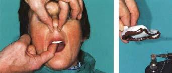
Set the patient in up right position. For mandibular impression the dentist stand in front of the patient
|
|
|
- Frederica Harper
- 5 years ago
- Views:
Transcription
1
2 Set the patient in up right position For mandibular impression the dentist stand in front of the patient For maxillary impression the dentist stand behind the patient
3 Denture retention relies on a number of factors including developing an adequate border seal around the denture against the soft tissues. This is achieved by extending the denture so that it just begins to displace the movable soft tissues at the periphery of the denture. Obviously such extensions cannot impinge on areas of muscle activity or the denture will be displaced in function. Thus, there is a need to record a dynamic shape of the oral soft tissues.
4 Objectives To shape the border of impression in order to allow the muscles to function in harmony with the denture. To improve the border seal of the denture.
5 Requirements of materials used for border molding Should have sufficient strength. Should allow some preshaping of the form of the borders. Should have a setting time of 3-5 min. Should retain adequate flow while seating in the mouth. Should not cause excessive displacement of the tissues of the vestibule. Should be readily trimmed and shaped so that excess material can be carved and the border shaped before the final impression is made.
6 Materials used for border molding Stick compound. Autopolymerizing acrylic resin. Polyether impression paste. Impression waxes. Periopack. Tissue conditioners.
7 Methods According to manipulation: 1. Manual or digital manipulation. 2. Functional manipulation. 3. Combination of both. According to technique: 1. Open mouth technique. 2. Closed mouth technique. 3. Single border molding. 4. Incremental border molding.
8 Manual or digital manipulation The contour of the denture borders is obtained by the dentist with digital (finger) manipulation of lips and cheeks of patient within functional limits. Functional manipulation The contour of the denture borders is obtained by the functional movements provided by the patient.
9 Open mouth technique In this technique the tissues are recorded in their undisplaced position. The patients mouth is partly opened and tray is held in position. Closed mouth technique In this technique the tissues are recorded in their functional and displaced position. The patient applies pressure by closing against occlusion rims or teeth and executes muscle actions such as swallowing, grinning or pursing the lips while the impression material flow.


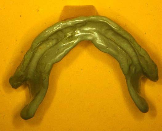
10 Incremental technique Use stick compound Soften over flame by rolling repeatedly to avoid over heating and burning.


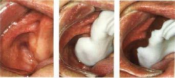
11 The softened stick compound is flowed along the border of the required segment of the tray. The tray is tempered in warm water, placed carefully in patient mouth and necessary movements performed by dentist and patient.

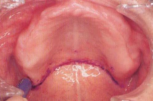
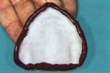
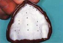
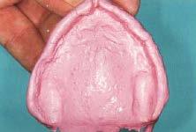
12 Clean the tray. The borders should be round and smooth with full extension.
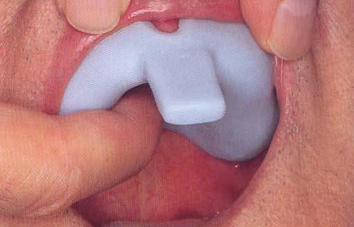
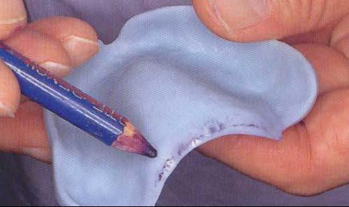
13 Single border molding Application of the material to the borders of the special tray (all borders) in one time and taking the impression of the borders, poly ether impression material usually used with this method.
14 techniques
15 The residual ridge may be said to have two forms: The anatomic form The anatomic form is the surface contour of the ridge when it is not supporting an occlusal load. The functional form The functional form of the residual ridge is the surface contour of the ridge when it is supporting a functional load.
16 The ideal residual ridge to support a denture base would consist of cortical bone that covers relatively dense cancellous bone with a broad rounded crest and high vertical slopes, and covered by firm, dense, fibrous connective tissue.


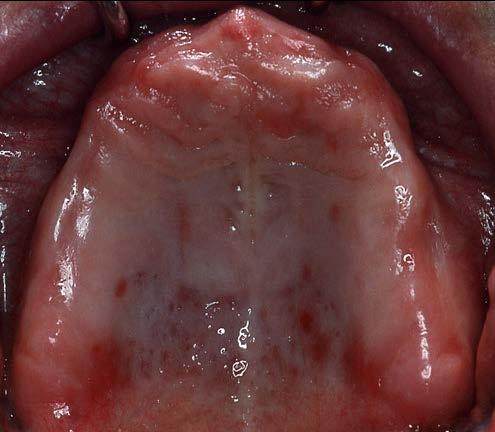
17 Maxilla The crestal area of the residual ridge will become the primary stressbearing area, consist primarily of cancellous bone, unlike in the mandible, oral tissue that overlies the maxillary residual alveolar bone is usually of a firm, dense nature (similar to the mucosa of the hard palate). Then the immediate buccal and lingual slopes of the ridge also considered primary stressbearing area.


18 Palatal tissue between the medial palatal raphae and the lingual slope of the posterior edentulous ridge are readily displaceable and cannot be considered as primary stress-bearing sites. They considered secondary stress bearing areas.


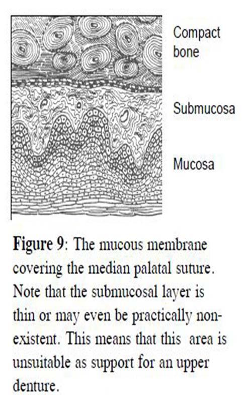
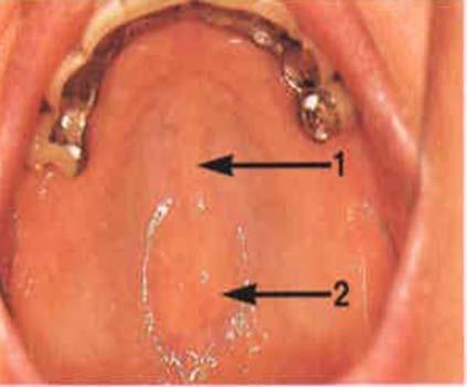

19 Incisive foramen & median palatal raphe considered as releif areas.

20 mandible The buccal shelf region (bounded by the external oblique line and crest of alveolar ridge) seems to be better suited for a primary stress-bearing role, because it is covered by relatively firm, dense, fibrous connective tissue supported by cortical bone. Then slopes of the residual ridge also considered primary stress-bearing area.

21 The crest of the mandibular residual ridge considered a secondary stress-bearing area, because the crest of the bony mandibular residual ridge is most often cancellous, and the lining mucosa not firmly attached especially in the posterior part.
22 Also referred as close mouth technique. Record the tissues in there functional position. The patient applies pressure by closing against occlusal rims or teeth. Patient exerts pressure and executes muscle actions. Close fit special tray with border molding. Impression compound, waxes, & soft liners. Displaced soft tissues unseated denture base. Pressure limits normal blood supply resorption.
23 Also referred as open mouth technique. Record the tissues in there undisplaced position. dentist exerts pressure. Spaced special tray with no border seal. Close adaptation of the denture base to the undistorted mucosa. Border seal retention.
24 Extension for maximum coverage within tissue tolerance. Light pressure or intimate contact with the movable loosely attached tissues in the vestibules. Minimum pressure. Impression compound tray & zinc oxide eugenol paste. Special tray (special design) with border refinement. Selective Pressure Impression Method use those portions of the residual ridge that can withstand additional stress (primary stress bearing area) and at the same time relieve the tissue of the residual ridge that cannot withstand functional loads.
25 Close fit special tray No space between the tray and oral tissue of residual ridge No spacer is used Used for mucodynamic impression technique Spaced special tray Space between the tray and oral tissue of residual ridge Spacer is used (wax) Used for mucostatic impression technique
26 Mucocompressive technique No spacer is used Mucostatic technique Maxilla: full coverage spacer with tissue stops in canine and molar region Mandible: full coverage spacer with tissue stops in canine and buccal shelf area Selective pressure technique Maxilla: relief areas (incisive papilla and mid palatine raphe) and over flabby tissues if present. Mandible: crest of the ridge
27


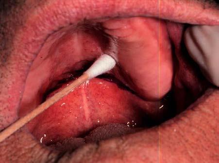
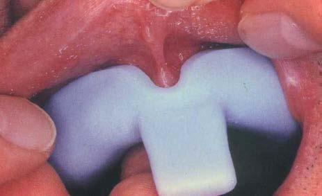
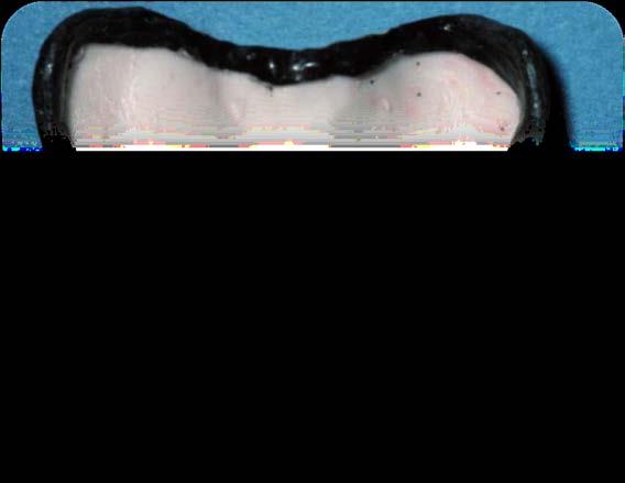
28 Frenal area Lip movement near labial frenum is vertical labial notch long & narrow. Muscle movements around buccal frenum are horizontal & vertical buccal frenum wide V shaped. Anterior movement should be recorded by pursing the lips.


29 Buccal vestibular area If border molding in the buccal space is inadequate, the denture will lose its seal because of the ingress of air under the denture base when the buccal vestibule is opened such as during laughing.

30 Coronoid process During border molding of the distobuccal border, ask the patient to move the jaw laterally from side to side. Else the coronoid process of the mandible may come into contact during function causing pain or discomfort.

31 Posterior border area The hamular notch is favorable landmark for placing the posterior border, which is the most important part for retention of upper denture. Bases short of the hamular notch will end on the thin - nonflexible tissue of the tuberosity and will consequently lack retention. POSTERIOR EXTENT OF DENTURE IN THIS REGION SHOULD END IN THE HAMULAR NOTCH & NOT EXTEND OVER THE HAMULAR PROCESS AS THIS CAN LEAD TO SEVERE PAIN DURING DENTURE WEAR

32 Pterygo-Mandibular Raphe Connects from the hamulus to the mylohyoid ridge When prominent, can cause pain, or loosening Requires relief groove if prominent

33 The vibrating line an imaginary line across the posterior part of the palate marking the division between the movable and immovable tissues of the soft palate. This can be identified when the movable tissues are functioning.
34 Prevents air passage between the tissues and denture base. Serves Primarily in denture retention by making contact with anterior portion of soft palate. Reduces patients awareness about the area hence decrease gag reflex. Prevents food accumulation between posterior border of denture and the soft palate. Compensates for polymerization shrinkage of denture base resin
35 Vibrating line The position of fovea palatine also influences the position of posterior border of the denture. Denture can extend 1-2mm across it. In patients with thick saliva, the fovea palatine should be left uncovered or else thick saliva flowing between the tissue and the denture can increase the hydrostatic pressure and displace the denture. Its an imaginary line drawn across the palate that marks the beginning of motion in the soft palate, when the individual says ah. Extends from one hamular notch to the other. It passes about 2mm in front of the fovea palatine. This line should lie on the soft palate. Distal end of the denture must cover the tuberosities and extend into hamular notches. It should end 1-2mm posterior to the vibrating line.
36 Anterior Vibrating Line: Its an imaginary line lying at the junction between the immovable tissues over the hard palate and slightly movable tissues of the soft palate The anterior vibrating line is cupid bow shaped due to projection of posterior nasal spine. It can be located by asking the patient to perform the valsalva maneuver. Posterior Vibrating Line: Its an imaginary line at the junction of the aponeurosis of tensor veli palati muscles and muscular portion of soft palate This line is usually straight. Its recorded by asking the patient to say ah in short but normal non-vigorous fashion.
37 1- Conventional Tech: By using special tray
38
39 3- Arbitrary Scrapping Of The Master Cast: Ant. & post. Vibrating lines are visualized by examining the pt s mouth and approximately marked on master cast. The lab technician scraped 0.5 to 1mm of stone in posterior palatal seal area of the master cast and fabricates the denture. This technique is inaccurate and not physiological and should be avoided.

40
41


42 Buccal flange area Buccal shelf, it is bounded medially by the alveolar crest, anteriorly by the buccal frenum, laterally by external oblique ridge and distally by the retromolar pad. The buccal shelf is covered with dense cortical bone and is also a wide area lying perpendicular to the direction of occlusal forces so it considered primary support bearing area in the mandibular arch. Extertnal oblique ridge, a bony ridge outsides the buccal shelf, runs anteroposteriorly. The denture border extend 1-2 mm beyond it.


43 If the denture border is extended beyond the external oblique ridge, the denture base will cover the buccinator muscle fibers. However the lower muscle bundle is loose and in active, it will not dislodge the denture. The muscle fibers run anteroposteriorly, paralleling the denture border and function in horizontal direction. If the denture border under extended in this area, it leads to food accumulation in the buccal sulcus and under the denture base.


44 Buccal frenum Clearance in this area should be provided for the normal action of the frenum.


45 Mylohyoid ridge area A denture border short of the mylohyoid ridges digs into the residual ridge and causes pain. If shortened, the denture border will impinge again upon the ridge. Border molding of the mylohyoid ridge area should be performed 4-6 mm below this ridge. Later the impression surface of the denture on the mylohyoid ridge area is relived. As the mylohyoid muscle runs anteroinferiorly even when it contracts maximally, it allows extension of the denture beyond the mylohyoid ridges.
")


46 Residual Ridge Resorption (RRR) Mylohyoid ridge Mylohyoid ridge can cause ulcers if it is a sharp Mucosa in this region is poorly keratinized and prone to trauma


47 Retromolar pad area The denture must retromolar pads. cover 2/3 of the It is composed of a firm fibrous connective tissue papilla in its anterior half and soft tissue containing molar glands in the posterior half. The position of the pads remains constant, even after the natural teeth are extracted. As the temporalis muscle fibers attach to the distal portion of the retromolar pad, stimulation from this muscle prevents the pad from resorbing. These facts ensure that the pads are an excellent guide for determining and setting the plane of occlusion between upper and lower denture teeth. The pads serve as bilateral, distal support for a mandibular denture. Covering the pads with the denture base helps reduce the rate of alveolar ridge resorption.


48 Retromylohyoid fossa It is space distal to mylohyoid muscle. Bounded by the mylohyoid muscle anteriorly, retromolar pad laterally, the superior constrictor muscle posterolaterally, the palatoglossus muscle posteromedially, & the tongue medially. During border molding, the border in this area is pushed into the retromylohyoid fossa by the strong intrinsic & extrinsic tongue muscles Thus it will show the so-called S curve as viewed from impression surface.


49 Sub lingual gland area Sub lingual gland lies above the mylohyoid muscle & is raised by the muscle during swallowing. If the denture border made short to relieve the raised sublingual gland, a space will occur between the denture border & the mucosa when the muscle is at rest & thus peripheral seal will be lost. The sublingual gland serves as a cushion, so neither lifts the denture nor causes the mucosa to be traumatized by the denture.


50 Anterior lingual flange area The border of the impression in this area is mainly influenced by the lingual frenum & the genioglossus muscle. To provide adequate clearance in this area the patient is instructed to lick the lower lip by moving tip of the tongue from side to side.


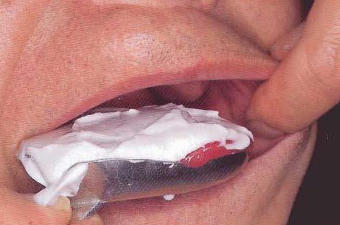
51 Labial flange area When the lip is pulled too much during border molding, the vestibule becomes shallow as the mentalis muscle attachment is higher than the base of the labial vestibule. On biting the operator fingers, the masticatory muscles become tense and the lips become relaxed as a reflex, then the impression is made in this siuation.
52 Objectives Impression of the flabby area made with pressure free technique. Impression of the sound edentulous arch that can withstand the occlusal load made with pressure technique.


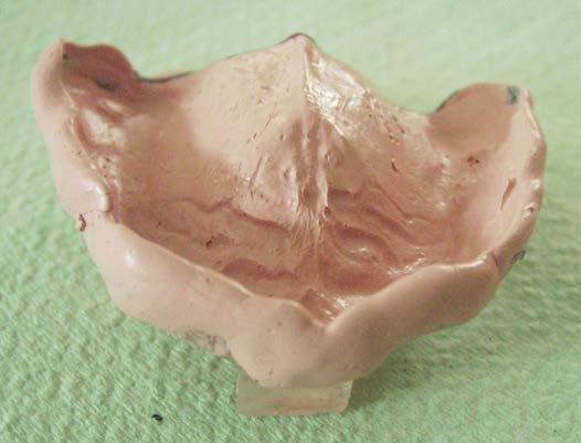
53 Flabby ridge Wax spacer Spaced special tray Final impression with impression plaster


54 Flabby anterior ridge A window is made in special tray Border molding of the special tray


55 Final impression of the entire ridge except the flabby ridge with ZOE Final impression of the anterior flabby ridge with impression plaster without pressure
56
57

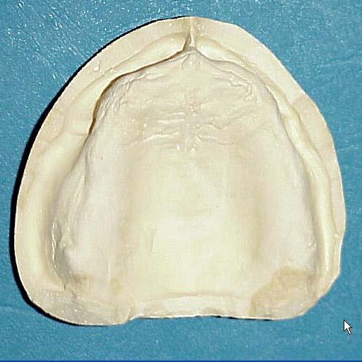
58 MAXILLARY IMPRESSION a. incisive papilla b. palatal rugae c. median palatine raphe J K A B d. maxillary tuberosity e. pterygomaxillary notch f. fovea palatini and vibrating line area I C i. residual alveolar ridge j. buccal frenum D D k. labial frenum E E

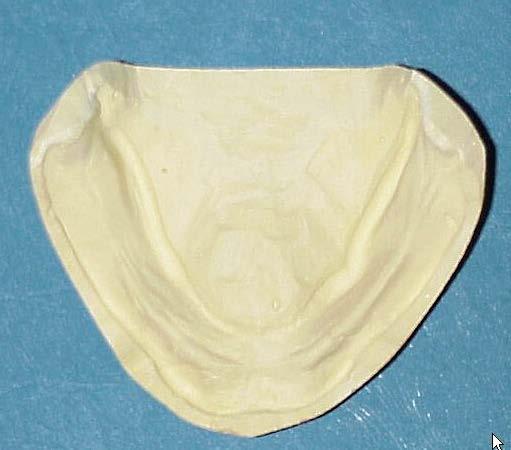
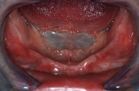
59 MANDIBULAR IMPRESSION a. retromolar pad b. mylohyoid (internal oblique line) c. masseteric notch d. residual alveolar ridge e. lingual frenum f. external oblique line g. buccal frenum h. labial frenum D A G E B G A F C H
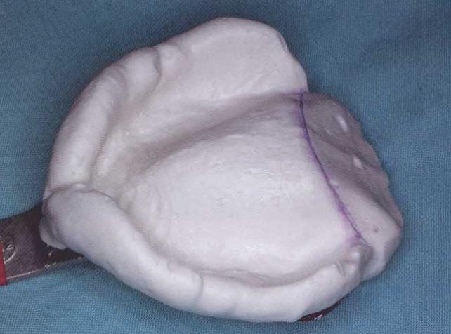
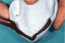
DEVELOPING ANALOGUE/SUBTITUTE FOR THE MANDIBULAR DENTURE BEARING AREA. Dr Muhammad Rizwan Memon FCPS Assistant Professor
 DEVELOPING ANALOGUE/SUBTITUTE FOR THE MANDIBULAR DENTURE BEARING AREA Dr Muhammad Rizwan Memon FCPS Assistant Professor Crest of Residual Ridge Buccal Shelf Shape of supporting structure Mylohyoid Ridge
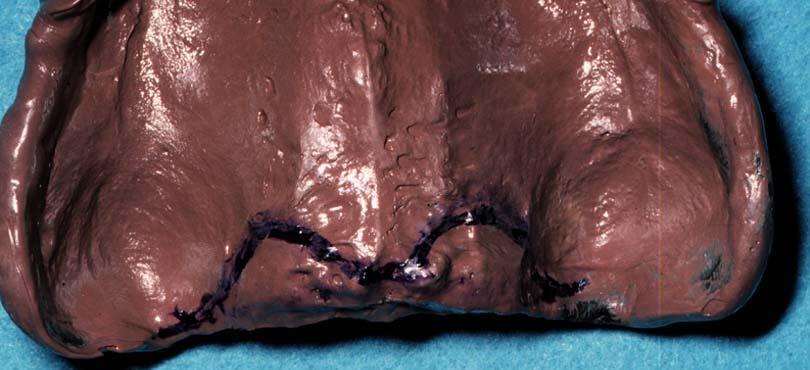
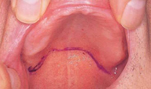
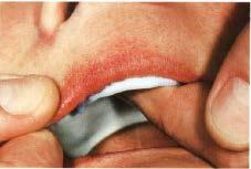
Figure (2-6): Labial frenum and labial notch.
: Labial frenum and labial notch.") The anatomy of the edentulous ridge in the maxilla and mandible is very important for the design of a complete denture. The consistency of the mucosa and architecture of the underlying bone is different
The anatomy of the edentulous ridge in the maxilla and mandible is very important for the design of a complete denture. The consistency of the mucosa and architecture of the underlying bone is different
Upper arch. 1Prosthodontics. Dr.Bassam Ali Al-Turaihi. Basic anatomy & & landmark of denture & mouth
 1Prosthodontics Lecture 2 Dr.Bassam Ali Al-Turaihi Basic anatomy & & landmark of denture & mouth Upper arch Palatine process of maxilla: it form the anterior three quarter of the hard palate. Horizontal
DEVELOPING ANALOGUE SUBTITUTE FOR THE MAXILLARY DENTURE BEARING AREA. Dr Muhammad Rizwan Memon FCPS Assistant Professor
 DEVELOPING ANALOGUE SUBTITUTE FOR THE MAXILLARY DENTURE BEARING AREA Dr Muhammad Rizwan Memon FCPS Assistant Professor Mucous membrane Residual Ridge Incisive papilla Rugae area Mid palatine raphe Hard
Oral cavity landmarks
 By: Dr. Ahmed Rabah Oral cavity landmarks The knowledge of oral anatomy and physiology will help the operator and provides enough landmarks to act as positive guide during denture construction. This subject
ARAB AMERICAN UNIVERSITY. Lab. Manual. Prosthetic Dentistry1; Removable Prosthodontics. 3 rd year
 ARAB AMERICAN UNIVERSITY Lab. Manual Prosthetic Dentistry1; Removable Prosthodontics 3 rd year Department of Fixed and removable prosthetic Dentistry Faculty of Dentistry 2012/2013 Course Instructor Dr.
IMPRESSION MAKING (IN COMPLETE DENTURES)
") IMPRESSION MAKING (IN COMPLETE DENTURES) DR ZURYATI AB GHANI BDS (WALES), Grad Dip Clin Dent (Adelaide), Doctor in Clinical Dentistry (prosthodontics), Adelaide, FRACDS 17.06.2007 Impressions An impression
IMPRESSION MAKING (IN COMPLETE DENTURES) DR ZURYATI AB GHANI BDS (WALES), Grad Dip Clin Dent (Adelaide), Doctor in Clinical Dentistry (prosthodontics), Adelaide, FRACDS 17.06.2007 Impressions An impression
Complete denture impressions
 Lec.5 Complete denture impressions د. غسان الطائي Dental Impression: a negative imprint of an oral structure used to produce a positive replica of the structure to be used as a permanent record or in the
THE BIOMECHANICAL BASIS OF RETENTION IN COMPLETE DENTURES
 THE BIOMECHANICAL BASIS OF RETENTION IN COMPLETE DENTURES Factors affecting the retention of dentures Retention is the resistance of the denture to removal along its path of insertion. Strictly speaking,
Post insertion problems in complete denture.
 Page1 Lect. 8 Prosthodontics Dr. Intisar J. Ismail 5 th class 22/11/2016 Post insertion problems in complete denture. Loss of natural teeth and subsequent alveolar resorption has a significant impact on
Post insertion problems in complete denture
 د. زينب الجمالي Lec. 3 Post insertion problems in complete denture Loss of natural teeth &subsequent alveolar resorption has a significant impact on appearance &function. CD fabrication techniques, &placement
Arrangement of the artificial teeth:
 Lecture Prosthodontic Dr. Osama Arrangement of the artificial teeth: It s the placement of the teeth on a denture with definite objective in mind or it s the setting of teeth on temporary bases. Rules
Denture Troubleshooting Guide
 Denture Troubleshooting Guide Technical bulletin from National Dentex Comfort Sore spot in vestibule upper or lower denture Sore spot in upper post dam. (posterior limit of upper) Single sore spots on
Difference between Provider Centric Approach and Patient Centric Approach in Complete Denture Impression
 Difference between Provider Centric Approach and Patient Centric Approach in Complete Denture Impression Shinichi Nukazawa Journal of the Academy of Clinical Dentistry Vol.29 No.1-2 combined edition: 18-26,
Pre prosthetic surgery
 Pre prosthetic surgery The surgical procedures designed to facilitate fabrication of a prosthesis or to improve the prognosis of prosthodontics care. AIMS OF PRE PROSTHETIC SURGERY 1-provide adequate bony
Techniques of local anesthesia in the mandible
 Techniques of local anesthesia in the mandible The technique of choice for anesthesia of the mandible is the block injection and this is attributed to the absence of the advantages which are present in
Vertical relation: It is the amount of separation between the maxilla and
 Vertical relations Vertical relation: It is the amount of separation between the maxilla and the mandible in a frontal plane. Vertical dimension: It is the distance between two selected points, one on
Prosthodontics Dr.Yassen H.
 Prosthodontics Dr.Yassen H. Lecture -2- Anatomy & Physiology Related to Prosthodontics (Myology) Muscles are divided or classified into: 1. Muscles of facial expression. 2. Suprahyoid muscles. 3. Infrahyoid
Nagri D et al. Linear occlusion and Neutral Zone recording for severely resorbed ridges
 Doi:10.21276/ledent.2018.02.01.02 Case Report LINEAR OCCLUSION AND NEUTRAL ZONE RECORDING USING TISSUE CONDITIONER REPORT OF A SEVERELY RESORBED RIDGE Divya Nagri, 1Ashish Kakkar, 2Neeraj Mittal, 3Lovely
Samantha W. Chou, D.M.D N. Southport Ave. Chicago, Illinois Phone: Fax:
 Samantha W. Chou, D.M.D. 2325 N. Southport Ave. Chicago, Illinois 60614 Phone: 312-608-6881 Fax: 773-296-0601 Samanthawchou@gmail.com What is our role as the dentist? "We live in a culture in which people
Basic Anatomy and Physiology of the Lips and Oral Cavity. Dr. Faghih
 Basic Anatomy and Physiology of the Lips and Oral Cavity Dr. Faghih It is divided into seven specific subsites : 1. Lips 2. dentoalveolar ridges 3. oral tongue 4. retromolar trigone 5. floor of mouth 6.
Making the Unstable Stable-A Case Series of Management of the Flabby Ridge
 IOSR Journal of Dental and Medical Sciences (IOSR-JDMS) e-issn: 2279-0853, p-issn: 2279-0861.Volume 14, Issue 6 Ver. VIII (Jun. 2015), PP 67-71 www.iosrjournals.org Making the Unstable Stable-A Case Series
Component parts of Chrome Cobalt Removable Partial Denture
 Lec. 5 د.بسام الطريحي Component parts of Chrome Cobalt Removable Partial Denture Major connectors: Are either bars or plates, the difference between them is in the amount of tissue covers. Plates are broad
Horizontal jaw relations: The relationship of mandible to maxilla in a
 Horizontal relations Horizontal jaw relations: The relationship of mandible to maxilla in a horizontal plane (in anteroposterior and side to side direction). a- Protruded or forward relation. b-lateral
Horizontal Jaw Relation
 Horizontal Jaw Relation Horizontal Jaw Relation It is the relationship of the mandible to the maxilla in a horizontal plane. It can also be described as the relationship of the mandible to the maxilla
Lips and labial mucosa
 Lips and labial mucosa External portion of the lips: the vermilion border and the skin Vermilion border : the exposed red portion of the lip, covered by mucous membrane, no mucous glands Boundary: the
Secondary, master, (functional impression) taking methods Dr. Károlyházy Katalin Semmelweis Univ. Budapest, Department of Prosthodontics Head of the
 taking methods Dr. Károlyházy Katalin Semmelweis Univ. Budapest, Department of Prosthodontics Head of the") Secondary, master, (functional impression) taking methods Dr. Károlyházy Katalin Semmelweis Univ. Budapest, Department of Prosthodontics Head of the Dept.: Prof. Dr.Hermann Péter Med.Habil. 2017. Secondary
Secondary, master, (functional impression) taking methods Dr. Károlyházy Katalin Semmelweis Univ. Budapest, Department of Prosthodontics Head of the Dept.: Prof. Dr.Hermann Péter Med.Habil. 2017. Secondary
Subdivided into Vestibule & Oral cavity proper
 Extends from the lips to the oropharyngeal isthmus The oropharyngeal isthmus: Is the junction of mouth and pharynx. Is bounded: Above by the soft palate and the palatoglossal folds Below by the dorsum
Oral cavity : consist of two parts: the oral vestibule and the oral cavity proper. Oral vestibule : is slit like space between.
 Oral cavity Oral cavity : consist of two parts: the oral vestibule and the oral cavity proper Oral vestibule : is slit like space between the teeth, buccal gingiva, lips, and cheeks 1 Oral cavity Oral
The Anatomical Study of the Sinew String Observed on the Buccal Mucosa of Mandibular Second Molar and Posterior of Retromolar Pad
 The Anatomical Study of the Sinew String Observed on the Buccal Mucosa of Mandibular Second Molar and Posterior of Retromolar Pad Seiichiro Someya Vol.28 No.1-2 combined edition: 14-20, 2008 (Japanese)
SPECIAL TRAY. Dr. Barbara Kispélyi Associate Professor. Semmelweis University, Faculty of Dentistry Department of Prosthodontics
 SPECIAL TRAY Dr. Barbara Kispélyi Associate Professor Semmelweis University, Faculty of Dentistry Department of Prosthodontics Primary impression Primary cast Special tray From web OUTLINE Primary impression
Principles of. By: Dr. Ahmad Rabah
 Principles of By: Dr. Ahmad Rabah 1. Utilize what's present: Whenever possible, select a design that fits the teeth and soft tissues, rather than choosing one that requires tissue alteration. When minimal
Arrangement of posterior artificial teeth Standardized parameters Curve of Wilson Curve of Spee
 . Arrangement of posterior artificial teeth Posterior teeth are set up in tight centric occlusion. The mandibular teeth are set in the wax occlusion rim over the residual ridge in their ideal buccolingual
Indirect retainers. 1 i
 8 1 i Indirect retainers Factors Influencing Effectiveness Indirect Retainers Auxiliary Functions Indirect Retainers Forms Indirect Retainers Auxiliary occlusal rest Canine extensions fiom occlusal rests
MANAGEMENT OF LAX TISSUES TO IMPROVE POSSESSION BY WINDOW TECHNIQUE. Dept.of Prosthodontics, A.M.E.S Dental College&Hospital, Raichur
 MANAGEMENT OF LAX TISSUES TO IMPROVE POSSESSION BY WINDOW TECHNIQUE. Subashani T *, Sunil Dhaded ** Dept.of Prosthodontics, A.M.E.S Dental College&Hospital, Raichur - 584103 *-Sr.Lecturer **-Professor
Lect. 14 Prosthodontics Dr. Osama
 Lect. 14 Prosthodontics Dr. Osama Principles of Removable Partial Denture Design Difference in Prosthesis Support and Influence on Design: For a tooth-supported prosthesis, the movement potential is less
4/15/2015. Secondary Impressions: Anatomic and Physiologic Impressions. April, 8, Impressions for tooth-supported RPDs
 Secondary Impressions: Anatomic and Physiologic Impressions Impressions for tooth-supported RPDs April, 8, 2015 Impressions for tooth and tissue supported RPDs In tooth-supported removable partial denture
General characteristics of Complete Removable Dentures (also Full Dentures)
") General characteristics of Complete Removable Dentures (also Full Dentures) Prosthetic treatment in totally edentulous patients is the most difficult kind of restorative treatment. There are a lot of individual
General characteristics of Complete Removable Dentures (also Full Dentures) Prosthetic treatment in totally edentulous patients is the most difficult kind of restorative treatment. There are a lot of individual
Prof. Dr. Hikmat J. Al-Judy B.D.S., M.Sc., Ph.D in Prosthodontic, College of Dentistry/ Baghdad University/ Baghdad, Iraq.
 IOSR Journal of Dental and Medical Sciences (IOSR-JDMS) e-issn: 2279-0853, p-issn: 2279-0861.Volume 14, Issue 7 Ver. V (July. 2015), PP 35-40 www.iosrjournals.org Comparison of the effect of sectional
3. The Jaw and Related Structures
 Overview and objectives of this dissection 3. The Jaw and Related Structures The goal of this dissection is to observe the muscles of jaw raising. You will also have the opportunity to observe several
Bones Ethmoid bone Inferior nasal concha Lacrimal bone Maxilla Nasal bone Palatine bone Vomer Zygomatic bone Mandible
 splanchnocranium - Consists of part of skull that is derived from branchial arches - The facial bones are the bones of the anterior and lower human skull Bones Ethmoid bone Inferior nasal concha Lacrimal
Dr.Sepideh Falah-kooshki
 Dr.Sepideh Falah-kooshki MAXILLA Premaxillary/median palatal suture (radiolucent). Incisive fossa and foramen (radiolucent). Nasal passages (radiolucent). Nasal septum (radiopaque). Anterior nasal spine
Dental Morphology and Vocabulary
 Dental Morphology and Vocabulary Palate Palate Palate 1 2 Hard Palate Rugae Hard Palate Palate Palate Soft Palate Palate Palate Soft Palate 4 Palate Hard Palate Soft Palate Maxillary Arch (Maxilla) (Uppers)
CLASSIFICATIONS. Established in 1994 as a subcommittee of the. Prosthodontic Care Committee
 CLASSIFICATIONS Established in 1994 as a subcommittee of the Prosthodontic Care Committee Committee Members Thomas J. McGarry, DDS, Chair Arthur Nimmo, DDS James F. Skiba, DDS Christopher R. Smith, DDS
A comparative evaluation of three different techniques for single step border molding
 ORIGINAL RESEARCH A comparative evaluation of three different techniques for single step border molding Kheur M 1, Jambhekar S 2, Sethi T 3, Kheur S 4 1Professor, 3 PG student, Dept. of Prosthodontics,
Accu-Dent. impression system. Procedural guide. System 2 TM. For removable dentate impressions
 Accu-Dent impression system System 2 TM Procedural guide System 2 TM For removable dentate impressions The Complete System 2 TM Accu-Dent s System 2 is designed for partials, immediate dentures, orthodontics,
J. 0. AKINOSI, B.D.s., F.D.S.R.C.S.
 British Journal of Oral Surgery 15 (1977-78) 83-87 A NEW APPROACH TO THE MANDIBULAR NERVE BLOCK J. 0. AKINOSI, B.D.s., F.D.S.R.C.S. Department of Oral Surgery and Pathology, College of Medicine, Lagos
Interim Denture Interim Complete Dental Prosthesis Clinical Steps
 Interim Denture Interim Complete Dental Prosthesis Clinical Steps Diagnostic Appointment Comprehensive Exam Extra oral Intra oral Address: Main complaint Esthetic concerns Other concerns Discuss (if present)
Mandibular ridge changes after adaptation. An issue of shortened dental arch to be considered from changes of soft tissues after unattended tooth loss
 (Journal of Dental Outlook, Vol.110, Vol.6:1021~1027, Japan 2007. An issue of shortened dental arch to be considered from changes of soft tissues after unattended tooth loss Dr. Jiro Abe Abe Dental Clinic
Rules & regulations and Requirements
 Rules & regulations and Requirements Regarding the attendance in the lectures, any student exceed 15% 0f the total will not allow to set the final exam. Please the rules not allow any student to sign in
Conservative prosthodontic procedures to improve mandibular denture stability in an atrophic mandibular ridge
 Review Article Conservative prosthodontic procedures to improve mandibular denture stability in an atrophic mandibular ridge D. R. Prithviraj, Vishal Singh, Sarvanan Kumar 1, D. P. Shruti Department of
Impression Manual.
 Impression Manual www.paladigitaldentures.com Contents Page 1 Requirements for Impressions Complete Denture Single Arch Maxillary Denture Single Arch Mandibular Denture Maxillary Impression Maxillary
Try-in of the Trial Denture by Dr. Mahmoud Ramadan
 Published 1/25/2009 Try-in of the Trial Denture by Dr. Mahmoud Ramadan Definition: Preliminary insertion of complete denture wax up (trial denture) to determine the fit, esthetics, maxillomandibular relations
PROSTHODONTIC REHABILITATION OF A SEVERELY RESORBED MANDIBULAR RIDGE USING NEUTRAL ZONE TECHNIQUE: A CASE REPORT
 CASE REPORT (e) ISSN Online: 2321-9599 (p) ISSN Print: 2348-6805 PROSTHODONTIC REHABILITATION OF A SEVERELY RESORBED MANDIBULAR RIDGE USING NEUTRAL ZONE TECHNIQUE: A CASE REPORT Teny Fernandez 1, Sheela
Fundamentals of technique Types of local anaesthesia Topical or surface anaesthesia
 Fundamentals of technique The importance of a quiet, confident, and friendly manner towards all patients so physical comfort is also essential for the co-operation of the patient and the ease of operation
Complete Denture Clinic Complete Removable Dental Prosthesis Procedures
 Complete Denture Clinic Complete Removable Dental Prosthesis Procedures Diagnostic Appointment Comprehensive Exam Extra oral Intra oral Discuss current dentures Main complaint Esthetic concerns Prosthetic
Anatomy of Oral Cavity DR. MAAN AL-ABBASI
 Anatomy of Oral Cavity DR. MAAN AL-ABBASI By the end of this lecture you should be able to: 1. Differentiate different parts of the oral cavity 2. Describe the blood and nerve supply of mucosa and muscles
Dr.Mikulás Krisztina. Fabrication of the trial denture, and the try in procedure
 Dr.Mikulás Krisztina Fabrication of the trial denture, and the try in procedure the correct shape for the labial, buccal and palatal surfaces Adjusting the upper record rim Before starting adjustment-upper
Infratemporal fossa: Tikrit University college of Dentistry Dr.Ban I.S. head & neck Anatomy 2 nd y.
 Infratemporal fossa: This is a space lying beneath the base of the skull between the lateral wall of the pharynx and the ramus of the mandible. It is also referred to as the parapharyngeal or lateral pharyngeal
PH-04A: Clinical Photography Production Checklist With A Small Camera
 PH-04A: Clinical Photography Production Checklist With A Small Camera Operator Name Total 0-49, Passing 39 Your Score Patient Name Date of Series Instructions: Evaluate your Series of photographs first.
How to provide intraoral scans to SomnoMed for the production of SomnoDent device.
 How to provide intraoral scans to SomnoMed for the production of SomnoDent device. KEY QUESTIONS: 1. Where do I send my Case? Send intra-oral scan files (maxilla and mandible in protrusive bite) and an
Temporal region. temporal & infratemporal fossae. Zhou Hong Ying Dept. of Anatomy
 Temporal region temporal & infratemporal fossae Zhou Hong Ying Dept. of Anatomy Temporal region is divided by zygomatic arch into temporal & infratemporal fossae. Temporal Fossa Infratemporal fossa Temporal
MODIFIED FUNCTIONAL IMPRESSION TECHNIQUE FOR RESORBED MANDIBULAR RIDGE: TWO CASE STUDIES
 Prosthetic dentistry MODIFIED FUNCTIONAL IMPRESSION TECHNIQUE FOR RESORBED MANDIBULAR RIDGE: TWO CASE STUDIES Divia CHUGH 1, Siddharth PHULL 2, Arpan RANA 1, Yashendra SAINI 1 1 Resident (Prosthodontics)
Neutral Zone Approach for Rehabilitation of Severely Atrophic Ridge
 CASE REPORT Neutral Zone Approach for Rehabilitation of Severely Atrophic Ridge Amit Porwal, Preet Jain, Siddesh.P.Birader, Santosh Nelogi, Naveen.H.C Abstract One of the most commonly faced problems among
DENT Advanced Topics in Removable Prosthodontics, Winter 2008
 University of Michigan Deep Blue deepblue.lib.umich.edu 2008-01 DENT 718 - Advanced Topics in Removable Prosthodontics, Winter 2008 Shotwell, Jeffrey Shotwell, J. (2008, April 23) Advanced Topics in Removable
Prosthodontic Management of Compromised Mandibular Ridge Using Modified Functional..
 IOSR Journal of Dental and Medical Sciences (IOSR-JDMS) e-issn: 2279-0853, p-issn: 2279-0861.Volume 15, Issue 6 Ver. IX (June 2016), PP 55-59 www.iosrjournals.org Prosthodontic Management of Compromised
Connect your Scanner to SomnoMed Canada. SOMGauge Protrusive Bite Recording - Manual. Scanning Impressions - Lower and Upper
 IOS Instructions How to create and submit the best scans to SomnoMed Canada for the creation of a custom SomnoDent Sleep Apnea Appliance Its a simple process: STEP 1 Connect your Scanner to SomnoMed Canada
MM01-Facebow And Bite Registration Procedure Checklist
 MM01-Facebow And Bite Registration Procedure Checklist Bite fork Registration OK Redo 1. Estimate the shape of the patient s arch 2. Place the bite tabs on the bite fork 3. Warm the compound in hot water
 ISSN 1560-1587 81 7.18 11.51 9.21 72.84 4.94 22.22 4 Exploring the distance between upper central incisor edge and incisive papilla in Taiwanese population Sheau-Jiuan Huang 1, Tsau-Mau Chou 1, Huey-Er
Oral Surgery. Basic Techniques of Dental Local Anesthesia. A variety of techniques used in administration and deposition of local anesthesia:
 Oral Surgery Lecture: 9 Dr. Saif Saadedeen Basic Techniques of Dental Local Anesthesia A variety of techniques used in administration and deposition of local anesthesia: 1. Topical anesthesia 2. Infiltration
SURVEYING OF REMOVABLE PARITAL DENTURES FEB, 11, 2015
 SURVEYING OF REMOVABLE PARITAL DENTURES FEB, 11, 2015 Dental Surveyor: It is a mechanical device used to determine the relative parallelism of the teeth surfaces and the undercuts areas in relation to
Removable Partial Denture Manual. Robert W. Loney, DMD, MS 2008
 Removable Partial Denture Manual Robert W. Loney, DMD, MS 2008 Removable Partial Denture Manual Robert W. Loney, DMD, MS 2008 Table of Contents - i Table of Contents Introduction to Removable Partial
Investigating the maxillary buccal vestibule
 Journal of Dental Sciences (2014) 9, 125e129 Available online at www.sciencedirect.com journal homepage: www.e-jds.com ORIGINAL ARTICLE Investigating the maxillary buccal vestibule Jen-Hao Chen a,b,f,
Impression techniques for removable partial dentures: A review
 Review Article DOI: 10.18231/2455-8486.2017.0012 Impression techniques for removable partial dentures: A review Reeta Jain 1,*, Supriya 2, Shweta 3, Kimmi 4 1 Professor & HOD, 2,3,4 PG Student, Dept. of
Methods of determining vertical dimension of occlusion
 Methods of determining vertical dimension of occlusion 1) Pre-extraction records a) Willis gauge This device could used to measure V D O before teeth extraction and then recorded in the patient record.
-Ibrahim Al-Naser. -Dr Al- Muhtaseb. 1 P a g e
 -1 -Ibrahim Al-Naser - -Dr Al- Muhtaseb 1 P a g e The Digestive System The doctor started the lecture by talking about the class rules. The GI system is an organ system, it is divided into: The Alimentary
Biology and Medicine Jain et al., Biol Med (Aligarh) 2016, 8:5
 2016, 8:5") Biology and Medicine Jain et al., 2016, 8:5 http://dx.doi.org/10.4172/0974-8369.1000307 Review Article Open Access A Clinical Review of Spacer Design for Conventional Complete Denture Ashish R. Jain*,
Biology and Medicine Jain et al., 2016, 8:5 http://dx.doi.org/10.4172/0974-8369.1000307 Review Article Open Access A Clinical Review of Spacer Design for Conventional Complete Denture Ashish R. Jain*,
Concepts of occlusion Balanced occlusion. Monoplane occlusion. Lingualized occlusion. Figure (10-1)
") Any contact between teeth of opposing dental arches; usually, referring to contact between the occlusal surface. The static relationship between the incising or masticatory surfaces of the maxillary or
Any contact between teeth of opposing dental arches; usually, referring to contact between the occlusal surface. The static relationship between the incising or masticatory surfaces of the maxillary or
-SQA-SCOTTISH QUALIFICATIONS AUTHORITY. Hanover House 24 Douglas Street GLASGOW G2 7NG NATIONAL CERTIFICATE MODULE DESCRIPTOR
 -SQA-SCOTTISH QUALIFICATIONS AUTHORITY Hanover House 24 Douglas Street GLASGOW G2 7NG NATIONAL CERTIFICATE MODULE DESCRIPTOR -Module Number- 0069143 -- -Superclass- PF -Title- DENTAL SPECIAL TRAYS -DESCRIPTION-
Department(S) And Institution(S) Department. Of Prosthodontics & Dental Materials, Indira Gandhi Government Dental College, Jammu, J&K, India.
 And Institution(S) Department. Of Prosthodontics & Dental Materials, Indira Gandhi Government Dental College, Jammu, J&K, India.") IOSR Journal of Dental and Medical Sciences (IOSR-JDMS) e-issn: 2279-0853, p-issn: 2279-0861.Volume 15, Issue 6 Ver. IX (June 2016), PP 70-74 www.iosrjournals.org Frequency and Location of Ulcerations
IOSR Journal of Dental and Medical Sciences (IOSR-JDMS) e-issn: 2279-0853, p-issn: 2279-0861.Volume 15, Issue 6 Ver. IX (June 2016), PP 70-74 www.iosrjournals.org Frequency and Location of Ulcerations
ISPUB.COM. Habitual Centric: A Case Report. Manisha, N Kathuria, A Gupta, N Gupta INTRODUCTION CASE REPORT
 ISPUB.COM The Internet Journal of Geriatrics and Gerontology Volume 6 Number 2 Habitual Centric: A Case Report Manisha, N Kathuria, A Gupta, N Gupta Citation Manisha, N Kathuria, A Gupta, N Gupta. Habitual
Stiff alginate for denture impressions, why?
 Stiff alginate for denture impressions, why? Every dentist or dental prosthetist knows how difficult it is to make a well-fitting, stable denture. What they do not always realise, however, is the importance
Relining and Rebasing of complete denture
 Relining and Rebasing of complete denture It is the procedures used to resurface the tissue-side of a denture with new material layer, thus producing an accurate adaptation to the denture foundation area.
Prosthetic Options in Implant Dentistry. Hakimeh Siadat, DDS, MSc Associate Professor
 Prosthetic Options in Dentistry Hakimeh Siadat, DDS, MSc Associate Professor Dental Research Center, Department of Prosthodontics & Dental s Faculty of Dentistry, Tehran University of Medical Sciences
Face-Bow Instructions
 M Panadent Corporation 580 S. Rancho Avenue Colton, California 92324, USA Tel: (909) 783-1841 USA & Canada (800) 368-9777 The following procedure is followed when Panadent Bite-Tab compound discs are used
The Gerber Articulator and System of Full Denture Construction
 The Gerber Articulator and System of Full Denture Construction Part 2(b) Setting-up the Teeth and Finishing the Dentures By G. E. White, Technical Instructor, University of Sheffield Dental School The
Occlusion in complete denture
 Occlusion in complete denture Occlusion is a concept that is pertinent to all dental patients wheather they have their own teeth or not.it is a term used to describe the contact relationship between the
Rehabilitation of Resorbed Mandibular Ridge with Implant Supported Overdenture- A Clinical Report
 Rehabilitation of Resorbed Mandibular Ridge with Implant Supported Overdenture- A Clinical Report 1 2 1 1 Mittal R, Saxena D, Rao S, Kumar M Abstract: Statement of Problem: Complete denture rehabilitation
Muscles of mastication [part 1]
![Muscles of mastication [part 1]](/thumbs/76/73586850.jpg "Muscles of mastication [part 1]") Muscles of mastication [part 1] In this lecture well have the muscles of mastication, neuromuscular function, and its relationship to the occlusion morphology. The fourth determinant of occlusion is the
Muscles of mastication [part 1] In this lecture well have the muscles of mastication, neuromuscular function, and its relationship to the occlusion morphology. The fourth determinant of occlusion is the
Prosthetic V. Removable dentures I.
 Prosthetic V. Removable dentures I. Removable dentures Partial Complete (full) lenka.roubalikova@tiscali.cz 2 Prosthetic dentistry replacement of Damaged teeth reconstruction of the crown (inlays, crowns)
AccuDent XD. A beautiful smile makes a great impression... Office Procedure Guide. ...but it takes a great impression to make a beautiful smile.
 AccuDent XD Alginate Impression System Office Procedure Guide A beautiful smile makes a great impression......but it takes a great impression to make a beautiful smile. EXPLORE. SHOP. LEARN. Accu-Dent
Management of maxillary flabby tissue with two part tray technique a case report
 Original article: Management of maxillary flabby tissue with two part tray technique a case report DR. RUPAL J. SHAH, DR. SANJAY LAGDIVE, DR. NAJMUNNISHA SABUGAR, DR. DHARA KOTAK, DR. KIRAN HADIYA Department
Stock Tray Modification for Two-Stage Impression Technique for Recording Flabby Ridges: Report of a Case Series
 I J Pre Clin Dent Res 2014;2(1):75-79 January-March All rights reserved International Journal of Preventive & Clinical Dental Research Stock Tray Modification for Two-Stage Impression Technique for Recording
Selection and arrangement of teeth in rpd
 Selection and arrangement of teeth in rpd upon completion of the articulator mounting and a thorough assessment of the occlusal requirements, the practitioner should be able to perform the proper arrangement
A comparison of posterior palatal seal creation in complete dentures by students at the University of Pretoria and private practitioners
 A comparison of posterior palatal seal creation in complete dentures by students at the University of Pretoria and private practitioners M Lekay-Adams A comparison of posterior palatal seal creation in
Anatomy Sheet: Oral cavity Done by: rasha Rakan edited by: khansaa Mahmoud
 Anatomy Sheet: Oral cavity Done by: rasha Rakan edited by: khansaa Mahmoud The oral cavity has 2 parts: 1. Oral vestibule: outer part that consists of outside the teeth, between the teeth, the cheeks and
Jaw relations and jaw relation records
 Lecture 11 Prosthodontics Dr. Osama Jaw relations and jaw relation records Jaw relations can be classified into 3 categories 1-Orientation jaw relation 2-Vertical jaw relation 3-Horizontal jaw relation
AVADENT-WAGNER EZ GUIDE PROTOCOL
 APPOINTMENT A. Final Impressions 1 APPOINTMENT 2 APPOINTMENT 3 D. Seat Wagner EZ Guide Try-In B. Papillameter Measurement E. Refine Anterior Teeth C. Tooth Mould Selection F. Interocclusal Record G. Deliver
6610 NE 181st Street, Suite #1, Kenmore, WA
 660 NE 8st Street, Suite #, Kenmore, WA 9808 www.northshoredentalacademy.com.08.900 READ CHAPTER The Professional Dental Assistant (p.-9) No Key Terms Recall Questions:,,,, and 6 CLASS SYLLABUS DAY READ