A Guide to Clinical Differential Diagnosis of Oral Mucosal Lesions
|
|
|
- Hilary Alexander
- 5 years ago
- Views:
Transcription
1 Continuing Education Brought to you by A Guide to Clinical Differential Diagnosis of Oral Mucosal Lesions Course Author(s): Michael W. Finkelstein, DDS, MS; Emily Lanzel, DDS, MS; John W. Hellstein, DDS, MS CE Credits: 4 hours Intended Audience: Dentists, Dental Hygienists, Dental Assistants, Dental Students, Dental Hygiene Students, Dental Assistant Students Date Course Online: 06/04/2002 Last Revision Date: 03/27/2017 Course Expiration Date: 03/26/2020 Cost: Free Method: Self-instructional AGD Subject Code(s): 730 Online Course: Disclaimer: Participants must always be aware of the hazards of using limited knowledge in integrating new techniques or procedures into their practice. Only sound evidence-based dentistry should be used in patient therapy. Introduction The primary goal of this course is to help you learn the process of clinical differential diagnosis of diseases and lesions of the oral mucosa. The first step in successful therapeutic management of a patient with an oral mucosal disease or lesion depends upon creating a differential diagnosis. This course also includes both an interactive and downloadable decision tree to assist in the diagnosis. ADA CERP The Procter & Gamble Company is an ADA CERP Recognized Provider. ADA CERP is a service of the American Dental Association to assist dental professionals in identifying quality providers of continuing dental education. ADA CERP does not approve or endorse individual courses or instructors, nor does it imply acceptance of credit hours by boards of dentistry. Concerns or complaints about a CE provider may be directed to the provider or to ADA CERP at: Approved PACE Program Provider The Procter & Gamble Company is designated as an Approved PACE Program Provider by the Academy of General Dentistry. The formal continuing education programs of this program provider are accepted by AGD for Fellowship, Mastership, and Membership Maintenance Credit. Approval does not imply acceptance by a state or provincial board of dentistry or AGD endorsement. The current term of approval extends from 8/1/2017 to 7/31/2021. Provider ID#
2 Conflict of Interest Disclosure Statement The late Dr. Finkelstein reported no conflicts of interest associated with this course when he last updated it. The staff at P&G expresses our condolences regarding the loss of Dr. Finkelstein on December 28, He was a passionate advocate for dental education who reached tens of thousands of students and patients with his knowledge of oral pathology that spanned a career of over 30 years at the University of Iowa. Dr. Lanzel and Dr. Hellstein report no conflicts of interest associated with this course. Course Contents Overview Learning Objectives Part I: Introduction to Clinical Differential Diagnosis How to Use the Decision Tree Benign and Malignant Tumors Part II: Surface Lesions of Oral Mucosa White Surface Lesions Surface Debris White Lesions White Lesions Due to Subepithelial Change Pigmented Surface Lesions of Oral Mucosa Localized Pigmented Surface Lesions of Oral Mucosa Extravascular Blood Lesions Melanocytic Lesions Tattoo Vesicular-Ulcerated-Erythematous Surface Lesions of Oral Mucosa Hereditary Diseases: Epidermolysis Bullosa Autoimmune Diseases Idiopathic Diseases Viral Diseases Mycotic Diseases-Candidosis (Candidiasis) Part III: Soft Tissue Enlargements of Oral Mucosa Reactive Soft Tissue Enlargements Soft Tissue Tumors Benign Epithelial Tumors of Oral Mucosa Benign Mesenchymal Tumors of Oral Mucosa Benign Salivary Gland Neoplasms of Oral Mucosa Cysts of Oral Mucosa Malignant Neoplasms of Oral Mucosa Part IV: Summary of Clinical Features of Oral Mucosal Lesions Table 1. White Surface Lesions of Oral Mucosa Table 2. Localized Pigmented Surface Lesions of Oral Mucosa Table 3. Vesicular-Ulcerated-Erythematous Surface Lesions of Oral Mucosa Table 4. Soft Tissue Enlargements Table 5. Benign Epithelial Tumors Table 6. Benign Mesenchymal Tumors Table 7. Benign Salivary Gland Tumors Table 8. Soft Tissue Cysts Course Test References About the Authors Overview Oral pathology is a visual specialty, and clinical images can facilitate your learning the clinical features of oral mucosal lesions. Several atlases are recommended in the Additional Resources section of this course. The textual material in this course is designed to be used with The Oral Pathology Image Database (Atlas). Please note that lesions or diseases discussed in the textual material that have clinical images available on The Oral Pathology Image Database are designated with *. Learning Objectives Upon completion of this course, the dental professional should be able to: Classify oral lesions into surface lesions and soft tissue enlargements using a decision tree (flowchart). Describe the clinical features that are characteristic of each class of oral mucosal lesions in the decision tree, including: White surface lesions - epithelial thickening, surface debris, and subepithelial change Generalized pigmented surface lesions Localized pigmented surface lesions - intravascular blood, extravascular blood, melanin pigment, and tattoo Vesicular-ulcerated-erythematous surface lesions - hereditary, autoimmune, viral, mycotic, and idiopathic Reactive soft tissue enlargements of oral mucosa Benign tumors of oral mucosa - epithelial, mesenchymal, and salivary gland 2
3 Malignant neoplasms of oral mucosa Cysts of oral mucosa Describe the characteristic or unique clinical features of the most common and/or important diseases of the oral mucosa. Perform a step-by-step clinical differential diagnosis, using the decision tree, for patients with oral mucosal lesions. Part I: Introduction to Clinical Differential Diagnosis Diagnosing lesions of the oral mucosa is necessary for the proper management of patients. Clinical differential diagnosis is the cognitive process of applying logic and knowledge, in a series of step-by-step decisions, to create a list of possible diagnoses. Differential diagnosis should be approached on the basis of exclusion. All lesions that cannot be excluded represent the initial differential diagnosis and are the basis for ordering tests and procedures to narrow the diagnosis. Guessing what the one best diagnosis is for an oral lesion can be dangerous for the patient because serious conditions can be overlooked. It is helpful for clinicians to organize their knowledge of oral pathology using a system that simulates the clinical appearance of oral lesions. A decision tree is a flowchart that organizes information so that the user can make a series of step-by-step decisions and arrive at a logical conclusion (Figure 1). How to Use the Decision Tree To use the decision tree, the clinician begins at the left side of the tree, makes the first decision, and proceeds to the right. The names of individual lesions are listed on the far right of the tree. Any lesion or group of lesions that cannot be excluded becomes part of the clinical differential diagnosis. The first decision to make when using the decision tree is whether the lesion is a surface lesion or soft tissue enlargement. Surface lesions consist of lesions that involve the epithelium and superficial connective tissue of mucosa and skin. They do not exceed 2-3 mm in thickness. Surface lesions are divided into three categories based on their clinical appearance: white, pigmented, and vesicularulcerated-erythematous. Each of these categories is further subclassified as shown in Tables 1-3. Soft tissue enlargements are swellings or masses that are divided into two categories: reactive and tumors in Table 4. The term tumor is used in the clinical sense of an enlargement and is not based on microscopic criteria or basic pathologic process. For example, irritation fibroma is classified as a tumor because this lesion is persistent and progressively increases in size, although most people agree that the true pathogenesis is that of a reactive process secondary to chronic irritation. Reactive soft tissue enlargements may increase and decrease (fluctuate) in size and usually eventually regress. Reactive enlargements are often, but not always, tender or painful and usually have a more rapid growth rate (measured in hours to weeks) than tumors. Some reactive enlargements begin as a diffuse lesion and become more localized with time. Sometimes reactive lesions are associated with tender lymph nodes and systemic manifestations, such as fever and malaise. Once it is decided that a soft tissue enlargement is reactive, the next step is to determine what the lesion is reacting to, such as bacterial, viral, or fungal infections or chemical or physical injury. Soft tissue tumors are characterized by being persistent and progressive; they do not resolve without treatment. They are usually not painful early in their development, and the growth rate varies from weeks to years. Soft tissue enlargements are swellings or masses that are divided into two categories: reactive and tumors in Table 4. The term tumor is used in the clinical sense of an enlargement and is not based on microscopic criteria or basic pathologic process. For example, irritation fibroma is classified as a tumor because this lesion is persistent and progressively increases in size, although most people agree that the true pathogenesis is that of a reactive process secondary to chronic irritation. Reactive soft tissue enlargements may increase and decrease (fluctuate) in size and usually eventually regress. Reactive enlargements 3
4 White Decision Tree for Oral Mucosa Lesions (Revised 3/08) Epithelial thickening Surface debris Subepithelial Candidosis Burn Congenital keratotic cysts Scar Fordyce granules Geographic tongue (erythema migrans) Hairy tongue Leukoedema White sponge nevus Hairy leukoplakia Lichen planus Nicotinic stomatitis Hyperkeratosis Epithelial dysplasia Carcinoma-in-situ Squamous cell carcinoma Clinical Impression Surface Lesions of Oral Mucosa Soft Tissue Enlargements Pigmented Vesicular-ulceratederythematous Reactive Tumor or Neoplasm Localized Generalized Benign Cysts Malignant Intravascular Extravasated blood Melanocytic Tattoo Hemangioma Varix Hereditary Addison s disease Heavy metal ingestion Peutz-Jeghers syndrome Neurofibromatosis Polyostotic fibrous dysplasia Pregnancy Medication Smoker s melanosis Kaposi s sarcoma Hematoma Ecchymosis Petechiae Oral melanotic macule Nevus Melanoma Ephelis Parulis/sinus track Periodontal abscess Mucocele (mucous extravasation phenomenon) Fibrous hyperplasia Inflammatory papillary hyperplasia Necrotizing sialometaplasia Epithelial Mesenchymal Salivary gland Gingival cyst of adult Lymphoepithelial cyst Epidermoid/dermoid cyst Thyroglossal tract cyst Squamous cell carcinoma Verrucous carcinoma Lymphoma Metastatic carcinoma Melanoma Sarcoma Salivary gland adenocarcinoma Hereditary Viral Autoimmune Idiopathic Mycotic Pleomorphic adenoma Papillary cystadenoma lymphomatosum Adenoid cystic carcinoma Acinic cell adenocarcinoma Mucoepidermoid carcinoma, low grade Polymorphous low-grade adenocarcinoma Monomorphic adenoma Epidermolysis bullosa Herpes simplex Herpangina Hand, foot and mouth disease Herpes zoster Infectious mononucleosis Varicella (Chickenpox) Aphthous ulcers Erythema multiforme Erythroplasia (erythroplakia) Medication-Induced mucositis Contact stomatitis Candidosis Adenoid cystic carcinoma Mucoepidermoid carcinoma Polymorphous low-grade adenocarcinoma Acinic cell adenocarcinoma Papilloma Verruca vulgaris Condyloma acuminatum Fibroma Peripheral ossifying fibroma Leiomyoma Rhabdomyoma Peripheral giant cell granuloma Hemangioma Lymphangioma Pyogenic granuloma Lipoma Neuroma Neurofibroma Schwannoma Granular cell tumor Congenital epulis Primary herpes Recurrent herpes Erosive lichen planus Mucous membrane pemphigoid Pemphigus vulgaris Bullous pemphigoid Lupus erythematosus Epithelial dysplasia Carcinoma in situ Squamous cell carcinoma Irritation fibroma Epulis fissuratum 2008 University of Iowa, College of Dentistry Figure 1. Oral Mucosa Lesions Decision Tree. 4
5 are often, but not always, tender or painful and usually have a more rapid growth rate (measured in hours to weeks) than tumors. Some reactive enlargements begin as a diffuse lesion and become more localized with time. Sometimes reactive lesions are associated with tender lymph nodes and systemic manifestations, such as fever and malaise. Once it is decided that a soft tissue enlargement is reactive, the next step is to determine what the lesion is reacting to, such as bacterial, viral, or fungal infections or chemical or physical injury. Soft tissue tumors are characterized by being persistent and progressive; they do not resolve without treatment. They are usually not painful early in their development, and the growth rate varies from weeks to years. Benign and Malignant Tumors If a soft tissue enlargement appears to be a tumor, the clinician must next determine if the enlargement is benign or malignant. Benign tumors are typically better defined or circumscribed and have a slower growth rate, measured in months and years, than malignant neoplasms. Malignant neoplasms are more likely to be painful and cause ulceration of the overlying epithelium than benign lesions. Since malignant neoplasms invade or infiltrate surrounding muscle, nerve, blood vessels, and connective tissue, they are fixed or adherent to surrounding structures during palpation. Some benign tumors are also fixed to surrounding structures, but other benign tumors are surrounded by a fibrous connective tissue capsule, which may allow the lesion to be moved within the tissue independent of surrounding structures. Benign tumors can be subdivided into four categories: epithelial, mesenchymal, salivary gland tumors, and cysts of soft tissue. Although soft tissue cysts are not tumors, their historical and clinical features resemble those of benign tumors. Each of these categories is further subclassified as shown in Tables 5-8. It should be emphasized that the clinical descriptions above are general guidelines, and exceptions occur. Removal of the lesion and microscopic examination of the tissue is the only way to arrive at a definitive diagnosis. Part II: Surface Lesions of Oral Mucosa Remember that surface lesions of oral mucosa consist of lesions that involve the epithelium and/or superficial connective tissue. They do not exceed 2-3 mm in thickness. Clinically, surface lesions are flat or slightly thickened rather than being swellings or enlargements. We initially divide surface lesions into three categories based on their clinical appearance: white, pigmented, and vesicular-ulceratederythematous. White Surface Lesions of Oral Mucosa Surface lesions of oral mucosa that appear white, tan, or light yellow are divided into three groups based on their clinical features: 1. White lesions due to epithelial thickening 2. White lesions due to accumulation of necrotic debris on the mucosal surface 3. White lesions due to subepithelial changes in the connective tissue. Epithelial thickening white lesions appear white because the pink to red color of the blood vessels in the underlying connective tissue is masked by the increased thickness of the epithelium. These lesions are asymptomatic, rough to palpation, and cannot be rubbed off with a gauze. They appear flat white when dried. Three of the epithelial thickening white lesions occur on the tongue: hairy tongue, hairy leukoplakia, and geographic tongue (erythema migrans). Hairy tongue* is the result of the accumulation of keratin on the dorsal surface of the tongue. Numerous causes have been proposed, but lack of mechanical stimulation to the dorsal tongue due to poor oral hygiene and/or a soft diet are probably the most important causes. The lesion presents as elongated filiform papillae having a hair-like appearance. The papillae are typically stained brown, black or other colors depending on the patient s diet and habits. Hairy tongue is typically not painful. Hairy tongue is not a 5


 is a common, harmless lesion that can typically be diagnosed by its clinical features.")

6 serious condition, but warrants treatment for cosmetic and hygienic reasons. Treatment involves using a toothbrush, tongue blade, or tongue scraper to brush or scrape the dorsal surface of the tongue. The prognosis is good. Hairy tongue Erythema migrans Nicotine stomatitis White sponge nevus Hairy leukoplakia is caused by Epstein-Barr virus and presents as unilateral or bilateral, asymptomatic, white, rough patches, usually on the lateral surfaces of the tongue. It most commonly occurs in HIV positive patients but can also be found in any immunocompromised patient. Hairy leukoplakia does not require treatment, but it should alert the clinician that the patient is immunocompromised. White Surface Lesions Erythema migrans (geographic tongue, benign migratory glossitis) is a common, harmless lesion that can typically be diagnosed by its clinical features. It presents as multiple red patches surrounded by a thickened, irregular, white border. A lesion will resolve in one area and appear in other areas (migrate). This condition is usually not painful and requires no treatment. If the patient complains of pain or burning with the lesions, a diagnosis of concomitant candidosis or burning mouth syndrome should be considered. Rarely, lesions of erythema migrans can be found on oral mucosal surfaces other than the tongue. Nicotine stomatitis* is an epithelial thickening lesion of the hard palate caused by heat from smoking a pipe, cigar, or occasionally cigarettes. The lesion is white, rough, asymptomatic, and leathery appearing and contains numerous red dots or macules. The red macules represent inflamed salivary gland duct orifices. Nicotine stomatitis is not considered a premalignant lesion and does not need to be biopsied. However, the patient should be encouraged to stop smoking, and the oral mucosa should be evaluated periodically. The prognosis for nicotine stomatitis is good, but the patient is at increased risk to develop cancer in other locations in the upper aerodigestive tract. White sponge nevus* is a genetic disorder, usually congenital or developing in childhood. The oral mucosa is diffusely white, rough, 6


7 thickened and folded. The most common location is the buccal mucosa bilaterally, but other oral mucosal areas may be involved. Nasal, pharyngeal, and anogenital mucosa may be affected. The condition is not painful. Other family members often have the condition. The clinical features and history are diagnostic. This condition is benign and requires no treatment. The prognosis is excellent. Leukoedema is a generalized white change of oral mucosa which is probably a variation of normal rather than a disease. The cause is unknown. It occurs much more commonly in blacks than whites. Leukoedema is diffuse and symmetrically distributed on the buccal mucosa and may extend onto the labial mucosa. The appearance is graywhite, opaque, or milky. It can be smooth to palpation or wrinkled, and it does not rub off. A characteristic clinical feature is that the white appearance decreases when the buccal mucosa is stretched. Leukoedema is asymptomatic, and the patient is unaware of its presence. Leukoedema is diagnosed clinically, and a biopsy is not required. No treatment is necessary. It is a benign lesion and is not premalignant. Lichen planus* is a chronic inflammatory disease involving skin and oral mucosa. It represents an immune abnormality involving T lymphocytes sensitized to antigens in the overlying stratified squamous epithelium. Often it is associated with medications the patient is taking, and it is then called a lichenoid mucositis secondary to medications. Classic lichen planus and drug-related lichenoid mucositis appear identical clinically and microscopically. Skin lesions of lichen planus consist of pruritic (itching), erythematous to light purple patches, sometimes with an overlying network pattern of white lines or striations. Oral lesions most commonly appear as white epithelial thickening arranged in a network pattern (Wickham striae) with erythema of the surrounding mucosa. White patches, erythematous erosions, and ulcers may also occur. The white lesions are not painful, but the erosions and ulcers are usually painful. Lichen planus almost always has multiple lesions bilaterally, with the buccal mucosa commonly involved. Oral lesions may occur with or without skin lesions. Asymptomatic lesions require no treatment other than inspection during annual dental visits. Topical and/or systemic corticosteroids Lichen planus Skin lesions of lichen planus 7


8 will almost always control, but not cure, painful erosions and ulcers of lichen planus. If suspected lichen planus is refractory to traditional treatment, an incisional biopsy may be required for definitive diagnosis. The term leukoplakia refers to a clinically white mucosal thickening lesion that cannot be further defined. Most leukoplakia will be shown microscopically to be hyperkeratosis, with or without epithelial dysplasia, carcinoma in situ, or superficially invasive squamous cell carcinoma. Leukoplakia is a clinical description not a diagnosis, and the term will not be used further in this discussion. Hyperkeratosis Hyperkeratosis (focal keratosis)* is a microscopic term meaning increased thickness of the keratin layer of stratified squamous epithelium with no microscopic evidence of atypical epithelial cells. Clinically, hyperkeratotic lesions appear as white, rough, non-painful patches that do not rub off. They are often secondary to chronic irritation, such as biting or tobacco use. Hyperkeratotic lesions on oral mucosal surfaces that are normally keratinized, such as dorsum of the tongue, hard palate, and attached gingiva, sometimes represent a physiologic response (callus) to chronic irritation. These lesions will usually resolve if the irritant is removed. Hyperkeratotic lesions on surfaces that are normally nonkeratinized are potentially more serious and should be biopsied if they do not resolve if irritants are removed. Remember, however, that dysplasia, carcinoma in situ, and squamous cell carcinoma can occur on any oral mucosal surface. Epithelial dysplasia is atypical or abnormal growth of the stratified squamous epithelium lining a mucosal surface. It is a diagnosis that must be made microscopically. These lesions appear clinically as white, rough, nonpainful areas, or non-painful red patches ( erythroplakia or erythroplasia ), or patches that demonstrate both red and white areas. Because these lesions are asymptomatic, the patient is usually not aware of them. Some lesions diagnosed as epithelial dysplasia will progress to squamous cell carcinoma, while Carcinoma in situ others will resolve. Since it is impossible to determine by microscopic examination which lesions will progress or resolve, treatment is complete surgical excision, if possible, and follow-up. Carcinoma in situ* is cancer of the oral epithelium which is confined to the epithelial layer. It presents most commonly as a persistent red plaque (erythroplakia) or a mixed white and red plaque. It may also appear as a white plaque. Complete removal is the treatment. When completely removed, the prognosis is excellent, although the patient is at increased risk of developing new lesions at other locations on the oral mucosa. Squamous cell carcinoma* is the most common malignant neoplasm of the oral cavity. Tobacco and alcohol are the most common risk factors, but squamous cell carcinoma can occur in patients with no known risk factors. Squamous cell carcinoma can occur anywhere on the oral mucosa, but is most common on the ventral and lateral surfaces of the tongue, 8


9 floor of the mouth, soft palate, tonsillar pillar area, and retromolar trigone areas. Superficially invasive, or early, squamous cell carcinoma lesions appear as surface lesions rather than soft tissue enlargements. They are almost invariably non-painful, and thus patients do not know they have a lesion. Early lesions may be white rough epithelial thickening lesions, red persistent non-painful lesions, or a combination of the two. It is important to recognize squamous cell carcinoma in its early stages when cure is possible without disfiguring surgery. The main treatment for oral squamous cell carcinoma is complete surgical excision. Lymph node dissection is performed when lymph nodes are involved. Radiation therapy is often used as an adjunct to surgery. Chemotherapy is reserved for palliative therapy. Squamous cell carcinoma Surface Debris White Lesions Surface debris white lesions are associated with necrosis of the overlying epithelium. The necrotic epithelium can be removed with a gauze leaving an erythematous or ulcerated base. Surface debris lesions are usually painful. A burn of oral mucosa can be caused by heat or chemicals. It presents as a painful ulcer covered by a white to yellow surface. Often the patient can provide a history of burn to confirm the diagnosis. A burn will resolve spontaneously. Fibrin clot Candidosis (candidiasis) Fibrin clot* refers to coagulated protein present on the surface of an ulcer. A fibrin clot appears clinically as a tan or yellow surface lesion usually surrounded by an erythematous halo associated with the ulcer. It can be rubbed off. There may be a history of injury or a mucosal disease associated with ulcers. Management should be aimed at treating the cause of the ulcer, as the fibrin clot resolves with the ulcer. Candidosis (candidiasis)* is a common cause of oral discomfort. Predisposing factors include immunosuppression, antibiotic therapy, xerostomia and use of dentures. Oral lesions may appear as white plaques, which rub off 9


10 leaving an erythematous base. Diffuse painful erythematous mucosa is another common presentation. A wide variety of topical and systemic antifungal agents are used for management. White Lesions Due to Subepithelial Change White lesions due to subepithelial change have normal overlying epithelium, but changes in the connective tissue partially mask blood vessels and cause the area to appear white, yellow or tan. These lesions have a smooth translucent surface, do not rub off, and are not painful. Gingivial cyst of the newborn* is also known as dental lamina cyst of the newborn. This is an epithelial inclusion cyst found on the attached alveolar mucosa of infants. It presents as an asymptomatic white thickened surface lesion. Similar cysts occur on the hard palate. No treatment is necessary as the lesions resolve spontaneously within several weeks after birth. Fordyce granules* appear as flat or slightly elevated, yellow clusters, most commonly located on the buccal mucosa and lip. They represent sebaceous glands. Fordyce granules are harmless and require no treatment. Scarring (subepithelial fibrosis) of the oral mucosa can appear as white surface lesions with a smooth surface. They are non-painful and do not rub off. Diagnosis is made by history of trauma or surgery to the area. No treatment is necessary. Pigmented Surface Lesions of Oral Mucosa Pigmented surface lesions of oral mucosa appear blue, brown, or black. They are classified as generalized lesions, which are diffuse and multifocal, and localized lesions, which are unilateral and involve only one or several locations. Note that some soft tissue enlargements are pigmented, but they are discussed under Soft Tissue Enlargements. Generalized Pigmented Surface Lesions of Oral Mucosa are bilateral, multiple and diffuse. There are numerous causes for generalized pigmentations, varying from common to rare, and the most important are discussed below. Gingivial cyst of newborn Fordyce granules Hereditary (racial, ethnic, physiologic)* pigmentation is the most common type of generalized pigmentation. The pigmentation is diffuse, symmetrical, and most apparent on the gingiva and labial mucosa. In general, the extent of oral mucosal pigmentation is directly related to the extent of skin pigmentation. Pregnancy can lead to multiple melanotic macules on oral mucosa and facial skin (melasma or chloasma). No treatment is necessary for the melanin pigmentation, and it typically fades after the pregnancy. Numerous medications*, such as quinine drugs used in the treatment of systemic lupus erythematosus, can cause diffuse pigmented lesions. Smoker s melanosis is caused by stimulation of melanin production by melanocytes due to chemical substances in cigarette smoke. 10


11 Neurofibromatosis Addison disease The anterior facial gingiva is most commonly involved, although any oral mucosal site can demonstrate this. Often smoker s melanosis can be clinically diagnosed by correlating a history of smoking with the location and distribution of the pigmentation. If the diagnosis is not evident, then biopsy is indicated. No therapy other than smoking cessation is necessary once the diagnosis has been made. Ingestion of, or exposure to, heavy metals*, such as lead, mercury, gold, arsenic and bismuth, can lead to diffuse pigmentation of oral mucosa. The pigmentation may be dark blue, gray or black and commonly involves the marginal gingiva. Diffuse mucosal ulceration and a metallic taste may also be noted. Extraoral manifestations may be a clue to the diagnosis, and include dermatitis, tremors, mental changes, headache, fatigue, and gastrointestinal upset. Management of suspected heavy metal intoxication involves referral for diagnostic workup. Peutz-Jeghers syndrome is a genetic condition characterized by numerous freckle-like lesions on the skin of the hands and around the mouth, nose and anogenital region. Intra-oral freckles may involve the lips, tongue and buccal mucosa. Patients also have multiple polyps, mainly in the small intestine. The polyps sometimes result in intestinal obstruction. Patients have an increased risk of gastrointestinal carcinoma but the polyps are not premalignant. Newly diagnosed patients with this syndrome should be referred for evaluation of the gastrointestinal tract. Neurofibromatosis (von Recklinghausen disease of skin) is a genetic disease with multiple subtypes. Type I is the most common type and is characterized by multiple neurofibromas. The neurofibromas vary in size, number, and may be well circumscribed or diffuse. Melanotic macules called café-au-lait spots, at least 1.5 cm in diameter and numbering 6 or more, are diagnostic of neurofibromatosis. Axillary freckles are also common. Numerous other systemic manifestations may be present in neurofibromatosis. Central nervous system abnormalities are especially prominent. There is no definitive treatment for neurofibromatosis. See also the discussion of neurofibromatosis with neurofibroma. Polyostotic fibrous dysplasia is a systemic syndrome in which diffuse bony lesions of fibrous dysplasia involve multiple areas of the skeleton. The McCune-Albright syndrome includes polyostotic fibrous dysplasia, caféau-lait melanotic macules, and endocrine abnormalities, such as precocious puberty in females. The Jaffe-Lichtenstein syndrome includes polyostotic fibrous dysplasia plus café-au-lait pigmentation without endocrine abnormalities. Note that the vast majority of cases of fibrous dysplasia of the jaws occur as a solitary (monostotic) lesion rather than as part of the polyostotic syndrome. Monostotic fibrous dysplasia does not have generalized café-au-lait pigmentations. In Addison disease* the adrenal cortex is destroyed, resulting in decreased production of cortisol, aldosterone and adrenal androgens. Signs and symptoms include weakness, anorexia, nausea, vomiting, diarrhea, abdominal 11


12 pain, decreased serum sodium, and hypotension. Diffuse pigmentation of skin and oral mucosa typically occur in Addison disease. Treatment consists of replacement therapy with glucocorticoids and mineral corticoids. The prognosis is good with appropriate therapy. Localized Pigmented Surface Lesions of Oral Mucosa Localized pigmented surface lesions are divided into 4 categories based on their cause and clinical features. Intravascular Blood Lesions appear red, blue or purple due to an increased amount of blood within blood vessels as a result of increased number or size of blood vessels. Firm palpation of the lesions causes them to blanch because the blood is displaced. Hemangioma* is a proliferation of blood vessels which usually is noted at birth or early childhood. It may be well circumscribed or diffuse. The arteriovenous malformation is a different lesion. It represents a direct communication between an artery and a vein, and it will demonstrate a thrill and bruit. A hemangioma requires no treatment unless it is a functional or cosmetic problem. Many hemangiomas will regress spontaneously during childhood. Incision of an arteriovenous malformation may lead to fatal hemorrhage. A varix* is a dilated vein or venule. It presents as a relatively small, localized, elevated, blue or purple lesion. It is compressible and blanches upon pressure unless a thrombus has formed within it. A thrombosed varix is firm and does not blanch. Varices are most common on the ventral surface of the tongue, floor of the mouth, lips, and buccal mucosa. Varices increase in number with age and may also be the result of trauma. Once a varix has been diagnosed it needs no further treatment. A thrombosed varix often cannot be clinically distinguished from a nevus, and biopsy and microscopic examination are necessary to establish a definitive diagnosis. Kaposi sarcoma* is a malignant vascular neoplasm most commonly seen in patients with HIV infection, organ transplants or other Hemangioma Varix Kaposi sarcoma causes of immune suppression. It appears as a flat or slightly elevated, blue to purple plaque on skin and oral mucosa. The lesion may develop into a compressible soft tissue enlargement that sometimes blanches on pressure. Oral lesions occur most commonly on the hard palate and gingiva. Treatment for oral Kaposi sarcoma includes systemic or intralesional chemotherapy and surgical excision. Prognosis depends on the systemic health status of the patient. 12


13 deficiency of platelets and/or clotting factors and viral infections. An ecchymosis is typically flat and red, purple, or blue in color. If the ecchymosis is due to trauma, then it will resolve spontaneously and no treatment is necessary. If it is secondary to a systemic disease, then further workup is indicated. Ecchymosis A hematoma* is the result of hemorrhage, with pooling of blood in the connective tissue. A hematoma causes thickening or enlargement of the mucosa. It is purple to black in color. No treatment is necessary once a diagnosis is made. A hematoma will resolve spontaneously in several weeks to over a month. Petechiae* are round, red, pinpoint areas of hemorrhage. Petechiae are usually caused by trauma, viral infection, or a bleeding problem. They resolve over a few weeks. Petechiae do not require treatment. Investigation of the cause of petechiae may be indicated. Hematoma Melanocytic Lesions Melanocytic lesions appear brown or black due to the deposition of melanin. Ephelis* is a freckle. It is flat, brown or black in color, and occurs on sun-exposed surfaces. It is due to increased production of melanin by melanocytes. An ephelis requires no treatment. Petechiae Extravascular Blood Lesions Extravascular Blood Lesions are due to the presence of blood outside of blood vessels. They do not blanch and typically resolve within a month. The patient often has a history of trauma or bleeding problem. Ecchymosis* is a bruise. It occurs due to hemorrhage and accumulation of blood in the connective tissue. It is usually the result of trauma, but may also be secondary to Oral melanotic macule* is a localized pigmented lesion associated with increased melanin pigmentation of the stratified squamous epithelium. It is asymptomatic, flat and not thickened, and appears similar to an ephelis (freckle) of skin. It is a harmless lesion, but its significance lies in distinguishing it from nevus or early melanoma. A biopsy should be performed if any doubt exists about the diagnosis. If an oral pigmented lesion is not thickened, but is larger in diameter, has any variation in color, cannot be diagnosed as tattoo based on radiographic findings, or has irregular borders it should be excised. Melanocytic Nevus* is a benign proliferation of nevus cells (melanocytes). Nevi of skin first appear in childhood and progress through a 13


, and diameter greater than 6 mm.")

14 series of clinical and microscopic stages. Most people have between 10 and 40 nevi on their skin. Nevi of skin that have uniform color and borders and are not changing in size or surface texture are not considered premalignant lesions and do not need to be removed unless they are chronically irritated or are a cosmetic problem. Nevi of oral mucosa are relatively rare. They occur most commonly on the gingiva and hard palate. Nevi of oral mucosa should be completely excised because they cannot be differentiated from melanoma based on their clinical features. Melanoma* is a malignant neoplasm of melanocytes. Melanoma of skin has increased significantly in incidence, while melanoma of oral mucosa is relatively rare. The most important clinical features of cutaneous melanoma are asymmetry of the lesion, variation in color (brown, black, red, white, blue), and diameter greater than 6 mm. Oral melanoma begins as an irregular, brown to black macule. Later the lesion will develop thickening and sometimes ulceration. The most common locations are the hard palate, gingiva, and alveolar ridge. It is not possible to distinguish an oral melanocytic nevus from early melanoma. If oral nevus and/or melanoma are included in the clinical differential diagnosis, then a biopsy is indicated. Biopsy is also indicated for flat, non-thickened pigmentations that are changing or have atypical color, borders, or size. Treatment for melanoma is complete surgical excision. The thickness of the lesion and depth of invasion are the most important prognostic factors. Tattoo Tattoo* is a localized pigmented area caused by implantation of foreign material into skin or oral mucosa. Oral tattoos are usually caused by amalgam particles or graphite in lead pencils. A tattoo is localized, dark gray to black, non-tender, and either macular or slightly thickened. A tattoo sometimes increases in size due to ingestion of the foreign material by phagocytes and then migration of these cells. Some tattoos can be visualized on a radiograph, but absence of radiographic evidence of amalgam particles does not Oral melanotic macule Nevus Melanoma Tattoo 14

15 Nikolsky sign exclude the diagnosis of tattoo. Obviously, some tattoos are intentional artistic endeavors and do not cause a diagnostic challenge. The typical small, localized, non-thickened tattoo does not require treatment, once a diagnosis is made. A tattoo that is thickened and does not have amalgam particles evident on a radiograph should be biopsied so that nevus and melanoma can be excluded. Vesicular-Ulcerated-Erythematous Surface Lesions of Oral Mucosa Numerous diseases cause ulcers of the oral mucosa. Once an ulcer forms, regardless of the disease, it results in discomfort. For that reason, differential diagnosis of oral mucosal ulcers is important to both patients and health care providers. Sometimes ulcers are preceded by blisters, but it is often impossible to determine if a blister was present because blisters in the oral cavity rapidly rupture. Small blisters (2-5 mm) are called vesicles, whereas larger blisters (greater than 5 mm) are called bullae (singular bulla). In some diseases applying lateral pressure to an area of normal appearing skin or mucosa may cause formation of a blister. This phenomenon is known as a Nikolsky sign. A Nikolsky sign may be present in epidermolysis bullosa, pemphigus, mucous membrane pemphigoid, lichen planus and lupus erythematosus. Not all patients with these diseases demonstrate a Nikolsky sign. A thorough history should be obtained from patients with vesicular-ulcerative diseases and should include the following questions: 1. How long have the lesions been present? This helps distinguish between acute and chronic diseases. Genetic diseases are often present from birth or early childhood. 2. Are the lesions recurrent? If yes: a. How often do they recur? b. How long does it take for each lesion to heal? Recurrent oral ulcers that heal in the same amount of time for a particular patient are characteristic of aphthous ulcers and recurrent herpes. c. Do they recur in the same locations? Recurrent herpetic lesions typically recur in the same location. 3. Has the patient noticed blisters? If blisters are seen, the following diseases can be excluded from the differential diagnosis: aphthous ulcers, ulcers of infectious mononucleosis, traumatic ulcers, and ulcers due to bacteria. 4. Has the patient noticed lesions on the skin, eyes, or genitals? Some systemic diseases may occur with extraoral lesions. 5. Has the patient had fever, malaise, lymphadenopathy in association with the lesions? A positive response may indicate an infectious agent, usually viral, caused the lesion. 6. What medications does the patient take? Medications may cause oral ulcers. 7. Have other family members had similar lesions? Epidermolysis bullosa is usually a familial disease. Vesicular-ulcerative-erythematous lesions are categorized based on their cause, if known. Lesions are classified as hereditary, autoimmune, viral, mycotic (candidosis or candidiasis), and idiopathic (unknown cause). Bacteria rarely cause oral ulcers and are not discussed here. Hereditary Diseases: Epidermolysis Bullosa Epidermolysis bullosa* refers to a group of mostly inherited diseases which cause blisters and ulcers of skin and sometimes oral mucosa. In almost all patients the lesions begin at birth or early childhood, and there is often a familial history of the condition. A Nikolsky sign may be present. Lesions of skin are consistently present. Some forms of epidermolysis bullosa can result in scarring and restricted mouth opening. Enamel defects of the teeth can also be present. 15

16 There is no cure for this group of diseases. The severity varies from mild (in the simplex form) to fatal (in the junctional and recessive forms). Antibiotics may be necessary to control infection associated with blisters and ulcers. Autoimmune Diseases Autoimmune diseases are characterized by blisters and painful ulcers of slow onset. The lesions may get better and worse, but they are persistent and chronic. Lesions do not heal in a predictable period of time. Lymphadenopathy is typically not present. Pemphigus vulgaris* is a painful autoimmune disease in which the patient forms antibodies to a component of desmosomes located in the stratified squamous epithelium. This results in loss of adherence of epithelial cells and the formation of intraepithelial blisters. The blisters are fragile and quickly rupture forming painful ulcers or erosions which heal slowly. Large areas of skin and mucosa can be involved and may cause serious problems with infection. A Nikolsky sign may be present. Oral lesions eventually form in almost all patients and may be the initial site of lesions. Pemphigus usually begins in mid to late adult life. Management of pemphigus vulgaris involves an incisional biopsy to establish a definite microscopic diagnosis. Immunofluorescence studies of biopsy material are necessary to make a definitive diagnosis of pemphigus. The disease is treated aggressively with corticosteroids or other immunosuppressive drugs. Without treatment the disease can be fatal. With aggressive treatment, the disease can often be managed successfully, but may still be fatal due to complications associated with the medications. In mucous membrane pemphigoid (cicatricial pemphigoid)* antibodies are directed against antigens in the basement membrane. This causes a separation between the surface epithelium and the underlying connective tissue, resulting in a subepithelial blister. Mucous membrane pemphigoid involves primarily oral mucosa, but mucosa of the nose, pharynx and vagina may also be involved, as well as the skin. A Nikolsky sign Mucous membrane pemphigoid may be present. Mucosal and skin blisters rupture to form painful ulcers. Involvement of the gingiva can lead to sloughing of the epithelium, sometimes called desquamative gingivitis. Desquamative gingivitis can also be present in lichen planus and pemphigus. Mucous membrane pemphigoid is also known as cicatricial pemphigoid because the ulcers can cause scarring of the mucosa and conjunctiva leading to blindness. It is most common in middle-aged and older adults. Without treatment it usually requires weeks to months for the ulcers to heal. Mucous membrane pemphigoid may appear clinically similar to pemphigus vulgaris and erosive lichen planus. An incisional biopsy with immunofluorescence studies is important to establish a definitive diagnosis. Mucous membrane pemphigoid cannot be cured, but topical and systemic corticosteroids, as well as other medications, are used to control the disease. The disease has exacerbations and remissions, with or without treatment. Bullous pemphigoid is a chronic autoimmune disease that involves the skin and, less commonly, the mucosa. The autoimmune reaction is directed against antigens in the basement membrane resulting in separation between the surface epithelium and the underlying connective tissue. Lesions consist of vesicles and bullae that rupture to form painful ulcers. Oral lesions in bullous pemphigoid resemble those of mucous membrane pemphigoid. Bullous pemphigoid 16

. Both types can have skin and oral mucosal lesions.")
17 Idiopathic Diseases Idiopathic diseases are those of unknown or poorly understood cause. These diseases do not have common historical and clinical features as a class or group, and thus each disease must be considered individually in the differential diagnosis. Lupus erythematous Aphthous ulcers does not cause scarring and tends not to be as chronic as mucous membrane pemphigoid. Bullous pemphigoid is managed with systemic corticosteroid medications. The prognosis is good. Lupus erythematosus* is an autoimmune disease that may be systemic or involve only skin and mucosa (discoid lupus). Both types can have skin and oral mucosal lesions. Skin lesions may present as erythematous or purple patches, sometimes covered with scales. The center of the lesion may become atrophic and scarred. Lesions are most common on sunexposed surfaces. Skin lesions usually precede oral mucosal lesions. Oral lesions consist of erythematous patches, painful ulcers and erosions, and white rough epithelial thickening lesions. Candidiasis is common in patients with lupus. An incisional biopsy and direct immunofluorescence testing is usually required to establish the diagnosis of skin and oral mucosal lesions of lupus. Topical and systemic corticosteroids are used to control, but not cure, the lesions. Aphthous ulcers* are a common cause of recurrent oral discomfort. The ulcers have an abrupt onset and resolve in a predictable amount of time for each patient, usually 7-14 days. Aphthous ulcers occur on nonkeratinized mucosal surfaces, such as buccal and labial mucosa, ventral surface of the tongue, floor of the mouth, and soft palate. A familial history is sometimes reported. The ulcers may be menstrually related. Major aphthous ulcers are larger and of longer duration than typical aphthae and often heal with scarring. Herpetiform aphthae refer to multiple crops of small aphthous ulcers. Aphthous ulcers are most commonly treated with topical corticosteroids, such as triamcinolone acetonide, 0.1% mouthrinse. A short burst of systemic corticosteroids may be needed for persistent lesions. Intralesional injection of corticosteroids can also be of value for a larger, deeper lesion. It is important to explain to the patient that the goal of therapy is to control the lesions, and that there is currently no definitive cure. Erosive lichen planus* is a chronic inflammatory disease involving skin and oral mucosa. It represents an immune abnormality involving T lymphocytes directed against antigens in the overlying stratified squamous epithelium. Occasionally lichen planus is associated with medications the patient is taking. The skin lesions consist of pruritic (itching), erythematous to light purple patches, sometimes with white striations. Oral lesions most commonly appear as white epithelial thickening arranged in a network pattern (Wickham striae) with erythema of the surrounding mucosa. White patches, erythematous erosions, and ulcers may occur with, or in place of, the striae. The white lesions are asymptomatic, but the erosions and ulcers are usually painful. Lichen planus almost always has multiple lesions bilaterally, 17


18 with the buccal mucosa commonly involved. Oral lesions may occur without skin lesions. Asymptomatic lesions require no treatment other than inspection during annual dental visits. Topical and/or systemic corticosteroids will almost always control, but not cure, painful erosions and ulcers of lichen planus. If suspected lichen planus is refractory to traditional treatment, an incisional biopsy may be required for definitive diagnosis. Erythema multiforme* is an idiopathic disease that involves an immunologic abnormality. It may be triggered by infection, especially with herpes simplex virus, or drugs, such as antibiotics. It is characterized by the acute onset of blisters and ulcers of skin and oral mucosa. The appearance of the skin lesions is variable. target or iris lesions of the skin are characteristic but not present in all cases, and consist of a blister surrounded by erythematous rings. Oral mucosal blisters and ulcers are present in multiple locations and are painful. Hemorrhagic crusting of the lips is often present. Fever, malaise, and pharyngitis may precede the lesions. Stevens-Johnson syndrome is a severe form of erythema multiforme and demonstrates conjunctivitis and genital ulcers in addition to mucocutaneous lesions. Topical and/ or systemic corticosteroids may be useful in treatment. Offending drugs should be discontinued. If it appears to be associated with herpes simplex infection, then antiviral medications may be used. Erythema multiforme is typically benign and resolves spontaneously, although it may recur. Rarely, Stevens-Johnson syndrome may be fatal. Medication-induced mucositis*: A number of different medications cause oral mucosal lesions that do not appear to be allergic reactions but rather represent a toxic sideeffect of the medication. These mucosal lesions can present as nonspecific ulcers, erosions or may resemble erosive lichen planus. They occur on both keratinized and nonkeratinized mucosal surfaces, and are chronic lesions. They do not necessarily appear immediately after the patient begins taking the medication. Erythema multiforme Medication-induced mucositis The first step in management is diagnosis. An incisional biopsy usually shows a nonspecific ulcer but may be useful in excluding other causes of chronic mucositis, such as erosive lichen planus and mucous membrane pemphigoid. After the diagnosis is made, the dentist should consult with the patient s physician to see if a different medication can be used. Once the offending medication is withdrawn, the lesions resolve. Contact stomatitis: Numerous chemical agents in food, candy, chewing gum, toothpaste, and mouthrinses can cause chronic mucositis. Flavoring agents, especially cinnamon flavoring, are common causes of contact stomatitis. The clinical features of contact stomatitis from cinnamon flavoring include mucosal pain, burning, erythema, edema, erosion, and ulceration. Hyperkeratosis often covers the erythematous areas producing a shaggy thickened surface. The lesions are 18


 may be present.")

19 diagnosed based on the clinical features and a history of cinnamon exposure. The lesions typically resolve within one week following discontinuation of the cinnamon products. Rarely, amalgam restoration or metal materials in crowns can cause a lichen planus-like reaction in the adjacent mucosa. Images of Primary Herpetic Gingivostomatitis: Erythroplasia (erythroplakia) is a clinical term corresponding microscopically to epithelial dysplasia, carcinoma in situ*, or superficially invasive squamous cell carcinoma*. It appears clinically as asymptomatic, persistent, erythematous, velvety, focal to diffuse mucosal areas. Because these lesions are asymptomatic, the patient is almost never aware of them. Viral Diseases Viral diseases typically have an acute or abrupt onset of multiple lesions. Systemic manifestations (including fever, malaise, lymphadenopathy, diarrhea, lymphocytosis) may be present. It is important to realize, however, that not all patients with viral diseases have systemic manifestations. All the viral diseases mentioned below, except for infectious mononucleosis, have a vesicle stage. Vesicles rupture rapidly and are often not apparent to patients or clinicians. Herpes simplex virus* (HSV) types 1 and 2 commonly infect skin and oral mucosa. Type 1 preferentially involves mucosa and skin above the waist, while type 2 usually infects the genital area, but occasionally the pattern is reversed. Individuals infected with HSV will harbor latent virus in regional nerve ganglia for the remainder of their lives. Primary symptomatic infection with HSV involving the mouth is called primary herpetic gingivostomatitis*. Although primary herpes is most common in children, it can certainly occur in older adults without antibody to HSV. Signs and symptoms of primary herpes include abrupt onset of fever, malaise, tender lymphadenopathy of the head and neck, and vesicles and ulcers anywhere on oral mucosa, the pharynx, lips and perioral skin. The gingiva is typically enlarged and erythematous. The lesions are painful, making it difficult to eat and drink. The lesions resolve spontaneously, usually within days. 19


20 Primary Herpetic Gingivostomatitis Herpes Labialis Recurrent Herpetic Lesion A number of systemic and topical antiviral drugs are available for patients needing treatment. An important consideration for drug therapy is that the earlier the treatment is initiated, the better the outcome. Supportive and symptomatic treatment includes maintaining hydration, especially in children, and systemic and topical analgesics. The best documented causes of recurrent herpetic lesions* are ultraviolet radiation, mechanical trauma, and immunosuppression. Recurrent herpes has vesicles and ulcers occurring on keratinized mucosal surfaces. The lesions are grouped in a tight cluster. Often a sudden prodrome of pain, tingling, or numbness precedes the onset of lesions. The frequency of recurrence varies with the individual. Resolution of lesions varies from 1 to several weeks but is constant for each person. Since herpetic lesions resolve spontaneously within a relatively short period of time, many patients do not request or require treatment. Varicella (chickenpox)* is the primary infection with the varicella-zoster virus. The disease begins with malaise, fever, pharyngitis, and lymphadenopathy. A pruritic skin rash begins on the face and trunk and spreads to the extremities. The skin lesions begin as vesicles which rupture and form crusts. They occur in successive waves or crops. Oral lesions may occur as vesicles which rupture to form non-painful ulcers. Varicella is usually a relatively mild, although annoying, infection in immunocompetent children. It tends to have more severe clinical features in adults. It can be quite serious in immunocompromised patients. Treatment is usually supportive and symptomatic in immunocompetent children. Antiviral medications, such as acyclovir, famciclovir, and valacyclovir, are useful in immunocompromised patients, adults, and infants. A vaccine for varicella is available. It appears to be highly effective, but the duration of immunity is not known. Herpes zoster, or shingles*, represents reactivation of the varicella-zoster virus in a person previously infected. Zoster tends not to recur as frequently as herpes simplex. Zoster typically begins with the abrupt onset of pain, tingling, or numbness in the distribution of a sensory nerve. It is important to note that neuralgia associated with the prodrome stage of zoster may initially involve one or more teeth in a quadrant, thus simulating a toothache. Patients reporting pain in teeth that show no clinical or radiographic abnormalities should NOT have dental procedures performed on them in an attempt to eliminate the pain. 20


21 Varicella (chickenpox) Herpangina Herpes zoster Neuralgia in the prodrome stage of zoster is followed by vesicles and ulcers similar in appearance to those caused by herpes simplex. Because the lesions follow a nerve distribution, they extend to the midline and stop. The vesicles and ulcers of zoster usually resolve within several weeks. However, the neuralgia may be extremely severe and persist for weeks to months. Antiviral drugs, when given early in the course of the disease, appear to be beneficial in reducing the neuralgia. Herpangina*, most commonly caused by Coxsackievirus A, presents with acute onset of mild fever, malaise, lymphadenopathy, pharyngitis, nausea and diarrhea. Many cases have no systemic manifestations. Oral lesions include vesicles and ulcers of the posterior oral mucosa, especially the soft palate and tonsillar pillar areas. Management includes analgesics, rest and encouraging the patient to drink plenty of fluids. Herpangina is typically a mild disease and resolves within approximately a week. Herpangina Hand, foot and mouth disease* is an infection caused by Coxsackievirus A or B. It has abrupt onset of mild fever and pharyngitis. Oral lesions consist of vesicles and ulcers that may involve any area of oral mucosa. Skin lesions consist of erythematous macules and vesicles of the palms, soles, fingers and toes. The disease typically resolves within a week. Management includes analgesics, rest and encouraging the patient to drink plenty of fluids. The prognosis is good. Infectious mononucleosis* is a viral infection caused by Epstein-Barr virus. Infections in children may be asymptomatic. Symptomatic patients may demonstrate pharyngitis, cervical lymphadenopathy, fever, malaise, enlargement of liver and spleen, and sometimes a skin rash. Oral lesions are sometimes present and include palatal petechiae, hyperplasia of palatal tonsils, necrosis of surface mucosa overlying tonsils, and necrotizing ulcerative gingivitis. Diagnosis is confirmed by serologic testing for heterophil 21
 is a potentially serious viral infection which can cause fever, malaise, cough, lymphadenopathy, pharyngitis, and an erythematous maculopapular skin rash.")

 Candidosis (candidiasis) is caused by overgrowth of candidal organisms due to systemic and/or local factors.")
: Red mucosa accompanied by burning or pain. Angular cheilitis: Cracks, crusts, pain in commissure area.")
22 Hand, foot and mouth disease Infectious mononucleosis antibodies. Infectious mononucleosis is treated with rest and analgesics. It usually resolves within 4 to 6 weeks. Rubeola (measles) is a potentially serious viral infection which can cause fever, malaise, cough, lymphadenopathy, pharyngitis, and an erythematous maculopapular skin rash. Oral lesions occur early in the disease and consist of red macules with white centers on the buccal mucosa, known as Koplik spots. The most important aspect of rubeola is the potential for complications to arise, including pneumonia and encephalitis. Vaccination is critically important. For infected patients, rest and supportive care is indicated. Rubeola is currently rare because of mandatory vaccination programs. Mycotic Diseases-Candidosis (Candidiasis) Candidosis (candidiasis) is caused by overgrowth of candidal organisms due to systemic and/or local factors. Candidosis (Candidiasis) Chronic Atrophic Candidosis Candidosis can occur in a variety of clinical forms: Pseudomembranous (thrush): White plaques which rub off leaving an erythematous base. Pain, burning, and taste alterations are common. Acute atrophic (erythematous): Red mucosa accompanied by burning or pain. Angular cheilitis: Cracks, crusts, pain in commissure area. Chronic atrophic candidosis: Erythematous mucosa confined to denture bearing mucosa. Variably painful. Chronic hyperplastic candidosis: White epithelial thickening similar to leukoplakia. Median rhomboid glossitis: Erythematous patch anterior to circumvallate papillae. 22

 and may increase and decrease (fluctuate) in size and usually eventually regress.")


23 Mucocutaneous candidosis: May involve skin, oral and vaginal mucosa and nails. May be familial. Part III: Soft Tissue Enlargements of Oral Mucosa Soft tissue enlargements are swellings or masses that are divided into two categories: reactive enlargements and soft tissue tumors. Reactive Soft Tissue Enlargements Reactive soft tissue enlargements are caused by injury, such as infections, physical trauma, chemical trauma, or allergic reactions. Reactive soft tissue enlargements usually have a rapid onset (short duration) and may increase and decrease (fluctuate) in size and usually eventually regress. Reactive enlargements are often, but not always, tender or painful and usually have a more rapid growth rate (measured in hours to weeks) than tumors. Sometimes patients with reactive enlargements will be able to report the source of injury. Sometimes reactive lesions are associated with tender lymph nodes and systemic manifestations, such as fever and malaise. Once it is decided that a soft tissue enlargement is reactive, the next step is to determine what the lesion is reacting to, such as bacterial, viral, or fungal infections or chemical or physical injury. Chronic Hyperplastic Candidosis Median Rhomboid Glossitis Some examples of reactive soft tissue enlargements: Mucocele (salivary extravasation phenomenon) Necrotizing sialometaplasia Periodontal abscess Radicular (periapical) abscess Fibrous hyperplasia Inflammatory papillary hyperplasia Salivary extravasation phenomenon, usually called a mucocele occurs when a salivary gland duct is ruptured and mucus forms a pool in the surrounding connective tissue. It most commonly occurs in locations that are easily traumatized, such as the lower lip. Clinical features include a well localized, compressible or fluctuant soft tissue enlargement. The patient may report that the lesion increases and decreases (fluctuates) in size. The surface Mucocele 23


24 Necrotizing sialometaplasia Periodontal abscess Periapical abscess leading to parulis of the lesion may be blue to purple or normal in color. Sometimes these lesions resolve spontaneously. Persistent lesions should be excised to minimize recurrence. Lesions must be examined microscopically to exclude salivary gland tumors. Necrotizing sialometaplasia is a reactive lesion of salivary gland origin. The cause is local ischemia producing infarction of salivary acini. The ensuing inflammation causes squamous metaplasia of ducts and hyperplasia of the surface stratified squamous epithelium. The vast majority of cases occur on the posterior lateral hard palate. The lesion begins acutely with swelling and pain or numbness. Eventually an ulcer forms within the enlargement. Necrotizing sialometaplasia resembles squamous cell carcinoma and/or mucoepidermoid carcinoma clinically and microscopically. Treatment is incisional biopsy and microscopic diagnosis. No further treatment is necessary once the diagnosis is established, as the lesion resolves spontaneously in weeks to several months. Periodontal abscess is an accumulation of pus within a periodontal pocket. It may be associated with pain, tenderness to palpation of the gingiva, tooth mobility, and erythema of the overlying gingiva. Radiographs may reveal loss of alveolar bone. Probing the pocket often allows release of purulent material. Tender lymphadenopathy may accompany a periodontal abscess. Treatment consists of draining the purulent material and debriding the pocket. The presence of fever and malaise indicates a systemic infection, and the patient should be placed on antibiotics. Follow-up periodontal therapy is necessary. Periapical abscess is an accumulation of purulent material in the periapical region of a tooth with a necrotic pulp. The involved tooth may or may not be symptomatic. As a periapical abscess becomes larger it can perforate the cortical bone, accumulate in the connective tissue, and form a compressible, painful soft tissue enlargement. Sometimes the purulent material perforates the overlying oral epithelium and forms a channel (sinus track) through which the material may drain into the oral cavity. Parulis ( gumboil ) is the soft tissue enlargement resulting from the accumulation of acute and chronic inflammation and granulation tissue at the opening of the sinus track onto the oral mucosa. Treatment of periapical abscess involves treatment of the involved tooth by root canal therapy or extraction. Inflammatory papillary hyperplasia represents an overgrowth of epithelial and fibrous connective tissue, usually as a response to chronic irritation from a denture. The lesion 24


25 occurs on the hard palate or mandibular alveolar mucosa underneath a denture. The surface of the lesion is bumpy, nodular, or velvety, and often erythematous. The patient may complain of pain or burning in association with the lesion, or the lesion may be asymptomatic. Lesions often have concomitant overgrowth of candidal organisms. The patient should remove the denture as much as possible, and the lesion should be reevaluated. If the papillary overgrowth is minimal, the lesion does not need to be removed. More extensive lesions should be excised. The denture should be remade and the patient educated about removing the denture at night and cleaning the denture. If candidosis is present it should be treated with a topical antifungal ointment or cream applied to the inner surface of the denture. Soft Tissue Tumors Soft tissue tumors are characterized by being persistent and progressive; they do not resolve without treatment. They are usually not painful early in their development, and the growth rate varies from weeks to years. If a soft tissue enlargement appears to be a tumor, the clinician must next determine if the enlargement is benign or malignant. Benign tumors are typically better defined or circumscribed and have a slower growth rate, measured in months and years, than malignant neoplasms. Malignant neoplasms are more likely to be painful and cause ulceration of the overlying epithelium than benign lesions. Malignant neoplasms are more rapidly growing, with growth rate measured in weeks to months. Since malignant neoplasms have the potential to invade or infiltrate surrounding muscle, nerve, blood vessels, and connective tissue, they are fixed or adherent to surrounding structures during palpation. Some benign tumors are also fixed to surrounding structures, but others are surrounded by a fibrous connective tissue capsule, which may allow them to be moved within the tissue independent of surrounding structures (freelymovable). If located in the area of teeth, benign tumors are more likely to move teeth, while Inflammatory papillary hyperplasia Papilloma malignant lesions loosen teeth. It is important to note that occasionally malignant lesions, especially of salivary gland origin, have clinical features that are deceptively benign. Benign Epithelial Tumors of Oral Mucosa Benign tumors of squamous epithelium covered in this course are firm, non-tender, white to tan, and have a rough, cauliflower, or warty surface. They are fixed to the surface mucosa. Papilloma, verruca, and condyloma are warts caused by human papillomavirus. Papilloma (squamous papilloma)* is a benign epithelial enlargement that is caused by human papilloma virus infection. It is firm, nonpainful, and pedunculated. It has a rough white cauliflower or warty surface with numerous fingerlike processes. It arises from the surface stratified squamous epithelium, is exophytic, and it does not invade underlying tissue. Excisional biopsy including the base of the lesion is the treatment. Recurrence is unlikely. 25


26 Condyloma acuminatum* is a warty soft tissue enlargement caused by human papilloma virus. It is a sexually transmitted disease and is most common in the anogenital region. It often has multiple lesions. Condyloma is treated by surgical excision. The patient s sexual partner should also be treated. Condyloma acuminatum Benign Mesenchymal Tumors of Oral Mucosa For purposes of this discussion, mesenchymal tumors are composed of fibrous connective tissue, smooth muscle, skeletal muscle, blood and lymphatic vessels, adipose tissue, and peripheral nerve tissue. Unless otherwise noted in the following descriptions, benign mesenchymal tumors clinically present as wellcircumscribed, persistent, slowly growing, nontender, soft tissue enlargements. Irritation fibroma, epulis fissuratum, and peripheral ossifying fibroma represent an overgrowth of fibrous connective tissue. Irritation fibroma Epulis fissuratum Verruca vulgaris* is a benign epithelial enlargement of skin and mucosa caused by human papilloma virus. It is asymptomatic, exophytic, and has a broad base. The surface is white, rough, and warty. Verruca may be solitary or multiple. Verrucae may spread to other body surfaces by autoinoculation. Sometimes they resolve spontaneously. Verrucae on the skin are usually treated by liquid nitrogen, chemical agents, or surgical excision. Oral verrucae are treated by excisional biopsy. Irritation fibroma* is a common reactive soft tissue enlargement due to chronic irritation or trauma. It most commonly presents as an exophytic, dome-shaped enlargement which may be firm or compressible to palpation. The mucosa overlying the lesion may be normal or ulcerated due to trauma. It is most common on the buccal and labial mucosa. Treatment for irritation fibroma is excisional biopsy and microscopic diagnosis. Recurrence is uncommon. Epulis fissuratum (inflammatory fibrous hyperplasia)* represents hyperplasia of dense connective tissue due to chronic irritation from the flange of a denture. It appears as an enlargement in the vestibule. Often a fissure will be present in the lesion, corresponding to the location of the denture flange. Treatment consists of surgical excision of the mass and microscopic diagnosis, and usually remaking or relining the denture. Peripheral ossifying fibroma (peripheral fibroma)*, is a reactive soft tissue enlargement arising from cells of the periodontal ligament. It is always located on the gingiva or attached alveolar mucosa, often ulcerated, and may be 26


27 red or have a normal mucosal color. It is most common in adolescents through young adults. An interesting feature microscopically is that peripheral ossifying fibroma frequently forms a mineralized product within a cellular fibrous stroma. Treatment is excisional biopsy and microscopic diagnosis. The lesion should be removed down to periosteum. It has a good prognosis although recurrence rates up to 16% have been reported. Treatment for recurrent lesions is re-excision. Schwannoma* is a benign neoplasm of Schwann cells. It is firm, encapsulated, and often freely moveable. Treatment is excisional biopsy and microscopic diagnosis. Recurrence is uncommon. Neurofibroma is also a benign neoplasm of Schwann cells. Neurofibroma most commonly occurs as a solitary lesion, but multiple neurofibromas are a characteristic feature of neurofibromatosis. Solitary neurofibroma is fixed to surrounding structures and may be firm or compressible upon palpation. Treatment consists of excisional biopsy and microscopic diagnosis. Recurrence is not expected. Traumatic (or amputation) neuroma* represents a reactive proliferation of nerve bundles following severing of a nerve. It arises most commonly in locations containing relatively large peripheral nerves, such as the mental foramen, tongue and lower lip. Neuroma is often, but not always, painful to palpation. Multiple neuromas unassociated with trauma are part of multiple endocrine neoplasia type 2B syndrome. Treatment consists of excisional biopsy and microscopic diagnosis. Lesions usually do not recur. Granular cell tumor* is a benign neoplasm previously called granular cell myoblastoma. The tumor cells are of Schwann cell origin. The lesion is fixed to surrounding structures. The most common location is the dorsum of the tongue. Microscopically, the lesion often appears infiltrative, however, conservative excision and microscopic diagnosis is usually curative. Peripheral ossifying fibroma Schwannoma Granular cell tumor Rhabdomyoma of the oral mucosa is a rare benign neoplasm of skeletal muscle origin. It is located only where skeletal muscle is found. The most common location is the tongue. It is fixed to surrounding structures. Treatment is excisional biopsy and microscopic diagnosis. Congenital epulis* is a benign soft tissue enlargement that occurs on the attached alveolar mucosa of infants. Almost 90% of 27


28 these lesions occur in females. Treatment is surgical excision and microscopic diagnosis. Prognosis is excellent. The following benign mesenchymal tumors have clinical features of vascular lesions: peripheral giant cell granuloma, pyogenic granuloma, hemangioma, leiomyoma, and sometimes peripheral ossifying fibroma as discussed above. A vascular soft tissue enlargement is red, blue, or purple and blanches upon pressure. Congenital epulis Peripheral giant cell granuloma Pyogenic granuloma Hemangioma Peripheral giant cell granuloma* is a reactive soft tissue enlargement that occurs only on gingiva or attached alveolar mucosa. Treatment is excisional biopsy and microscopic diagnosis. The microscopic features consist of giant cells that are identical to those of central giant cell granuloma. Pyogenic granuloma* is a soft tissue enlargement that develops in reaction to minor injury or irritation. It can be found on any oral mucosal surface at any age, but is most common on the gingiva in children and pregnant females. Pyogenic granuloma is compressible, can be lobulated and is often pedunculated. Ulceration is frequently present. The initial growth rate is quite rapid. In lesions of longer duration collagen replaces much of the vascularity and the lesion begins to resemble an irritation fibroma. Treatment is excisional biopsy and microscopic diagnosis. Recurrence is not unusual, and recurrent lesions should be re-excised. For pregnant patients excision can be deferred until after the pregnancy is completed. Hemangioma* is a proliferation of blood vessels which usually is noted at birth or early childhood. It may be well circumscribed or diffuse. The arteriovenous malformation is a different lesion. It represents a direct communication between an artery and a vein, and it will demonstrate a thrill and bruit. A hemangioma requires no treatment unless it is a functional or cosmetic problem. Many hemangiomas will regress spontaneously during childhood. Incision of an arteriovenous malformation may lead to fatal hemorrhage. Leiomyoma is a benign neoplasm of smooth muscle. In the oral cavity it arises from smooth muscle in the wall of blood vessels. It is firm and sometimes has a vascular appearance. Treatment 28

29 is excisional biopsy and microscopic diagnosis. It does not tend to recur. Lymphangioma is a developmental overgrowth of lymphatic vessels and not a true neoplasm. In almost all cases lymphangioma is present at birth or appears during the first 2 years of life. The most common locations are the neck and the tongue. Tongue lesions can cause macroglossia, leading to problems with eating and speaking. Tongue lesions are usually compressible and fixed to surrounding structures. The mucosa overlying the lesion often has multiple nodules resembling small vesicles. Lymphangiomas are unlikely to undergo spontaneous regression. Surgical removal of the lesion is difficult if the lesion is poorly circumscribed. The prognosis for most patients is good, but occasionally lymphangioma can cause airway obstruction and be life-threatening. Benign Salivary Gland Neoplasms of Oral Mucosa Benign tumors of salivary gland origin are typically encapsulated, slowly growing, and non-tender. The overlying mucosa is normal in appearance unless it has been traumatized. Salivary gland tumors can originate throughout the oral mucosal except for the following locations: midline and anterior hard palate, gingiva, and attached alveolar mucosa. It is important to remember that some malignant salivary gland neoplasms can sometimes be slowly growing, well circumscribed and non-tender, and thus simulate a benign tumor. Tumors that can mimic a benign neoplasm include adenoid cystic carcinoma, polymorphous low-grade adenocarcinoma, low-grade mucoepidermoid carcinoma and acinic cell adenocarcinoma. Pleomorphic adenoma*, also known as mixed tumor, is the most common tumor of salivary gland origin. The parotid gland is the most common location. The posterior lateral quadrant of the hard palate is the most common location for tumors of minor salivary glands, but it may be found in any mucosal region that contains salivary glands. Pleomorphic adenoma has clinical features similar to many other benign tumors arising Pleomorphic adenoma from salivary glands and mesenchymal tissue. Complete surgical removal and microscopic diagnosis is the treatment. Very rarely carcinoma arises in a pre-existing pleomorphic adenoma. Monomorphic adenoma is a generic term that refers to a group of benign salivary gland neoplasms with microscopic features different from pleomorphic adenoma. This term is seldom used currently. Instead, the specific name of the adenoma is used, such as canalicular adenoma, basal cell adenoma, oncocytoma and others. Treatment is excisional biopsy and microscopic diagnosis. Complete excision of the lesion results in cure. Papillary cystadenoma lymphomatosum, also known as Warthin tumor, probably arises from salivary gland tissue trapped within lymph nodes. Almost all cases arise in the parotid gland, and it is more common in older adults. It has been reported to occur bilaterally in 5 to 14% of cases. Surgical removal and microscopic diagnosis is the treatment for papillary cystadenoma lymphomatosum. Some authorities believe that the lesion is often multicentric, accounting for an approximately 10% recurrence (or persistence) rate. The prognosis is good. Polymorphous low-grade adenocarcinoma* is a malignant neoplasm of salivary gland origin. It is included in the category of benign salivary neoplasms because it is usually slowly growing, of long duration, and not painful. This tumor occurs almost exclusively in minor salivary glands. Treatment is wide surgical 29


30 Occasionally it can be accompanied by pain or paresthesia. Complete surgical excision and microscopic diagnosis is the recommended treatment. Lesions in the major glands may require removal of the entire gland. Pleomorphic adenoma Mucoepidermoid carcinoma Epidermoid/dermoid cyst excision. Metastasis to cervical lymph nodes is not common, and distant metastasis is rare. If the lesion recurs it can often be successfully treated by another surgical excision. The prognosis is relatively good, and few cases are fatal. Acinic cell adenocarcinoma is a malignant salivary gland neoplasm that is most common in the parotid gland. It typically presents as a slowly growing well circumscribed lesion that can be confused with a benign tumor. Adenoid cystic carcinoma* is an adenocarcinoma of salivary gland origin. It may mimic a benign tumor, but it may also present with pain. The tumor has a tendency to invade nerves. Treatment is complete surgical excision. Adenoid cystic carcinoma is characterized by recurrence and metastasis and has a very poor 20-year survival rate. Mucoepidermoid carcinoma* is a carcinoma of salivary gland origin that contains mucous cells and squamous cells. Mucoepidermoid carcinoma has a range of microscopic features that generally correlate with the clinical behavior. Lesions that are composed predominantly of mucous cells (low-grade lesions) are slowly-growing, compressible, and relatively less aggressive. Lesions composed predominantly of squamous cells (high-grade lesions) tend to be more rapidly growing, firm, and infiltrative. It should be emphasized that all mucoepidermoid carcinomas are malignant neoplasms and have the potential to recur and metastasize. Treatment is complete surgical excision and microscopic diagnosis. The prognosis depends upon the stage (extent of spread of the tumor) and microscopic grade. Low-grade tumors have a relatively good prognosis, but high-grade tumors have a prognosis similar to squamous cell carcinoma. Cysts of Oral Mucosa Cysts of oral mucosa have similar historical and clinical features as benign mesenchymal and salivary gland tumors and are often included in the differential diagnosis of these benign tumors. Cysts are typically well-circumscribed, compressible, non-tender, and slowly growing. The overlying mucosa is normal unless traumatized. Location is often helpful in the differential diagnosis of possible soft tissue cysts. Epidermoid/dermoid cyst* is most commonly seen in the skin and oral mucosa. The most common location in the oral cavity is the floor of the mouth. It is doughy to palpation. 30


31 Treatment is surgical removal and microscopic diagnosis. Gingival cyst* of the adult is found in the gingiva anterior to the first molars, with a marked predilection for the mandibular caninepremolar region. Simple surgical excision and microscopic diagnosis is the recommended treatment. The prognosis is excellent. Lymphoepithelial cyst* develops when epithelium entrapped within oral lymphoid tissue undergoes cystic transformation. The typical lymphoepithelial cyst is a small (less than 1 cm), well-circumscribed, yellow or white, soft tissue nodule located in the floor of the mouth or ventral-lateral surface of the tongue. Treatment for lymphoepithelial cyst is excisional biopsy and microscopic diagnosis. There is little tendency for recurrence. Thyroglossal tract cyst develops from epithelial remnants of the thyroglossal duct that extends from the foramen cecum to the thyroid gland. Cysts can form anywhere along this tract, but most commonly develop in the neck inferior to the hyoid bone. They are nonpainful, movable, and fluctuant. Complete surgical excision which removes the cyst, part of the hyoid bone and portions of surrounding muscle, is the recommended treatment. Recurrence is a possibility. Malignant Neoplasms of Oral Mucosa A challenge in differential diagnosis of soft tissue enlargements is the distinction between malignant and reactive lesions. Both can be rapidly growing and painful. The key distinction is that malignant neoplasms are persistent and progressive, while reactive lesions fluctuate in size or eventually regress. Reactive lesions may be associated with soft, tender lymph nodes, while lymph nodes involved with metastatic malignant neoplasms are firm and non-tender. Squamous cell carcinoma* is the most common malignant neoplasm of the oral cavity. Tobacco and alcohol use have been identified as risk factors, but squamous cell carcinoma can occur in patients with no known risk factors. This tumor can occur anywhere on the oral mucosa, but it is most Gingival cyst Lymphoepithelial cyst common on the ventral and lateral surfaces of the tongue, floor of the mouth, soft palate, tonsillar pillar area, and retromolar trigone. Advanced squamous cell carcinoma presents as an indurated (hard) tumor mass fixed to surrounding structures. It is often ulcerated and may be painful. It may be associated with cervical lymphadenopathy representing metastatic lesions. Early squamous cell carcinoma and its precursor lesions are almost invariably asymptomatic, and thus patients do not know they have a lesion. Early lesions may be white rough epithelial thickening lesions (leukoplakia), red persistent non-painful lesions (erythroplakia) or a combination of the two. It is important to discover squamous cell carcinoma in its early stages when cure is possible without disfiguring surgery. The main treatment is complete surgical excision. Lymph node dissection is performed when lymphadenopathy is evident. Radiation therapy is often used as an adjunct to surgery. Chemotherapy is reserved for palliative therapy. Verrucous carcinoma is a slowly- 31


32 Salivary gland adenocarcinoma Lymphomas growing, low-grade variation of squamous cell carcinoma. The lesion has a rough warty surface and is usually asymptomatic. Verrucous carcinoma can invade underlying tissue but almost never metastasizes. It has a good prognosis compared to typical oral squamous cell carcinoma. Salivary gland adenocarcinoma includes polymorphous low-grade adenocarcinoma, adenoid cystic carcinoma, acinic cell adenocarcinoma, mucoepidermoid carcinoma, carcinoma arising in pleomorphic adenoma, and a number of other lesions. These lesions may grow rapidly or slowly and present with pain and paresthesia or be asymptomatic. They all demonstrate infiltrative growth. Treatment is generally complete surgical excision. Prognosis depends upon the stage or extent of the tumor and its microscopic features. Lymphomas* are a diverse group of malignant neoplasms of lymphocytes and their precursors. They form solid tumor masses and usually arise within lymphoid tissue. Lymphomas are subdivided into Hodgkin disease and non-hodgkin lymphomas. The most common presentation of Hodgkin s disease in the head and neck area is persistent, progressive enlargement of cervical and supraclavicular lymph nodes. Hodgkin disease only rarely has intraoral lesions. Non-Hodgkin lymphomas include numerous different lesions that may arise in lymph nodes or in extranodal sites. Lesions arising in lymph nodes are nontender, slowly enlarging masses that eventually become multiple fixed enlargements. Extranodal lymphoma in the oral cavity may be the first manifestation of lymphoma or may be part of a disseminated process. Extranodal oral lymphoma of soft tissue is typically a nontender, poorly circumscribed, compressible, soft tissue enlargement, sometimes with erythema and ulceration of the overlying mucosa. The most common sites are Waldeyer ring, posterior hard palate, buccal mucosa, or gingiva. Lesions may also arise within the jaws. Jaw lesions have clinical features similar to other malignancies of bone. Malaise, fever, and weight loss may accompany both Hodgkin disease and non-hodgkin lymphoma. The management of lymphoma involves biopsy of the lesion to obtain a definitive diagnosis. This is followed by staging to determine the extent of the disease. Chemotherapy and/or radiation therapy are used for treatment. The prognosis is extremely variable. Carcinomas metastatic to oral soft tissue: Metastatic neoplasms to the oral cavity make up only 1% of all oral cancers, and these tumors are found much more frequently in the bone of the jaws than in the oral soft tissues. The vast majority of tumors that metastasize to the oral cavity are adenocarcinomas. The most common primary locations of these tumors include breast, lung, kidney, gastrointestinal tract (stomach and colon), thyroid and prostate. The most common oral mucosal locations for metastatic carcinoma are the gingiva and tongue. Early lesions are dome-shaped nodules with a smooth, normal-appearing surface. These lesions may appear benign clinically. Later, the surface may become ulcerated and 32
, and leiomyosarcoma (smooth muscle origin).")
33 necrotic, and the lesion may bleed easily. These lesions appear malignant clinically. Sarcomas are relatively rare malignant neoplasms of non-epithelial tissue. Sarcomas may arise in soft tissue or bone. Examples include fibrosarcoma, rhabdomyosarcoma (skeletal muscle origin), and leiomyosarcoma (smooth muscle origin). Sarcomas generally are rapidly growing, poorly circumscribed, infiltrative, and cause ulceration of the overlying tissue. Treatment is usually surgical removal combined with chemotherapy and/ or radiation therapy. The prognosis depends upon the stage of the disease and microscopic features. Melanomas are relatively rare in the oral cavity. They are discussed in the section on localized pigmented surface lesions. Part IV: Summary of Clinical Features of Oral Mucosal Lesions The following pages present the Summary of Clinical Features of Oral Mucosal Lesions tables: Table 1. White Surface Lesions of Oral Mucosa Table 2. Localized Pigmented Surface Lesions of Oral Mucosa Table 3. Vesicular-Ulcerated-Erythematous Surface Lesions of Oral Mucosa Table 4. Soft Tissue Enlargements Table 5. Benign Epithelial Tumors Table 6. Benign Mesenchymal Tumors Table 7. Benign Salivary Gland Tumors Table 8. Soft Tissue Cysts Table 1. White Surface Lesions of Oral Mucosa. 33

34 Table 1. White Surface Lesions of Oral Mucosa. (Continued) 34

35 Table 2. Localized Pigmented Surface Lesions of Oral Mucosa. 35

36 Table 3. Vesicular-Ulcerated-Erythematous Surface Lesions of Oral Mucosa. 36
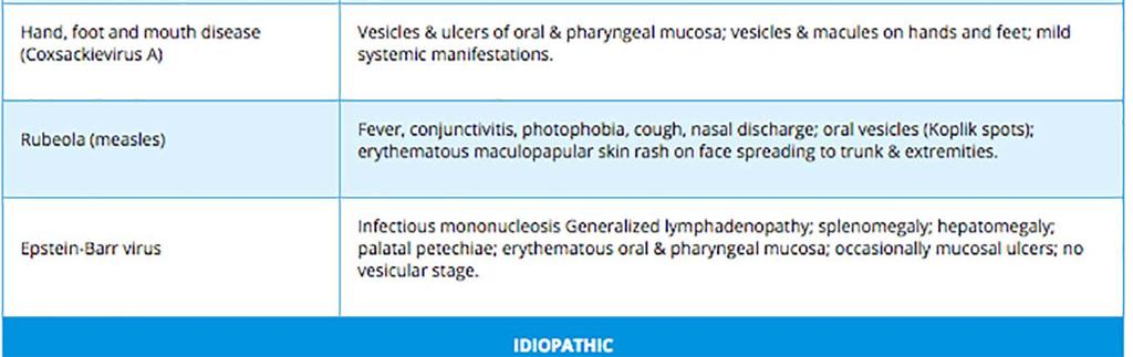
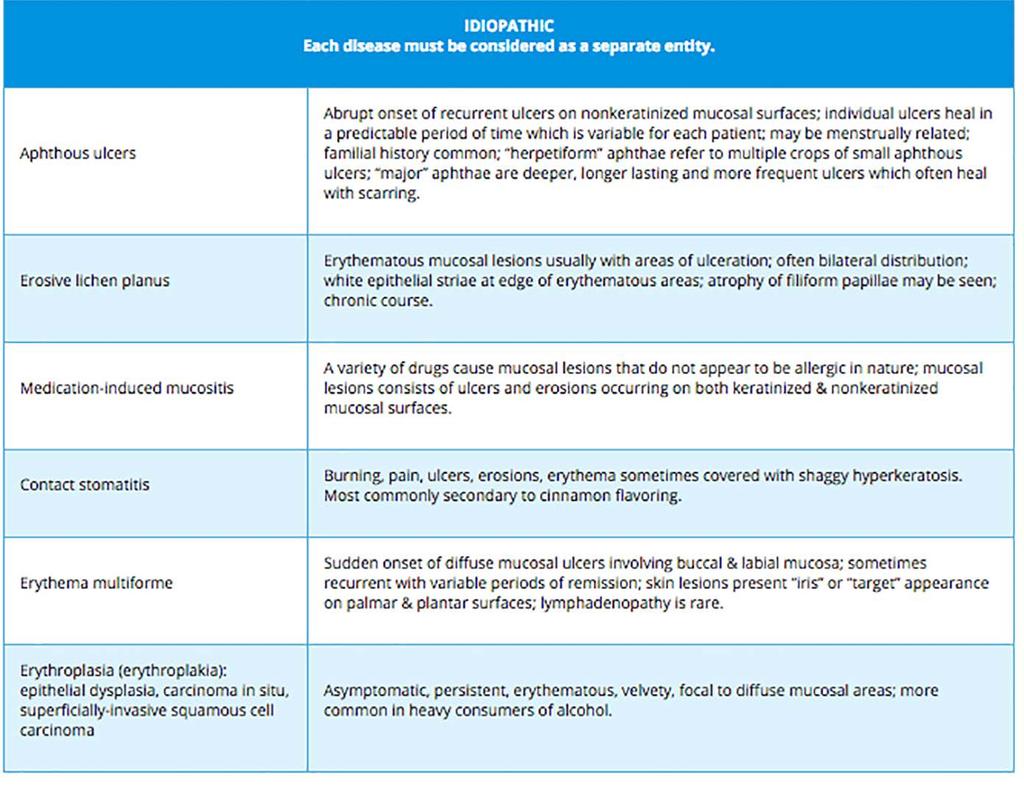
37 Table 3. Vesicular-Ulcerated-Erythematous Surface Lesions of Oral Mucosa. 37


38 Table 4. Soft Tissue Enlargements. Table 5. Benign Epithelial Tumors. 38

39 Table 6. Benign Mesenchymal Tumors. 39


40 Table 7. Benign Salivary Gland Tumors. Table 8. Soft Tissue Cysts. 40
41 Conclusion Successful management of patients with lesions of oral mucosa can be accomplished by dental health care providers if a step-by-step approach is used to gather information and apply it in a systematic manner to diagnostic decision trees. This course provides general guidelines that can be used to eliminate or exclude most oral lesions from the clinical differential diagnosis. Those lesions that cannot be excluded constitute the clinical differential diagnosis. The value of formulating a clinical differential diagnosis for an oral lesion is that it eliminates guessing at the best diagnosis and thus guides the next steps in managing the patient s lesion. Determining the definitive or final diagnosis of an oral lesion often requires additional testing, such as obtaining a biopsy specimen and having it microscopically diagnosed. 41
42 Course Test Preview To receive Continuing Education credit for this course, you must complete the online test. Please go to: 1. A 19-year-old woman has painful ulcers on the labial mucosa and buccal mucosa of 4 days duration. She has had similar ulcers on previous occasions, and each time the lesions healed in approximately 7 days. The best diagnosis is. A. aphthous ulcers B. mucous membrane pemphigoid C. recurrent herpes D. primary herpes E. pemphigus 2. A 67-year-old woman has a chronic sore mouth of 2 years duration. The lesions consist of multiple persistent ulcers adjacent to white rough thickened areas which do not rub off and are arranged in a striated pattern. The lesions are bilateral and involve the buccal mucosa, lateral borders of the tongue, and gingiva. Of the following, the best diagnosis is. A. epidermolysis bullosa B. pemphigus C. herpangina D. lichen planus E. recurrent herpes 3. A patient has multiple, asymptomatic, irregular, flat patches on the dorsum of the tongue. Each patch has a red center and an irregular white periphery. The patient reports that the lesions come and go. The best diagnosis is. A. lichen planus B. geographic tongue (erythema migrans) C. epithelial dysplasia D. pemphigus E. superficially invasive squamous cell carcinoma 4. A 16-year-old woman has a compressible, nontender, 5x5 mm soft tissue enlargement of the lower labial mucosa. The lesion has a blue mucosal surface which does not blanch upon palpation. The patient states that she has been aware of the lesion for 2 months and that it has increased and decreased in size during this time. Of the following, the best diagnosis is. A. peripheral ossifying fibroma B. pleomorphic adenoma (mixed tumor) C. pyogenic granuloma D. hemangioma E. mucocele 5. A 13-year-old female patient has mild fever, lymphadenopathy and vesicles and ulcers of the soft palate and tonsillar pillars bilaterally, of 5 days duration. No other oral lesions are present. The best diagnosis is. A. herpangina B. herpes zoster C. recurrent herpes D. primary herpes E. infectious mononucleosis 42
43 6. A patient has a white, nontender, 5 x 6 mm, soft tissue enlargement on the right soft palate of at least 2 years duration. The surface is rough and resembles a wart. The lesion is pedunculated. The best diagnosis is. A. irritation fibroma B. lipoma C. papilloma D. epulis fissuratum E. mucocele 7. A 44-year-old man has rough, white, nonpainful lesions which do not rub off located bilaterally on the buccal mucosa, floor of the mouth, and hard and soft palates. He states that he has been aware of the lesions since childhood. Of the following, the best diagnosis is. A. white sponge nevus B. lichen planus C. leukoedema D. squamous cell carcinoma 8. A 25-year-old woman has a 5x6 mm, nontender, compressible soft tissue enlargement located on the interdental papilla between teeth #10 and #11. The lesion is erythematous and bleeds easily. She states that she has been aware of the lesion for approximately 3 days. The patient is in her second trimester of pregnancy. Of the following, the best diagnosis is. A. papilloma B. irritation fibroma C. pleomorphic adenoma D. pyogenic granuloma E. hemangioma 9. Which white surface lesion rubs off? A. Epithelial dysplasia B. Subepithelial fibrosis C. Burn D. Fordyce granules E. Hairy leukoplakia 10. Which of the following lesions is asymptomatic and smooth to palpation? A. Burn B. Familial epithelial hyperplasia C. Fibrin clot D. Nicotinic stomatitis E. Scarring 11. Which pigmented surface lesion is consistently flat and not thickened? A. Oral melanotic macule B. Hemangioma C. Kaposi sarcoma D. Melanoma E. Varix 43
44 12. Which one of these clinical features would be most helpful in distinguishing hematoma from nevus? A. Color of the lesion B. Duration of the lesion C. Whether the lesion blanches upon pressure D. Whether the lesion is thickened E. Whether the lesion is painful 13. A patient has a thickened, compressible, blue pigmentation of the buccal mucosa that blanches upon pressure. Which of the following should be included in the clinical differential diagnosis? A. Ecchymosis B. Petechiae C. Lentigo D. Tattoo E. Varix 14. Which of the following diseases initially forms oral vesicles? A. Aphthous ulcers B. Infectious mononucleosis C. Mucous membrane (cicatricial) pemphigoid D. Toxic mucositis 15. Which of the following lesions have/has an abrupt or sudden onset? A. Pemphigus B. Recurrent herpes C. Mucous membrane pemphigoid D. Lichen planus 16. Which disease has the worst prognosis? A. Pemphigus vulgaris B. Mucous membrane pemphigoid C. Bullous pemphigoid D. Discoid lupus erythematosus E. Toxic mucositis 17. A 35-year-old man has painful ulcers on the lips and buccal mucosal and asymptomatic macules and vesicles on the face, hands, and trunk. He has a mild fever but no lymphadenopathy. The lesions had an acute onset 3 days ago. The best diagnosis is. A. pemphigus B. lichen planus C. erythema multiforme D. toxic epidermal necrolysis E. primary herpes 18. Which disease typically begins with the abrupt onset of pain or altered sensation followed by vesicles and ulcers unilaterally in the distribution of a peripheral nerve? A. Herpes zoster B. Herpangina C. Primary herpes D. Carcinoma-in-situ E. Varicella 44
45 19. Which of the following lesions is usually congenital? A. Hemangioma B. Dermoid cyst C. Irritation fibroma D. Pleomorphic adenoma E. Papilloma 20. Which of the following lesions is/are encapsulated? A. Neurofibroma B. Rhabdomyoma C. Schwannoma D. Neuroma E. Granular cell tumor 21. Which of the following lesions is/are compressible to palpation? A. Keratoacanthoma B. Verruca vulgaris C. Granular cell tumor D. Peripheral fibroma E. Low-grade mucoepidermoid carcinoma 45
:63-6. 3. Neville BW. Oral and maxillofacial pathology, 3rd ed. St. Louis, MO. Saunders/Elsevier. 2009. 4. Neville BW, Damm DD, White DH.")

46 References 1. Darling MR, Daley TD. Blistering mucocutaneous diseases of the oral mucosa a review: part 1. Mucous membrane pemphigoid. J Can Dent Assoc Dec;71(11): Darling MR, Daley TD. Blistering mucocutaneous diseases of the oral mucosa a review: part 2. Pemphigus vulgaris. J Can Dent Assoc Feb;72(1): Neville BW. Oral and maxillofacial pathology, 3rd ed. St. Louis, MO. Saunders/Elsevier Neville BW, Damm DD, White DH. Color atlas of clinical oral pathology, 2nd ed. Baltimore, MD. Williams & Wilkins Regezi JA, Sciubba JJ, Jordon RCK. Oral pathology : clinical pathologic correlations, 4th ed. St. Louis, MO. Saunders Eversole LR, Wysocki GP. Contemporary oral and maxillofacial pathology, 2nd ed. St. Louis, MO. Mosby Scully C, Felix DH. Oral Medicine update for the dental practitioner: oral white patches. Br Dent J Nov 12;199(9): doi: /sj.bdj Vincent SD, Lilly GE. Clinical, historic, and therapeutic features of aphthous stomatitis. Literature review and open clinical trial employing steroids. Oral Surg Oral Med Oral Pathol Jul;74(1): Vincent SD, Lilly GE, Baker KA. Clinical, historic, and therapeutic features of cicatricial pemphigoid. A literature review and open therapeutic trial with corticosteroids. Oral Surg Oral Med Oral Pathol Oct;76(4): Woo S, Sonis ST. RRecurrent aphthous ulcers: a review of diagnosis and treatment. J Am Dent Assoc Aug;127(8): Additional Resources Oral Pathology Image Database Interactive Decision Tree for Oral Mucosal Lesions About the Authors Michael W. Finkelstein, DDS, MS The staff at P&G expresses our condolences regarding the loss of Dr. Finkelstein on December 28, We will miss him. Dr. Finkelstein received his DDS degree and MS degree in Oral Pathology from the University of Iowa College of Dentistry. He was a faculty member in the Department of Oral Pathology, Radiology and Medicine at the University of Iowa since Areas of Research: Developed and evaluated patient case studies and other materials for teaching clinical diagnosis and development of clinical problem-solving skills. Histopathologic investigation of proliferative verrucous leukoplakia. Emily Lanzel, DDS, MS Dr. Lanzel received her DDS degree and MS degree in Oral Pathology from the University of Iowa College of Dentistry. She has been a faculty member in the Department of Oral Pathology, Radiology and Medicine at the University of Iowa since emily-lanzel@uiowa.edu 46

47 John W. Hellstein, DDS, MS Dr. John W. Hellstein graduated from the School of Dentistry at University of Missouri at Kansas City in He received an MS in Stomatology and a Certificate in Oral Pathology from the University of Iowa in Since 2002, he has been appointed within the Department of Oral and Maxillofacial Pathology, Medicine and Radiology and is currently Professor of Oral and Maxillofacial Pathology at the University of Iowa. john-hellstein@uiowa.edu 47
Vascular. Extravasated blood. Melanocytic. Tattoo. Epidermolysis bullosa. Lichen planus. Pemphigoid Pemphigus Lupus. Candidosis. Surface Epithelial
 Oral Soft Tissue Pathology Epithelial Thickening (white) Combination Erythema migrans Epithelial atrophy (red) Surface Lesions Clinical Impression Enlargements Surface Debris Pigmented Vesicular Ulcerated
Pigmented lesions of the Oral cavity
 Oral medicine أ.م.د احسان عبد هللا كميل Pigmented lesions of the Oral cavity Pigmented oral lesions are a large group of disorders in which the dark or brown color is the essential clinical characteristic.
LESIONS OF THE ORAL CAVITY ORAL CAVITY. Oral Cavity Subsites 4/10/2013 LIPS TEETH GINGIVA ORAL MUCOUS MEMBRANES PALATE TONGUE ORAL LYMPHOID TISSUES
 LESIONS OF THE ORAL CAVITY David I. Kutler, MD, FACS Associate Professor Division of Head and Neck Surgery Department of Otolaryngology HNS Weill Cornell Medical Center ORAL CAVITY LIPS TEETH GINGIVA ORAL
Contents. 3 Diagnostic Tests and Studies Introduction Examination... 27
 Contents 1 Normal Anatomy... 1 1.1 Introduction... 1 1.2 Surface Landmarks... 1 1.3 Oral Mucosa... 3 1.4 Tongue... 5 1.5 Floor of Mouth... 6 1.6 Palate... 6 1.7 Dentition... 7 1.8 Temporomandibular Joint...
The Oral Cavity. Image source:
 The Oral Cavity Anatomy Image source: http://anatomyforlayla.blogspot.co.za/2007/04/blog-post.html The major structures of the oral cavity are the lips, the teeth, the alveolar ridges (bony areas that
4Ps LUMPS AND BUMPS B.L.&T. BUMPS, LUMPS, AND TATTOOS. Most Common BUMP in the oral cavity Fibroma INTERDENTAL PAPILLAE LESIONS
 B.L.&T. BUMPS, LUMPS, AND TATTOOS LUMPS AND BUMPS DIFFERENTIAL DIAGNOSIS FOR LUMPS AND BUMPS Traumatic Fibroma Papilloma Epulis Fissuratum Inflammatory Papillary Hyperplasia Lesions of Attached Gingiva
Contents. 1 Normal Anatomy Introduction... 17
 Contents 1 Normal Anatomy... 1 Introduction... 1 Surface Landmarks... 1 Oral Mucosa... 1 Tongue... 4 Floor of Mouth... 6 Palate... 7 Dentition... 7 Temporomandibular Joint... 9 Innervation... 10 Jaws and
Index. Dent Clin N Am 49 (2005) Note: Page numbers of article titles are in boldface type.
 Note: Page numbers of article titles are in boldface type.") Dent Clin N Am 49 (2005) 273 278 Index Note: Page numbers of article titles are in boldface type. A Acanthosis nigricans, familial, 251 Amalgam tattoo, 197 198 Amphotericin B, 62 Ankyloglossia, 11 Anti-inflammatory
Dent Clin N Am 49 (2005) 273 278 Index Note: Page numbers of article titles are in boldface type. A Acanthosis nigricans, familial, 251 Amalgam tattoo, 197 198 Amphotericin B, 62 Ankyloglossia, 11 Anti-inflammatory
الطلاوة = Leukoplakia LEUKOPLAKIA
 LEUKOPLAKIA Leukoplakia is a clinical term that refers to a predominantly white lesion of the oral mucosa that cannot be rubbed off or characterized by any other definable lesion or known disease. 130
That. Name QUIZ. 60 SEPTEMBER 2017 // dentaltown.com
 QUIZ Name That General dentists are first in the line of practitioners that patients see for an oral lesion evaluation; therefore, a sound understanding of oral mucosal diseases and their clinical presentation
TANYA A. WRIGHT, DDS OBJECTIVES
 TANYA A. WRIGHT, DDS OBJECTIVES One will be able to recognize pathological entities One will be able to establish a reasonable differential diagnosis One will be able to identify various types of lesions
Oral Health & HIV. Professor Sudeshni Naidoo Department of Community Dentistry University of the Western Cape
 Oral Health & HIV Professor Sudeshni Naidoo Department of Community Dentistry University of the Western Cape Importance & relevance of Oral HIV Lesions >70% of HIV+ve patients present with oral manifestations
Lesions & Lifestyles
 Lesions & Lifestyles attended a 3 hour Continuing Education Seminar on Oral Pathology presented by Nancy Dewhirst, RDH,BS on (date) at (location):. Course material is directly related patient care. Notes:
Diseases of oral cavity
 Diseases of oral cavity Diseases of Teeth and Supporting Structures Inflammatory/Reactive Lesions Infections Oral Manifestations of Systemic Disease Precancerous and Cancerous Lesions Odontogenic Cysts
Oral Medicine. Dr. Qianming Ian CHEN
 Oral Medicine Dr. Qianming Ian CHEN ORAL MEDICINE Oral medicine is the specialty of dentistry that is concerned with the oral health care of medically compromised patients and with the diagnosis and nonsurgical
Differential Diagnosis of Oral Lesions. An Interactive Lecture Using Audience Response Polling. John L. Alonge, MS, DDS
 Differential Diagnosis of Oral Lesions An Interactive Lecture Using Audience Response Polling John L. Alonge, MS, DDS Goals 1. Review the diagnostic process needed to formulate a differential diagnosis
Classification: 1. Infective: 2. Traumatic: 3. Idiopathic: Recurrent Aphthous Stomatitis (RAS) 4. Associated with systemic disease:
 4. Associated with systemic disease:") Classification: 1. Infective: 2. Traumatic: 3. Idiopathic: Recurrent Aphthous Stomatitis (RAS) 4. Associated with systemic disease: Hematological GIT Behcet s HIV 5. Associated with dermatological diseases:
Classification: 1. Infective: 2. Traumatic: 3. Idiopathic: Recurrent Aphthous Stomatitis (RAS) 4. Associated with systemic disease: Hematological GIT Behcet s HIV 5. Associated with dermatological diseases:
Oral Cancer Dr Christine Goodall Consultant Oral Surgeon University of Glasgow Dental School
 Oral Cancer Dr Christine Goodall Consultant Oral Surgeon University of Glasgow Dental School christine.goodall@glasgow.ac.uk Locations Lip, mouth, oropharynx Tongue, floor of mouth, buccal mucosa, palate,
Autoimmune Diseases with Oral Manifestations
 Autoimmune Diseases with Oral Manifestations Martin S. Greenberg DDS, FDS RCSEd Professor Emeritus Department of Oral Medicine University of Pennsylvania Disclosure Statement I have no actual or potential
Stomatitis.
 Stomatitis http://www.entusa.com/oral_photographs/20080102-stomatitis-palate_small.jpg Oral inflammation and ulcers, known as stomatitis, may be mild and localized or severe and widespread. They are invariably
SALIVARY GLAND DISEASES. Omar alnoubani MD,MRCS
 SALIVARY GLAND DISEASES Omar alnoubani MD,MRCS Salivary Glands Overview Parotid gland Sublingual gland Submandibular gland Salivary glands - Types 3 Major Salivary Glands Parotid Submandibular Sublingual
NEOPLASMS OF THE SURFACE EPITHELIUM (KERATINOCYTES)
") NEOPLASMS OF THE SURFACE EPITHELIUM (KERATINOCYTES) Papillary Lesions Precancerous Lesions Keratinocyte Proliferations Carcinomas Melanotic Lesions Melanomas Normal Mucosa Keratin layer Spinous layer Basal
NEOPLASMS OF THE SURFACE EPITHELIUM (KERATINOCYTES) Papillary Lesions Precancerous Lesions Keratinocyte Proliferations Carcinomas Melanotic Lesions Melanomas Normal Mucosa Keratin layer Spinous layer Basal
Dr Rodney Itaki Lecturer Division of Pathology Anatomical Pathology Discipline
 Oral Lesions & Oral Cancer Dr Rodney Itaki Lecturer Division of Pathology Anatomical Pathology Discipline University of Papua New Guinea School of Medicine & Health Sciences Division of Pathology Overview
Shawn A. McClure D.M.D., M.D. Miami Oral & Maxillofacial Surgeon Associate Professor, Director of Research Department of Oral & Maxillofacial Surgery
 Shawn A. McClure D.M.D., M.D. Miami Oral & Maxillofacial Surgeon Associate Professor, Director of Research Department of Oral & Maxillofacial Surgery NOVA/NSU COLLEGE OF DENTAL MEDICINE 1 Diseases of Head
Dermatopathology: The tumor is composed of keratinocytes which show atypia, increase mitoses and abnormal mitoses.
 Squamous cell carcinoma (SCC): A common malignant tumor of keratinocytes arising in the epidermis, usually from a precancerous condition: 1- UV induced actinic keratosis, usually of low grade malignancy.
Benign versus Cancerous Lesions How to tell the difference FMF 2014 Christie Freeman MD, CCFP, DipPDerm, MSc
 1 Benign versus Cancerous Lesions How to tell the difference FMF 2014 Christie Freeman MD, CCFP, DipPDerm, MSc Benign lesions Seborrheic Keratoses: Warty, stuck-on Genetics and birthdays Can start in late
REF: Chap 1 (Pemphigus vulgaris/etiology and
 Chapter 1: Vesiculobullous Diseases Test Bank MULTIPLE CHOICE 1. Intercellular deposits of IgG are consistently found in oral epithelium in which of the following? a. Cicatricial pemphigoid b. Lichen planus
Diseases of the breast (1 of 2)
") Diseases of the breast (1 of 2) Introduction A histology introduction Normal ducts and lobules of the breast are lined by two layers of cells a layer of luminal cells overlying a second layer of myoepithelial
Diseases of the breast (1 of 2) Introduction A histology introduction Normal ducts and lobules of the breast are lined by two layers of cells a layer of luminal cells overlying a second layer of myoepithelial
ANS: C REF: Chap 1 (Pemphigus vulgaris/etiology and pathogenesis), p 11
, p 11") Chapter 1: Vesiculobullous Diseases Test Bank MULTIPLE CHOICE 1. Intercellular deposits of IgG are consistently found in oral epithelium in which of the following? a. Cicatricial pemphigoid b. Lichen planus
Chapter 1: Vesiculobullous Diseases Test Bank MULTIPLE CHOICE 1. Intercellular deposits of IgG are consistently found in oral epithelium in which of the following? a. Cicatricial pemphigoid b. Lichen planus
Role of the Dental Hygienist in Oral Pathology. Role of the Dental Hygienist in Oral Pathology. Cancers of the Oral Cavity.
 Gum Gardeners Study Club April 25, 2016 Early Detection of Oral Cancer Cindy Kleinegger, DDS, MS NW Oral Pathology Tigard, OR nworalpathology.com Role of the Dental Hygienist in Oral Pathology Work closely
Premalignant lesions may expose to a promoting. factor & may be induced to undergo malignant. Carcinoma in situ displays the cytologic features of
 بسم رلاهللا Def. Premalignant lesions may expose to a promoting factor & may be induced to undergo malignant transformation. Carcinoma in situ displays the cytologic features of malignancy without invasion
CAMOSUN COLLEGE School of Health & Human Services Dental Programs. DHYG 321 Oral Science COURSE OUTLINE
 CAMOSUN COLLEGE School of Health & Human Services Dental Programs 2013 COURSE OUTLINE 1. Instructor Information (a) Instructor Lynne Viczko (b) Office hours Posted on office door (c) Location Dental Building
IN THE NAME OF GOD. Dr.kheirandish DDS,MSC Oral and maxillofacial pathology
 IN THE NAME OF GOD Dr.kheirandish DDS,MSC Oral and maxillofacial pathology Dermatologic Diseases Chapter 16 ECTODERMAL DYSPLASIA o Two or more ectodermally derived anatomic structures fail to develop o
Diseases of the vulva
 Diseases of the vulva 1. Bartholin Cyst - Infection of the Bartholin gland produces an acute inflammation within the gland (adenitis) and may result in an abscess. Bartholin duct cysts - Are relatively
Differential Diagnosis of Oral Masses. Palatal Lesions
 Differential Diagnosis of Oral Masses Palatal Lesions Palatal Masses Periapical Abscess Torus Palatinus Mucocele Lymphoid Hyperplasia Adenomatous Hyperplasia Benign Salivary Neoplasms Malignant Salivary
Oral cavity cancer accounts for approximately 3% of all malignancies and is a significant worldwide health problem.
 Oral cavity cancer accounts for approximately 3% of all malignancies and is a significant worldwide health problem. Majority are SCC ( 5-year survival rate only about 50-60% ) Many SCC arrive from premalignant
Benign Oral cavity lesions. Mohammed ALESSA MBBS,FRCSC Assistant Professor Consultant Otolaryngology, Head & Neck Surgery
 Benign Oral cavity lesions Mohammed ALESSA MBBS,FRCSC Assistant Professor Consultant Otolaryngology, Head & Neck Surgery Anatomy Histology Physiology Pathology Clinical cases Introduction The oral cavity
Differential Diagnosis of Oral Ulcerations
 Differential Diagnosis of Oral Ulcerations Dr. Nagamani Narayana Department of Oral Biology University of Nebraska Medical Center College of Dentistry Objectives Differential diagnosis of oral ulcerations
Medical History. Oral Medicine and General Medicine
 Medical History Oral Medicine and General Medicine Gingivitis herpetica acuta NECROTIZÁLÓ SIALOMETAPLASIA SOOR Medical History The life expectancy has recently increased and increasing By dental prevention
Salivary Glands 3/7/2017
 Salivary Glands 3/7/2017 Goals and objectives Focus on the entities unique to H&N Common board type facts Information for your future practice Salivary Glands Salivary Glands Major gland. Paratid. Submandibular.
Dental Care and Health An Update. Dr. Ranjini Pillai, DDS, MPH, FAGD, FICOI
 Dental Care and Health An Update Dr. Ranjini Pillai, DDS, MPH, FAGD, FICOI WHO s Definition of Health? Health is a state of complete physical, mental, and social wellbeing and not merely the absence of
Differential Diagnosis of Oral Masses. Gingival Lesions
 Differential Diagnosis of Oral Masses Gingival Lesions Gingival/Alveolar Ridge Masses Parulis Periodontal Abscess Tori and Exostoses Reactive Proliferations Peripheral Odontogenic Cysts Peripheral Odontogenic
04/09/2018. Squamous Cell Neoplasia and Precursor Lesions. Agenda. Squamous Dysplasia. Squamo-proliferative lesions. Architectural features
 Squamous Cell Neoplasia and Precursor Lesions Jennifer L. Hunt, MD, MEd Aubrey J. Hough Jr, MD, Endowed Professor of Pathology Chair of Pathology and Laboratory Medicine University of Arkansas for Medical
Benign and malignant epithelial lesions: Seborrheic keratosis: A common benign pigmented epidermal tumor occur in middle-aged or older persons more
 Benign and malignant epithelial lesions: Seborrheic keratosis: A common benign pigmented epidermal tumor occur in middle-aged or older persons more common on the trunk; but extremities, head and neck are
Periocular Malignancies
 Periocular Malignancies Andrew Gurwood, O.D., F.A.A.O., Dipl. Marc Myers, O.D., F.A.A.O. Drs. Myers and Gurwood have no financial interests to disclose. Course Description Discussion of the most common
Oral Cancer and Common Oral Lesions seen in HIV Seropositive Patients. Gwen Cohen Brown DDS, FAAOMP Professor New York City College of Technology
 Oral Cancer and Common Oral Lesions seen in HIV Seropositive Patients Gwen Cohen Brown DDS, FAAOMP Professor New York City College of Technology Program Objectives Recognize the oral health needs of the
BLOCK 12 Viruses of the ENT
 BLOCK 12 Viruses of the ENT Acute infections Introduction Pharyngitis, Common cold, Sinusitis, Otitis media Recurrent infections Herpes zoster oticus Chronic infections HIV and ENT manifestations Neoplasms
IT S FUNDAMENTAL MY DEAR WATSON! A SHERLOCKIAN APPROACH TO DERMATOLOGY
 IT S FUNDAMENTAL MY DEAR WATSON! A SHERLOCKIAN APPROACH TO DERMATOLOGY Skin, Bones, and other Private Parts Symposium Dermatology Lectures by Debra Shelby, PhD, DNP, FNP-BC, FADNP, FAANP Debra Shelby,
Skin and Body Membranes
 4 Skin and Body Membranes PowerPoint Lecture Slide Presentation by Jerry L. Cook, Sam Houston University ESSENTIALS OF HUMAN ANATOMY & PHYSIOLOGY EIGHTH EDITION ELAINE N. MARIEB Skin and Body Membranes
Oral Tumors in Dogs Gingival Enlargement
 Oral Tumors in Dogs Is that lump you re seeing in your dog s mouth normal? Or is it something to be concerned about? The easiest way to know for sure is to have it evaluated by a veterinarian. When you
Malignant Melanoma Early Stage. A guide for patients
 This melanoma patient brochure is designed to help educate melanoma patients and their caregivers. It was developed under the guidance of Dr. Michael Smylie, Professor, Department of Oncology, University
DISORDERS OF THE SALIVARY GLANDS Neoplasms Dr.M.Baskaran Selvapathy S IV
 DISORDERS OF THE SALIVARY GLANDS Neoplasms Dr.M.Baskaran Selvapathy S IV NEOPLASMS A) Epithelial I. Benign Pleomorphic adenoma( Mixed tumour) Adenolymphoma (Warthin s tumour) Oxyphil adenoma (Oncocytoma)
My Journey into the World of Salivary Gland Sebaceous Neoplasms
 My Journey into the World of Salivary Gland Sebaceous Neoplasms Douglas R. Gnepp Warren Alpert Medical School at Brown University Rhode Island Hospital Pathology Department Providence RI Asked to present
Clinical characteristics
 Skin Cancer Fernando Vega, MD Seattle Healing Arts Clinical characteristics Precancerous lesions Common skin cancers ACTINIC KERATOSIS Precancerous skin lesions Actinic keratoses Dysplastic melanocytic
LEUKOPLAKIA Definition Epidemiology Clinical presentation
 LEUKOPLAKIA Definition Leukoplakia is the most common premalignant or "potentially malignant" lesion of the oral mucosa. Leukoplakia is a predominantly white lesion of the oral mucosa than cannot be clinicopathologically
MANSOURA UNIVERSITY FACULTY OF DENTISTRY ORAL PATHOLOGY DEPT
 MANSOURA UNIVERSITY FACULTY OF DENTISTRY ORAL PATHOLOGY DEPT THIRD YEAR Course Director: Dr. Nadia M. Lotfy Professor of Oral Pathology Dr. Manal Mohamed Zyada Associate Professor of Oral Pathology Oral
Dysplasia, Mimics and Other Controversies
 Dysplasia, Mimics and Other Controversies Mary S. Richardson, MD Dept. of Pathology Medical University of South Carolina Charleston, SC Notice of Faculty Disclosure In accordance with ACGME guidelines,
Oral Cancer FAQs. What is oral cancer? How many people are diagnosed with oral cancer each year?
 Oral Cancer FAQs What is oral cancer? Oral cancer or oral cavity cancer, is cancer that starts in the mouth. Areas affected by this type of cancer are the lips, the inside lining of the lips and cheeks
CHEILITIS GRANULOMATOSA
 CHEILITIS GRANULOMATOSA Report of two Cases with Clinical and Diagnostic Implications Presented By Dr. Amar Sholapurkar Under the guidance of Dr.Ausaf Ahsan Department of Oral Medicine & Radiology, MCODS,
Histopathology: skin pathology
 Histopathology: skin pathology These presentations are to help you identify, and to test yourself on identifying, basic histopathological features. They do not contain the additional factual information
WOMEN'S INTERAGENCY HIV STUDY ORAL PROTOCOL FORM OP 4: ORAL MUCOSAL TISSUE EXAM
 WOMEN'S INTERAGENCY HIV STUDY ORAL PROTOCOL FORM OP 4: ORAL MUCOSAL TISSUE EXAM COMPLETING THE FORM GENERAL INFORMATION Affix the Participant ID label in the space indicated. Record the visit number. Be
 2015 course one self-study course The Ohio State University College of Dentistry is a recognized provider for ADA CERP credit. ADA CERP is a service of the American Dental Association to assist dental
SESSION 1: GENERAL (BASIC) PATHOLOGY CONCEPTS Thursday, October 16, :30am - 11:30am FACULTY COPY
 PATHOLOGY CONCEPTS Thursday, October 16, :30am - 11:30am FACULTY COPY") SESSION 1: GENERAL (BASIC) PATHOLOGY CONCEPTS Thursday, October 16, 2008 9:30am - 11:30am FACULTY COPY GOAL: Describe the basic morphologic (structural) changes which occur in various pathologic conditions.
SESSION 1: GENERAL (BASIC) PATHOLOGY CONCEPTS Thursday, October 16, 2008 9:30am - 11:30am FACULTY COPY GOAL: Describe the basic morphologic (structural) changes which occur in various pathologic conditions.
LENTIGO SIMPLEX. Epidemiology
 LENTIGO SIMPLEX Epidemiology The frequency of lentigo simplex in children and adults has not been determined. There does not appear to be a racial or gender predilection. Lentigo simplex is the most common
Pigmented Oral Lesions: Clues to Identifying the Potentially Malignant. lesions may mimic melanoma.
 ABSTRACT: Oral nevi, melanotic macules on the lip, atypical amalgam tattoos, melanoacanthomas, and lesions caused by embedded foreign bodies or by perforating injuries of the oral mucosa all require biopsy,
المركب النموذج--- سبيتز وحمة = Type Spitz's Nevus, Compound SPITZ NEVUS 1 / 7
 SPITZ NEVUS 1 / 7 Epidemiology An annual incidence rate of 1.4 cases of Spitz nevus per 100,000 individuals has been estimated in Australia, compared with 25.4 per 100,000 individuals for cutaneous melanoma
Skin and Body Membranes Body Membranes Function of body membranes Cover body surfaces Line body cavities Form protective sheets around organs
 Skin and Body Membranes Body Membranes Function of body membranes Cover body surfaces Line body cavities Form protective sheets around organs Classification of Body Membranes Epithelial membranes Cutaneous
WHITE LESIONS OF THE ORAL CAVITY - diagnostic appraisal & management strategies
 WHITE LESIONS OF THE ORAL CAVITY - diagnostic appraisal & management strategies * Joshy V.R ** Hari.S * Reader, Dept of Oral Pathology, Yenepoya Dental College, Yenepoya University, Mangalore 575 018.
RECORD or PRINT THE CONFIRMATION ID This unique ID is displayed upon successful submission of your answer form.
 2014 course two self-study course The Ohio State University College of Dentistry is a recognized provider for ADA, CERP, and AGD Fellowship, Mastership and Maintenance credit. ADA CERP is a service of
Allergic contact stomatitis is a rare disorder,
 Allergic Contact Stomatitis: A Case Report and Review of Literature P Lokesh, T Rooban, Joshua Elizabeth, K Umadevi, K Ranganathan Abstract Allergic contact stomatitis is a well-recognized entity, which
Carcinoma of Unknown Primary site (CUP) in HEAD & NECK SURGERY
 in HEAD & NECK SURGERY") Carcinoma of Unknown Primary site (CUP) in HEAD & NECK SURGERY SEARCHING FOR THE PRIMARY? P r o f J P P r e t o r i u s H e a d : C l i n i c a l U n i t C r i t i c a l C a r e U n i v e r s i t y O f
Carcinoma of Unknown Primary site (CUP) in HEAD & NECK SURGERY SEARCHING FOR THE PRIMARY? P r o f J P P r e t o r i u s H e a d : C l i n i c a l U n i t C r i t i c a l C a r e U n i v e r s i t y O f
Diagnostic Detectives: Catching common culprits and spotting a mimic in Oral Medicine, Oral Radiology and Oral Pathology. Outline
 Diagnostic Detectives: Catching common culprits and spotting a mimic in Oral Medicine, Oral Radiology and Oral Pathology Who is your detective alter ego? Dr. Kristina Perschbacher Oral Pathology and Oral
Kings College London Dental Institute. Guy s & St Thomas NHS Foundation Trust Oral Medicine Unit. Disease Activity Scoring sheets
 Kings College London Dental Institute Guy s & St Thomas NHS Foundation Trust ral Medicine Unit Disease Activity Scoring sheets Clinical scoring systems for oral mucosal Diseases The routine clinical management
2018 Oregon Dental Conference Course Handout Denis Lynch, DDS, PhD
 2018 Oregon Dental Conference Course Handout Denis Lynch, DDS, PhD Course 9148: Diagnosis and Treatment of Recurrent Oral Ulcers Friday, April 6 9 am - 12 pm Diagnosis and Treatment of Recurrent Oral Ulcers
Diagnostic aids of oral cancer
 Diagnostic aids of oral cancer The World Health Organization has clearly indentified prevention and early detection as major objectives in the control of the oral cancer. At the present time, screening
Index. oralmaxsurgery.theclinics.com. Note: Page numbers of article titles are in boldface type.
 Index Note: Page numbers of article titles are in boldface type. A Adenomatoid odontogenic tumor, pediatric, 50 51 Ameloblastic carcinoma, pediatric, 17, 49 Ameloblastic fibro-odontoma, pediatric, 54 Ameloblastic
Atlas of Eyelid and Conjunctival Tumors
 Atlas of Eyelid and Conjunctival Tumors Jerry A. Shields, M.D. Director, Ocular Oncology Service Wills Eye Hospital Professor of Ophthalmology Thomas Jefferson University Philadelphia, Pennsylvania Carol
INFLAMMATORY DISEASES PART I. Immunopathology Part I
 INFLAMMATORY DISEASES PART I Immunopathology Part I Nonspecific & T Cell Mediated Mucosal Inflammatory Lesions Nonspecific and Idiopathic Mucositis Hypersensitivity and Autoimmune T cell mediated Immunoglobulin
Problem diagnoses. Current issues in Anatomic pathology. Problem Diagnoses in Tumors of the Oral Cavity 5/29/2009
 Current issues in Anatomic pathology Problem Diagnoses in Tumors of the Oral Cavity Richard Jordan DDS PhD FRCPath Professor of Oral Pathology & Pathology Director, UCSF Oral Pathology Diagnostic Laboratory
A Rare case of Tubercular Gingivitis Case Report
 Case Report A Rare case of Tubercular Gingivitis Case Report *Dr. Ansh Chugh 1, Dr. Firoz A Hakkim 2, Dr. Rajesh. V 3, Dr. Raghava Sharma 4 1: JUNIOR RESIDENT IN GENERAL MEDICINE 2: SENIOR RESIDENT IN
IN THE NAME OF GOD Dr. Kheirandish Oral and maxillofacial pathology
 IN THE NAME OF GOD Dr. Kheirandish Oral and maxillofacial pathology ORAL FOCAL MUCINOSIS Uncommon Tumorlike Cutaneous myxoid cyst Overproduction of hyaluronic acid by firoblasts Young adults Female Gingiva
ORAL MELANOMA Definition Epidemiology Clinical Presentation
 ORAL MELANOMA Definition Melanoma is a highly malignant neoplasia, arising from melanocytes, the cells that produce the brownish pigment melanin. Melanin is the determinant in skin colour and protects
WHITE LESIONS OF THE UPPER AIRWAY
 WHITE LESIONS OF THE UPPER AIRWAY WHITE LESION CONFIGURATIONS Solitary vrs Multifocal Flat Plaque Verrucous/rippled Lacey White with red component Papular (curdled milk plaques) Pseudomembranous PLAQUES
Chapter 2 Variants of Normal and Common Benign Conditions
 Chapter 2 Variants of Normal and Common Benign Conditions Summary Fundamental to diagnosing oral pathologic conditions is the ability to recognize the spectrum of clinical findings that represents variation
ORAL MELANOTIC NEVI: A CASE REPORT AND REVIEW OF LITERATURE
 ORAL MELANOTIC NEVI: A CASE REPORT AND REVIEW OF LITERATURE Abstract: *V.T Beena **Isha Chauhan *** R. Heera ***R. Rajeev Oral melanotic nevi are uncommon oral lesions causing focal. Melanotic nevi are
General information about skin cancer
 Skin Cancer General information about skin cancer Key points Skin cancer is a disease in which malignant (cancer) cells form in the tissues of the skin. There are different types of cancer that start in
Pathology of the skin. 2nd Department of Pathology, Semmelweis University
 Pathology of the skin 2nd Department of Pathology, Semmelweis University Histology of the skin Epidermis: Stratum corneum Stratum granulosum Stratum spinosum Stratum basale Dermis: papillary and reticular
Integumentary System
 Integumentary System The integumentary system is commonly known as the Skin Largest organ of human body 10% total body weight and would cover over 20 square feet Functions of Skin 1. Protection Barrier
MUCOCUTANEOUS LESIONS Normal structures in epithelium cell adhesion to each other and to underlying connective tissue:
 ORAL DERMATOSES AND MUCOSAL/GINGIVAL LESIONS MUCOCUTANEOUS LESIONS Normal structures in epithelium cell adhesion to each other and to underlying connective tissue: Diagram taken from: Oral and Maxillofacial
Oral Medicine FULL Referral Guide (FRG)
") Oral Medicine FULL Referral Guide (FRG) Yorkshire & the Humber February 2017 Version 1 Contents Introduction... 1 Oral Medicine Referral Decision Process 1 Step 1. Oral Medicine Condition?... 2 A. Soft
Oral Medicine FULL Referral Guide (FRG) Yorkshire & the Humber February 2017 Version 1 Contents Introduction... 1 Oral Medicine Referral Decision Process 1 Step 1. Oral Medicine Condition?... 2 A. Soft
Diagnostic sieve. Looking Beyond the Vermillion Border. Time bombs for medical GPs! Normal oral mucosa
 Sat 12 June 2010 Millennium WS 28 + 38 2.00-2.55; 3.05-4.00 PM Looking Beyond the Vermillion Border Laurence J. Walsh BDSc, PhD, DDSc, FFOP(RCPA), GCEd, FICD, FPFA, FADI, FIADFE The University of Queensland
ACCURATE DIAGNOSIS IS THE ONLY TRUE CORNERSTONE ON WHICH RATIONAL TREATMENT CAN BE BUILT. C Noyek
 ACCURATE DIAGNOSIS IS THE ONLY TRUE CORNERSTONE ON WHICH RATIONAL TREATMENT CAN BE BUILT. C Noyek Oral diagnostics Definition of the discipline That area of dentistry, the which deals with gathering, recording
Skin and Body Membranes
 Essentials of Human Anatomy & Physiology Elaine N. Marieb Seventh Edition Chapter 4 Skin and Body Membranes Slides 4.1 4.32 Lecture Slides in PowerPoint by Jerry L. Cook Skin and Body Membranes Function
Oral Cancer- Improving Early Detection
 Oral Cancer- Improving Early Detection GDC Recommended Subject Aims: To give an overview of the dental team's role in detecting the early signs of oral cancer; to give an overview of the risk factors associated
DISORDERS OF THE BREAST Dated. FIBROADENOSIS Other common names: mastitis, fibrocystic disease, cystic mammary dysplasia.
 DISORDERS OF THE BREAST Dated BENIGN BREAST DISORDERS (Essential Surg 2 nd Ed, pp 540) FIBROADENOSIS Other common names: mastitis, fibrocystic disease, cystic mammary dysplasia. Fibroadenosis is the distortion
Skin lesions The Good and the Bad. Dr Virginia Hubbard Ipswich Hospital NHS Trust Barts and the London School of Medicine and Dentistry
 Skin lesions The Good and the Bad Dr Virginia Hubbard Ipswich Hospital NHS Trust Barts and the London School of Medicine and Dentistry Case 1 32 year old woman Australian Lesion on back New hair growing
THE TONGUE. In Clinical Diagnosis
 THE TONGUE In Clinical Diagnosis A Colour Atlas of The Tongue in Clinical Diagnosis D.W. BEAVEN S.E. BROOKS BATES Guide to Physical Examination Lynn s. Bickley רויטל רחימי, תום כספי פנימית ב', תל-השומר
VIRUS. Viral infection causing, or associated with diseases of the oral mucosa : Herpes Simpleks 1 & 2
 VIRUS Viral infection causing, or associated with diseases of the oral mucosa : VIRUS Herpes Simpleks 1 & 2 Varicella - Zoster Coxsakie A PENYAKIT Primary Gingivostomatitis Herpetica Herpes Labialis Recurrent
Oral Medicine Update for the dental practitioner Oral white patches
 IN BRIEF Most white lesions in the mouth are inconsequential and caused by friction or trauma. However, cancer and some systemic diseases such as lichen planus and candidosis may present in this way. Biopsy