Simulators of melanoma
|
|
|
- Hester Helena Holland
- 5 years ago
- Views:
Transcription

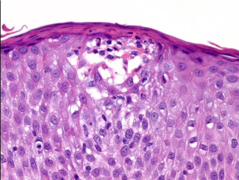
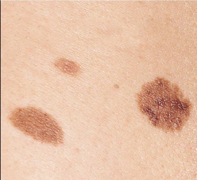
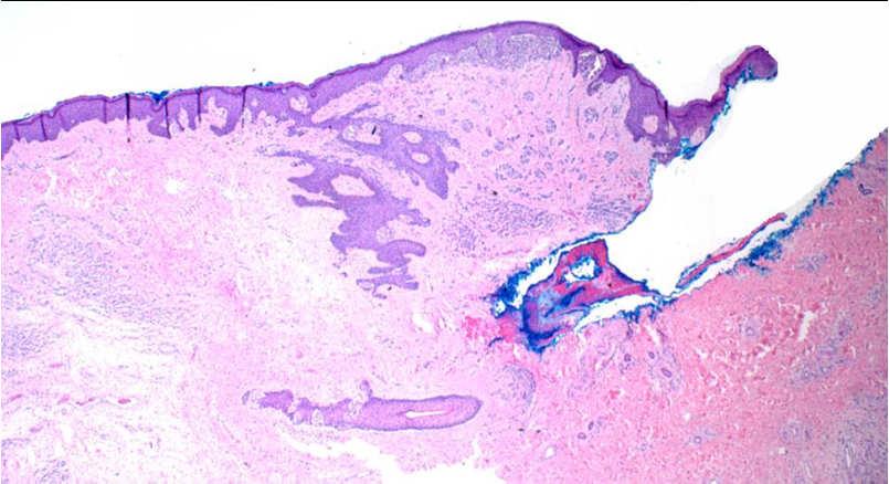
1 Simulators of melanoma Philip E. LeBoit, M.D. Depts. of Pathology and Dermatology University of California, San Francisco Simulators of melanoma Simulators of melanoma in situ Melanocytic Non-melanocytic Simulators of invasive melanoma Melanocytic Non-melanocytic Simple lentigo with suprabasilar scatter 1
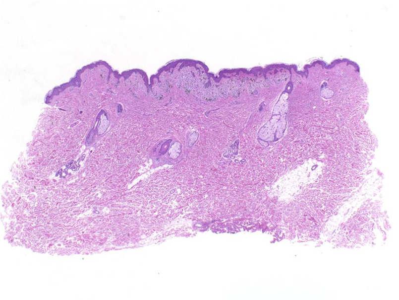
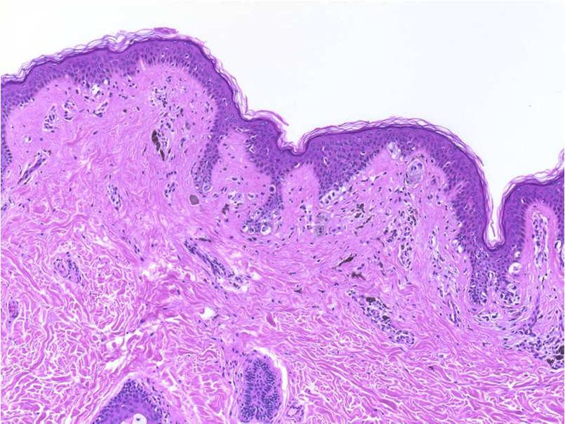
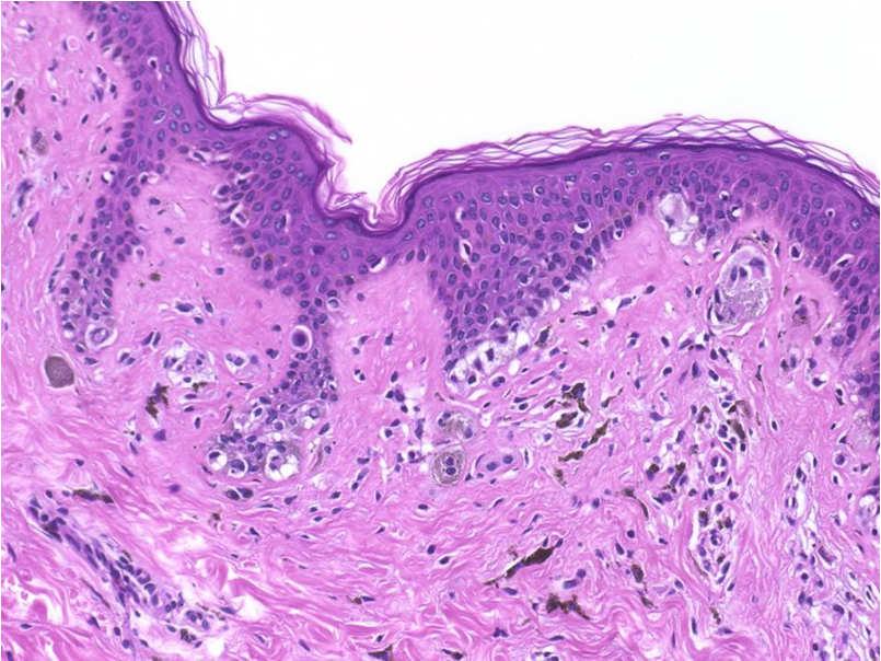
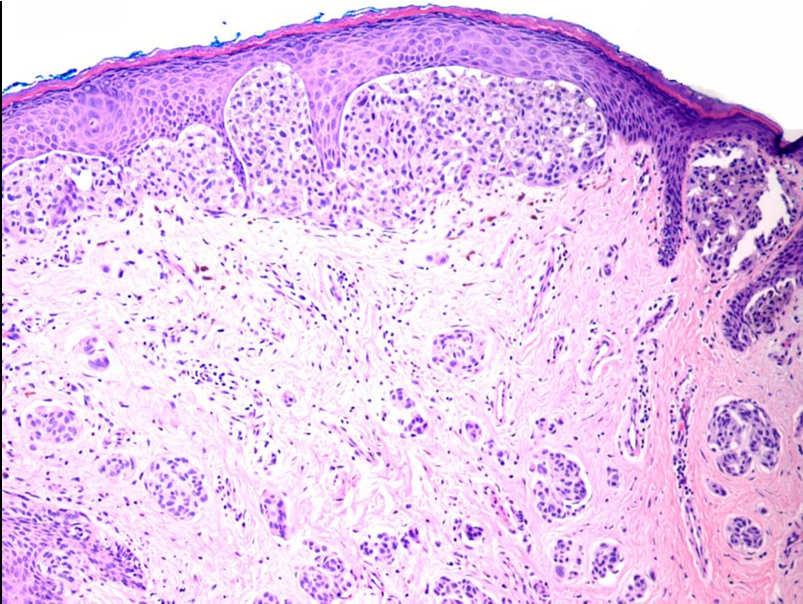
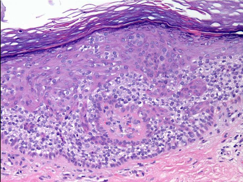
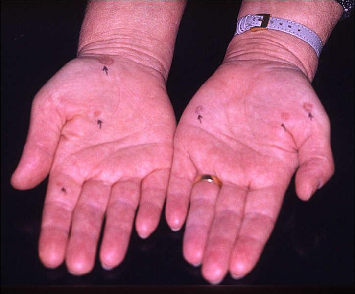
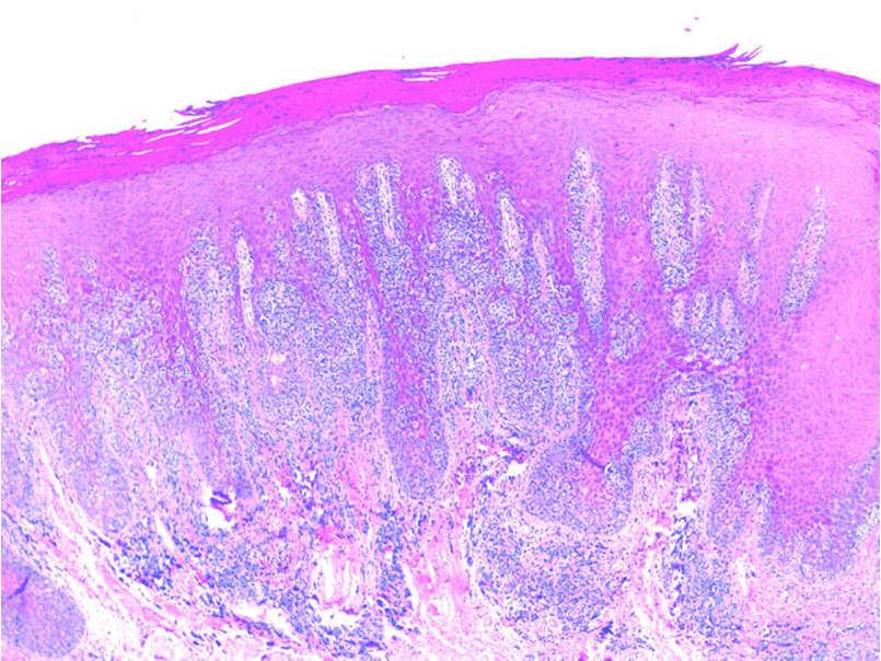
2 Melanocytic nevi on acral skin 2

3 What makes a site special? Nevi on the scalp of children Everone is special -Barney Everyone is different. Few people are special. - Jonah M. Peterschild,
 Back (two patterns) Flexural Ear Conjunctiva Spitz acral")
4 Nevi on special sites Acral-volar Nail matrix/bed Genitalia Scalp (children) Back (two patterns) Flexural Ear Conjunctiva Spitz acral Spitz ear 4
5 5
6 6


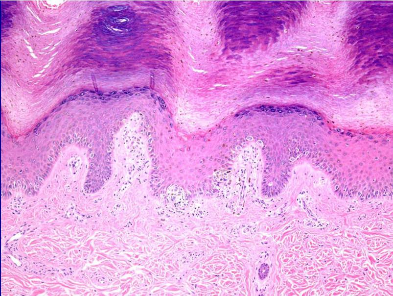
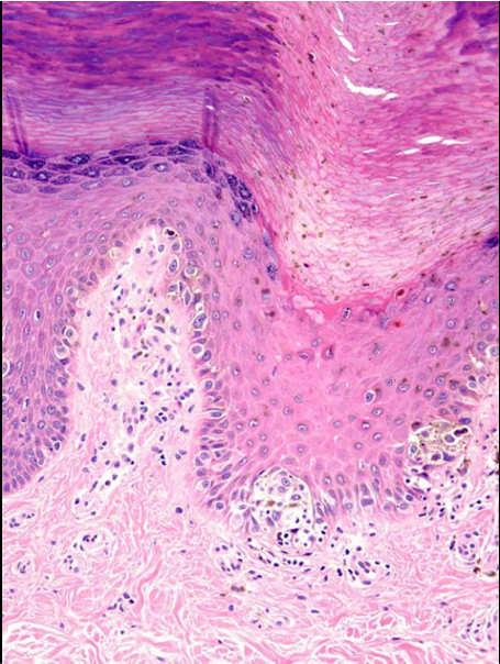
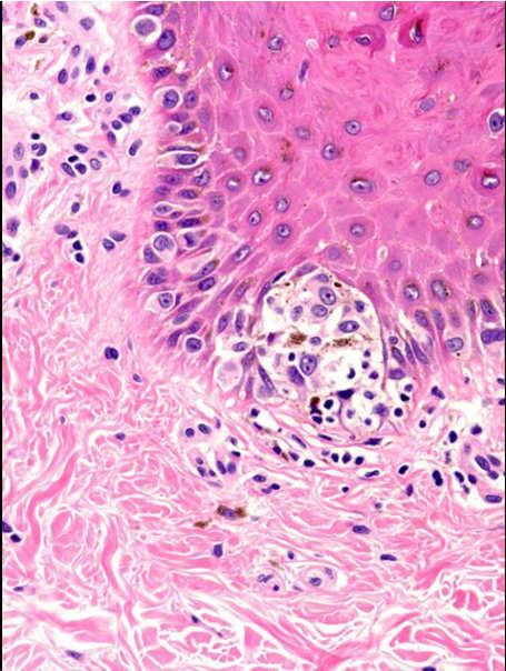
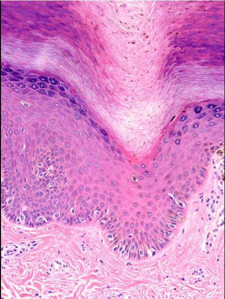
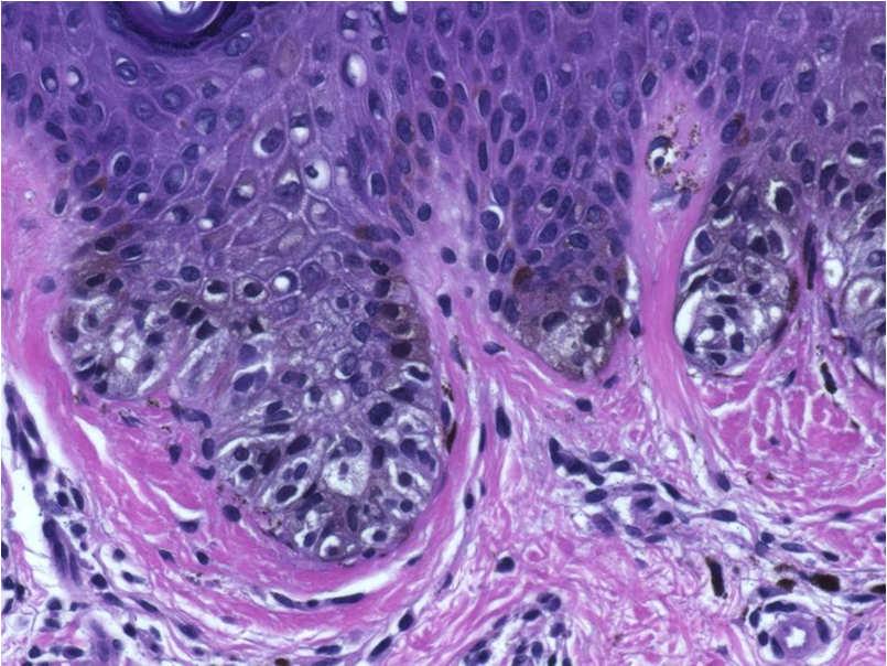
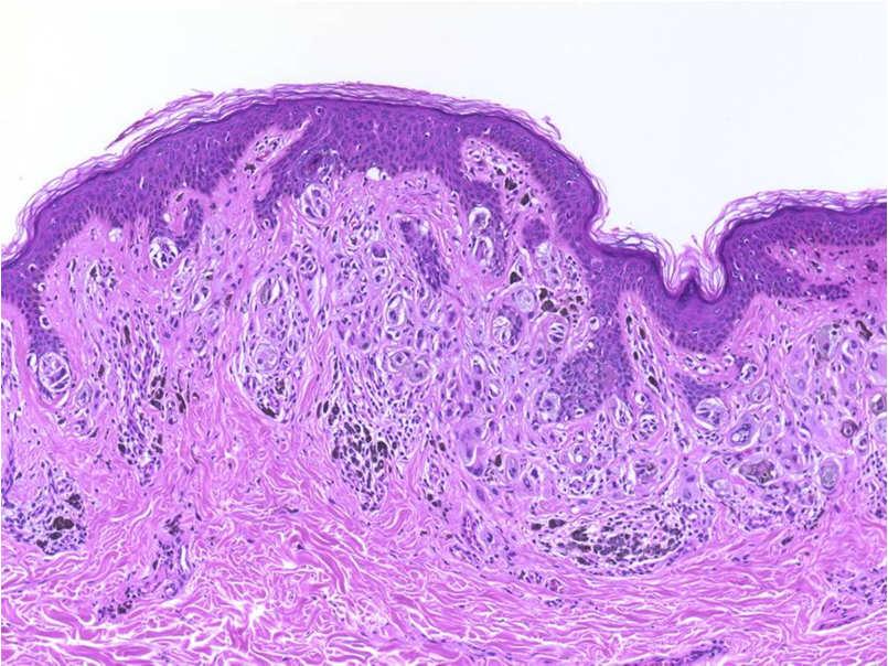
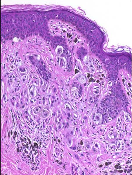
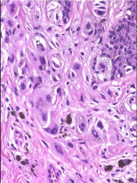
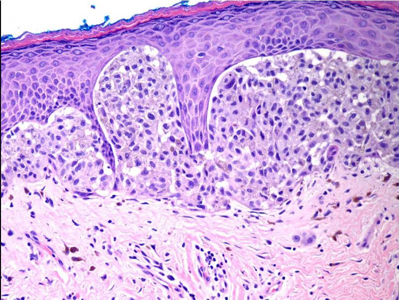
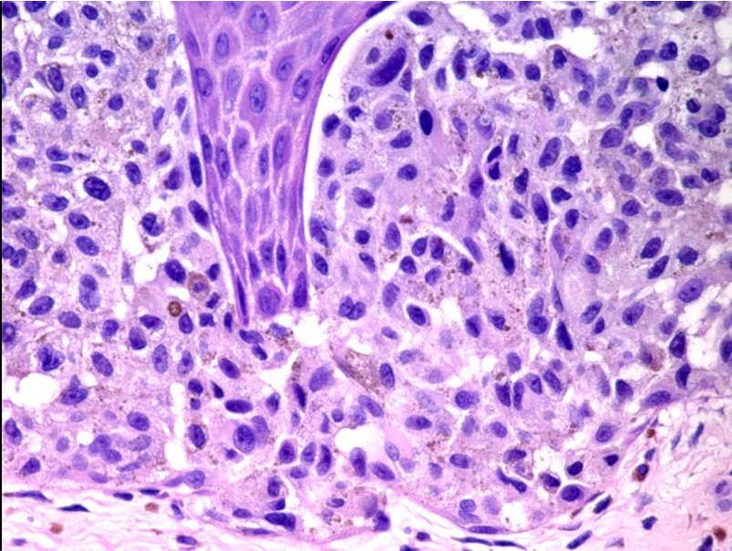
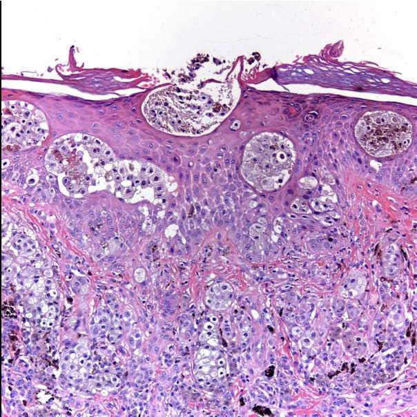
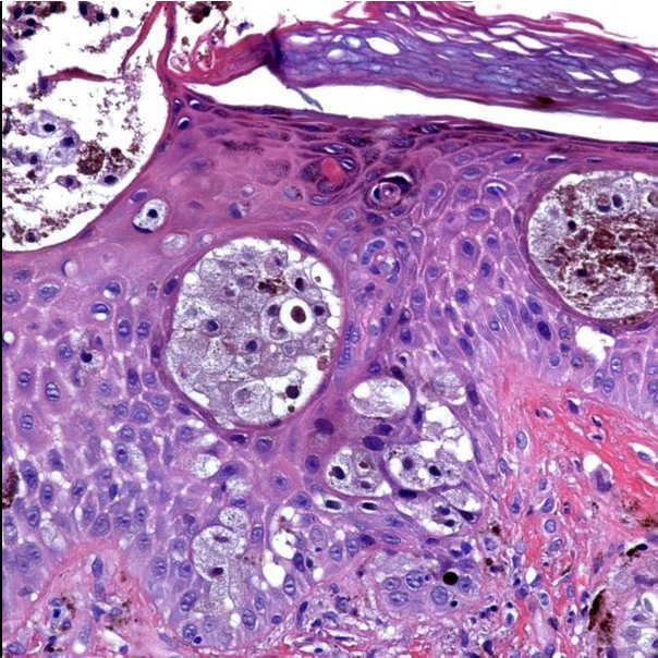
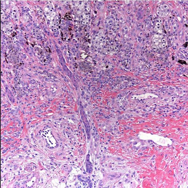
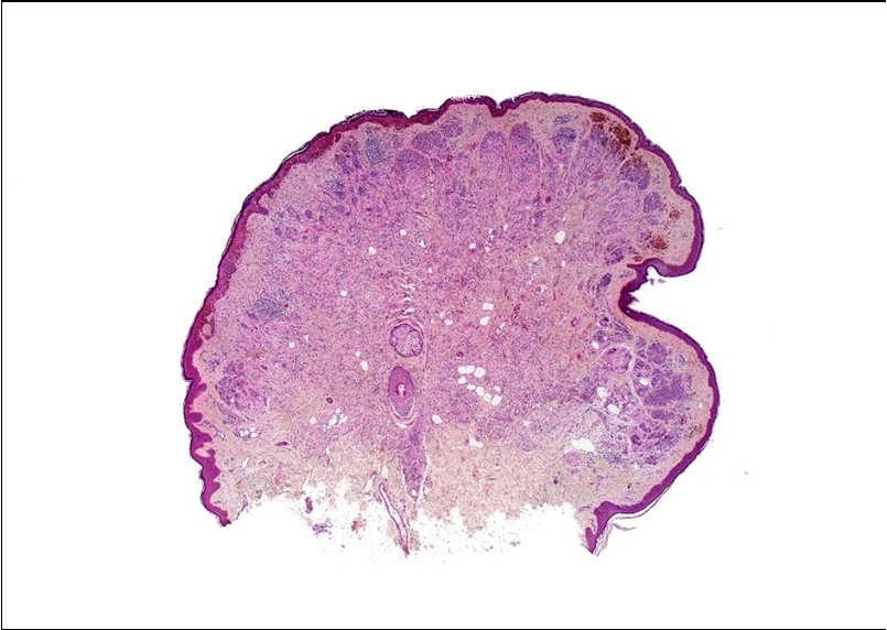
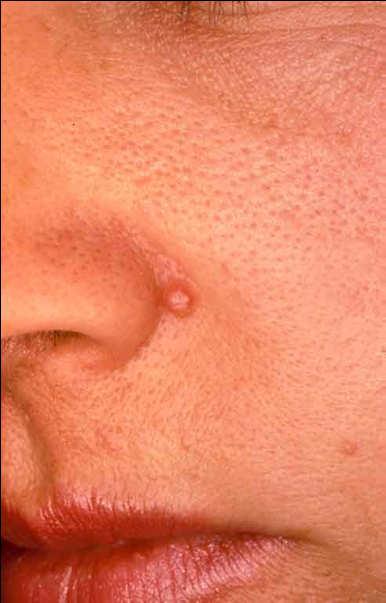
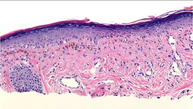
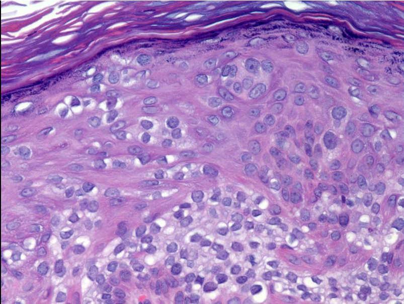
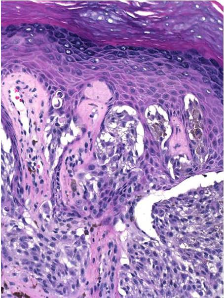
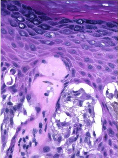
7 Melanocytic Acral Nevus with Intraepidermal Ascent of Cells 7
8 8


9 The American Journal of Surgical Pathology 27(3): ; Mar
10 10
11 11
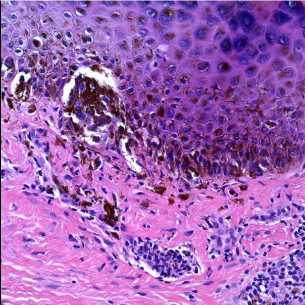
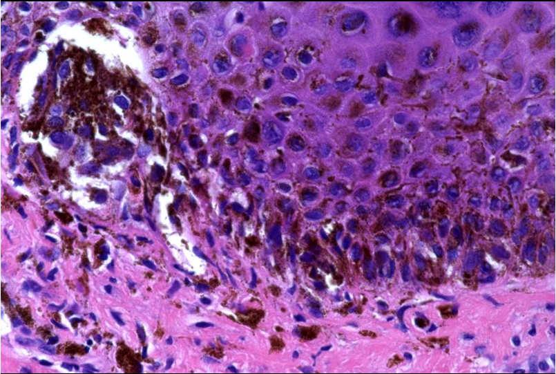
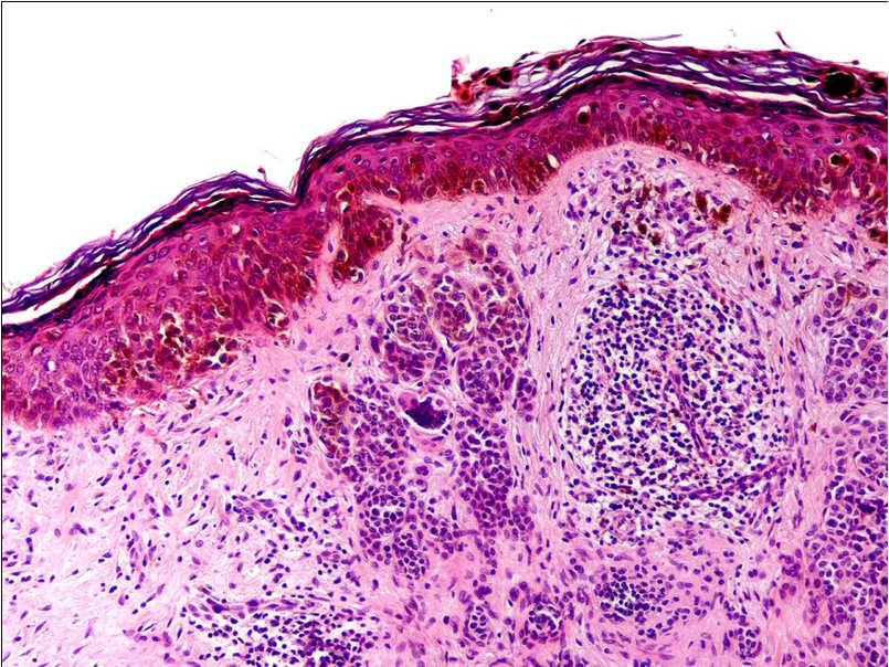
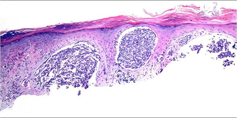
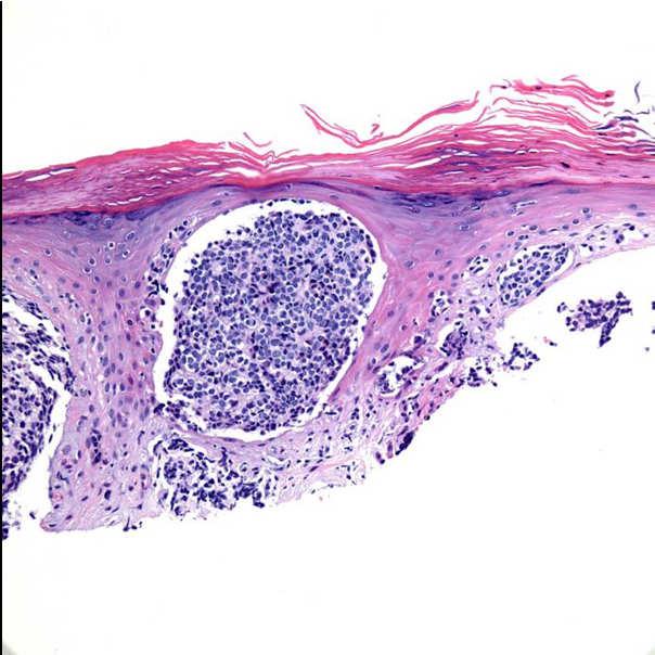
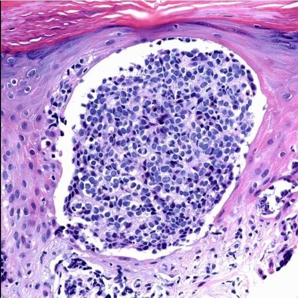
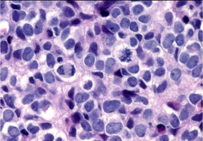
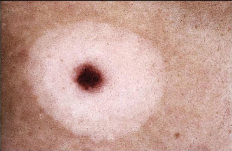
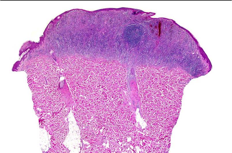
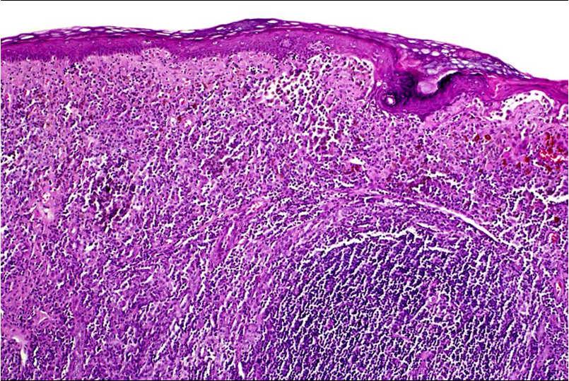
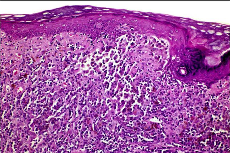
12 Inflamed melanocytic nevi 12


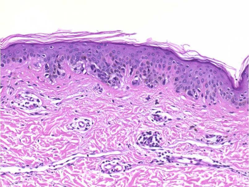
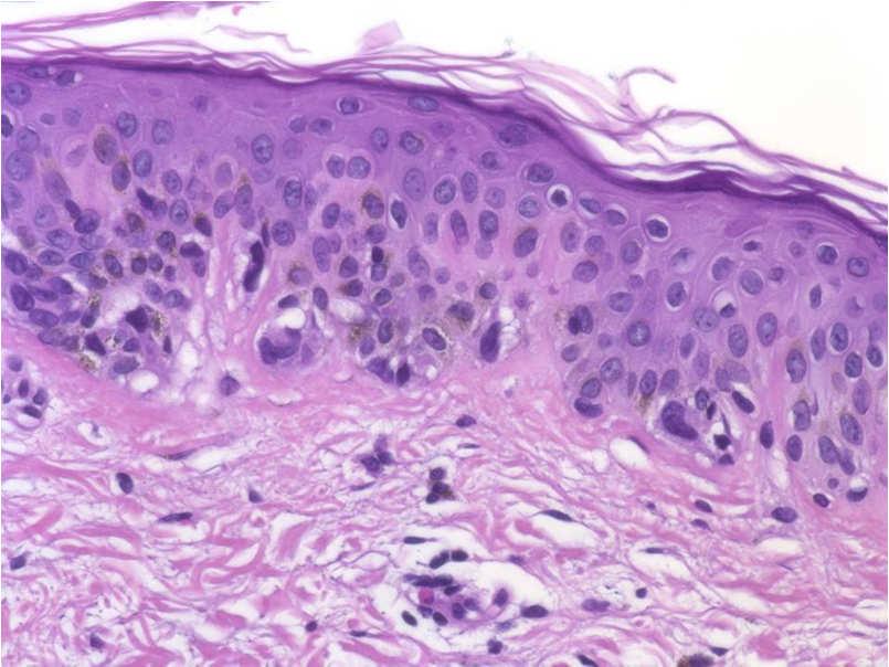
13 Inflamed melanocytic nevi Rapid epidermopoiesis can lead to upward scatter of single melanocytes Inflammation can result in increased pigment synthesis (increased pigmentation, more cytoplasm) No proof that inflammation can cause nuclear atypia, mitotic activity 13
14 14


15 A peculiar papulosquamous eruption involving pigmented nevi LB Meyerson, Arch Dermatol 1971, 103:510-2 Two patients with multiple pruritic papulosquamous lesions Scaly and red aspects resolved with topical steroid treatment Pigmented nevi persisted Speculated about koebnerization of pityriaisis rosea as cause 15
16 16

 Halo eczema surrounding seborrheic keratoses (Australas.")
17 Meyerson s nevus Subsequent contributions Co-existence of Meyerson s and Sutton s nevus after sunburn (Dermatol. 1994;189:269-70) Halo eczema- resolution after excision of central lesion alone (Clin Exp Dermatol 1991; 16:66-7) Halo eczema surrounding seborrheic keratoses (Australas. J Dermatol. 1990; 30: 73-6 Nevus with lichen sclerosus 17
18 18
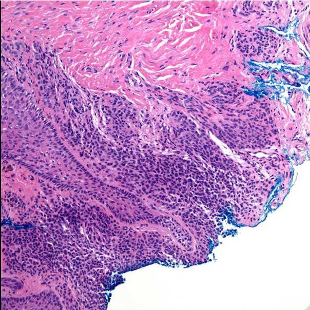
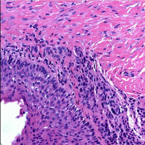
19 19


20 What s ailing this nevus? 20
21 21
22 Dysplastic nevi 22
23 23
24 24
25 25

26 Superficial Spitz nevus 26
27 27

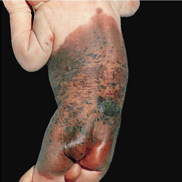
28 Inflamed or traumatized junctional Reed nevus 28
29 29
30 30
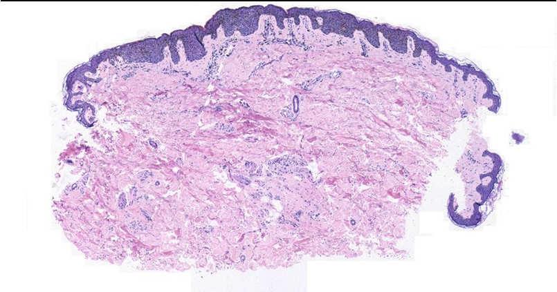
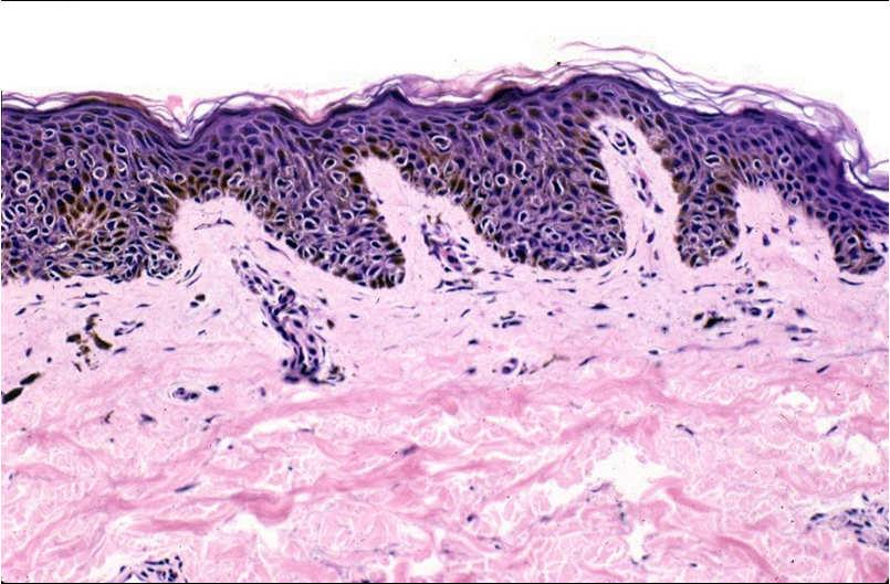
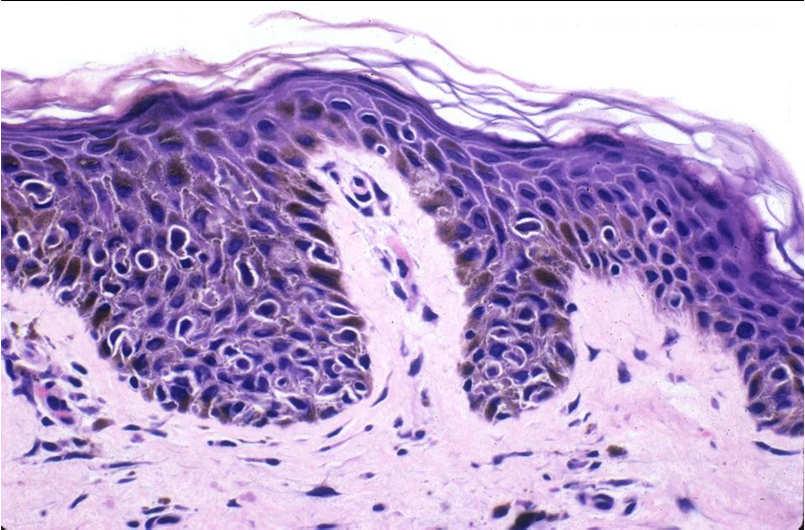
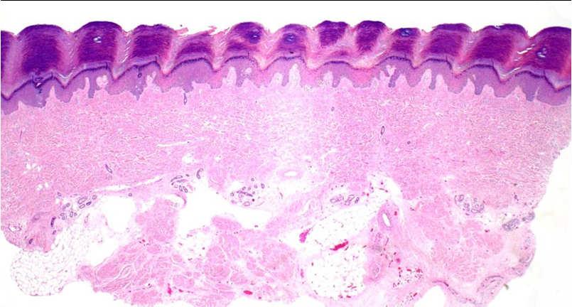
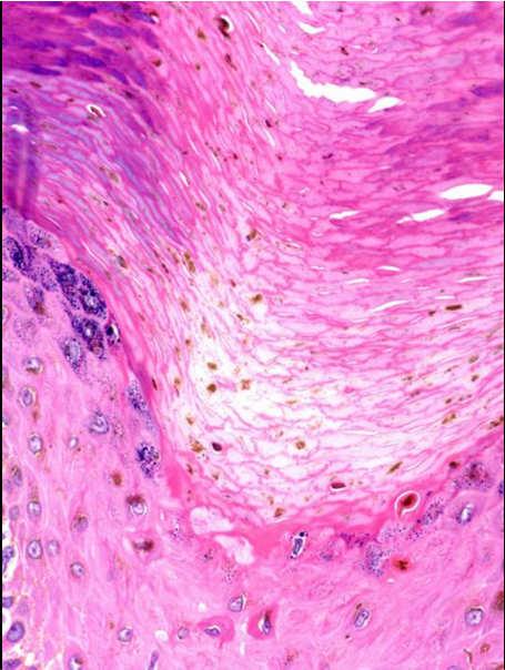
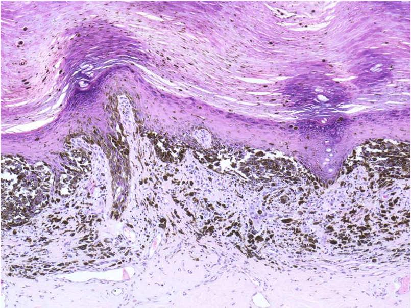
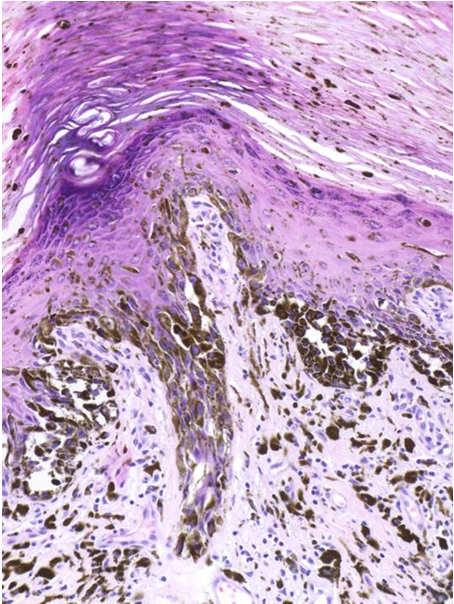
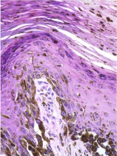
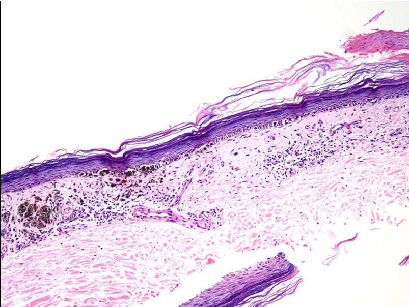
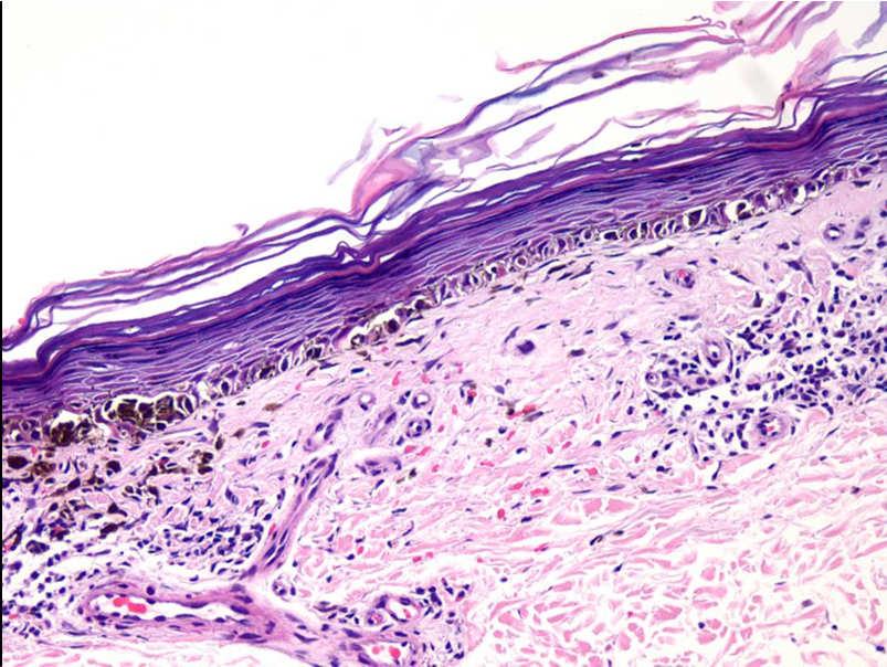
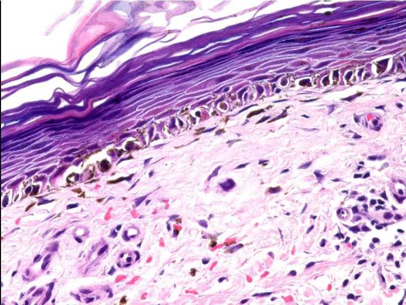
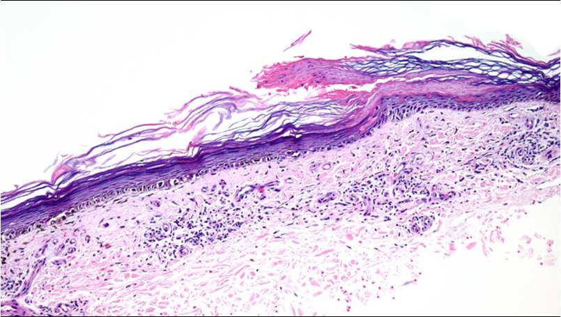
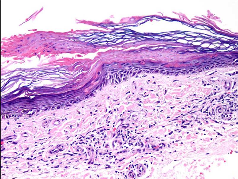
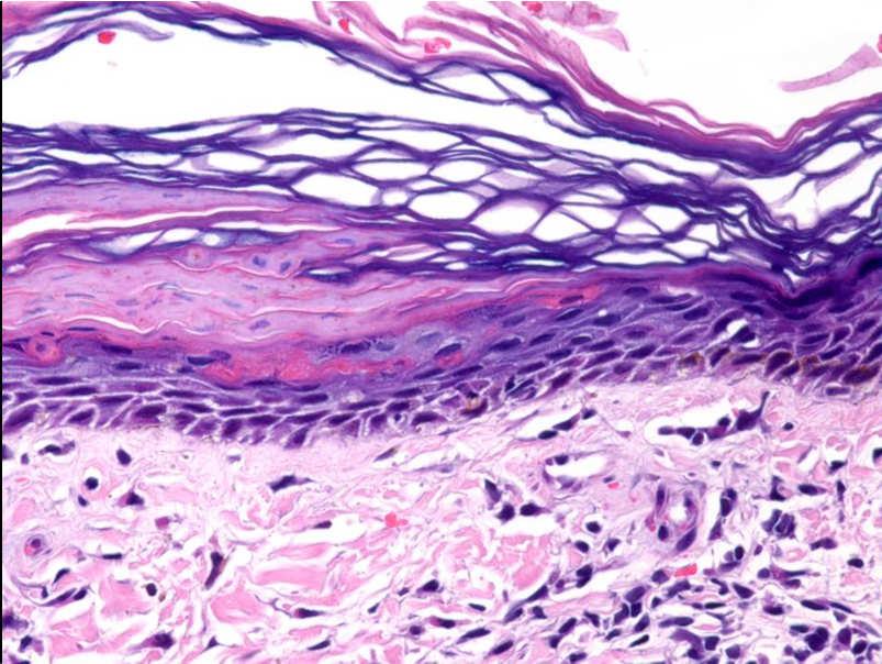
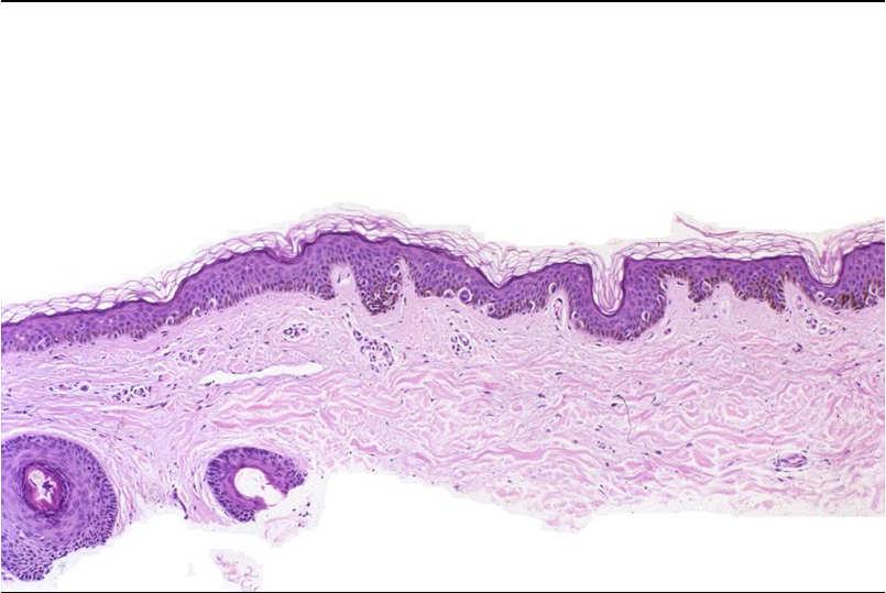
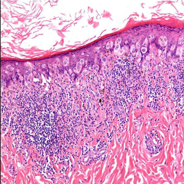
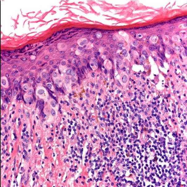
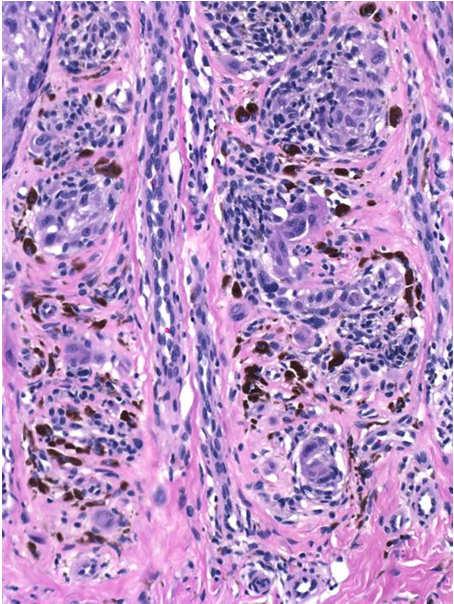
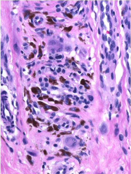
31 31

32 Persistent/recurrent nevi 32
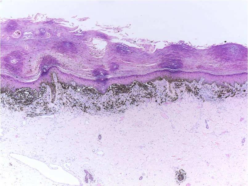
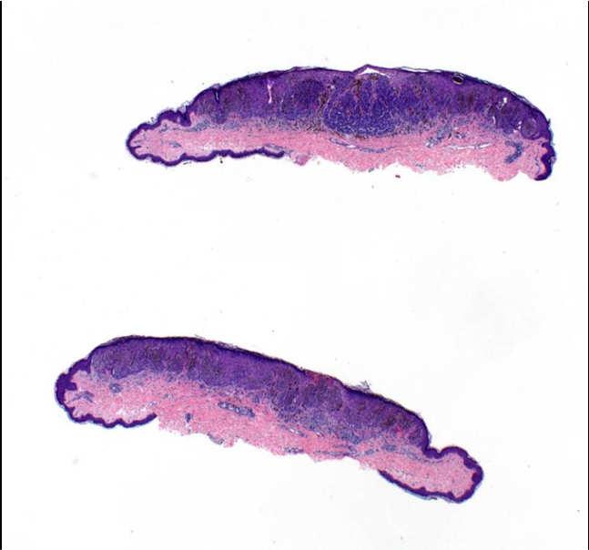
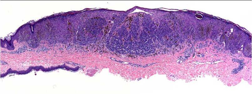
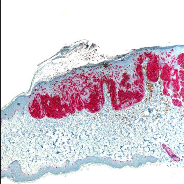
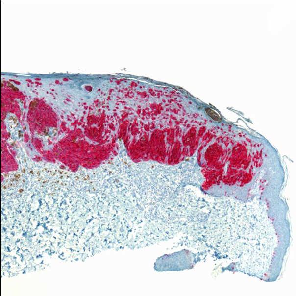
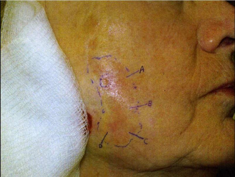
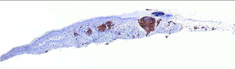
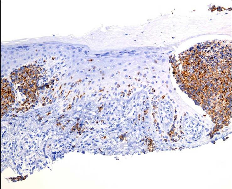
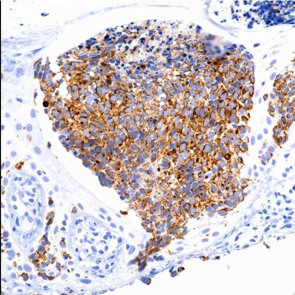
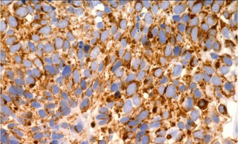
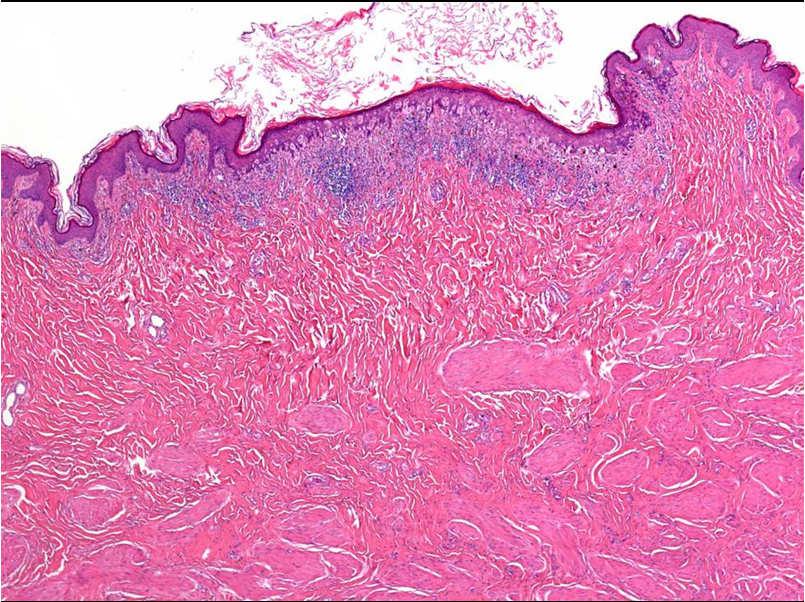
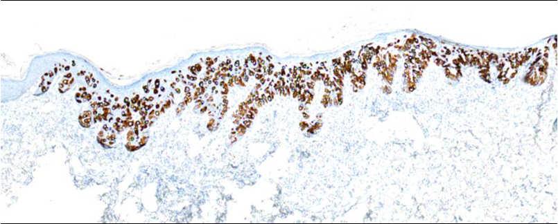
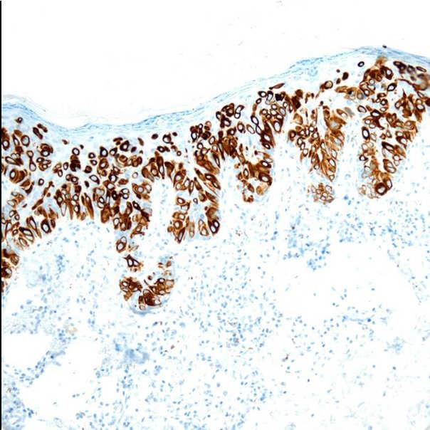
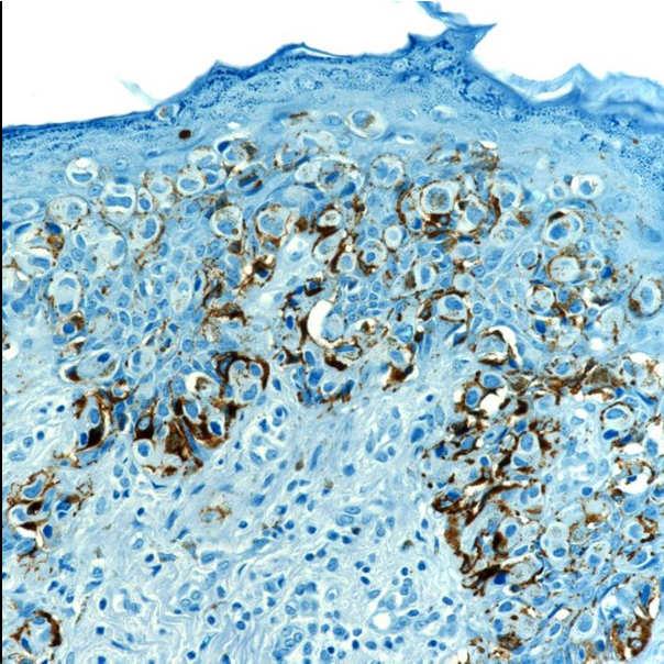
33 33

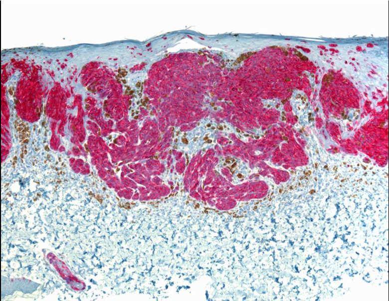
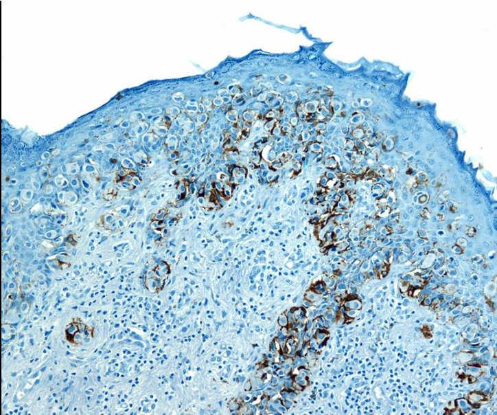
34 Genital nevi 34

35 Masson,
36 36

37 Pagetoid scatter in congenital nevi in neonates 37
38 38

39 Large single melanocytes overlying intradermal nevi 39


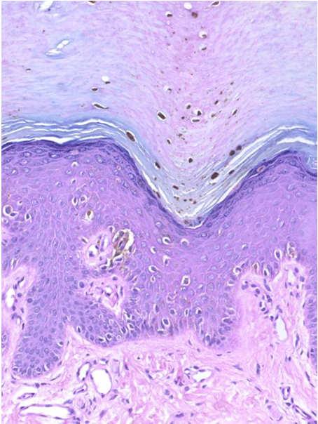
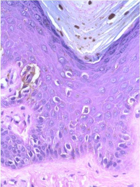
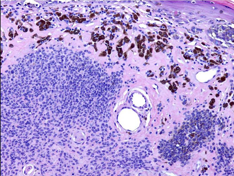
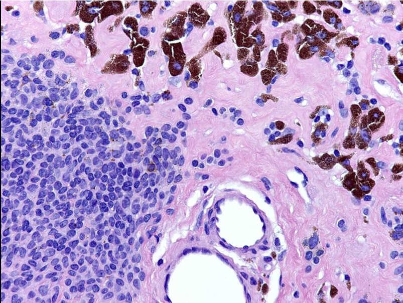
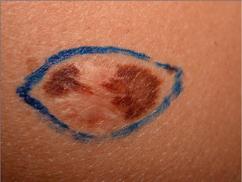
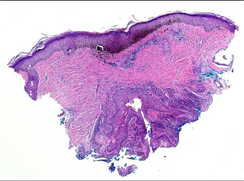
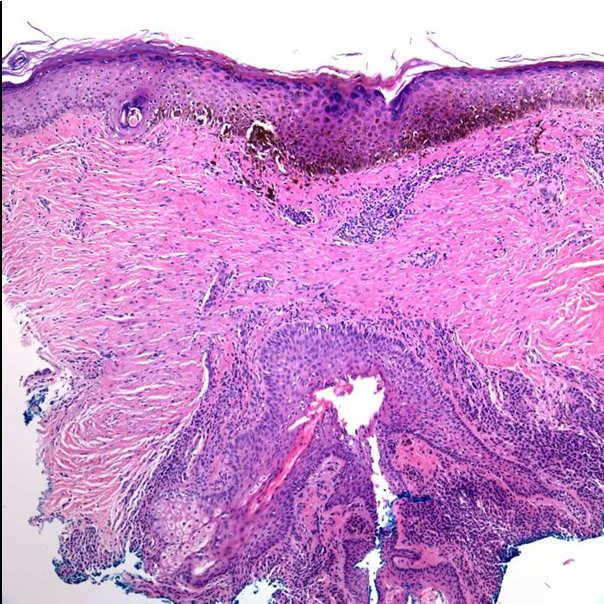
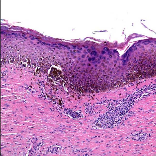
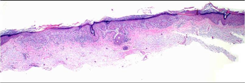
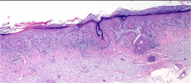
40 Large single melanocytes in sun damaged skin: Actinic melanocytosis Actinic melanocytosis Gilchrest et al J.Invest.Dermatol. Paired biopsies, exposed and non-exposed skin, adjacent sites donors aged yr Density of DOPA+ melanocytes ~2x in exposed > nonexposed skin at all ages, suggesting an irreversible effect of sun exposure Melanocyte density declined ~ 6 to 8% /decade in both sites Dopa-positivity of individual melanocytes was consistently greater in chronically exposed skin > nonexposed skin of same subject and did not vary with age 40
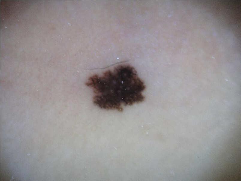
 Prominent dendrites in melanoma in situ, sometimes More irregular pigmentation Adnexal involvement more marked and more deep in melanoma in situ")
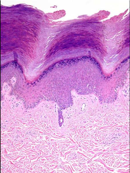
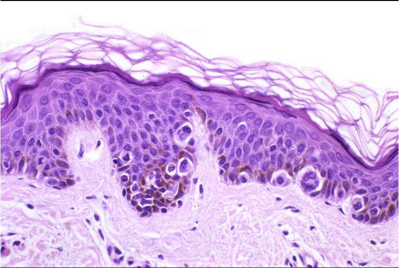
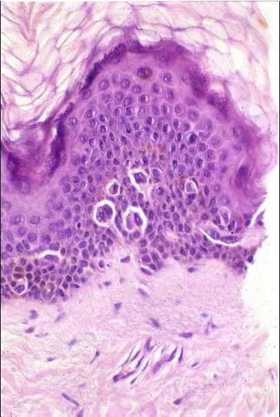
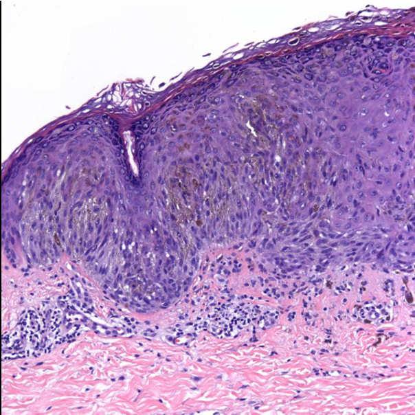
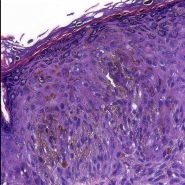
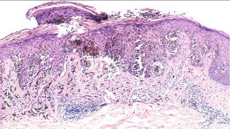
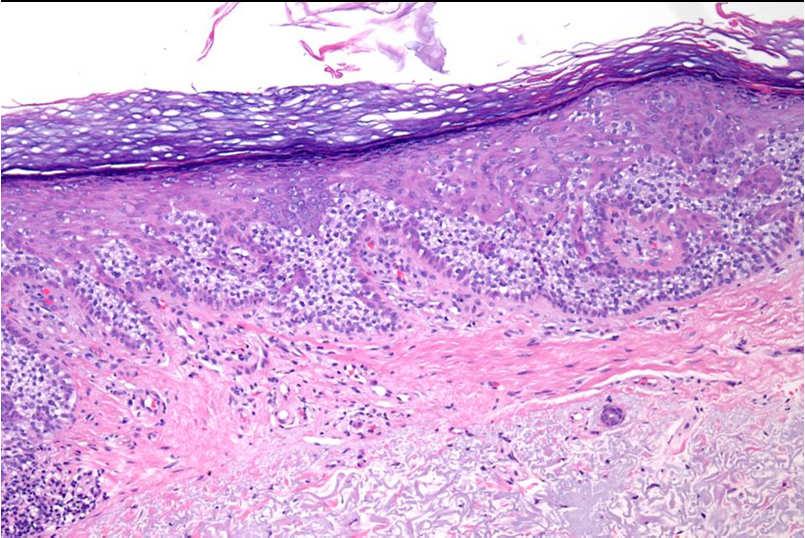
41 Features that point to melanoma in situ over actinic melanocytosis : Irregular intervals between melanocytes Nests may be present (but sometimes levels needed to show them) Pagetoid spread may be present (but sometimes levels needed to show it) Prominent dendrites in melanoma in situ, sometimes More irregular pigmentation Adnexal involvement more marked and more deep in melanoma in situ (there may be increased melanocytes in follicular infundibula and acrosyringia in actinic melanocytosis) Immunostains do not help distinguish MIS from actinic melanocytosis (HMB45, Ki-67) Weyers W, Bonczkowitz M, Weyers I, Bittinger A, Schill WB. Melanoma in situ versus melanocytic hyperplasia in sun-damaged skin. Assessment of the significance of histopathologic criteria for differential diagnosis.am J Dermatopathol Dec;18(6):

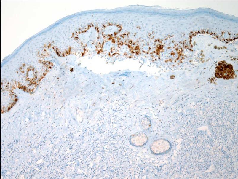
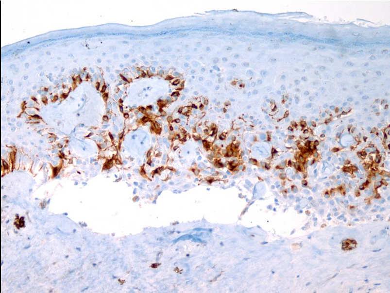
 Arch Dermatol.")

42 Typical melanoma antigen recognized by T cells 1-staining normal sun-exposed skin with a focal area of increased melanocyte density (arrows) (original magnification x200) Arch Dermatol Jul;142(7): Hendi, A. et al. Arch Dermatol 2006;142: Copyright restrictions may apply. 42
")

43 Melanoma antigen recognized by T cells 1-staining normal sun-exposed skin shows extension of melanocytes along the follicle (original magnification x200) Biopsy of the contralateral side? Hendi, A. et al. Arch Dermatol 2006;142: Copyright restrictions may apply. Non-melanocytic simulators of melanoma in situ 43
44 Angiofibromas with prominent junctional melanocytes 44


45 Pagetoid Bowen s disease 45
46 46
47 Pagetoid reticulosis 47


48 Pageoid spread in Merkel cell carcinoma 48
49 49
50 50
51 Pagetoid dyskeratosis 51

52 Paget s disease And extramammary Paget s disease 52
53 53

54 Spitz nevus 54
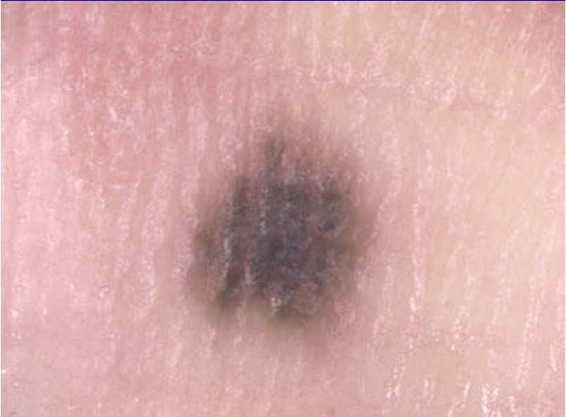
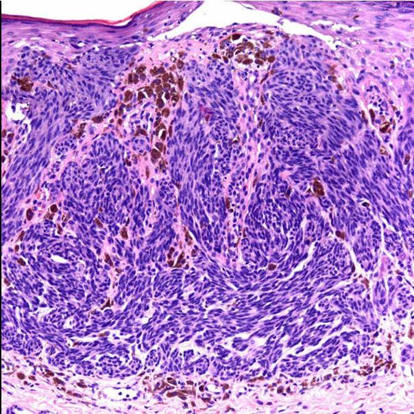
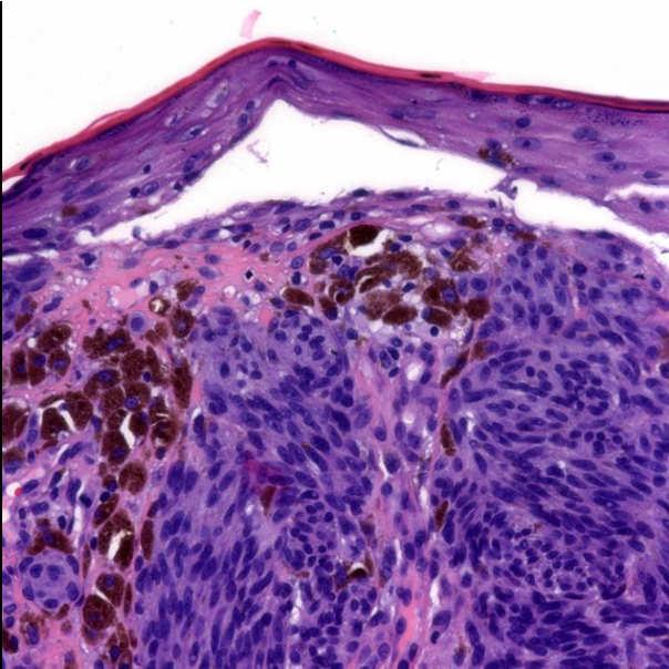
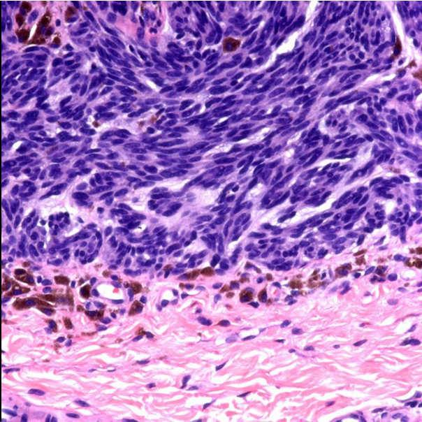
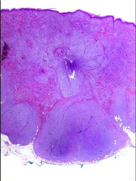
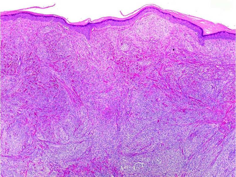
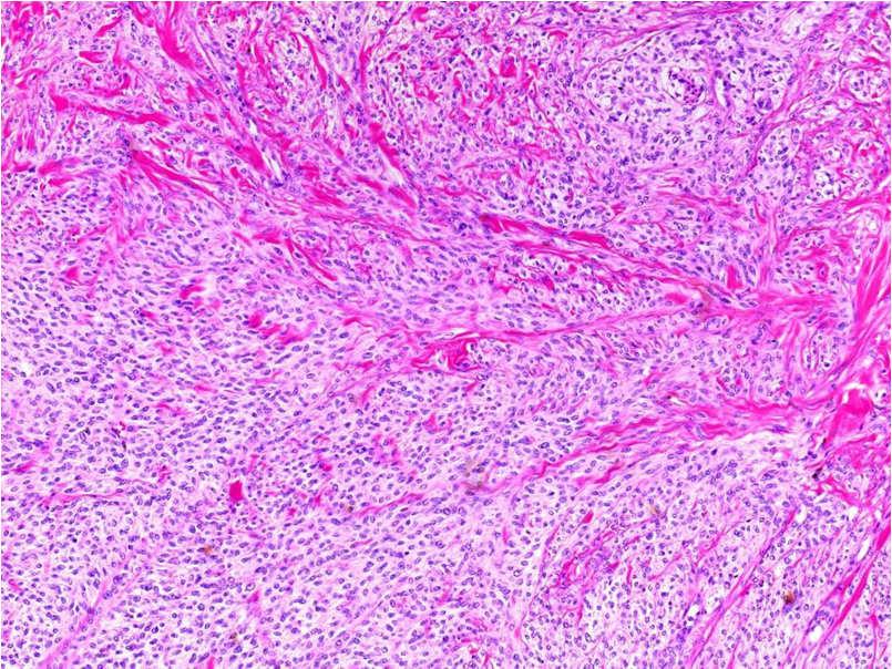
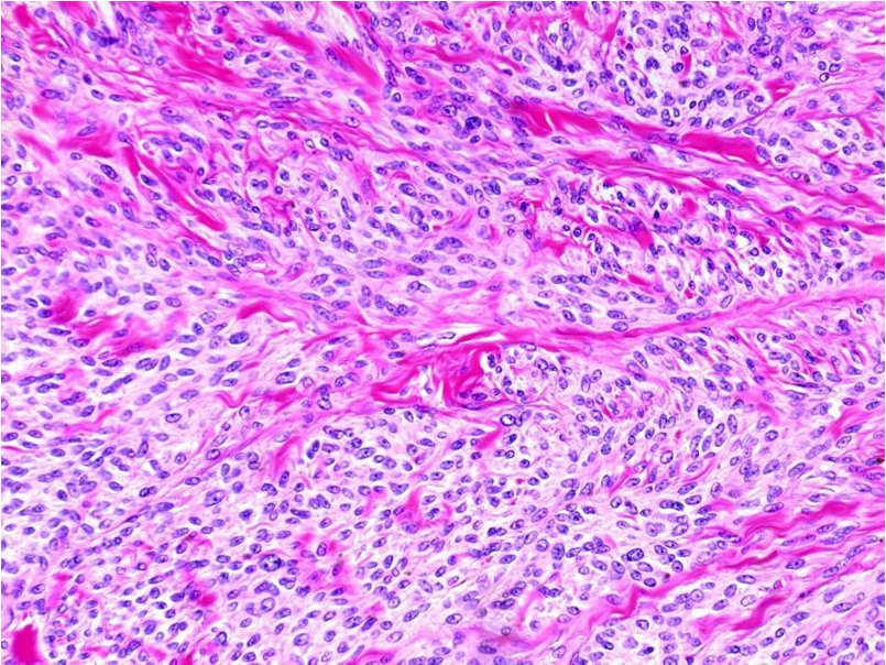
55 55
56 56
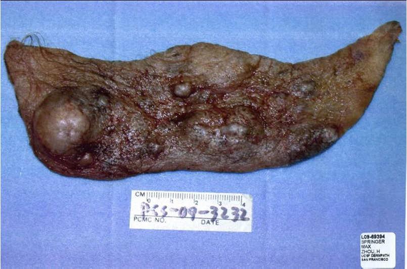
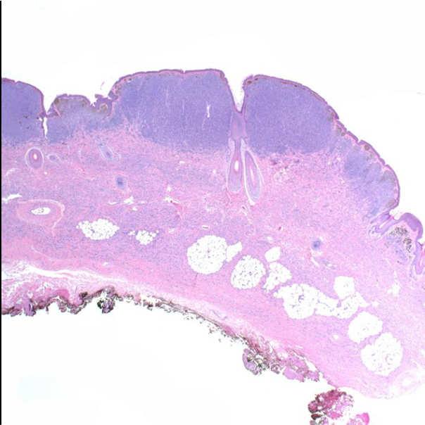
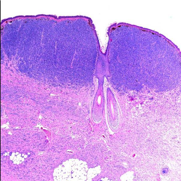
57 57

58 Spitz nevus- genomics Usually b-raf wild type H-ras mutated in a minority Most have normal CGH profile 20% with 11p gain Blue nevus, esp. cellular 58
59 59
60 60
61 61
62 Deep penetrating nevus 62
63 63


64 Combined nevi Combined nevi Small round and blue Small round and Spitz Spitz and blue Deep penetrating and small round 64

65 5/28/
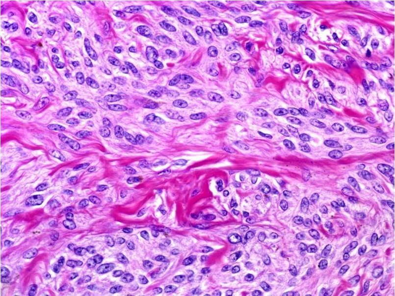
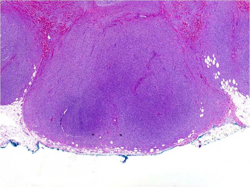
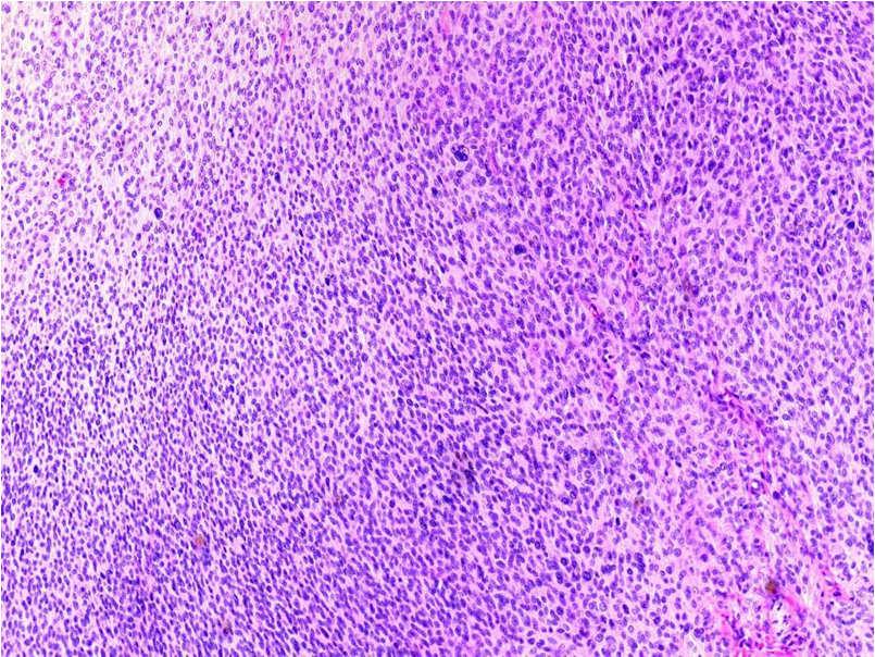
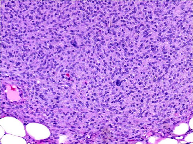
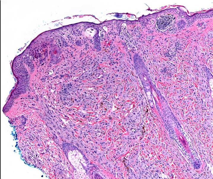
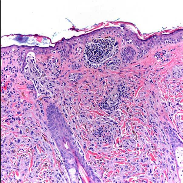
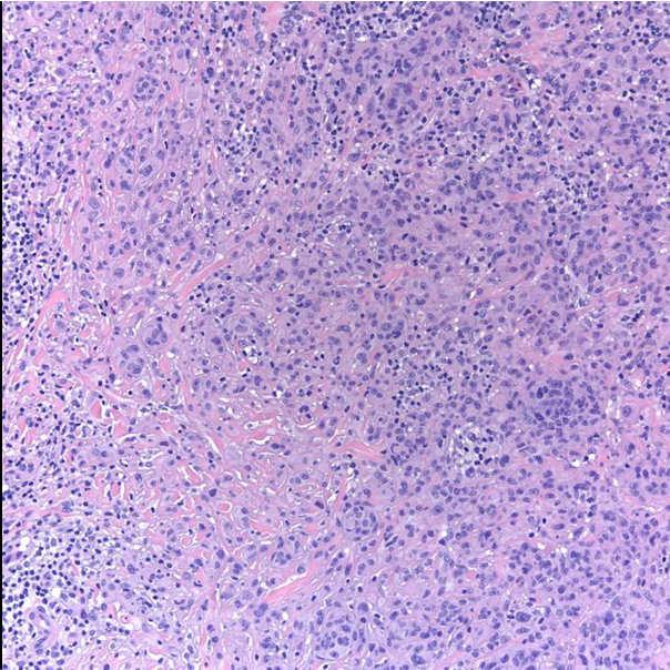
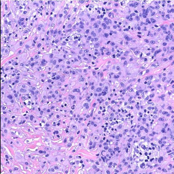
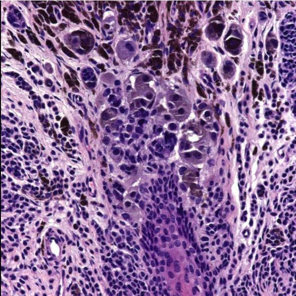
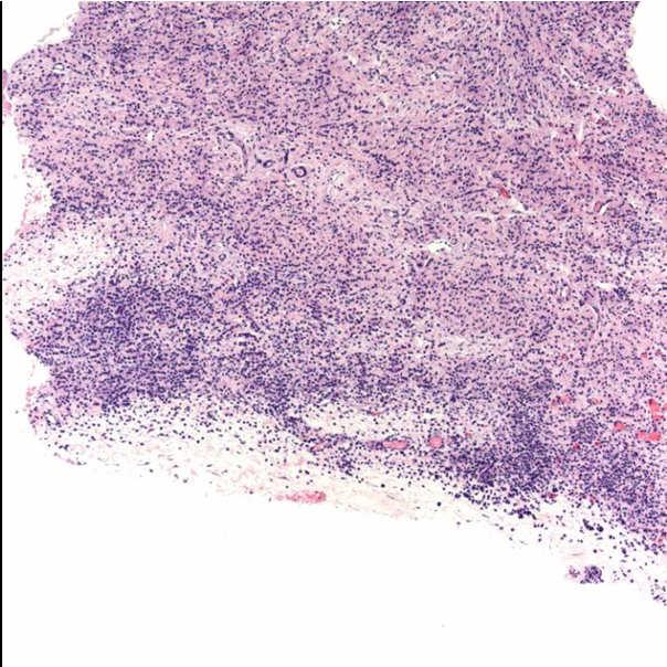
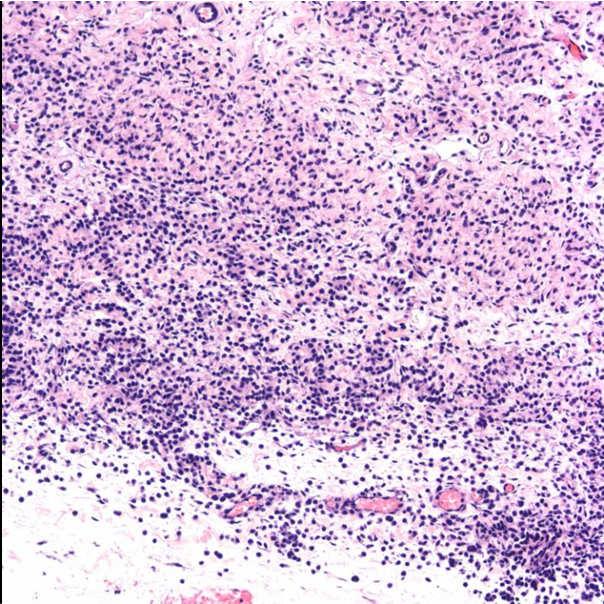
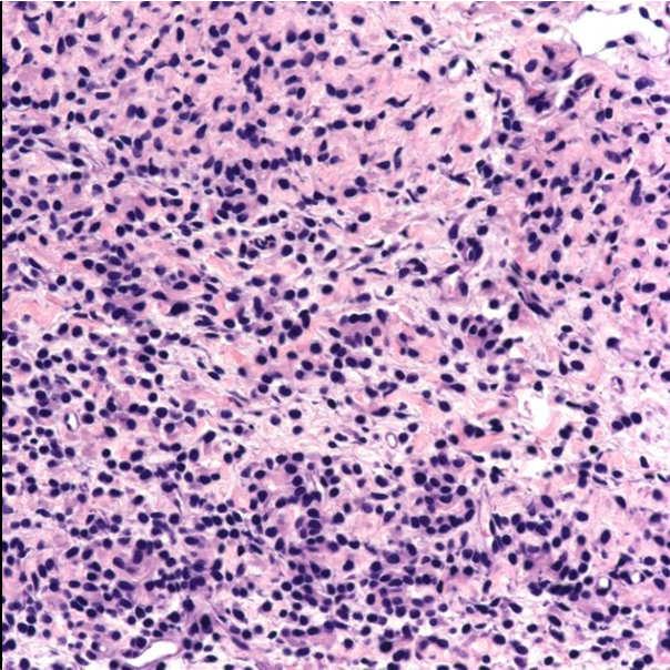
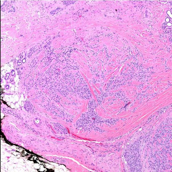
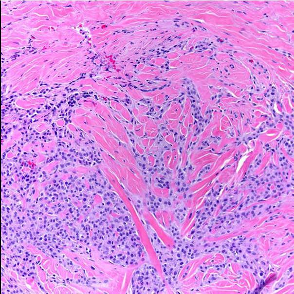
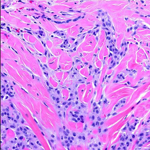
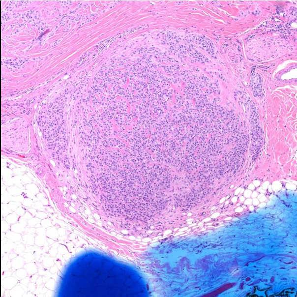
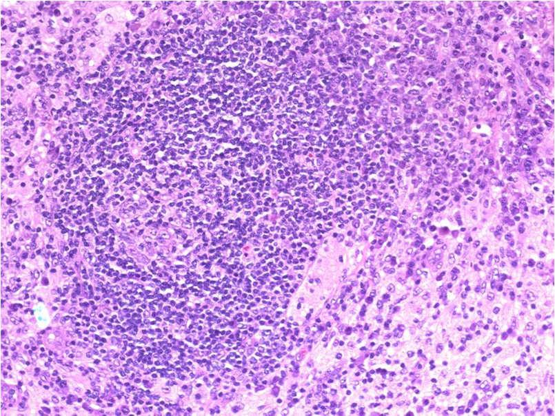
66 66


67 Ancient nevus FIGURE 2 Ancient Melanocytic Nevus: A Simulator of Malignant Melanoma. Kerl, Helmut; Wolf, Ingrid; Kerl, Katrin; Cerroni, Lorenzo; Kutzner, Heinz; Argenyi, Zsolt American Journal of Dermatopathology. 33(2): , April DOI: /IAE.0b013e31817f7b58 FIGURE 2. Ancient melanocytic nevus. Dome-shaped asymmetric brownish tumor with a bluish cast Lippincott Williams & Wilkins, Inc. Published by Lippincott Williams & Wilkins, Inc. 2 67

68 Halo nevus 68
69 Proliferative nodules in congential nevi 69
70 70
71 71
72 72
73 73
 not seen in proliferative nodules Prognostic values of CGH not yet 74")
74 Cytogenetic changes in proliferative nodules Papules have no cytogenetic abnormalities Nodular proliferations usually normal, or with losses or gains of whole chromosomes Focused losses and gains (typical of melanoma) not seen in proliferative nodules Prognostic values of CGH not yet 74


75 Non-melanocytic simulators of melanoma in the dermis Cellular and epithelioid schwannoma Cellular neurothekeoma Atypical fibroxanthoma Desmoplastic leiomyosarcoma and squamous cell carcinoma Pigmented dermatofibrosarcoma protuberans Pigmented basal and sqaumous cell carcinoma Pigmented matricoma Rosai-Dorfman disease Monsel s granuloma Carcinoma metastastic to nevus Pigmented metastatic carcinoma Cellular neurothekeoma 75
76 76
77 77

78 Rosai-Dorfman Disease Cutaneous Rosai-Dorfman Disease Rare: about 60 cases Papules, plaques, nodules, many sites Rare presentations: acneiform, vasculitic, panniculitic, pigmented macules, in zoster scars, bilateral cauliflower ear deformities Affects older patients, different epidemiology Often no significant symptoms, rare laboratory anomalies* Spontaneous remissions, and/or recurrence 78
79 79
80 80
Benign and malignant epithelial lesions: Seborrheic keratosis: A common benign pigmented epidermal tumor occur in middle-aged or older persons more
 Benign and malignant epithelial lesions: Seborrheic keratosis: A common benign pigmented epidermal tumor occur in middle-aged or older persons more common on the trunk; but extremities, head and neck are
Malignant tumors of melanocytes: Part 1. Deba P Sarma, MD., Omaha
 Malignant tumors of melanocytes: Part 1 Deba P Sarma, MD., Omaha The melanocytic tumor is one of the most difficult and confusing areas in Dematopathology. It is true that most (95%) of such lesions are
Benign versus Cancerous Lesions How to tell the difference FMF 2014 Christie Freeman MD, CCFP, DipPDerm, MSc
 1 Benign versus Cancerous Lesions How to tell the difference FMF 2014 Christie Freeman MD, CCFP, DipPDerm, MSc Benign lesions Seborrheic Keratoses: Warty, stuck-on Genetics and birthdays Can start in late
Diagnoses of Cases 1. Lentigo, other melanosis and the acquired nevus 2. Variations on the acquired nevus 3. Dermal melanocytosis
 Diagnoses of Cases 1. Lentigo, other melanosis and the acquired nevus 1 1A. Lentigo simplex 4 1B. Psoralens and ultraviolet A (PUVA) lentigo 6 1C. Solar lentigo 8 1D. Café au lait macule 10 1E. Ink-spot
Desmoplastic Melanoma R/O BCC. Clinical Information. 74 y.o. man with lesion on left side of neck r/o BCC
 R/O BCC Sabine Kohler, M.D. Professor of Pathology and Dermatology Dermatopathology Service Stanford University School of Medicine Clinical Information 74 y.o. man with lesion on left side of neck r/o
Melanocytic Lesions: Use of Immunohistochemistry and Special Studies Napa Valley 2018
 Melanocytic Lesions: Use of Immunohistochemistry and Special Studies Napa Valley 2018 Victor G. Prieto, MD, PhD Professor Depts. of Pathology and Dermatology University of Texas - MD Anderson Cancer Center
Pathology of the skin. 2nd Department of Pathology, Semmelweis University
 Pathology of the skin 2nd Department of Pathology, Semmelweis University Histology of the skin Epidermis: Stratum corneum Stratum granulosum Stratum spinosum Stratum basale Dermis: papillary and reticular
Dermatopathology. Dr. Rafael Botella Estrada. Hospital La Fe de Valencia
 Dermatopathology Dr. Rafael Botella Estrada. Hospital La Fe de Valencia Melanoma and mimics Dr. Martin Mihm Malignant lesions result from the accumulation of mutations Class I lesions (benign) Class II
Basal cell carcinoma 5/28/2011
 Goal of this Presentation A practical approach to the diagnosis of cutaneous carcinomas and their mimics Thaddeus Mully, MD University of California San Francisco To review common non-melanoma skin cancers
Dermatopathology: The tumor is composed of keratinocytes which show atypia, increase mitoses and abnormal mitoses.
 Squamous cell carcinoma (SCC): A common malignant tumor of keratinocytes arising in the epidermis, usually from a precancerous condition: 1- UV induced actinic keratosis, usually of low grade malignancy.
David B. Troxel, MD. Common Medicolegal Situations: Misdiagnosis of Melanoma
 Common Medicolegal Situations: Misdiagnosis of Melanoma David B. Troxel, MD Medical Director, The Doctors Company, Napa, California Clinical Professor Emeritus, University of California at Berkeley Past
STUDY. Melanocytes in Long-standing Sun-Exposed Skin
 STUDY Melanocytes in Long-standing Sun-Exposed Skin Quantitative Analysis Using the MART-1 Immunostain Ali Hendi, MD; David G. Brodland, MD; John A. Zitelli, MD Objective: To help distinguish early melanoma
Glenn D. Goldman, MD. Fletcher Allen Health Care. University of Vermont College of Medicine
 Glenn D. Goldman, MD Fletcher Allen Health Care University of Vermont College of Medicine Recognize and identify the main types of skin cancer Understand how and why Mohs surgery is utilized for the treatment
Conflict of Interest 9/2/2014. Pathogenesis and Comparison of Atypical Spitz Nevi vs Benign Spitz, and Childhood Melanoma
 Pathogenesis and Comparison of Atypical Spitz Nevi vs Benign Spitz, and Childhood Melanoma Martin C. Mihm Jr., M.D., F.A.C.P. Harvard Medical School Brigham and Women s Hospital Dana Farber Cancer Center
المركب النموذج--- سبيتز وحمة = Type Spitz's Nevus, Compound SPITZ NEVUS 1 / 7
 SPITZ NEVUS 1 / 7 Epidemiology An annual incidence rate of 1.4 cases of Spitz nevus per 100,000 individuals has been estimated in Australia, compared with 25.4 per 100,000 individuals for cutaneous melanoma
IT S FUNDAMENTAL MY DEAR WATSON! A SHERLOCKIAN APPROACH TO DERMATOLOGY
 IT S FUNDAMENTAL MY DEAR WATSON! A SHERLOCKIAN APPROACH TO DERMATOLOGY Skin, Bones, and other Private Parts Symposium Dermatology Lectures by Debra Shelby, PhD, DNP, FNP-BC, FADNP, FAANP Debra Shelby,
Dermoscopy: Recognizing Top Five Common In- Office Diagnoses
 Dermoscopy: Recognizing Top Five Common In- Office Diagnoses Vu A. Ngo, DO Department of Family Medicine and Dermatology Choctaw Nation Health Services Authority Learning Objectives Introduction to dermoscopy
MECHANISMS OF HUMAN DISEASE: LABORATORY SESSION PATHOLOGY OF THE SKIN LAB. Friday, February 13, :30 am 11:00 am
 MECHANISMS OF HUMAN DISEASE: LABORATORY SESSION PATHOLOGY OF THE SKIN LAB Friday, February 13, 2009 9:30 am 11:00 am FACULTY COPY GOALS: Describe the basic clinical and morphologic features of various
MECHANISMS OF HUMAN DISEASE: LABORATORY SESSION PATHOLOGY OF THE SKIN LAB. Friday, February 12, :30 am 11:00 am
 MECHANISMS OF HUMAN DISEASE: LABORATORY SESSION PATHOLOGY OF THE SKIN LAB Friday, February 12, 2012 9:30 am 11:00 am FACULTY COPY GOALS: Describe the basic clinical and morphologic features of various
BAP-oma & BEYOND MICHAEL A NOWAK, MD
 BAP-oma & BEYOND MICHAEL A NOWAK, MD CONFLICTS No conflicts with the content of this lecture BAP-oma Wiesner 2011: Families with multiple tan dome-shaped papules of head, neck, trunk, and extremities.
Dermatology for the PCP Deanna G. Brown, MD, FAAD Susong Dermatology Consulting Staff at CHI Memorial
 Dermatology for the PCP Deanna G. Brown, MD, FAAD Susong Dermatology Consulting Staff at CHI Memorial Cutaneous Oncology for the PCP Deanna G. Brown, MD, FAAD Susong Dermatology Consulting Staff at CHI
PHILIP E. LEBOIT. Histological Diagnosis of Nevi and Melanoma
 GUIDO MASSI PHILIP E. LEBOIT Histological Diagnosis of Nevi and Melanoma GUIDO MASSI PHILIP E. LEBOIT Histological Diagnosis of Nevi and Melanoma With Contributions by PAOLA PASQUINI and FRANCESCO FEDERICO,
A PRACTICAL APPROACH TO ATYPICAL MELANOCYTIC LESIONS BIJAN HAGHIGHI M.D, DIRECTOR OF DERMATOPATHOLOGY, ST. JOSEPH HOSPITAL
 A PRACTICAL APPROACH TO ATYPICAL MELANOCYTIC LESIONS BIJAN HAGHIGHI M.D, DIRECTOR OF DERMATOPATHOLOGY, ST. JOSEPH HOSPITAL OBJECTIVES Discuss current trends and changing concepts in our understanding of
Female 18. Deeply pigmented lesion on trunk.?warty naevus?seborrhoeic keratosis?malignant melanoma. The best diagnosis is:
 Female 18. Deeply pigmented lesion on trunk.?warty naevus?seborrhoeic keratosis?malignant melanoma. The best diagnosis is: A. deep penetrating naevus B. naevoid malignant melanoma C. pigment synthesising
PATHOLOGY OF THE SKIN 2. Tumours of the skin
 PATHOLOGY OF THE SKIN 2. Tumours of the skin Máirín E. McMenamin MB MRCPI FRCPath Dip (Dermatopathol) RCPath St. James s Hospital and University of Dublin, Trinity College Tumour (Neoplasia) Benign or
Clinical characteristics
 Skin Cancer Fernando Vega, MD Seattle Healing Arts Clinical characteristics Precancerous lesions Common skin cancers ACTINIC KERATOSIS Precancerous skin lesions Actinic keratoses Dysplastic melanocytic
Chapter 6 Squamous Cell Carcinoma: Variants and Challenges
 Chapter 6 Squamous Cell Carcinoma: Variants and Challenges Michael B. Morgan EPIDEMIOLOGY: Second most common skin cancer, rare in the dark-skinned races. ETIOLOGY: Ultraviolet light, HPV infection. PATHOGENESIS:
أملس عضلي غرن = Leiomyosarcoma. Leiomyosarcoma 1 / 5
 Leiomyosarcoma 1 / 5 EPIDEMIOLOGY Exact incidence is unknown, but older studies suggest that leiomyosarcomas comprise approximately 3 percent of soft-tissue sarcomas. Superficial leiomyosarcoma occurs
Index. Springer-Verlag Berlin Heidelberg 2017 J.A. Plaza, V.G. Prieto, Pathology of Pigmented Skin Lesions, DOI /
 A Acral lentiginous (mucosal lentiginous) melanoma, 483 Acral lentiginous melanoma (ALM) asymmetric and irregular lentiginous junctional growth, 431 clinical features, 427 428 differential diagnosis, 428
Melanocytic lesions on Genital Skin Melanoma vs. Melanocytic Nevus, Revisited. Timothy H. McCalmont, MD University of California, San Francisco
 Melanocytic lesions on Genital Skin Melanoma vs. Melanocytic Nevus, Revisited Timothy H. McCalmont, MD, San Francisco I. IS IT BENIGN OR IS IT MALIGNANT? One of the commonest determinations we make as
Management of pediatric melanocytic lesions
 Open Journal of Clinical & Medical Case Reports Management of pediatric melanocytic lesions Volume 3 (2017) Issue 8 ISSN 2379-1039 Jin Kim, BS; Emmanuel Gabriel MD, PhD; Weiguo Liu MD, PhD; Lin Lin MD,
Cutaneous Malignancies: A Primer COPYRIGHT. Marissa Heller, M.D.
 Cutaneous Malignancies: A Primer Marissa Heller, M.D. Associate Director of Dermatologic Surgery Department of Dermatology Beth Israel Deaconess Medical Center December 10, 2016 Skin Cancer Non-melanoma
Glenn D. Goldman, MD. University of Vermont Medical Center. University of Vermont College of Medicine
 Glenn D. Goldman, MD University of Vermont Medical Center University of Vermont College of Medicine Recognize and identify the main types of skin cancer and their precursors Identify and understand new
Maligna Melanoma and Atypical Fibroxanthoma: An Unusual Collision Tumour G Türkcü 1, A Keleş 1, U Alabalık 1, D Uçmak 2, H Büyükbayram 1 ABSTRACT
 Maligna Melanoma and Atypical Fibroxanthoma: An Unusual Collision Tumour G Türkcü 1, A Keleş 1, U Alabalık 1, D Uçmak 2, H Büyükbayram 1 ABSTRACT Two different neoplasia in the same biopsy material called
Common Benign Lesions and Skin Cancers. 22nd May 2015 Dr Mark Foley
 Common Benign Lesions and Skin Cancers 22nd May 2015 Dr Mark Foley Thank you for downloading this file. This intended to supplement the presentation given at the NZ Wound Care Conference, it is not intended
Atypical Nevi When to Re-excise. Catherine Barry, DO Dermatopathologist
 Atypical Nevi When to Re-excise Catherine Barry, DO Dermatopathologist Why talk about skin cancer? Because it s the most common type of cancer! Non-melanoma Skin Cancers Basal Cell Carcinoma Squamous Cell
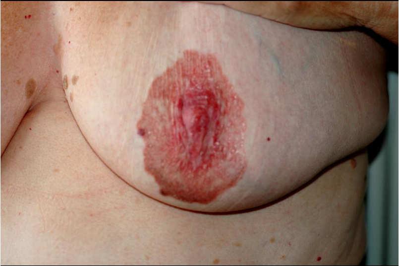
Paget. Pigmented Extramammary Paget's Disease. Mei-ju Ko Cheng-Hsiang Hsiao* Chung-Jen Tseng Yi-Hua Liao
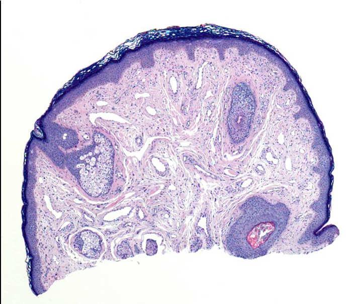
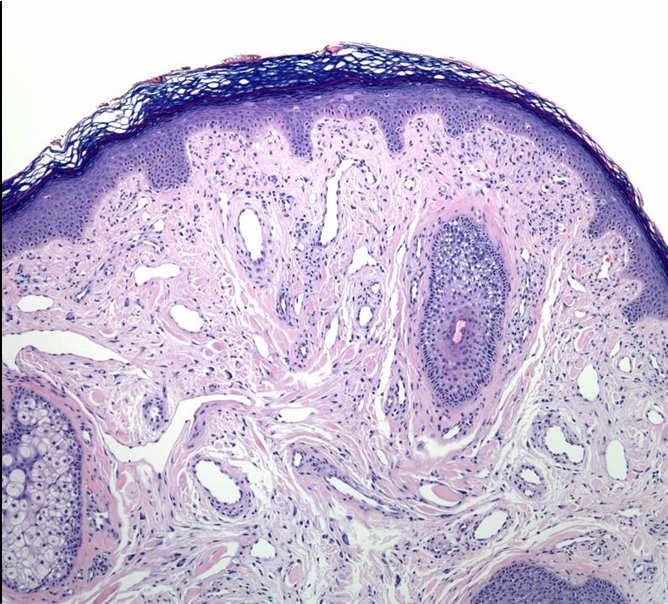
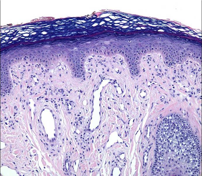
 Paget * * Pigmented Extramammary Paget's Disease Mei-ju Ko Cheng-Hsiang Hsiao* Chung-Jen Tseng Yi-Hua Liao Extramammary Paget's disease (EMPD) usually presents as an erythematous, scaly patch with crusts,
Multiple Primary Melanoma in a Thai Male: A Case Report
 Case Report Multiple Primary Melanoma in a Thai Male: A Case Report J Med Assoc Thai 2014; 97 (Suppl. 2): S234-S238 Full text. e-journal: http://www.jmatonline.com Kittisak Payapvipapong MD*, Pinyapat
Identifying Skin Cancer. Mary S. Stone MD Professor of Dermatology and Pathology University of Iowa Carver College of Medicine March, 2018
 Identifying Skin Cancer Mary S. Stone MD Professor of Dermatology and Pathology University of Iowa Carver College of Medicine March, 2018 American Cancer Society web site Skin Cancer Melanoma Non-Melanoma
Cancer Reporting for Dermatologists. Florida Department of Health Florida Cancer Data System. March 9, Agenda
 Cancer Reporting for Dermatologists Florida Department of Health Florida Cancer Data System March 9, 2011 Agenda Welcome Introductions Cancer Reporting in Florida BETA Participation Expectations Review
Histopathology of Melanoma
 THE YALE JOURNAL OF BIOLOGY AND MEDICINE 48, 409-416 (1975) Histopathology of Melanoma G. J. WALKER SMITH Department ofpathology, Yale University School ofmedicine, 333 Cedar Street, New Haven, Connecticut
Ways to get into trouble, ideas on avoiding trouble, and diagnostic approaches to keep trouble at bay
 Pitfalls in the diagnosis of melanocytic tumors Timothy McCalmont, MD University of California, San Francisco Ways to get into trouble, ideas on avoiding trouble, and diagnostic approaches to keep trouble
Regression 2/3/18. Histologically regression is characterized: melanosis fibrosis combination of both. Distribution: partial or focal!
 Regression Margaret Oliviero MSN, ARNP Harold S. Rabinovitz MD Histologically regression is characterized: melanosis fibrosis combination of both Distribution: partial or focal! Dermatoscopic terminology
VACAVILLE DERMATOLOGY
 Connecting the Dots on those Spots NANDAN V. KAMATH, M.D. VACAVILLE DERMATOLOGY Sources All of the photos were taken with permission from the Dermnet NZ website - Dermnet New Zealand after communicating
Gross Appearance & Histology of Skin Cancer. Kyle Mannion M.D. January 21, 2005
 Gross Appearance & Histology of Skin Cancer Kyle Mannion M.D. January 21, 2005 Actinic Keratosis 5-20% will develop squamous/basal cell ca Almost solely from solar damage Usually develop during 4 th decade
EARLY ONLINE RELEASE
 EARLY ONLINE RELEASE Note: This article was posted on the Archives Web site as an Early Online Release. Early Online Release articles have been peer reviewed, copyedited, and reviewed by the authors. Additional
Abrupt Intralesional Color Change on Dermoscopy as a New Indicator of Early Superficial Spreading Melanoma in a Japanese Woman
 Published online: June 24, 2015 1662 6567/15/0072 0123$39.50/0 This is an Open Access article licensed under the terms of the Creative Commons Attribution-NonCommercial 3.0 Unported license (CC BY-NC)
Melanocytes in nonlesional sun-exposed skin: A multicenter comparative study
 Melanocytes in nonlesional sun-exposed skin: A multicenter comparative study AliHendi,MD, a,b David A. Wada, MD, d M. Amanda Jacobs, MD, e JuliaE.Crook,PhD, c Kimberly R. Kortuem, MD, d Brent R. Weed,
MELANOCYTIC SIMULATORS OF MELANOMA Philip E. LeBoit, M.D. Departments of Pathology and Dermatology University of California, San Francisco
 MELANOCYTIC SIMULATORS OF MELANOMA Philip E. LeBoit, M.D. Departments of Pathology and Dermatology University of California, San Francisco Pathologists have become increasingly aware of the many and sometimes
There is NO single Melanoma Stain. > 6000 Mutations in Melanoma. What else can be done to discriminate atypical nevi from melanoma?
 Las Vegas Fall Clinical 2016: The Assessment and Diagnosis of Melanoma Whitney A. High, MD, JD, MEng Associate Professor, Dermatology & Pathology Director of Dermatopathology (Dermatology) University of
Histopathology: skin pathology
 Histopathology: skin pathology These presentations are to help you identify, and to test yourself on identifying, basic histopathological features. They do not contain the additional factual information
LENTIGO SIMPLEX. Epidemiology
 LENTIGO SIMPLEX Epidemiology The frequency of lentigo simplex in children and adults has not been determined. There does not appear to be a racial or gender predilection. Lentigo simplex is the most common
NON-MELANOCYTIC MELANOMA LOOK-ALIKES. Hideko Kamino, M.D. Dermatopathology Section New York University School of Medicine
 NON-MELANOCYTIC MELANOMA LOOK-ALIKES Hideko Kamino, M.D. Dermatopathology Section New York University School of Medicine There is a diagnostically important group of entities called non-melanocytic melanoma
F006 Imaging in Dermatology Melanocytic Neoplasia Clinical-Confocal-Pathological-Correlations
 F006 Imaging in Dermatology Melanocytic Neoplasia Clinical-Confocal-Pathological-Correlations Melissa Gill, MD SkinMedical Research and Diagnostics Dobbs Ferry, NY, USA Department of Pathology SUNY Downstate
Springer Healthcare. Staging and Diagnosing Cutaneous Melanoma. Concise Reference. Dirk Schadendorf, Corinna Kochs, Elisabeth Livingstone
 Concise Reference Staging and Diagnosing Cutaneous Melanoma Dirk Schadendorf, Corinna Kochs, Elisabeth Livingstone Extracted from Handbook of Cutaneous Melanoma: A Guide to Diagnosis and Treatment Published
SKIN CANCER. Most common cancer diagnosis 40% of all cancers
 SKIN CANCER Most common cancer diagnosis 40% of all cancers OBJECTIVES Review common and uncommon cancers of the skin. Special emphasis on melanoma and dysplastic nevus Review pathology/tnm/staging, which
Update on Spitzoid and Blue nevus-like melanocytic lesions Emphasis on molecular studies informing diagnosis, prognosis and therapy
 Update on Spitzoid and Blue nevus-like melanocytic lesions Emphasis on molecular studies informing diagnosis, prognosis and therapy Michael T. Tetzlaff MD, PhD Associate Professor Department of Pathology,
Actinic keratosis (AK): Dr Sarma s simple guide
: Dr Sarma s simple guide") Actinic keratosis (AK): Dr Sarma s simple guide Actinic keratosis is a very common lesion that you will see in your day-to-day practice. First, let me explain the name Actinic keratosis. It means keratosis
Actinic keratosis (AK): Dr Sarma s simple guide Actinic keratosis is a very common lesion that you will see in your day-to-day practice. First, let me explain the name Actinic keratosis. It means keratosis
LUMPS AND BUMPS: AN ORGANIZED APPROACH TO DIAGNOSIS AND MANAGEMENT
 LUMPS AND BUMPS: AN ORGANIZED APPROACH TO DIAGNOSIS AND MANAGEMENT Tammy P. Than, M.S., O.D., F.A.A.O. The University of Alabama at Birmingham / School of Optometry 1716 University Blvd. Birmingham, AL
Lid Lesions: Relax or Refer
 Lid Lesions: Relax or Refer Blair Lonsberry, MS, OD, MEd., FAAO Professor of Optometry Pacific University College of Optometry blonsberry@pacificu.edu Agenda Benign vs. Malignant lesions Benign Eyelid
Criteria for the diagnosis of nevus
 Index Criteria for the diagnosis of nevus Criteria for the diagnosis of nevus ABCD, 1, 301, 329, 451, 501, 502 Aberrant mongolian spot, 82, 114, 120 Abtropfung, 29, 157 Achromic melanoma in situ, 422 Achromic
Malignant tumors of melanocytes : Part 3. Deba P Sarma, MD., Omaha
 Malignant tumors of melanocytes : Part 3 Deba P Sarma, MD., Omaha Let s go over one case of melanoma using the following worksheet. Of the various essential information that needs to be included in the
Dermatological Manifestations in the Elderly. Sanjay Siddha Staff Dermatologist UHN & MSH
 Dermatological Manifestations in the Elderly Sanjay Siddha Staff Dermatologist UHN & MSH Disclosure No actual or potential conflicts of interest or commercial relationships to declare Objectives Recognize
Learning Objectives. Tanning. The Skin. Classic Features. Sun Reactive Skin Type Classification. Skin Cancers: Preventing, Screening and Treating
 Learning Objectives Skin Cancers: Preventing, Screening and Treating Robert A. Baldor, MD, FAAFP Professor, Family Medicine & Community Health University of Massachusetts Medical School Distinguish the
Cutaneous metastases. Thaddeus Mully. University of California, San Francisco Professor, Departments of Pathology and Dermatology
 Cutaneous metastases Thaddeus Mully University of California, San Francisco Professor, Departments of Pathology and Dermatology DISCLOSURE OF RELATIONSHIPS WITH INDUSTRY Thaddeus Mully Course C005 Essential
The Enigmatic Spitz Lesion
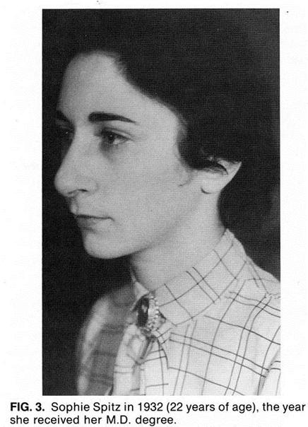
 The Enigmatic Spitz Lesion The Dawn of Spitz S Spitz Sophie Spitz Melanomas of Childhood ; Am J Pathol 1948 1910-1956 13 children (18 mo - 12 yrs) 12/13 had a benign clinical course Sophie Spitz Born 1910
Toby Maurer, MD University of California, San Francisco. Lifetime risk of an American developing melanoma
 Distinguishing Pigmented Skin Lesions and Melanoma Toby Maurer, MD University of California, San Francisco Epidemiology of Melanoma Lifetime risk of an American developing melanoma 1935: 1 in 1500 1980:
Signature nevi: individuals with multiple melanocytic nevi commonly have similar clinical and histologic patterns
 Dermatology Practical & Conceptual www.derm101.com Signature nevi: individuals with multiple melanocytic nevi commonly have similar clinical and histologic patterns Robert M. Hurwitz, M.D. 1, Larry J.
Supplementary Figure 1. Spitzoid Melanoma with PPFIBP1-MET fusion. (a) Histopathology (4x) shows a domed papule with melanocytes extending into the
 Histopathology (4x) shows a domed papule with melanocytes extending into the") Supplementary Figure 1. Spitzoid Melanoma with PPFIBP1-MET fusion. (a) Histopathology (4x) shows a domed papule with melanocytes extending into the deep dermis. (b) The melanocytes demonstrate abundant
Supplementary Figure 1. Spitzoid Melanoma with PPFIBP1-MET fusion. (a) Histopathology (4x) shows a domed papule with melanocytes extending into the deep dermis. (b) The melanocytes demonstrate abundant
NEOPLASMS OF THE SURFACE EPITHELIUM (KERATINOCYTES)
") NEOPLASMS OF THE SURFACE EPITHELIUM (KERATINOCYTES) Papillary Lesions Precancerous Lesions Keratinocyte Proliferations Carcinomas Melanotic Lesions Melanomas Normal Mucosa Keratin layer Spinous layer Basal
NEOPLASMS OF THE SURFACE EPITHELIUM (KERATINOCYTES) Papillary Lesions Precancerous Lesions Keratinocyte Proliferations Carcinomas Melanotic Lesions Melanomas Normal Mucosa Keratin layer Spinous layer Basal
Guy Perrot (Ги Перро)
") НАУЧНО-ПРАКТИЧЕСКАЯ КОНФЕРЕНЦИЯ (МАСТЕР-КЛАСС) «ПРАКТИЧЕСКИЕ АСПЕКТЫ ДИАГНОСТИКИ И ЛЕЧЕНИЯ МЕЛАНОМЫ КОЖИ» DIAGNOSTIC AND PITFALLS IN MELANOMA Guy Perrot (Ги Перро) MD PHD pathologist, University Hospital
НАУЧНО-ПРАКТИЧЕСКАЯ КОНФЕРЕНЦИЯ (МАСТЕР-КЛАСС) «ПРАКТИЧЕСКИЕ АСПЕКТЫ ДИАГНОСТИКИ И ЛЕЧЕНИЯ МЕЛАНОМЫ КОЖИ» DIAGNOSTIC AND PITFALLS IN MELANOMA Guy Perrot (Ги Перро) MD PHD pathologist, University Hospital
Malignant non-melanocytic lesions
 Malignant non-melanocytic lesions Course C023: Fundamentals of Dermoscopy March 4, 2019, 11:20 AM - 11:50 PM Room: 146B Jason B. Lee, MD Professor & Vice Chair Director of Dermatopathology & Pigmented
Dermatologica Sinica
 DERMATOLOGICA SINICA 31 (2013) 140e144 Contents lists available at SciVerse ScienceDirect Dermatologica Sinica journal homepage: http://www.derm-sinica.com CASE REPORT Atypical fibroxanthoma-like amelanotic
Important Decisions in Dermatopathology: The Clinico- Pathologic Correlation. Dermatopathology Specialists Needed. Changing Trends
 Important Decisions in Dermatopathology: The Clinico- Pathologic Correlation Uma Sundram, MD, PhD Departments of Pathology and Dermatology Stanford University May 29, 2008 Dermatopathology Specialists
Acral Melanoma in Japan
 Acral Melanoma in Japan MAKOTO SEUI, M.D., HIDEAKI TAKEMATSU, M.D., MICHIKO HOSOKAWA, M.D., MASAAKI OBATA, M.D., YASUSHI TOMITA, M.D., TAIZO KATO, M.D., MASAAKI TAKAHASHI, M.D., AND MARTIN C. MIHM, JR.,
Cluster designation 5 staining of normal and non-lymphoid neoplastic skin*
 J Cutan Pathol 2005: 32: 50 54 Copyright # Blackwell Munksgaard 2005 Blackwell Munksgaard. Printed in Denmark Journal of Cutaneous Pathology Cluster designation 5 staining of normal and non-lymphoid neoplastic
Large majority caused by sun exposure Often sun exposure before age 20 Persons who burn easily and tan poorly are at greatest risk.
 Basics of Skin Cancer Detection and Treatment of Non- Melanoma Skin Cancers Large majority caused by sun exposure Often sun exposure before age 20 Persons who burn easily and tan poorly are at greatest
Case 26 Male 37. Right jawline 5mm nodule?keloid. The best diagnosis is:
 Case 26 Male 37. Right jawline 5mm nodule?keloid. The best diagnosis is: A. Desmoplastic Spitz naevus B. Atypical Spitz Tumour C. Spitzoid melanoma D. Deep penetrating naevus E. Spitz naevus Case 26: M
Melanoma and Mimickers
 Melanoma and Mimickers Kara Walton, MD Assistant Professor of Dermatology and Dermatopathology Medical College of Wisconsin Disclosures No relevant financial disclosures 1 Objectives Recognize common benign
Melanoma Update: 8th Edition of AJCC Staging System
 Melanoma Update: 8th Edition of AJCC Staging System Rosalie Elenitsas, M.D. Professor of Dermatology Director, Dermatopathology University of Pennsylvania DISCLOSURE OF RELATIONSHIPS WITH INDUSTRY None
Case Report A Rare Cutaneous Adnexal Tumor: Malignant Proliferating Trichilemmal Tumor
 Case Reports in Medicine Volume 2015, Article ID 742920, 4 pages http://dx.doi.org/10.1155/2015/742920 Case Report A Rare Cutaneous Adnexal Tumor: Malignant Proliferating Trichilemmal Tumor Omer Alici,
Non-melanocytic Patterns
 Non-melanocytic Lesions Non-melanocytic Patterns Michelle Tarbox, MD Assistant Professor of Dermatology and Dermatopathology Texas Tech University Health Sciences Center 2018 Seborrheic keratoses Acanthotic
Non-Melanocytic Pattern Dermoscopy
 Non-Melanocytic Pattern Dermoscopy I have no conflicts of interest to disclose Except that I LOVE dermoscopy Michelle Tarbox, MD Assistant Professor of Dermatology and Dermatopathology Texas Tech University
American College of Mohs Surgery. Diagnostic Quality Control Program (Review of Answers)
") American College of Mohs Surgery Diagnostic Quality Control Program 2010 (Review of Answers) Question 1 A flesh-colored plaque on the right ala of an otherwise healthy 57 year-old male is referred for
American College of Mohs Surgery Diagnostic Quality Control Program 2010 (Review of Answers) Question 1 A flesh-colored plaque on the right ala of an otherwise healthy 57 year-old male is referred for
Brief Report. Shivanand Gundalli 1, Smita Kadadavar 1, Somil Singhania 1, Rutuja Kolekar 2 INTRODUCTION. Melanocytic Nevus
 Our Dermatology Online Histopathological spectrum of benign melanocytic nevi our experience in a tertiary care centre Shivanand Gundalli 1, Smita Kadadavar 1, Somil Singhania 1, Rutuja Kolekar 2 1 Department
Management of patients with melanocytic and non-melanocytic neoplasms
 Management of patients with melanocytic and non-melanocytic neoplasms Ashfaq Marghoob MD Harold Rabinovitz MD Margaret Oliviero ARNP Harald Kittler MD Jupiter Cancer Centrer Characteristic Dermoscopic
An Overview of Melanoma. Harriet Kluger, M.D. Associate Professor Section of Medical Oncology Yale Cancer Center
 An Overview of Melanoma Harriet Kluger, M.D. Associate Professor Section of Medical Oncology Yale Cancer Center Melanoma Statistics Median age at presentation 45-55 55 years Incidence: 2003 54,200 cases
Identifying Benign and Malignant Skin Lesions. No Disclosures. Common Benign Lesions. Benign Lesions 2/25/2018. Stucco Keratoses.
 Dermatology in Primary Care Identifying Benign and Malignant Skin Lesions Christy Quire Baker, APRN, FNP-BC, DCNP Dermatology Certified Nurse Practitioner No Disclosures Common Benign Lesions Seborrheic
Know who is at risk: LOOK! for ABCDs, rapidly changing lesions, do a biopsy when indicated
 Lindy P. Fox, MD Assistant Professor Director, Hospital Consultation Service Department of Dermatology University of California, San Francisco Applies to adults without history of malignancy or premalignant
Diagnostic Value of Fluorescence Method on Melanoma in Dogs
 Jpn J Vet Dermatol 2003; 9 (4): 159 164 Diagnostic Value of Fluorescence Method on Melanoma in Dogs Masahiko Nagata 1), Atsuhiko Hasegawa 2) 1) Animal Dermatology Center, ASC 2) Department of Pathobiology,
Associate Clinical Professor of Dermatology MUSC
 Re-excision of Moderately Dysplastic Nevi: Should we or shouldn t we? John C. Maize, Jr, M.D. Dermatologist and Dermatopathologist Trident Dermatology, Charleston SC Associate Clinical Professor of Dermatology
Diagnosis of Lentigo Maligna Melanoma. Steven Q. Wang, M.D. Memorial Sloan-Kettering Cancer Center Basking Ridge, NJ
 Diagnosis of Lentigo Maligna Melanoma Steven Q. Wang, M.D. Memorial Sloan-Kettering Cancer Center Basking Ridge, NJ Conflict of Interest: None Topics Epidemiology and Natural History Clinical and Histologic
OBSERVATION. Treatment of Lentigo Maligna (Melanoma In Situ) With the Immune Response Modifier Imiquimod
 With the Immune Response Modifier Imiquimod") OBSERVATION Treatment of Lentigo Maligna (Melanoma In Situ) With the Immune Response Modifier Imiquimod Ingrid H. Wolf, MD; Lorenzo Cerroni, MD; Kazuo Kodama, MD; Helmut Kerl, MD Background: Surgical excision
OBSERVATION Treatment of Lentigo Maligna (Melanoma In Situ) With the Immune Response Modifier Imiquimod Ingrid H. Wolf, MD; Lorenzo Cerroni, MD; Kazuo Kodama, MD; Helmut Kerl, MD Background: Surgical excision
Melanoma and the genes: Molecular alterations informing the diagnosis of melanocytic tumors
 Melanoma and the genes: Molecular alterations informing the diagnosis of melanocytic tumors Michael T. Tetzlaff MD, PhD Associate Professor Department of Pathology, Section of Dermatopathology Department
Cutaneous Adnexal Tumors
 Cutaneous Adnexal Tumors Lesions with Predominant Follicular Differentiation Special Emphasis on Basal Cell Carcinoma 2014-04-01 Prof. Dr. med. Katharina Glatz Pathologie Cutaneous Adnexal Tumors Hair
Malignant Melanoma Early Stage. A guide for patients
 This melanoma patient brochure is designed to help educate melanoma patients and their caregivers. It was developed under the guidance of Dr. Michael Smylie, Professor, Department of Oncology, University
Doctors of Optometry Course Notes
 Doctors of Optometry Course Notes OD19 1CE COPE: 43871-AS Eyelid Lumps and Bumps Sunday, February 26, 2017 2:40 pm 3:30 pm Regency C 3 rd Floor Presenter: Blair Lonsberry, OD, FAAO Dr. Lonsberry is a Full
Melanocytic proliferations in sundamaged
 Atypical Spitzoid Tumor: What Does It Mean And How Should It Be Managed? Melanocytic proliferations in sundamaged skin Jane L. Messina, Jane L. Messina MD International Melanoma Pathology Working Group