ARTICLE INFO ABSTRACT
|
|
|
- Austin Mason
- 5 years ago
- Views:
Transcription
![Melanocytic Pigmentation: A Single Manifestation of Myriad of Pathologies [PP: 05-09] Dr. Swapna Honwad Department of Oral Pathology dr.swapnahonwad@gmail.com Dr. Elsy P.](/docs-images/90/103902432/images/1-0.jpg "Simon Department of Endodontics Dr. Manjiri Joshi Consultant Dental Surgeon Calicut, Dr.")
1 Melanocytic Pigmentation: A Single Manifestation of Myriad of Pathologies [PP: 05-09] Dr. Swapna Honwad Department of Oral Pathology dr.swapnahonwad@gmail.com Dr. Elsy P. Simon Department of Endodontics Dr. Manjiri Joshi Consultant Dental Surgeon Calicut, Dr. Samir Ahmed Department of Oral Pathology ARTICLE INFO ABSTRACT The paper received on: 10/04/2016 Accepted after review on: 27/04/2016 Published on: 03/05/2016 Keywords: The colour of oral pigmentation can vary depending on the quantity and depth or location of the melanin pigment. Melanin is an endogenous pigment which is responsible for physiological pigmentation, produced by melanocytes. Melanin is also synthesized by nevus cells, which are derived from the neural crest and are found in the skin and mucosa. In the current article three cases have been discussed which showed different demonstrations of melanin pigmentation. First case was intra dermal nevus, a benign neoplasm of melanocytes. Histopathologically, aggregates of epitheloid cells showing peripheral cells filled with melanin pigment. Second case was malignant melanoma, a neoplastic condition of melanocytes presented on anterior palate. On histopathological examination the tissue revealed neoplastic melanocytes, deeper epitheloid cells with hyperchromatic nuclei with active mitosis. Third case was anaplastic condition of melanocytes, amelanotic melanoma, where non pigmented epitheloid cells exhibiting cellular atypia, loss of cohesiveness. Immunohistochemistry assisted in concluding final diagnosis. Oral Pigmentation, Melanin Pigment, Melanocytes, Intra Dermal Nevus, Epitheloid Cells Cite this article as: Honwad, S., Joshi, M., Simon, E. & Ahmed, S. (2016). Melanocytic Pigmentation: A Single Manifestation of Myriad of Pathologies. Case Reports in Odontology. 3(1), Retrieved from 1. Introduction Oral mucosa is not uniformly coloured. The colour varies in different physiological and pathological conditions 1,2. Pigments associated with mucosal discoloration could be classified as endogenous (e.g. melanin and blood-related pigments) and exogenous (e.g. metals and drug-related pigments) 3. Melanin is produced by melanocytes in the basal layer of the epithelium and is transferred to adjacent keratinocytes via membrane-bound organelles called melanosomes. Melanin is also synthesized by nevus cells, derived from the neural crest cells. Colour differences in different areas result from the relative activity of the melanocytes in producing melanin 3. The melanocytes are present in any region of the
2 oral cavity and can be present in reactive, benign or malignant lesions. Oral and perioral lesions of melanocytic origin, while uncommon can be very challenging to identify as neoplastic or non-neoplastic based on clinical appearance, making histopathological examination of all cases essential. However their variable microscopic presentation often makes even histopathological diagnosis a challenge. Thus the current article describes such challenging three melanocytic lesions which show myriad of pathologies. They are non-neoplastic intradermal nevi, neoplastic malignant melanoma, and anaplastic amelanotic melanoma. 2. Case Presentations 1. Intradermal Nevi: A 21 year old female patient reported with long standing, asymptomatic, solitary pigmented growth on left naso labial fold. The lesion was measuring 1cm in size, round in shape with regular borders and smooth surface [Figure: 1]. Complete excision was done. On histopathological examination specimen showed an encapsulated sub epithelial collection of epitheloid cells in lobular pattern. The peripheral cells of these aggregates showed melanin pigment. Cellular atypia was not observed [Figure: 2]. These histopathologic features decisive of benign neoplasm of melanocytes, intradermal nevus. Intradermal nevus harbour oncogenic serine/threonine-protein kinase B-Raf (BRAF), less commonly, neuroblastoma ras viral oncogene homolog (NRAS) mutations 5. Nevomelanocytes tend to cluster in compact so called theques 6. Regarding morphogenesis, the melanocytic proliferation can be divided into three phases: proliferation of benign neoplastic melanocytes along the submucosal mucosal junction (junctional nevus); migration of these cells to the underlying mesenchymal tissue (compound nevus); and loss of the junctional component of the nevi, so that all remaining nevomelanocytes are located within the sub epithelial connective tissue stroma (intra dermal nevus) Malignant Melanoma A 65 year old male patient reported with a solitary, pigmented proliferative growth on anterior maxillary region. The growth measured 7*4 cm in dimension, oval in shape. The lesion extends from distal gingiva in relation to 16 to gingiva of 26 and also involves labial mucosa and vestibule [Figure: 3]. Incisional biopsy was done for histopathogical examination. Swelling was soft in consistency and borders were indurated, bled on touch. Microscopic features revealed enlarged neoplastic melanocytes laden with melanin pigment in juxta epithelial connective tissue. Deeper connective tissue exhibited epitheloid cells with vesiculated hyperchromatic nuclei. Mitotic activity was also evident [Figure: 4]. Oral mucosal melanoma is rare. It accounts for less than 1% of all oral malignancies. It is characterized by proliferation of malignant melanocytes along the junction between the epithelium and connective tissue or may occur deep inside the connective tissue. The palate is the most common site, which account for about 40% of cases Amelanotic Melanoma 36 year old female patient presented with solitary, pedunculated growth on gingiva in relation to right maxillary premolar region. The lesion measures 2*1.5 cm in size, oval in shape with smooth surface and normal overlying mucosa. Page 6
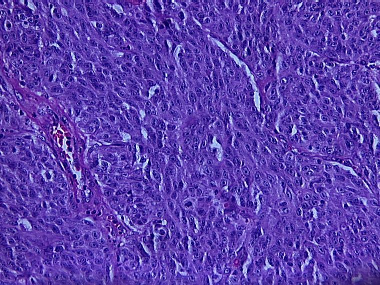
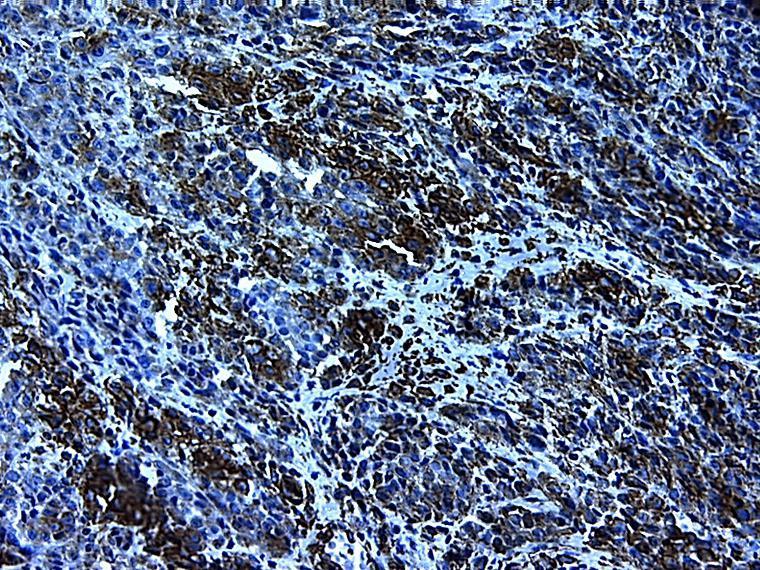
3 Clinically the lesion looked like reactive, or benign growth [Figure: 5]. On microscopic examination, tissue showed proliferating epitheloid cells arranged in nests. The cells exhibit cellular and nuclear pleomorphism. Abnormal and bizarre mitotic figures, cells unveiled loss of cohesiveness and infiltrating in deeper connective tissue were suggestive of malignant non epithelial neoplasm [Figure: 6]. Further investigations were done to get final diagnosis. Immunohistochemical slides showed strong positive for S-100, partial for vimentin, and diffuse for HMB-45[Figure: 7]. These findings indicate malignant condition of melanocytes, neural crest cell derivatives. 3. Discussion Melanocytes are dendritic cells derived from neural crest. They have various morphologies ranging from round, oval; fusiform to dendritic. Nevus cells are nondendritic melanocytes 3,7. Moles, or melanocytic nevi, are both markers of an increased risk of cutaneous melanoma and direct precursor lesions. Intradermal nevus can be differentiated from malignant melanoma, with good degree of certainty by clinical features like symmetric appearance, uniform pigmentation and slow rate of growth 8. Malignant melanoma can be diagnosed with ease when they present as extensively pigmented asymmetric growth with irregular borders. So histopathologically, while diagnosing various round, epitheloid, and spindle cell lesions, melanoma should be the differential until otherwise ruled out. Current strategies to minimize the morbidity and mortality associated with melanoma focus on scrutiny of individuals with identifiable moles and melanocytic nevi 8. Benign and atypical moles have been shown to exist in histologic contiguity with cutaneous melanomas, suggesting that these melanocytic proliferations are also susceptible to malignant transformation 9. Thus we conclude the melanocytic lesions myriad presentation need thorough clinical evaluation, keen histopathological examination and appropriate immunostaining as the need may be. References: 1. D.Eisen, Disorders of pigmentation in the oral cavity, Clin. Dermatol. 18(2000) A. Hemminki, The molecular basis and clinical aspects of Peutz Jeghers syndrome,cell.mol.lifesci.55(1999) Nanci A: Ten cate s oral histology- Development, structure and function: 8 th ed 2013; Elsevier 4. Kauzman A, Pavone M, Blanas N, Bradley G. Pigmented lesions of the oral cavity: review, differential diagnosis, and case presentation. J Can Dent Assoc. 2004;70: Gondak RO, Jorge RS, Jorge J, Lopes MA, Vargas PA; Oral pigmented lesions: Clinicopathologic features and review of the literature: Med Oral Patol Oral Cir Bucal Nov 1;17 (6):e Grichnik JM. Dermoscopy of melanocytic neoplasms: subpatterns of dysplastic/atypical nevi. Arch Dermatol. 2003;139: Rajendran R, B Shivapathasundaram: Shafer, Hine,Levy;Shafer s text book of oral pathology: 8 th ed. 2016; Elsevier 8. Tsao H, Bevona C, Goggins W, Quinn T; The Transformation Rate of Moles (Melanocytic Nevi) Into Cutaneous Melanoma: arch dermatol(vol 139), mar 2003: Page 7


4 9. Gruber SB, Barnhill RL, Stenn KS, Roush GC. Nevomelanocytic proliferations in association with cutaneous malignant melanoma: a multivariate analysis. J Am Acad Dermatol. 1989;21: Figures with Legends: Figure 1: Intradermal nevus Figure 4: Malignant Melanoma under Low and High Magnification Figure 2: H & E Staining Of Intradermal Nevus Figure 3: Malignant Melanoma Figure 5: Amelanotic Melanoma Page 8

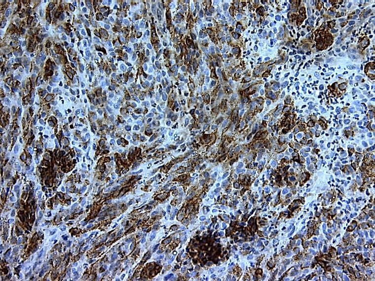
5 Figure 7: Vimantin and HMB 45 Immunohistochemical Staining Figure 6: H & E Staining of Amelanotic Melanoma Page 9
ORAL MELANOTIC NEVI: A CASE REPORT AND REVIEW OF LITERATURE
 ORAL MELANOTIC NEVI: A CASE REPORT AND REVIEW OF LITERATURE Abstract: *V.T Beena **Isha Chauhan *** R. Heera ***R. Rajeev Oral melanotic nevi are uncommon oral lesions causing focal. Melanotic nevi are
Benign and malignant epithelial lesions: Seborrheic keratosis: A common benign pigmented epidermal tumor occur in middle-aged or older persons more
 Benign and malignant epithelial lesions: Seborrheic keratosis: A common benign pigmented epidermal tumor occur in middle-aged or older persons more common on the trunk; but extremities, head and neck are
Desmoplastic Melanoma R/O BCC. Clinical Information. 74 y.o. man with lesion on left side of neck r/o BCC
 R/O BCC Sabine Kohler, M.D. Professor of Pathology and Dermatology Dermatopathology Service Stanford University School of Medicine Clinical Information 74 y.o. man with lesion on left side of neck r/o
Dermatopathology. Dr. Rafael Botella Estrada. Hospital La Fe de Valencia
 Dermatopathology Dr. Rafael Botella Estrada. Hospital La Fe de Valencia Melanoma and mimics Dr. Martin Mihm Malignant lesions result from the accumulation of mutations Class I lesions (benign) Class II
Pigmented lesions of the Oral cavity
 Oral medicine أ.م.د احسان عبد هللا كميل Pigmented lesions of the Oral cavity Pigmented oral lesions are a large group of disorders in which the dark or brown color is the essential clinical characteristic.
Financial disclosures
 Mesenchymal Neoplasms with Melanocytic Differentiation By Konstantinos Linos MD, FCAP, FASDP Bone, Soft Tissue and Dermatopathology Assistant Professor of Pathology Dartmouth-Hitchcock Medical Center Geisel
Morphologic characteristics of nevi associated with melanoma: a clinical, dermatoscopic and histopathologic analysis
 DERMATOLOGY PRACTICAL & CONCEPTUAL www.derm101.com Morphologic characteristics of nevi associated with melanoma: a clinical, dermatoscopic and histopathologic analysis Temeida Alendar 1, Harald Kittler
Malignant Peripheral Nerve Sheath Tumor
 C H A P T E R 120 Malignant Peripheral Nerve Sheath Tumor Currently, malignant peripheral nerve sheath tumor (MPNST) is the most commonly used generic name for the neoplasms known in the past as neurosarcoma,
Female 18. Deeply pigmented lesion on trunk.?warty naevus?seborrhoeic keratosis?malignant melanoma. The best diagnosis is:
 Female 18. Deeply pigmented lesion on trunk.?warty naevus?seborrhoeic keratosis?malignant melanoma. The best diagnosis is: A. deep penetrating naevus B. naevoid malignant melanoma C. pigment synthesising
Pathology of the skin. 2nd Department of Pathology, Semmelweis University
 Pathology of the skin 2nd Department of Pathology, Semmelweis University Histology of the skin Epidermis: Stratum corneum Stratum granulosum Stratum spinosum Stratum basale Dermis: papillary and reticular
Maligna Melanoma and Atypical Fibroxanthoma: An Unusual Collision Tumour G Türkcü 1, A Keleş 1, U Alabalık 1, D Uçmak 2, H Büyükbayram 1 ABSTRACT
 Maligna Melanoma and Atypical Fibroxanthoma: An Unusual Collision Tumour G Türkcü 1, A Keleş 1, U Alabalık 1, D Uçmak 2, H Büyükbayram 1 ABSTRACT Two different neoplasia in the same biopsy material called
Among the benign intraepithelial melanocytic proliferations, Inflamed Conjunctival Nevi. Histopathological Criteria. Resident Short Reviews
 Resident Short Reviews Inflamed conjunctival nevi (ICN) may suggest malignancy because of their rapid growth and atypical histology. The objective of this study was to characterize the diagnostic features
Malignant tumors of melanocytes: Part 1. Deba P Sarma, MD., Omaha
 Malignant tumors of melanocytes: Part 1 Deba P Sarma, MD., Omaha The melanocytic tumor is one of the most difficult and confusing areas in Dematopathology. It is true that most (95%) of such lesions are
المركب النموذج--- سبيتز وحمة = Type Spitz's Nevus, Compound SPITZ NEVUS 1 / 7
 SPITZ NEVUS 1 / 7 Epidemiology An annual incidence rate of 1.4 cases of Spitz nevus per 100,000 individuals has been estimated in Australia, compared with 25.4 per 100,000 individuals for cutaneous melanoma
Melanocytic Lesions: Use of Immunohistochemistry and Special Studies Napa Valley 2018
 Melanocytic Lesions: Use of Immunohistochemistry and Special Studies Napa Valley 2018 Victor G. Prieto, MD, PhD Professor Depts. of Pathology and Dermatology University of Texas - MD Anderson Cancer Center
Acquired melanocytic nevi in Egyptian patients: A clinicopathological study
 Acta Dermatovenerol APA Acta Dermatovenerologica Alpina, Pannonica et Adriatica ;:- doi:.8/v---8 Acquired melanocytic nevi in Egyptian patients: A clinicopathological study Mohamed A. El-Khalawany Abstract
Associate Clinical Professor of Dermatology MUSC
 Re-excision of Moderately Dysplastic Nevi: Should we or shouldn t we? John C. Maize, Jr, M.D. Dermatologist and Dermatopathologist Trident Dermatology, Charleston SC Associate Clinical Professor of Dermatology
Atypical Histologic Features in Melanocytic Nevi
 The American Journal of Dermatopathology 22(5): 391 396, 2000 2000 Lippincott Williams & Wilkins, Inc., Philadelphia Atypical Histologic Features in Melanocytic Nevi Carmelo Urso, M.D. The atypical histologic
Cytyc Corporation - Case Presentation Archive - March 2002
 FirstCyte Ductal Lavage History: 68 Year Old Female Gail Index: Unknown Clinical History: Negative Mammogram in 1995 6 yrs. later presents with bloody nipple discharge Subsequent suspicious mammogram Suspicious
The Relevance of Cytologic Atypia in Cutaneous Neural Tumors
 The Relevance of Cytologic Atypia in Cutaneous Neural Tumors Recent Findings - New Developments New Problems Zsolt B. Argenyi, M.D. Professor of Pathology & Dermatology Director of Dermatopathology Department
A Report of a Rare Case of Anaplastic Large Cell Lymphoma of the Oral Cavity
 AJMS Al Ameen J Med Sci (2 0 1 2 )5 (1 ):9 8-1 0 2 (A US National Library of Medicine enlisted journal) I S S N 0 9 7 4-1 1 4 3 C O D E N : A A J M B G CASE REPORT A Report of a Rare Case of Anaplastic
LENTIGO SIMPLEX. Epidemiology
 LENTIGO SIMPLEX Epidemiology The frequency of lentigo simplex in children and adults has not been determined. There does not appear to be a racial or gender predilection. Lentigo simplex is the most common
Atypical Nevi When to Re-excise. Catherine Barry, DO Dermatopathologist
 Atypical Nevi When to Re-excise Catherine Barry, DO Dermatopathologist Why talk about skin cancer? Because it s the most common type of cancer! Non-melanoma Skin Cancers Basal Cell Carcinoma Squamous Cell
Brief Report. Shivanand Gundalli 1, Smita Kadadavar 1, Somil Singhania 1, Rutuja Kolekar 2 INTRODUCTION. Melanocytic Nevus
 Our Dermatology Online Histopathological spectrum of benign melanocytic nevi our experience in a tertiary care centre Shivanand Gundalli 1, Smita Kadadavar 1, Somil Singhania 1, Rutuja Kolekar 2 1 Department
Dermatopathology: The tumor is composed of keratinocytes which show atypia, increase mitoses and abnormal mitoses.
 Squamous cell carcinoma (SCC): A common malignant tumor of keratinocytes arising in the epidermis, usually from a precancerous condition: 1- UV induced actinic keratosis, usually of low grade malignancy.
1/10/2018. Soft Tissue Tumors Showing Melanocytic Differentiation. Overview. Desmoplastic/ Spindle Cell Melanoma
 2016 MFMER slide-1 2016 MFMER slide-2 2016 MFMER slide-3 Soft Tissue Tumors Showing Melanocytic Differentiation Andrew L. Folpe, M.D. Professor of Laboratory Medicine and Pathology Mayo Clinic, Rochester,
Oral Amelanotic Melanoma of the Maxilla
 Case Report Nasrollah Saghravanian 1, Mahdi Pazouki 2, Maryam Zamanzadeh 3 1 Associated Professor, Department of Oromaxillofacial Pathologist, Oral and Maxillofacial Diseases Research Center, School of
Special slide seminar
 Special slide seminar Tomáš Rozkoš The Fingerland Department of Pathology Charles University Medical Faculty and Faculty Hospital in Hradec Králové Czech Republic Case history, 33 years old resistance
Neoplasia 2018 Lecture 2. Dr Heyam Awad MD, FRCPath
 Neoplasia 2018 Lecture 2 Dr Heyam Awad MD, FRCPath ILOS 1. List the differences between benign and malignant tumors. 2. Recognize the histological features of malignancy. 3. Define dysplasia and understand
NEOPLASMS OF THE SURFACE EPITHELIUM (KERATINOCYTES)
") NEOPLASMS OF THE SURFACE EPITHELIUM (KERATINOCYTES) Papillary Lesions Precancerous Lesions Keratinocyte Proliferations Carcinomas Melanotic Lesions Melanomas Normal Mucosa Keratin layer Spinous layer Basal
NEOPLASMS OF THE SURFACE EPITHELIUM (KERATINOCYTES) Papillary Lesions Precancerous Lesions Keratinocyte Proliferations Carcinomas Melanotic Lesions Melanomas Normal Mucosa Keratin layer Spinous layer Basal
Division of Pathology
 Case 38 Adult woman with a 35mm right breast lump at the 10 o clock position. Excision performed. (Case contributed by Dr Mihir Gudi, KKH) Division of Pathology Merlion, One Fullerton Singapore Diagnosis
ORAL MELANOMA Definition Epidemiology Clinical Presentation
 ORAL MELANOMA Definition Melanoma is a highly malignant neoplasia, arising from melanocytes, the cells that produce the brownish pigment melanin. Melanin is the determinant in skin colour and protects
Ameloblastic Carcinoma of the Mandible: a Rare Case Report
 IBIMA Publishing Journal of Research and Practice in Dentistry http://www.ibimapublishing.com/journals/dent/dent.html Vol. 2015 (2015), Article ID 672596, 6 pages DOI: 10.5171/2015.672596 Case Report Ameloblastic
World Articles of Ear, Nose and Throat Page 1
 World Articles of Ear, Nose and Throat ---------------------Page 1 Primary Malignant Melanoma of the Tongue: A Case Report Authors: Nanayakkara PR*, Arudchelvam JD** Ariyaratne JC*, Mendis K*, Jayasekera
IN THE NAME OF GOD Dr. Kheirandish Oral and maxillofacial pathology
 IN THE NAME OF GOD Dr. Kheirandish Oral and maxillofacial pathology ORAL FOCAL MUCINOSIS Uncommon Tumorlike Cutaneous myxoid cyst Overproduction of hyaluronic acid by firoblasts Young adults Female Gingiva
Multiple Primary Melanoma in a Thai Male: A Case Report
 Case Report Multiple Primary Melanoma in a Thai Male: A Case Report J Med Assoc Thai 2014; 97 (Suppl. 2): S234-S238 Full text. e-journal: http://www.jmatonline.com Kittisak Payapvipapong MD*, Pinyapat
Histopathology of Melanoma
 THE YALE JOURNAL OF BIOLOGY AND MEDICINE 48, 409-416 (1975) Histopathology of Melanoma G. J. WALKER SMITH Department ofpathology, Yale University School ofmedicine, 333 Cedar Street, New Haven, Connecticut
The Enigmatic Spitz Lesion
 The Enigmatic Spitz Lesion The Dawn of Spitz S Spitz Sophie Spitz Melanomas of Childhood ; Am J Pathol 1948 1910-1956 13 children (18 mo - 12 yrs) 12/13 had a benign clinical course Sophie Spitz Born 1910
Genetic Testing: When should it be ordered? Julie Schloemer, MD Dermatology
 Genetic Testing: When should it be ordered? Julie Schloemer, MD Dermatology Outline Germline testing CDKN2A BRCA2 BAP1 Somatic testing Gene expression profiling (GEP) BRAF Germline vs Somatic testing
MECHANISMS OF HUMAN DISEASE: LABORATORY SESSION PATHOLOGY OF THE SKIN LAB. Friday, February 13, :30 am 11:00 am
 MECHANISMS OF HUMAN DISEASE: LABORATORY SESSION PATHOLOGY OF THE SKIN LAB Friday, February 13, 2009 9:30 am 11:00 am FACULTY COPY GOALS: Describe the basic clinical and morphologic features of various
Disorders of Cell Growth & Neoplasia. Histopathology Lab
 Disorders of Cell Growth & Neoplasia Histopathology Lab Paul Hanna April 2010 Case #84 Clinical History: 5 yr-old, West Highland White terrier. skin mass from axillary region. has been present for the
Papillary Lesions of the Breast A Practical Approach to Diagnosis. (Arch Pathol Lab Med. 2016;140: ; doi: /arpa.
 Papillary Lesions of the Breast A Practical Approach to Diagnosis (Arch Pathol Lab Med. 2016;140:1052 1059; doi: 10.5858/arpa.2016-0219-RA) Papillary lesions of the breast Span the spectrum of benign,
Chapter 6 Squamous Cell Carcinoma: Variants and Challenges
 Chapter 6 Squamous Cell Carcinoma: Variants and Challenges Michael B. Morgan EPIDEMIOLOGY: Second most common skin cancer, rare in the dark-skinned races. ETIOLOGY: Ultraviolet light, HPV infection. PATHOGENESIS:
Case Series Oral lipomas: A report of two cases ABSTRACT
 International Journal of Medicine and Biomedical Research Volume 3 Issue 1 January April 2014 www.ijmbr.com Michael Joanna Publications Case Series Oral lipomas: A report of two cases Omisakin O.O 1*,
Histopathology: skin pathology
 Histopathology: skin pathology These presentations are to help you identify, and to test yourself on identifying, basic histopathological features. They do not contain the additional factual information
Dermoscopy: Recognizing Top Five Common In- Office Diagnoses
 Dermoscopy: Recognizing Top Five Common In- Office Diagnoses Vu A. Ngo, DO Department of Family Medicine and Dermatology Choctaw Nation Health Services Authority Learning Objectives Introduction to dermoscopy
Lichenoid Tissue Reaction in Malignant Melanoma A Potential Diagnostic Pitfall
 natomic Pathology / LICHENOID TISSUE RECTION IN MLIGNNT MELNOM Lichenoid Tissue Reaction in Malignant Melanoma Potential Diagnostic Pitfall CPT Scott R. Dalton, MC, US, 1,3 Capt Matt. aptista, USF, MC,
Finding Melanoma. Is not easy!
 Finding Melanoma Is not easy! Finding Melanoma Victoria mean depth at diagnosis is 1.5 mm. Melanoma 1.5mm Has Stage 1B Mortality 10% Melanoma Spotting a killer! Spotting a killer Visual Clues What are
number Done by Corrected by Doctor Maha Shomaf
 number 16 Done by Waseem Abo-Obeida Corrected by Zeina Assaf Doctor Maha Shomaf MALIGNANT NEOPLASMS The four fundamental features by which benign and malignant tumors can be distinguished are: 1- differentiation
Basics in Dermoscopy
 Basics in Dermoscopy Manal Bosseila Professor of Dermatology, Cairo University Member of European Academy Dermatology & Venereology EADV Member of International Dermoscopy Society IDS Member of Aesthetic
Papillary Lesions of the breast
 Papillary Lesions of the breast Emad Rakha Professor of Breast Pathology The University of Nottingham Papillary lesions of the breast are a heterogeneous group of disease, which are characterised by neoplastic
A PRACTICAL APPROACH TO ATYPICAL MELANOCYTIC LESIONS BIJAN HAGHIGHI M.D, DIRECTOR OF DERMATOPATHOLOGY, ST. JOSEPH HOSPITAL
 A PRACTICAL APPROACH TO ATYPICAL MELANOCYTIC LESIONS BIJAN HAGHIGHI M.D, DIRECTOR OF DERMATOPATHOLOGY, ST. JOSEPH HOSPITAL OBJECTIVES Discuss current trends and changing concepts in our understanding of
Pathology. Skin Tumor. Bayan N. Mohammad 15/10/2015. Mohammad al-orjani. Page 0 of 23
 #7 35 Pathology Skin Tumor Bayan N. Mohammad 15/10/2015 Mohammad al-orjani Page 0 of 23 بسم هللا الرحمن الرحيم GREETINGS This lecture is about skin tumors, all the slides are included and every slide will
Case Report A Rare Cutaneous Adnexal Tumor: Malignant Proliferating Trichilemmal Tumor
 Case Reports in Medicine Volume 2015, Article ID 742920, 4 pages http://dx.doi.org/10.1155/2015/742920 Case Report A Rare Cutaneous Adnexal Tumor: Malignant Proliferating Trichilemmal Tumor Omer Alici,
Nasal Cavity and Paranasal Sinuses
 Chapter 2 Nasal Cavity and Paranasal Sinuses Introduction Included in this chapter are nasal cavities, frontal sinus, ethmoid complex, sphenoid sinus, and maxillary sinuses. These cavities and sinuses
BASAL CELL CARCINOMA WITH ECCRINE DIFFERENTIATION: A RARE ENTITY Divvya B 1, Rehana Tippoo 2, P. Viswanathan 3, B. Krishnaswamy 4, A.
 BASAL CELL CARCINOMA WITH ECCRINE DIFFERENTIATION: A RARE ENTITY Divvya B 1, Rehana Tippoo 2, P. Viswanathan 3, B. Krishnaswamy 4, A. Anvar Ali 5 HOW TO CITE THIS ARTICLE: Divvya B, Rehana Tippoo, P. Viswanathan,
Morphologic Features of Melanocytes, Pigmented Keratinocytes, and Melanophages by In Vivo Confocal Scanning Laser Microscopy
 Morphologic Features of Melanocytes, Pigmented Keratinocytes, and Melanophages by In Vivo Confocal Scanning Laser Microscopy Klaus J. Busam, M.D., Carlos Charles, M.D., Grace Lee, M.D., Allan C Halpern,
Abrupt Intralesional Color Change on Dermoscopy as a New Indicator of Early Superficial Spreading Melanoma in a Japanese Woman
 Published online: June 24, 2015 1662 6567/15/0072 0123$39.50/0 This is an Open Access article licensed under the terms of the Creative Commons Attribution-NonCommercial 3.0 Unported license (CC BY-NC)
Ocular Neoplasia What s Common? What s New? Richard R Dubielzig
 Ocular Neoplasia What s Common? What s New? Richard R Dubielzig Orbit 288 6% Tumors of the globe make up 3225 out of 6110 total neoplasms = 53%. Tumors of the conjunctiva make up 1192 out of 6110 total
Primary Mucosal Desmoplastic Melanoma of Gingiva
 The Korean Journal of Pathology 2006; 40: 456-60 Primary Mucosal Desmoplastic Melanoma of Gingiva - A Case Report - Gawon Choi Jeong Won Kim Soon Yuhl Nam 1 Kyung-Ja Cho Departments of Pathology and 1
Asadi-Amoli et al Adenocarcinoma of RPE Iranian Journal of Ophthalmology - Volume 19, Number 4, 2007
 Adenocarcinoma of Retinal Pigment Epithelium Clinically Diagnosed as Malignant Melanoma; A Case Report with Unsystematic Review of Literature Fahimeh Asadi-Amoli, MD, 1 Hedyeh Moradi, MD 2 Mohammad-Taher
MECHANISMS OF HUMAN DISEASE: LABORATORY SESSION PATHOLOGY OF THE SKIN LAB. Friday, February 12, :30 am 11:00 am
 MECHANISMS OF HUMAN DISEASE: LABORATORY SESSION PATHOLOGY OF THE SKIN LAB Friday, February 12, 2012 9:30 am 11:00 am FACULTY COPY GOALS: Describe the basic clinical and morphologic features of various
Update on Spitzoid and Blue nevus-like melanocytic lesions Emphasis on molecular studies informing diagnosis, prognosis and therapy
 Update on Spitzoid and Blue nevus-like melanocytic lesions Emphasis on molecular studies informing diagnosis, prognosis and therapy Michael T. Tetzlaff MD, PhD Associate Professor Department of Pathology,
Simulators of melanoma
 Simulators of melanoma Philip E. LeBoit, M.D. Depts. of Pathology and Dermatology University of California, San Francisco Simulators of melanoma Simulators of melanoma in situ Melanocytic Non-melanocytic
Diagnostic Value of Fluorescence Method on Melanoma in Dogs
 Jpn J Vet Dermatol 2003; 9 (4): 159 164 Diagnostic Value of Fluorescence Method on Melanoma in Dogs Masahiko Nagata 1), Atsuhiko Hasegawa 2) 1) Animal Dermatology Center, ASC 2) Department of Pathobiology,
CINtec p16 INK4a Staining Atlas
 CINtec p16 INK4a Staining Atlas Rating Rating Positive The rating positive will be assigned if the p16 INK4a -stained slide shows a continuous staining of cells of the basal and parabasal cell layers of
Prepared By Jocelyn Palao and Layla Faqih
 Prepared By Jocelyn Palao and Layla Faqih The structure of the suspected atypical cell should always be compared to the structure of other similar, benign, cells which are present in the smears. The diagnosis
أملس عضلي غرن = Leiomyosarcoma. Leiomyosarcoma 1 / 5
 Leiomyosarcoma 1 / 5 EPIDEMIOLOGY Exact incidence is unknown, but older studies suggest that leiomyosarcomas comprise approximately 3 percent of soft-tissue sarcomas. Superficial leiomyosarcoma occurs
Management of pediatric melanocytic lesions
 Open Journal of Clinical & Medical Case Reports Management of pediatric melanocytic lesions Volume 3 (2017) Issue 8 ISSN 2379-1039 Jin Kim, BS; Emmanuel Gabriel MD, PhD; Weiguo Liu MD, PhD; Lin Lin MD,
David B. Troxel, MD. Common Medicolegal Situations: Misdiagnosis of Melanoma
 Common Medicolegal Situations: Misdiagnosis of Melanoma David B. Troxel, MD Medical Director, The Doctors Company, Napa, California Clinical Professor Emeritus, University of California at Berkeley Past
Senior lecturer Department of Oral Medicine and Radiology, Maharaja Ganga Singh Dental College and Research Center, Sriganganagar
 Medicine Case Report 1 2 2 2 3 Dr. Abhayjeet Singh, Dr. Ashish Kumar, Dr. Vikas Berwal, Dr. Pushpchander Swami, Dr. Manjit Kaur Singh A, Kumar A, Berwal V, Swami P, Kaur M.. J Periodontal Med Clin Pract
Case Report Clear Cell Basal Cell Carcinoma
 SAGE-Hindawi Access to Research Volume 2011, Article ID 386921, 4 pages doi:10.4061/2011/386921 Case Report Clear Cell Basal Cell Carcinoma Deba P. Sarma, 1 Daniel Olson, 1 Jennifer Olivella, 1 Tracey
Appendix : Dermoscopy
 Go Back to the Top To Order, Visit the Purchasing Page for Details APP Appendix : Dermoscopy Dermoscopy, also known as dermatoscopy, epiluminoscopy and epiluminescent microscopy, is an effective non-invasive
Nasal mucosal melanosis may act as a harbinger of melanoma: A case report
 Nasal mucosal melanosis may act as a harbinger of melanoma: A case report The Harvard community has made this article openly available. Please share how this access benefits you. Your story matters. Citation
CHONDROID CHORDOMA OF NASAL SEPTUM: A CASE REPORT Gayattre Kailas 1, Muniraju M 2, Archana Mathri 3
 CHONDROID CHORDOMA OF NASAL SEPTUM: A Gayattre Kailas 1, Muniraju M 2, Archana Mathri 3 HOW TO CITE THIS ARTICLE: Gayattre Kailas, Muniraju M, Archana Mathri. Chondroid Chordoma of Nasal Septum: A Case
F006 Imaging in Dermatology Melanocytic Neoplasia Clinical-Confocal-Pathological-Correlations
 F006 Imaging in Dermatology Melanocytic Neoplasia Clinical-Confocal-Pathological-Correlations Melissa Gill, MD SkinMedical Research and Diagnostics Dobbs Ferry, NY, USA Department of Pathology SUNY Downstate
Case 4 Diagnosis 2/21/2011 TGB
 Case 4 22 year old female presented with asymmetric enlargement of the left lobe of the thyroid gland. No information available relative to a prior fine needle aspiration biopsy. A left lobectomy was performed.
Case year old female presented with asymmetric enlargement of the left lobe of the thyroid
 Case 4 22 year old female presented with asymmetric enlargement of the left lobe of the thyroid gland. No information available relative to a prior fine needle aspiration biopsy. A left lobectomy was performed.
1 NORMAL HISTOLOGY AND METAPLASIAS
 1 NORMAL HISTOLOGY AND METAPLASIAS, MD Anatomy and Histology 1 Metaplasias 2 ANATOMY AND HISTOLOGY The female breast is composed of a branching duct system, which begins at the nipple with the major lactiferous
International Society of Gynecological Pathologists Symposium 2007
 International Society of Gynecological Pathologists Symposium 2007 Anais Malpica, M.D. Department of Pathology The University of Texas M.D. Anderson Cancer Center Grading of Ovarian Cancer Histologic grade
CASE REPORT Benign epithelioid peripheral nerve sheath tumour resembling schwannoma
 Malaysian J Pathol 2014; 36(3) : 217 221 CASE REPORT Benign epithelioid peripheral nerve sheath tumour resembling schwannoma Thejasvi KRISHNAMURTHY MD and SR NIVEDITHA MD, DNB Department of Pathology,
Ancient schwannoma of the mouth floor A case report and review
 Oral Oncology EXTRA (2006) 42, 281 285 available at www.sciencedirect.com journal homepage: http://intl.elsevierhealth.com/journal/ooex CASE REPORT Ancient schwannoma of the mouth floor A case report and
Mody. AIS vs. Invasive Adenocarcinoma of the Cervix
 Common Problems in Gynecologic Pathology Michael T. Deavers, M.D. Houston Methodist Hospital, Houston, Texas Common Problems in Gynecologic Pathology Adenocarcinoma in-situ (AIS) of the Cervix vs. Invasive
Guy Perrot (Ги Перро)
") НАУЧНО-ПРАКТИЧЕСКАЯ КОНФЕРЕНЦИЯ (МАСТЕР-КЛАСС) «ПРАКТИЧЕСКИЕ АСПЕКТЫ ДИАГНОСТИКИ И ЛЕЧЕНИЯ МЕЛАНОМЫ КОЖИ» DIAGNOSTIC AND PITFALLS IN MELANOMA Guy Perrot (Ги Перро) MD PHD pathologist, University Hospital
НАУЧНО-ПРАКТИЧЕСКАЯ КОНФЕРЕНЦИЯ (МАСТЕР-КЛАСС) «ПРАКТИЧЕСКИЕ АСПЕКТЫ ДИАГНОСТИКИ И ЛЕЧЕНИЯ МЕЛАНОМЫ КОЖИ» DIAGNOSTIC AND PITFALLS IN MELANOMA Guy Perrot (Ги Перро) MD PHD pathologist, University Hospital
We are IntechOpen, the world s leading publisher of Open Access books Built by scientists, for scientists. International authors and editors
 We are IntechOpen, the world s leading publisher of Open Access books Built by scientists, for scientists 3,500 108,000 1.7 M Open access books available International authors and editors Downloads Our
ELECTRON MICROSCOPY OF A SMALL PIGMENTED CUTANEOUS LESION*
 ELECTRON MICROSCOPY OF A SMALL PIGMENTED CUTANEOUS LESION* The description of the lesion in the title of this rcport is intentionally non-committal. Diagnosed clinically as a lentigo, it was removed as
Diplomate of the American Board of Pathology in Anatomic and Clinical Pathology
 A 33-year-old male with a left lower leg mass. Contributed by Shaoxiong Chen, MD, PhD Assistant Professor Indiana University School of Medicine/ IU Health Partners Department of Pathology and Laboratory
Clear Cell Sarcoma of the Foot: A Case Report of Malignant Melanoma of Soft Parts
 Clear Cell Sarcoma of the Foot: A Case Report of Malignant Melanoma of Soft Parts by Al Kline, DPM The Foot & Ankle Journal 1 (3): 3 A case report is presented describing a clear cell sarcoma of the foot
BREAST PATHOLOGY. Fibrocystic Changes
 BREAST PATHOLOGY Lesions of the breast are very common, and they present as palpable, sometimes painful, nodules or masses. Most of these lesions are benign. Breast cancer is the 2 nd most common cause
Benign versus Cancerous Lesions How to tell the difference FMF 2014 Christie Freeman MD, CCFP, DipPDerm, MSc
 1 Benign versus Cancerous Lesions How to tell the difference FMF 2014 Christie Freeman MD, CCFP, DipPDerm, MSc Benign lesions Seborrheic Keratoses: Warty, stuck-on Genetics and birthdays Can start in late
Basal cell carcinoma 5/28/2011
 Goal of this Presentation A practical approach to the diagnosis of cutaneous carcinomas and their mimics Thaddeus Mully, MD University of California San Francisco To review common non-melanoma skin cancers
Periocular Malignancies
 Periocular Malignancies Andrew Gurwood, O.D., F.A.A.O., Dipl. Marc Myers, O.D., F.A.A.O. Drs. Myers and Gurwood have no financial interests to disclose. Course Description Discussion of the most common
Pigmented Lesions of the Oral Mucosa
 Pigmented Lesions of the Oral Mucosa Eric T. Stoopler and Faizan Alawi Abstract Pigmented lesions of the oral mucosa are encountered on a routine basis in clinical practice. Oral health-care providers
Enterprise Interest Nothing to declare
 Enterprise Interest Nothing to declare Diagnoses one would not like to miss in soft tissue pathology early in your career Marta Sbaraglia, MD Department of Pathology Hospital of Treviso University of Padua
Gastrooesophageal reflux disease. Jera Jeruc Institute of pathology, Faculty of Medicine, Ljubljana, Slovenia
 Gastrooesophageal reflux disease Jera Jeruc Institute of pathology, Faculty of Medicine, Ljubljana, Slovenia Reflux esophagitis (RE) GERD: a spectrum of clinical conditions and histologic alterations resulting
Total body photography in high risk patients
 Total body photography in high risk patients Doug Grossman, MD, PhD Department of Dermatology Huntsman Cancer Institute University of Utah Summer AAD F032 Practical Considerations for Patients with Melanoma
Case RAC7783. M46. Ear. Mole. r/o MM.?Blue naevus RAC7783
 Case RAC7783. M46. Ear. Mole. r/o MM.?Blue naevus RAC7783 Pie Chart Participants N=74 Benign: 48 N=74 Blue naevus: 38 Intradermal: 12 DPN: 10 Compound 3 Clonal: 3; Spitz 2; Special Site: 1; Congenital:
Salivary Glands 3/7/2017
 Salivary Glands 3/7/2017 Goals and objectives Focus on the entities unique to H&N Common board type facts Information for your future practice Salivary Glands Salivary Glands Major gland. Paratid. Submandibular.
Melanocytic proliferations in sundamaged
 Atypical Spitzoid Tumor: What Does It Mean And How Should It Be Managed? Melanocytic proliferations in sundamaged skin Jane L. Messina, Jane L. Messina MD International Melanoma Pathology Working Group