Non-Melanocytic Pattern Dermoscopy
|
|
|
- Tobias Stafford
- 5 years ago
- Views:
Transcription


 Comma-like - IDN Dotted -Spitz Linear irregular -")

 D. Lines looped (hairpin vessels)(scc) E.")
 (SCCIS) H.")
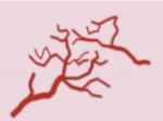
1 Non-Melanocytic Pattern Dermoscopy I have no conflicts of interest to disclose Except that I LOVE dermoscopy Michelle Tarbox, MD Assistant Professor of Dermatology and Dermatopathology Texas Tech University Health Sciences Center Non-Melanocytic Lesion Dermoscopy Often also non-pigmented Problem: No Pigment! Solution: Use your clues! Vascular structures Chrysalis structures Texture Structureless areas Scale Ulceration Vascular morphology J Am Acad Dermatol - 01-SEP-2010; 63(3) Comma-like - IDN Dotted -Spitz Linear irregular - AMM Hairpin SK, SCC, KA Glomerular - SCCIS Arborizing - BCC 3 4 Crown vessels Seb H Strawberry pattern - AK Milky red areas/globules Thick AMM A regular B in a string C clustered D radial E irregularly branched F irregular A. Dots (melanoma) B. Clods (lacunes) (hemangioma) C. Lines straight (ulcerated BCC) D. Lines looped (hairpin vessels)(scc) E. Lines curved (comma vessels) (dermal nevus) F. Lines serpentine (linear irregular vessels (BCC) G. Lines helical (corkscrew vessels) (SCCIS) H. Lines coiled (glomerular vessels) (SCCIS 5 6 J Am Acad Dermatol - 01-SEP-2010; 63(3) Rosendahl, Cliff et al. Dermatology Practical & Conceptual4.1 (2014): PMC. Web. 9 Sept
 (CCA) D. Linear (SCCIS) E.")
 (SCC) G.")


: 59 66. PMC.")

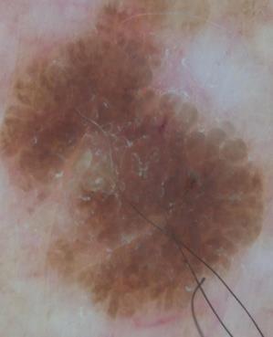
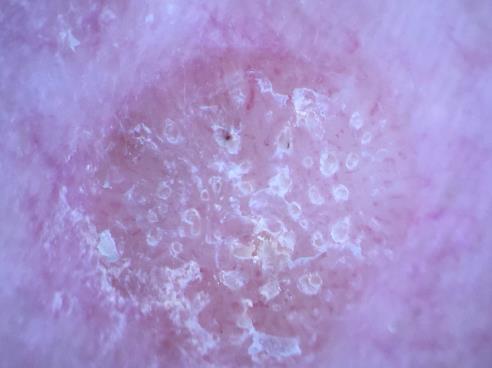
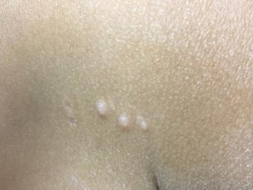
2 A. Random (non-specific) (SCCIS) B. Clustered (SCCIS) C. Serpiginous (string of pearls) (CCA) D. Linear (SCCIS) E. Centered (seborrheic keratosis) F. Radial (starburst) (SCC) G. Reticular (sun-damaged skin) Non-Melanocytic Lesions Benign Lesions Seborrheic keratoses Clear cell acanthoma Vascular lesions Angiomas Angiokeratomas Dermatofibromas Conventional Cellular Malignant Lesions Basal cell carcinoma Actinic keratoses Squamous cell carcinomas Merkel cell carcinoma H. Branched (arborizing) (BCC) Rosendahl, Cliff et al. Dermatology Practical & Conceptual4.1 (2014): PMC. Web. 9 Sept , patient granted special permission to show tattoo 9 10 Milia-like cysts = pseudo-horn cysts (black arrows) Comedo-like openings = comedo structures (red arrows) Multiple milia like cysts


3 13 14 Irregular crypts and comedo like openings Fissures and Ridges Wedge shaped clefts in the epidermis AKA gyri and sulci, fat fingers, or cerebriform pattern Can also be seen in melanocytic nevi with congenital patterns, and epidermal nevi

4 Fissures/ridges Fissures/ridges Congenital type nevus with Fissures/ridges

5


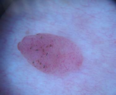
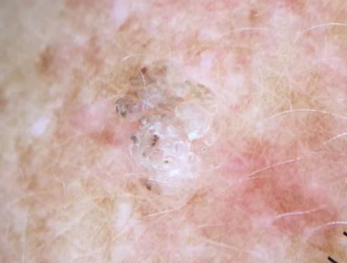
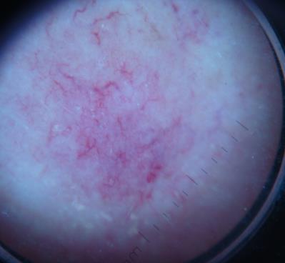
6 Clear cell acanthoma String of pearls vessels Glycogen rich keratinocytes


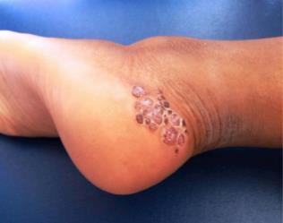
7 Hemangioma Red-blue homogeneous color Red-blue lacunes Angiokeratoma Multiple red to bluish-black well defined lacunae Blue-white veil: no diagnostic significance Red-blue lacunae, no pigment network Hyperkeratosis over thrombosed vessels Acral pseudolymphomatous angiokeratoma of children with rainbow pattern: A mimicker of Kaposi sarcoma Pinos León, Víctor Hugo et al. Journal of the American Academy of Dermatology, Volume 76, Issue 2, S25 - S


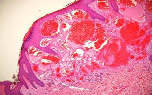
8 Multinucleate Cell Angiohistiocytoma Multinucleated cells in the superficial dermis with an angular outline (detailed in lower right corner, Hematoxylin & eosin, X100). Proliferation of small dilated vessels, slight interstitial infiltrate of spindle or dendritic perivascular cells, mostly lymphohistiocytic Rato M, Monteiro AF, Parente J, Aranha J. Case for diagnosis. Multinucleated cell angiohistiocytoma. Anais Brasileiros de Dermatologia. 2018;93(2):


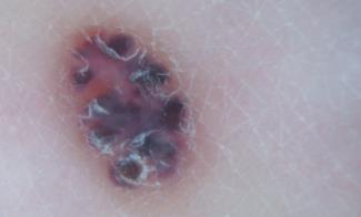
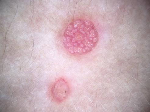
9 Dermatofibroma Central scar like pallor Surrounding delicate pigment network Chrysalis structures Diffuse reddish areas (attributed to the dilated vessels), whitish patches (associated with thickening of the collagen) and isolated peripheral areas with a fine reticulated appearance Cellular Dermatofibroma Nodule with central scar like pallor Surrounding delicate pigment network Chrysalis structures / central erosion/focal ulceration BROWN BROWN


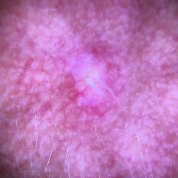
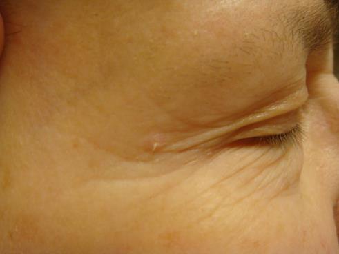
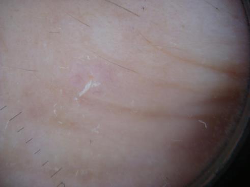
10 Ulceration without a clear history of trauma should lead to a biopsy Serum leaking from ulcerated areas may trap fibers of clothing or loose hair Adherent fiber ~ dermoscopic clue to ulceration
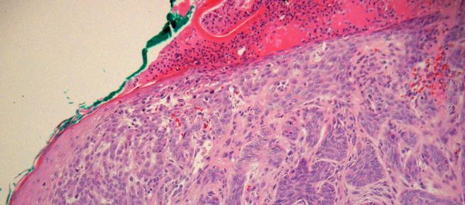
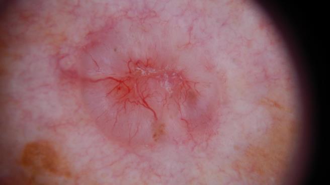
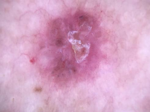
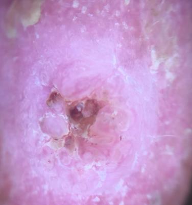
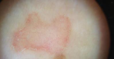
11 Ulceration Ulceration Seen in pigmented and non-pigmented basal cell carcinoma Arborizing vessels


12 Large Blue-Gray Ovoid Nests Well circumscribed, confluent, pigmented ovoid areas Larger than globules Not connected to larger tumor body Represent large nests of pigmented BCC Large Blue-Gray Ovoid Nests Benign intradermal vs Basal cell carcinoma Multiple Blue-Gray Globules Round, well circumscribed structures Smaller nests of pigmented basal cells Multiple Blue-Gray Globules


13 Maple Leaf Areas Nests of pigmented epithelial nodules of basal cell carcinoma Spoke-wheel areas Maple leaf areas Spoke-Wheel Areas Well circumscribed brown to gray-blue-brown radial projections meeting at a darker central hub Nests of basal cell carcinoma radiating from a follicular epithelium Chrysalis Structures AKA Crystalline structures or shiny white streaks (SWS) Only seen with polarized dermoscopy Most commonly seen in basal cell carcinoma and invasive melanomas, may be seen in dermatofibromas and scars In melanomas may reflect increased tumor thickness and regression BCC Invasive Melanoma Aneurysmal DF 13


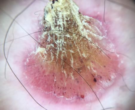
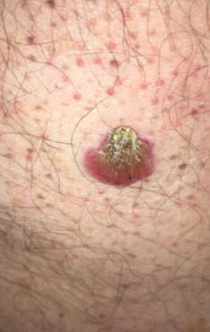
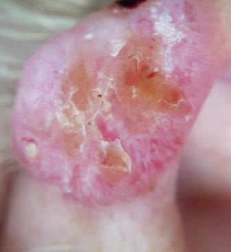
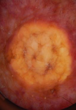
14 BCC BCC BCC Fibroepithelioma of Pinkus Chrysalis structures on Dermoscopy Dermatol Pract Concept 2013;4(1):9 Non pigmented Actinic Keratoses Pink-red pseudo-network surrounding follicles White-to-yellow surface scale Fine wavy vessels surrounding hair follicles Yellowish keratotic plugs in follicular ostia Strawberry-like pattern composed of reddish pseudo network around whitish keratin filled hair follicles Actinic keratosis 14


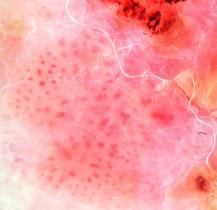
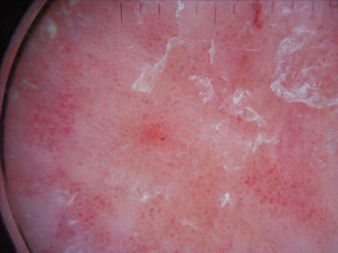

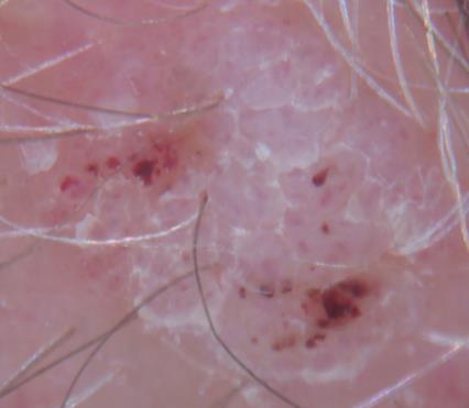
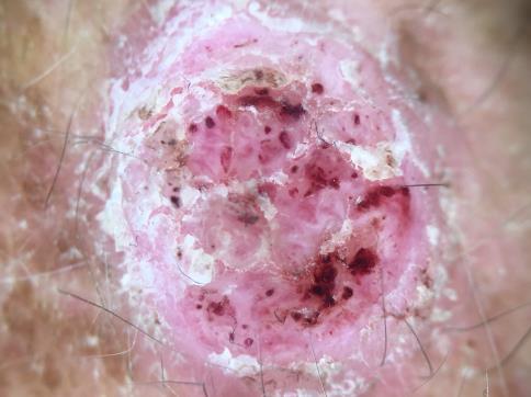
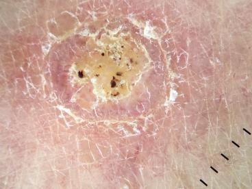
15 Pigmented actinic keratosis Pigmented actinic keratosis Squamous Cell Carcinoma in Situ Glomeruloid blood vessels Focal heme crust Scale

16

")
 vessels on a")


, arborizing (blue arrow), coiled (black")
 ->nmass of highly keratinized")


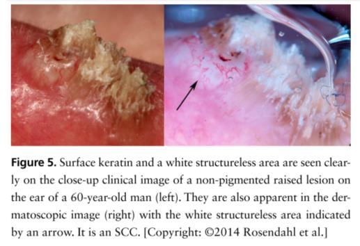
17 Dermoscopic features of squamous cell carcinoma of the tongue: It looks similar to cutaneous squamous cell carcinoma Lobules composed of central whitish yellow keratin Polymorphous vessels dotted (yellow arrow) Arborizing (blue arrow), glomerular (black circle), and linear irregular (red arrow) vessels on a yellowish white background Journal of the American Academy of Dermatology, Volume 75, Issue 2, 2016, e53 e Diagnosing squamous cell carcinoma of the lip using dermoscopy Central whitish yellow keratin surrounded by polymorphous vessels Dotted (yellow arrow), arborizing (blue arrow), coiled (black circle), and hairpin (red arrow) White structureless areas (black arrow) ->nmass of highly keratinized malignant squamous cells Thin white cylinder with a hair shaft emerging from the center of it This finding is produced by highly keratinized malignant squamous cells invading hair follicles. Journal of the American Academy of Dermatology, Volume 76, Issue 2, Supplement 1, 2017, S82 S83 [Copyright: 2014 Rosendahl et al.] Dermatol Pract Concept 2013;4(1):


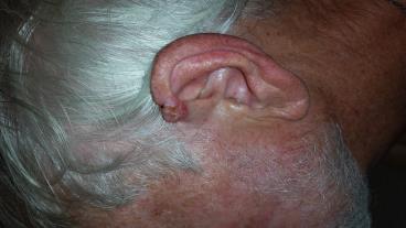
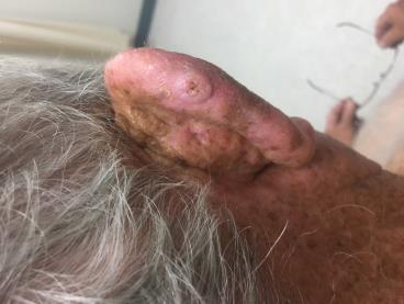
18 Merkel Cell Carcinoma
![Clinical and dermoscopic features of combined cutaneous squamous cell carcinoma (SCC)/neuroendocrine [Merkel cell] carcinoma (MCC)](/docs-images/95/124189316/images/19-0.jpg "Combined squamous/merkel cell carcinoma, A.")
 and large-diameter arborizing vessels at the periphery (larger lower")

19 Clinical and dermoscopic features of combined cutaneous squamous cell carcinoma (SCC)/neuroendocrine [Merkel cell] carcinoma (MCC) Combined squamous/merkel cell carcinoma, A. Erythematous nodule with adherent scale; background dermatoheliosis with atrophy and wrinkling, B. Dermoscopy with milky red areas centrally (small top arrow) and large-diameter arborizing vessels at the periphery (larger lower arrows) Journal of the American Academy of Dermatology, Volume 73, Issue 6, 2015, The most likely diagnosis is: A. Stasis dermatitis B. Actinic keratosis C. Porokeratosis D. Clear cell acanthoma E. Squamous cell carcinoma in situ The most likely diagnosis is: A. Stasis dermatitis B. Actinic keratosis C. Porokeratosis D. Clear cell acanthoma E. Squamous cell carcinoma in situ


20 Dermoscopic findings Glomeruloid blood vessels Focal heme crust Scale Diagnosis: Squamous Cell Carcinoma In Situ The most likely diagnosis is: A. Actinic keratosis B. Inflamed seborrheic keratosis C. Atypical nevus D. Clear cell acanthoma E. Basal cell carcinoma The most likely diagnosis is: A. Actinic keratosis B. Inflamed seborrheic keratosis C. Atypical nevus D. Clear cell acanthoma E. Basal cell carcinoma


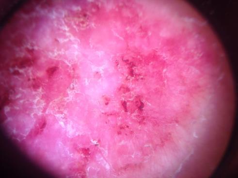
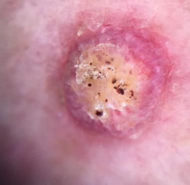
21 Dermoscopic features String of pearls blood vessels Diagnosis: Clear cell acanthoma The most likely diagnosis is: A. Basal cell carcionoma B. Inflamed seborrheic keratosis C. Amelanotic melanoma D. Merkel cell carcinoma E. Squamous cell carcinoma The most likely diagnosis is: A. Basal cell carcionoma B. Inflamed seborrheic keratosis C. Amelanotic melanoma D. Merkel cell carcinoma E. Squamous cell carcinoma


22 The most likely diagnosis is: A. Nodular melanoma B. Inflamed seborrheic keratosis C. Atypical nevus D. Angiokeratoma E. Pigmented basal cell carcinoma The most likely diagnosis is: A. Nodular melanoma B. Inflamed seborrheic keratosis C. Atypical nevus D. Angiokeratoma E. Pigmented basal cell carcinoma


23 Dermoscopic features Asymmetric Blue grey ovoid nests Arborizing telangiectasias Chrysalis structures Epidermal ulceration Diagnosis: Pigmented basal cell carcinoma Thank you! Michelle Tarbox, MD Assistant Professor of Dermatology Texas Tech University Health Sciences Center Scabies Triangle indicating the head of the mite


24 Fun Benign things

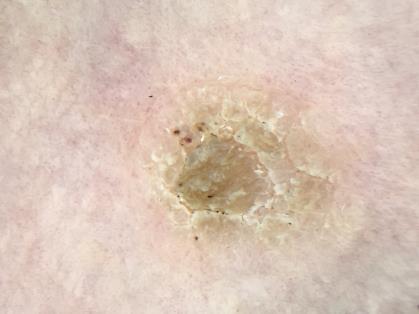
25 Porokeratosis White tract structure =cornoid lamella Central white area, red dots, globules and lines


26 Sebaceous Hyperplasia Aggregated white-yellow nodules ~ cumulous cloud Crown vessels (radial wreath-like) Banching vessels that extend towards the center of the lesion without crossing it


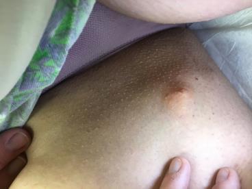
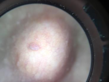
27 Nevus Sebaceous Accessory Nipple Central white area Central streak Faint pigmented network at the periphery Xanthogranuloma Orange-yellow background coloration with clouds of xanthomatous deposits


28 Thank you! Michelle Tarbox, MD Assistant Professor of Dermatology Texas Tech University Health Sciences Center
Non-melanocytic Patterns
 Non-melanocytic Lesions Non-melanocytic Patterns Michelle Tarbox, MD Assistant Professor of Dermatology and Dermatopathology Texas Tech University Health Sciences Center 2018 Seborrheic keratoses Acanthotic
comedo-like openings (clods, brown or orange & circles) milia-like cysts (dots or clods, white) 1/29/18 Dotted vessels are also commonly seen in SCC
 milia-like cysts (dots or clods, white) 1/29/18 Dotted vessels are also commonly seen in SCC") Brown circles Dotted vessels are also commonly seen in SCC Step1 1. Nevus (unequivocal) 2. DF/IDN 3. BCC 4. SCC Network Patchy network Peripheral network & central hypopigmentation DF: network with central
Brown circles Dotted vessels are also commonly seen in SCC Step1 1. Nevus (unequivocal) 2. DF/IDN 3. BCC 4. SCC Network Patchy network Peripheral network & central hypopigmentation DF: network with central
Dermoscopy: Recognizing Top Five Common In- Office Diagnoses
 Dermoscopy: Recognizing Top Five Common In- Office Diagnoses Vu A. Ngo, DO Department of Family Medicine and Dermatology Choctaw Nation Health Services Authority Learning Objectives Introduction to dermoscopy
Disclosure. Objectives. PAFP CME Conference Lou Mancano MD, FAAFP Reading Health System November 18, 2016
 PAFP CME Conference Lou Mancano MD, FAAFP Reading Health System November 18, 2016 1 Disclosure The speaker has no conflict of interest, financial agreement, or working affiliation with any group or organization.
It can be helpful in some cases of actinic keratosis, Bowen s disease and squamous cell carcinoma
 Dermoscopy Introduction, Terminology and Structures (to be read in conjunction with the Diagnostic Dermoscopic Algorithm) Copyright to Cunliffe TP (Jan. 2017) All rights reserved Introduction Dermoscopy
Appendix : Dermoscopy
 Go Back to the Top To Order, Visit the Purchasing Page for Details APP Appendix : Dermoscopy Dermoscopy, also known as dermatoscopy, epiluminoscopy and epiluminescent microscopy, is an effective non-invasive
Malignant non-melanocytic lesions
 Malignant non-melanocytic lesions Course C023: Fundamentals of Dermoscopy March 4, 2019, 11:20 AM - 11:50 PM Room: 146B Jason B. Lee, MD Professor & Vice Chair Director of Dermatopathology & Pigmented
Key factors in successfully integrating dermoscopy into your clinical practice
 Key factors in successfully integrating dermoscopy into your clinical practice S051 Dilemmas and challenges in skin cancer therapies and management Monday, March 4 th 2019 (9AM-12PM) Room 209A 10:56-11:09AM
Dermoscopy in everyday practice. What and Why? When in doubt cut it out? Trilokraj Tejasvi MD
 Dermoscopy in everyday practice Trilokraj Tejasvi MD Assistant Professor, Department of Dermatology, Director Teledermatology services, University of Michigan, Faculty Associate, GLOBAL REACH, Michigan
22/04/2015. Dermoscopy of Melanoma. Ilsphi Browne. Overview
 Dermoscopy of Melanoma Ilsphi Browne Overview The device Dermoscopic criteria (terminology) Colour Patterns Global features Local features Approach to diagnosing pigmented lesions Other uses in general
6/17/2018. Breaking Bad (Part 1) Dermoscopy of Brown(ish) Things. Bad?
 Dermoscopy of Brown(ish) Things. Bad?") Breaking Bad (Part 1) Dermoscopy of Brown(ish) Things Jennie T. Clarke, MD ssociate Professor of Dermatology University of Utah School of Medicine Bad? 1 Brown(ish) Things Bad Melanoma Pigmented basal
Breaking Bad (Part 1) Dermoscopy of Brown(ish) Things Jennie T. Clarke, MD ssociate Professor of Dermatology University of Utah School of Medicine Bad? 1 Brown(ish) Things Bad Melanoma Pigmented basal
Yes. Breaking Bad II: Dermoscopy of Pink-ish Things. Does it Fit? Yes 6/17/2018. Yes. Joslyn Kirby, MD, MS, MEd
 Breaking Bad II: Dermoscopy of Pink-ish Things Joslyn Kirby, MD, MS, MEd Yes Observe Yes Step 2. Fit a Benign Nevus Pattern? Does it Fit? Step 1: Melanocytic? pigment network, globules, homogeneous? No
Fundamentals of dermoscopy
 Fundamentals of dermoscopy Learning objectives Upon completion of this session, participants should be able to: describe the basic principles of dermoscopy identify features associated with pigmented and
Clinical and Dermoscopic Features of Thin Nodular Melanoma
 Clinical and Dermoscopic Features of Thin Nodular Melanoma A study of the International Dermoscopy Society Coordinator: Dr. Alexander J. Stratigos and colleagues, alstrat2@gmail.com ** Extended to May
Prediction without Pigment: a decision algorithm for non-pigmented skin malignancy
 DERMATOLOGY PRACTICAL & CONCEPTUAL www.derm101.com Prediction without Pigment: a decision algorithm for non-pigmented skin malignancy Cliff Rosendahl 1, Alan Cameron 1, Philipp Tschandl 2, Agata Bulinska
Benign versus Cancerous Lesions How to tell the difference FMF 2014 Christie Freeman MD, CCFP, DipPDerm, MSc
 1 Benign versus Cancerous Lesions How to tell the difference FMF 2014 Christie Freeman MD, CCFP, DipPDerm, MSc Benign lesions Seborrheic Keratoses: Warty, stuck-on Genetics and birthdays Can start in late
Basics in Dermoscopy
 Basics in Dermoscopy Manal Bosseila Professor of Dermatology, Cairo University Member of European Academy Dermatology & Venereology EADV Member of International Dermoscopy Society IDS Member of Aesthetic
Skin lesions The Good and the Bad. Dr Virginia Hubbard Ipswich Hospital NHS Trust Barts and the London School of Medicine and Dentistry
 Skin lesions The Good and the Bad Dr Virginia Hubbard Ipswich Hospital NHS Trust Barts and the London School of Medicine and Dentistry Case 1 32 year old woman Australian Lesion on back New hair growing
10/3/2018. Dermoscopy: Looking beneath the surface of the skin. Dermoscopy for Family Medicine 10/11/2018
 Dermoscopy for Family Medicine 10/11/2018 Jane M. Grant-Kels, MD, FAAD Founding Chair Emeritus, Dept of Dermatology Professor of Dermatology, Pathology & Pediatrics Director of the Cut Oncology Ctr & Melanoma
Dermoscopy STFM Richard Usatine, MD 5/2/16. Disclosure Statement: Some Dermatoscopes. Dermoscopy Video. Thanks to Dr.
 Disclosure Statement: Dermoscopy STFM 2016 Richard P. Usatine, MD, FAAFP Professor, Family and Community Medicine Professor, Dermatology and Cutaneous Surgery Medical Director, Clinic University of Texas
Introduction to Dermoscopy. Nicholas Compton, MD June 16, 2010
 Introduction to Dermoscopy Nicholas Compton, MD June 16, 2010 Overview What is dermoscopy Brief history Types of dermoscopy General approach to lesion of interest 2 step algorithm 3-point checklist Practice
Review of vasculature visualized on dermoscopy
 doi: 10.1111/1346-8138.13686 Journal of Dermatology 2017; 44: 525 532 REVIEW ARTICLE Review of vasculature visualized on dermoscopy Yaei TOGAWA Department of Dermatology, Chiba University Graduate School
Regression 2/3/18. Histologically regression is characterized: melanosis fibrosis combination of both. Distribution: partial or focal!
 Regression Margaret Oliviero MSN, ARNP Harold S. Rabinovitz MD Histologically regression is characterized: melanosis fibrosis combination of both Distribution: partial or focal! Dermatoscopic terminology
Dermoscopy. Enhanced Diagnostic Ability: Pigmented Lesions. Ted Rosen, MD Baylor College of Medicine Houston, Texas
 Dermoscopy Enhanced Diagnostic Ability: Pigmented Lesions Ted Rosen, MD Baylor College of Medicine Houston, Texas Faculty Disclosure Statement No conflicts relevant to this workshop! Sir William Osler
Dermoscopy, the use of a handheld
 ONLINE EXCLUSIVE Dermoscopy in family medicine: A primer Dermoscopy allows you to see deeper into the skin than with the naked eye. Here s how you can make use of it to spot malignant conditions sooner.
Common Benign Lesions and Skin Cancers. 22nd May 2015 Dr Mark Foley
 Common Benign Lesions and Skin Cancers 22nd May 2015 Dr Mark Foley Thank you for downloading this file. This intended to supplement the presentation given at the NZ Wound Care Conference, it is not intended
Introduction to Dermoscopy. Disclosure. Introduction
 Introduction to Dermoscopy 1 Disclosure Dr. Deborah Bren has no conflict of interest, financial agreement, or working affiliation with any group or organization. 2 Introduction Deborah A. Bren, DO Family
Benign and malignant epithelial lesions: Seborrheic keratosis: A common benign pigmented epidermal tumor occur in middle-aged or older persons more
 Benign and malignant epithelial lesions: Seborrheic keratosis: A common benign pigmented epidermal tumor occur in middle-aged or older persons more common on the trunk; but extremities, head and neck are
Dermatopathology: The tumor is composed of keratinocytes which show atypia, increase mitoses and abnormal mitoses.
 Squamous cell carcinoma (SCC): A common malignant tumor of keratinocytes arising in the epidermis, usually from a precancerous condition: 1- UV induced actinic keratosis, usually of low grade malignancy.
Dermoscopy. Sir William Osler. Dermoscopy. Dermoscopy. Melanoma USA Primary Care Update Faculty Disclosure Statement
 Diagnostic Ability: Pigmented Lesions Ted Rosen, MD Baylor College of Medicine Houston, Texas Enhanced 2010 Primary Care Update Faculty Disclosure Statement Ted Rosen, MD Speakers Bureau: Abbott, Amgen,
Pathology of the skin. 2nd Department of Pathology, Semmelweis University
 Pathology of the skin 2nd Department of Pathology, Semmelweis University Histology of the skin Epidermis: Stratum corneum Stratum granulosum Stratum spinosum Stratum basale Dermis: papillary and reticular
Dermoscopy-a BRIEF introduction
 Dermoscopy-a BRIEF introduction Aim of presentation -to tell you what dermoscopy is -to show some of what it can do -point the interested learner to further resources Overview of dermoscopy Dermoscopy
Dermoscopy Quiz 3-Point Checklist Algorithm
 Dermoscopy Quiz 3-Point Checklist Algorithm GLOBAL PATTERN Globular LOCAL CRITERIA Aggregated globules Milia-like cysts 3 POINT CHECK LIST Symmetrical No abnormal net Slight Blue-white veil BENIGN MELANOCYTIC
Features Causing Confusion between Basal Cell Carcinoma and Squamous Cell Carcinoma in Clinical Diagnosis
 TH Ryu, et al pissn 1013-9087ㆍeISSN 2005-3894 Ann Dermatol Vol. 30, No. 1, 2018 https://doi.org/10.5021/ad.2018.30.1.64 ORIGINAL ARTICLE Features Causing Confusion between Basal Cell Carcinoma and Squamous
المركب النموذج--- سبيتز وحمة = Type Spitz's Nevus, Compound SPITZ NEVUS 1 / 7
 SPITZ NEVUS 1 / 7 Epidemiology An annual incidence rate of 1.4 cases of Spitz nevus per 100,000 individuals has been estimated in Australia, compared with 25.4 per 100,000 individuals for cutaneous melanoma
Abrupt Intralesional Color Change on Dermoscopy as a New Indicator of Early Superficial Spreading Melanoma in a Japanese Woman
 Published online: June 24, 2015 1662 6567/15/0072 0123$39.50/0 This is an Open Access article licensed under the terms of the Creative Commons Attribution-NonCommercial 3.0 Unported license (CC BY-NC)
Actinic keratosis (AK): Dr Sarma s simple guide
: Dr Sarma s simple guide") Actinic keratosis (AK): Dr Sarma s simple guide Actinic keratosis is a very common lesion that you will see in your day-to-day practice. First, let me explain the name Actinic keratosis. It means keratosis
Actinic keratosis (AK): Dr Sarma s simple guide Actinic keratosis is a very common lesion that you will see in your day-to-day practice. First, let me explain the name Actinic keratosis. It means keratosis
50 interactive dermoscopic case discussions Dr Stephen Hayes
 50 interactive dermoscopic case discussions Dr Stephen Hayes Annotations will be found on your memory drive, as will 100 case discussions and other learning material Melanoma 2mm thick Ugly duckling-one
MODULE 1. LOCAL AND GENERAL CRITERIA IN PIGMENTED MELANOCYTIC LESIONS.
 DERMOSCOPY TEACHING PROGRAMME Dermoscopy Teaching Programme Module 1 MODULE 1. LOCAL AND GENERAL CRITERIA IN PIGMENTED MELANOCYTIC LESIONS. Dermoscopy is a non-invasive in vivo technique that provides
Dermoscopic Features of Non-Pigmented Eccrine Poromas in. Department of Dermatology, Shinshu University School of Medicine,
 Original article Dermoscopic Features of Non-Pigmented Eccrine Poromas in Association with their Histopathological Features Akane Minagawa, Hiroshi Koga,* Masaomi Takahashi, + Kenji Sano, + Ryuhei Okuyama,
Desmoplastic Melanoma R/O BCC. Clinical Information. 74 y.o. man with lesion on left side of neck r/o BCC
 R/O BCC Sabine Kohler, M.D. Professor of Pathology and Dermatology Dermatopathology Service Stanford University School of Medicine Clinical Information 74 y.o. man with lesion on left side of neck r/o
Basal cell carcinoma 5/28/2011
 Goal of this Presentation A practical approach to the diagnosis of cutaneous carcinomas and their mimics Thaddeus Mully, MD University of California San Francisco To review common non-melanoma skin cancers
Dermoscopy of non-pigmented skin lesions: a literature review
 Hong Kong J. Dermatol. Venereol. (2017) 25, 13-21 Review Article Dermoscopy of non-pigmented skin lesions: a literature review S Thomas, X Li, HP Soyer In this article, we will review benchmark dermoscopic
IT S FUNDAMENTAL MY DEAR WATSON! A SHERLOCKIAN APPROACH TO DERMATOLOGY
 IT S FUNDAMENTAL MY DEAR WATSON! A SHERLOCKIAN APPROACH TO DERMATOLOGY Skin, Bones, and other Private Parts Symposium Dermatology Lectures by Debra Shelby, PhD, DNP, FNP-BC, FADNP, FAANP Debra Shelby,
Revised Pattern Analysis: a method for the accurate diagnosis of pigmented skin lesions
 Dermatoscopy for Students A concise outline of: Revised Pattern Analysis: a method for the accurate diagnosis of pigmented skin lesions And Chaos and Clues: a decision algorithm for routine practice to
The impact of GP sub-specialisation and dermatoscopy use on diagnostic accuracy for melanomas in Australia
 The impact of GP sub-specialisation and dermatoscopy use on diagnostic accuracy for melanomas in Australia Cliff Rosendahl, Gail Williams, Diann Eley, Tobias Wilson, Greg Canning, Jeffrey Keir, Ian McColl,
Clinical characteristics
 Skin Cancer Fernando Vega, MD Seattle Healing Arts Clinical characteristics Precancerous lesions Common skin cancers ACTINIC KERATOSIS Precancerous skin lesions Actinic keratoses Dysplastic melanocytic
Dermoscopy. Synonyms. Dermoscopy. Definition. Dermoscopy opens up a world of colour and structure that can t be seen with the naked eye
 Synonyms Dermoscopy Australasian College of Dermatologists G.P Training Module Dermoscopy Dermatoscopy Epiluminescence microscopy Skin surface microscopy Incident light microscopy Oil immersion microscopy
VACAVILLE DERMATOLOGY
 Connecting the Dots on those Spots NANDAN V. KAMATH, M.D. VACAVILLE DERMATOLOGY Sources All of the photos were taken with permission from the Dermnet NZ website - Dermnet New Zealand after communicating
Chapter 6 Squamous Cell Carcinoma: Variants and Challenges
 Chapter 6 Squamous Cell Carcinoma: Variants and Challenges Michael B. Morgan EPIDEMIOLOGY: Second most common skin cancer, rare in the dark-skinned races. ETIOLOGY: Ultraviolet light, HPV infection. PATHOGENESIS:
Rosettes in actinic keratosis and squamous cell carcinoma: distribution, association to other dermoscopic signs and description of the rosette pattern
 DOI: 10.1111/jdv.14474 JEADV ORIGINAL ARTICLE Rosettes in actinic keratosis and squamous cell carcinoma: distribution, association to other dermoscopic signs and description of the rosette pattern B. Lozano-Masdemont,
LUMPS AND BUMPS: AN ORGANIZED APPROACH TO DIAGNOSIS AND MANAGEMENT
 LUMPS AND BUMPS: AN ORGANIZED APPROACH TO DIAGNOSIS AND MANAGEMENT Tammy P. Than, M.S., O.D., F.A.A.O. The University of Alabama at Birmingham / School of Optometry 1716 University Blvd. Birmingham, AL
Dermatology for the PCP Deanna G. Brown, MD, FAAD Susong Dermatology Consulting Staff at CHI Memorial
 Dermatology for the PCP Deanna G. Brown, MD, FAAD Susong Dermatology Consulting Staff at CHI Memorial Cutaneous Oncology for the PCP Deanna G. Brown, MD, FAAD Susong Dermatology Consulting Staff at CHI
Supplementary Online Content
 Supplementary Online Content Tschandl P, Rosendahl C, Akay BN, et al. Expert-level diagnosis of nonpigmented skin cancer by combined convolutional neural networks. JAMA Dermatol. Published online November
Principles of Anatomy and Physiology
 Principles of Anatomy and Physiology 14 th Edition CHAPTER 5 The Integumentary System Introduction The organs of the integumentary system include the skin and its accessory structures including hair, nails,
Cutaneous Malignancies: A Primer COPYRIGHT. Marissa Heller, M.D.
 Cutaneous Malignancies: A Primer Marissa Heller, M.D. Associate Director of Dermatologic Surgery Department of Dermatology Beth Israel Deaconess Medical Center December 10, 2016 Skin Cancer Non-melanoma
STUDY. Dermoscopy of Squamous Cell Carcinoma and Keratoacanthoma
 ONLINE FIRST STUDY Dermoscopy of Squamous Cell Carcinoma and Keratoacanthoma Cliff Rosendahl, MBBS; Alan Cameron, MBBS; Giuseppe Argenziano, MD; Iris Zalaudek, MD; Philipp Tschandl, MD; Harald Kittler,
What is Dermoscopy? Early Dermoscopes. Deciphering Dermoscopy: Terminology, Features & Algorithms 6/17/2018
 Deciphering Dermoscopy: Terminology, Features & Algorithms Where did it come from and why do we use it? Jennie T. Clarke, MD Associate Professor of Dermatology University of Utah School of Medicine What
Skin Cancer of the Nose: Common and Uncommon
 Skin Cancer of the Nose: Common and Uncommon Mark Russell, M.D. Associate Professor of Dermatology, Otolaryngology, and Pathology University of Virginia Objectives Review clinical presentations of select
Case Report Dermoscopy Clues in Pigmented Bowen s Disease
 Dermatology Research and Practice Volume 2010, Article ID 464821, 9 pages doi:10.1155/2010/464821 Case Report Dermoscopy Clues in Pigmented Bowen s Disease Daniela Gutiérrez-Mendoza, 1 Roberto Narro-Llorente,
Melanocytic Global Patterns Reticular Globular Cobblestone Homogeneous Starburst Multicomponent Nonspecific
 Step 1 Step 2 DERMOSCOPIC ANALYSIS CHECKLIST Melanocytic vs Nonmelanocytic Pigment network Brown dots / globules Homogeneous blue global pattern Acral patterns By default Melanocytic Global Patterns Reticular
Discoid Lupus Erythematosus
 S023 Hair and Scalp Dermoscopy Discoid Lupus Erythematosus Bruna Duque Estrada, M.D. Instituto de Dermatologia Prof. Rubem David Azulay Rio de Janeiro, Brazil. Disclosure of Relationship with Industry
BJD British Journal of Dermatology. Summary. What s already known about this topic? CLINICAL AND LABORATORY INVESTIGATIONS
 CLINICAL AND LABORATORY INVESTIGATIONS BJD British Journal of Dermatology Pigmented nodular melanoma: the predictive value of dermoscopic features using multivariate analysis M.A. Pizzichetta, 1 H. Kittler,
Lid Lesions: Relax or Refer
 Lid Lesions: Relax or Refer Blair Lonsberry, MS, OD, MEd., FAAO Professor of Optometry Pacific University College of Optometry blonsberry@pacificu.edu Agenda Benign vs. Malignant lesions Benign Eyelid
STUDY. Scott W. Menzies, MB,BS, PhD; Karin Westerhoff, MD; Harold Rabinovitz, MD; Alfred W. Kopf, MD; William H. McCarthy, MBBS, MEd; Brian Katz
 STUDY Surface Microscopy of Pigmented Basal Cell Carcinoma Scott W. Menzies, MB,BS, PhD; Karin Westerhoff, MD; Harold Rabinovitz, MD; Alfred W. Kopf, MD; William H. McCarthy, MBBS, MEd; Brian Katz Objectives:
INTRODUCTION HOUSEKEEPING June 11 th Dr John Adams Dermatologist/Dermoscopist MOLEMAP NZ/Australia MOLESAFE USA
 INTRODUCTION HOUSEKEEPING June 11 th 2015 Dr John Adams Dermatologist/Dermoscopist MOLEMAP NZ/Australia MOLESAFE USA Program Skin cancer statistics. Dermoscopy description and usefulness. Patient /lesion
Reports on Scientific Meetings
 Hong Kong J. Dermatol. Venereol. (2016) 24, 146-153 The Hong Kong Society of Dermatology and Venereology Annual Scientific Meeting 2016 Reported by BTH Chan, CT Chau, CW Chow, CC Koh, WYK Lam, BS Tong,
Cutaneous Adnexal Tumors
 Cutaneous Adnexal Tumors Lesions with Predominant Follicular Differentiation Special Emphasis on Basal Cell Carcinoma 2014-04-01 Prof. Dr. med. Katharina Glatz Pathologie Cutaneous Adnexal Tumors Hair
Accepted Article. Dermoscopic diagnosis of amelanotic/hypomelanotic melanoma
 Received Date : 19-May-2016 Revised Date : 01-Sep-2016 Accepted Date : 20-Sep-2016 Article type : Research Letter Dermoscopic diagnosis of amelanotic/hypomelanotic melanoma M.A. Pizzichetta, 1 H. Kittler,
Mole mapping and monitoring. Dr Stephen Hayes. Associate Specialist in Dermatology, University Hospital Southampton
 Mole mapping and monitoring Dr Stephen Hayes Associate Specialist in Dermatology, University Hospital Southampton Outline of presentation The melanoma epidemic Benefits of early detection Risks of the
MECHANISMS OF HUMAN DISEASE: LABORATORY SESSION PATHOLOGY OF THE SKIN LAB. Friday, February 12, :30 am 11:00 am
 MECHANISMS OF HUMAN DISEASE: LABORATORY SESSION PATHOLOGY OF THE SKIN LAB Friday, February 12, 2012 9:30 am 11:00 am FACULTY COPY GOALS: Describe the basic clinical and morphologic features of various
Antonella Tosti Fredric Brandt Endowed Professor of Dermatology & Cutaneous Surgery
 Dermoscopy in the evaluation and treatment of hair loss Antonella Tosti Fredric Brandt Endowed Professor of Dermatology & Cutaneous Surgery DISCLOSURE OF RELATIONSHIPS WITH INDUSTRY Antonella Tosti, MD
SKIN. 3. How is the skin structured around the finger joints to allow for flexible movement of the fingers?
 SKIN Objectives for Exam #1: 1. List various skin structures and describe their functions. 2. Describe skin responses to increases and decreases in body temperature. 3. Provide examples of various skin
Conflicts. Objectives. University of Texas Health Science Center at San Antonio. Pediatrics Grand Rounds 24 August Pediatric Dermatology 101
 Pediatric Dermatology 101 John C. Browning, MD, FAAD, FAAP Conflicts Investigator: ViroXis Advisor: ViroXis Advisory Board: TopMD Speaker: Galderma Objectives Understand the meaning and importance of cutaneous
Learning Objectives. Tanning. The Skin. Classic Features. Sun Reactive Skin Type Classification. Skin Cancers: Preventing, Screening and Treating
 Learning Objectives Skin Cancers: Preventing, Screening and Treating Robert A. Baldor, MD, FAAFP Professor, Family Medicine & Community Health University of Massachusetts Medical School Distinguish the
Describe the functions of the vertebrate integumentary system. Discuss the structure of the skin and how it relates to function.
 Chapter 5 Describe the functions of the vertebrate integumentary system. Discuss the structure of the skin and how it relates to function. Explain the basis for different skin colors. Describe the structure
Histopathology: skin pathology
 Histopathology: skin pathology These presentations are to help you identify, and to test yourself on identifying, basic histopathological features. They do not contain the additional factual information
EXPERIMENTAL THERMAL BURNS I. A study of the immediate and delayed histopathological changes of the skin.
 EXPERIMENTAL THERMAL BURNS I A study of the immediate and delayed histopathological changes of the skin. RJ Brennan, M.D. and B. Rovatti M.D. The purpose of this study was to determine the progressive
Squamous cell carcinoma: variation in dermatoscopic vascular features between well and non-well differentiated tumors
 DERMATOLOGY PRACTICAL & CONCEPTUAL www.derm101.com Squamous cell carcinoma: variation in dermatoscopic vascular features between well and non-well differentiated tumors John Pyne MBBS BOptom (Hons) MMed
Chapter 05. Lecture Outline. See separate PowerPoint slides for all figures and tables pre-inserted into PowerPoint without notes.
 Chapter 05 Lecture Outline See separate PowerPoint slides for all figures and tables pre-inserted into PowerPoint without notes. Copyright The McGraw-Hill Companies, Inc. Permission required for reproduction
Keratoacanthoma versus invasive squamous cell carcinoma: a comparison of dermatoscopic vascular features in 510 cases
 DERMATOLOGY PRACTICAL & CONCEPTUAL www.derm101.com Keratoacanthoma versus invasive squamous cell carcinoma: a comparison of dermatoscopic vascular features in 510 cases John H. Pyne 1, Graham Windrum 1,
MECHANISMS OF HUMAN DISEASE: LABORATORY SESSION PATHOLOGY OF THE SKIN LAB. Friday, February 13, :30 am 11:00 am
 MECHANISMS OF HUMAN DISEASE: LABORATORY SESSION PATHOLOGY OF THE SKIN LAB Friday, February 13, 2009 9:30 am 11:00 am FACULTY COPY GOALS: Describe the basic clinical and morphologic features of various
Malignant tumors of melanocytes: Part 1. Deba P Sarma, MD., Omaha
 Malignant tumors of melanocytes: Part 1 Deba P Sarma, MD., Omaha The melanocytic tumor is one of the most difficult and confusing areas in Dematopathology. It is true that most (95%) of such lesions are
Exenteration. Introduction. The skin. Epidermal malignancies 8/3/2017. Neglected basal cell carcinoma
 Jeremiah Tao, MD, FACS Director, Oculoplastic and Orbital Surgery Associate Professor, UC Irvine Neglected basal cell carcinoma Exenteration Introduction Chief question with any eyelid lesion: Suspicious
Integumentary System
 Integumentary System The integumentary system is commonly known as the Skin Largest organ of human body 10% total body weight and would cover over 20 square feet Functions of Skin 1. Protection Barrier
Acral and Mucosal Dermoscopy
 Acral and Mucosal Dermoscopy Caroline C. Kim, MD Assistant Professor, Department of Dermatology Harvard Medical School Director, Pigmented Lesion Clinic Associate Director, Cutaneous Oncology Program Beth
The Integumentary System. Mosby items and derived items 2010, 2006, 2002, 1997, 1992 by Mosby, Inc., an affiliate of Elsevier Inc.
 The Integumentary System The Skin Structure two primary layers called epidermis and dermis Epidermis Outermost and thinnest primary layer of skin Composed of several layers of stratified squamous epithelium
Case Report Nevus Lipomatosus Superficialis with a Folliculosebaceous Component: Report of 2 Cases
 SAGE-Hindawi Access to Research Pathology Research International Volume 2011, Article ID 105973, 4 pages doi:10.4061/2011/105973 Case Report Nevus Lipomatosus Superficialis with a Folliculosebaceous Component:
Pathology. Skin Tumor. Bayan N. Mohammad 15/10/2015. Mohammad al-orjani. Page 0 of 23
 #7 35 Pathology Skin Tumor Bayan N. Mohammad 15/10/2015 Mohammad al-orjani Page 0 of 23 بسم هللا الرحمن الرحيم GREETINGS This lecture is about skin tumors, all the slides are included and every slide will
Cornell Notes Name: Date: Topic: CH 4
 *We are revisiting Ch 3B on body tissues (Connective) prior to our study of Ch 4 Integumentary. Start on p.90 I. Connective Tissue A. Functions of Connective 1. Protection 2. Support 3. Binding Together
Teaching point. Case 1 2/3/18. Challenging Cases. Examples of challenging cases?
 Challenging Cases Examples of challenging cases? 1. Challenge in diagnosis 2. Challenge in monitoring an off label treatment 3. Challenge where clinical diagnosis does not match the pathology diagnosis
Periocular Malignancies
 Periocular Malignancies Andrew Gurwood, O.D., F.A.A.O., Dipl. Marc Myers, O.D., F.A.A.O. Drs. Myers and Gurwood have no financial interests to disclose. Course Description Discussion of the most common
Case Report A Case of Cystic Basal Cell Carcinoma Which Shows a Homogenous Blue/Black Area under Dermatoscopy
 Volume 20, Article ID 450472, 4 pages doi:0.55/20/450472 Case Report A Case of Cystic Basal Cell Carcinoma Which Shows a Homogenous Blue/Black Area under Dermatoscopy Akihiro Yoneta, Kohei Horimoto, Keiko
Unit 4 The Integumentary System
 Unit 4 The Integumentary System I. Classification of Body Membranes A. Epithelial Membranes (3) 1. Cutaneous Membrane > Stratified Squamous > Sits on Dense Connective Tissue > Skin: Epidermis & Dermis
Identifying Benign and Malignant Skin Lesions. No Disclosures. Common Benign Lesions. Benign Lesions 2/25/2018. Stucco Keratoses.
 Dermatology in Primary Care Identifying Benign and Malignant Skin Lesions Christy Quire Baker, APRN, FNP-BC, DCNP Dermatology Certified Nurse Practitioner No Disclosures Common Benign Lesions Seborrheic
Simulators of melanoma
 Simulators of melanoma Philip E. LeBoit, M.D. Depts. of Pathology and Dermatology University of California, San Francisco Simulators of melanoma Simulators of melanoma in situ Melanocytic Non-melanocytic
Triage amalgamated dermoscopic algorithm (TADA) for skin cancer screening
 for skin cancer screening") DERMATOLOGY PRACTICAL & CONCEPTUAL www.derm101.com Triage amalgamated dermoscopic algorithm (TADA) for skin cancer screening Tova Rogers 1, Maria Marino 1, Stephen W. Dusza 1, Shirin Bajaj 1, Michael A.
DERMATOLOGY PRACTICAL & CONCEPTUAL www.derm101.com Triage amalgamated dermoscopic algorithm (TADA) for skin cancer screening Tova Rogers 1, Maria Marino 1, Stephen W. Dusza 1, Shirin Bajaj 1, Michael A.
INVESTIGATION. The relation between dermoscopy and histopathology of basal cell carcinoma *
 INVESTIGATION The relation between dermoscopy and histopathology of basal cell carcinoma * 351 Nazan Emiroglu 1 Fatma Pelin Cengiz 1 Funda Kemeriz 2 DOI: http://dx.doi.org/10.1590/abd1806-4841.20153446