INFESTATION OF THE SKIN WITH DEMODEX
|
|
|
- Lucas Carpenter
- 6 years ago
- Views:
Transcription
1 INFESTATION OF THE SKIN WITH DEMODEX FOLLICULORUM ROBERT L. BRECKENRIDGE, M.D. Department of Pathology and Clinical Laboratories, Jefferson Medical College, Philadelphia 7, Pennsylvania Demodex folliculorum is a common parasite of the human skin. Since its discovery by Simon in 1843, it has been reported in hair follicles, meibomian and sebaceous glands, involving particularly the face, nose, lips and forehead of man and some domestic animals. 4 A number of lesions have been ascribed to this mite, including acne, comedones, senile keratosis, chalazion, blepharites and even epitheliomas. 6 Borrel 3 even believed that D. folliculorum played an important role in the transmission of leprosy. This parasite has also been said to produce demodicidosis, 7 a specific lesion of the scalp, and pityriasis folliculorum (Demodex). 1,2 Garven 6 suggested that it may be a factor in the production of 'sore nipples. The majority of authors, however, refute the pathogenicity of the parasite. Demodex folliculorum is a microscopic mite, measuring about 400 microns in length 9 and having an elongated, transparent body with 8 stumpy, molelike legs which have 3 joints (Fig. A). The capitulum is short and trapezoidal in shape. The mandibles and maxillae are styliform, and the palps are closely applied on the lower surface of the head. The abdomen is transversely striated circumferentially and tapers to the posterior rounded end. Respiratory organs are absent. The male sexual organ is well developed and usually projects from the genital orifice on the dorsal surface of the cephalothorax in the interval between legs I and II. 10 The introitus of the female is a longitudinal slit at the anterior end of the ventral side in front of the last pair of legs. The female lays eggs that are fusiform in shape, transparent, thin-shelled, and measure 75 by 35 microns. The capacity of the female for laying eggs has not been determined. 10 A 6-legged larva hatches from the egg and develops into an 8-legged adult after 4 moltings. It lives with its head directed internally in the hair follicles and sebaceous and meibomian glands. As many as 200 have been counted in 1 hair follicle. Usually more than one follicle is infested. Fanthom 6 estimated that they are present in the skin of half the population including children, particularly in the normal borders of the eyelids. They are not found in infants. The mode of transmission is not well understood. The present study was undertaken to establish the morphologic characteristics of the mite in tissues and to clarify the importance of this ubiquitous parasite in the production of symptoms and various skin diseases. MATERIAL A total of 1435 skin biopsies, which had been seen in our laboratory during the past 4 years, was examined for D. folliculorum. Except for special stains, Received for publication December 13, Dr. Breckenridge is Associate Director of Clinical Laboratories. 348
2 INFESTATION AVITH DEM OBEX FOLLICULORUM 349 only those sections Avere studied AA'hich Avere stained originally Avith hematoxylin and eosin. Usually only one slide Avas made from a single case. The majority of the lesions in AA'hich the mite was seen Avere epitheliomas (86), sebaceous cysts (19) or nevi (40), the 3 most common skin biopsies in our hospital. The remaining sections included a variety of skin diseases. The recognition of D. folliculorum is based upon the morphologic characteristics of the mite in the skin. The cuticle of the mite may be mistaken for keratin, but it has transverse striations and the body has a definite contour (Fig. A). Frequently leg and head parts are present. The blue-staining germ cells are quite characteristic and are seen more readily in cross-sections (Fig. B). They are smaller and less regular in outline and staining qualities than Pityrosporum ovale Avith Avhich they might be confused. 8 After a little practice, the mites are easily identified on scanning the section Avith the IOAV poaver of the microscope and searching the hair follicles and sebaceous or meibomian glands. Because most arthropods contain glycoprotein, the Hotchkiss-McManus stain was used in examining some of the sections. The vermiform mite took a positive stain and stood out clearly from the surrounding tissue (Fig. C). This stain proved to be ideal for studying the mite, but is not required for its identification. RESULTS Distribution. Demodex folliculorum was found in 186 skin sections of 1435 examined. It Avas found most commonly on the face (146), especially about the nose (37 of the 146), AA'here sebaceous glands are most numerous, and also on the neck (8), thorax (15), buttock (2) and penis (1). Fourteen of the sections Avere not specified as to site of excision. None Avas found in biopsies from the extremities. Of interest Avas the presence of D. folliculorum in the circumcised foreskin removed from a 70-year-old man. The specimen also shoaa'ed erythroplasia of Queyrat. Another interesting instance Avas the discovery of the mite in the skin overlying a pilonidal cyst. Careful search failed to reveal the presence of the mite either in this pilonidal cyst or in others examined. This Avas analogous to the findings in the sebaceous cysts studied. D. folliculorum Avas seen in 7 of 32 sections of nipple examined. It Avas found in the sebaceous glands and rarely in the main collecting ducts near the surface. There Avas no dilatation, inflammation or other evidence of disease in the area adjacent to the mite. Pathologic changes. The mite AA'as always seen AA'ith its head directed internally in the hair follicles alongside the hair shaft. Its presence produced dilatation of the follicle and obstruction to the normal AOAV of sebum over the shaft and the cutaneous surface. The hair*shafts Avere broken off, and the epidermis adjacent to the infesting mite Avas hyperkeratotic. The mite Avas encased in a sheath of keratin Avhich frequently projected above the surface as a small excrescence (Fig. D). The histologic changes often simulated those seen in comedones and acne. In some sections the mite AA r as contained in an epithelial-lined cyst in Avhich the hair shaft greav in a curled manner. In a few cases the parasite Avas
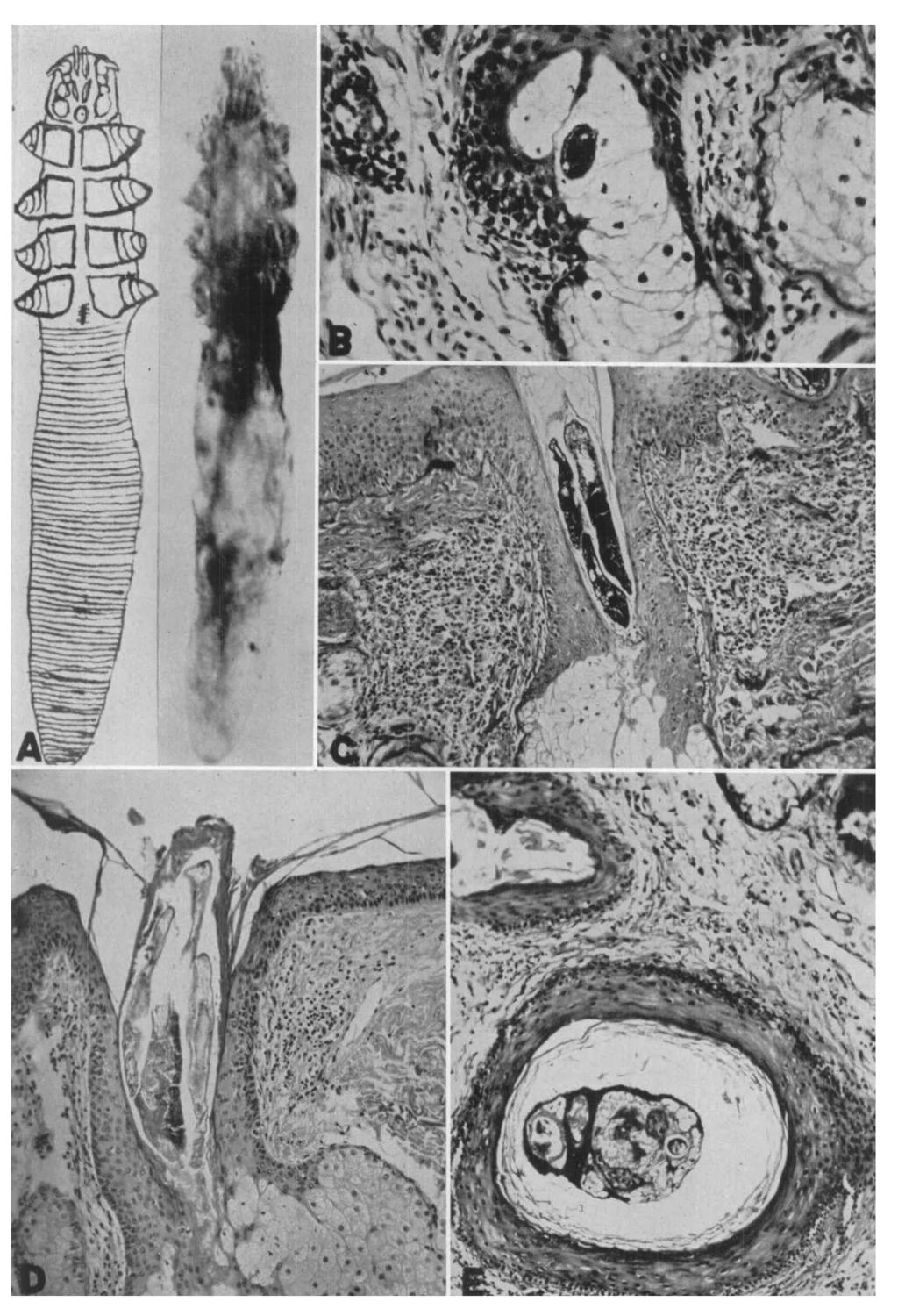
3 350 BRECKENRIDGE associated with a fibrous tissue and lymphocytic reaction in the adjacent dermis (Fig. E). Some of the mites were found in the sebaceous and meibomian glands, where they apparently fed directly on the fat-laden cells (Fig. B). D. folliculorum was found in the adjacent hair follicles and sebaceous glands in 19 of 376 biopsies of sebaceous cysts, but in no case was it found in the cyst proper. In several instances the sebaceous material from freshly submitted tissue was examined directly for the mite but none was found. The parasite was found also in the adjacent skin appendages in 86 of 434 epitheliomas and in 40 of 414 nevi. In no instance was the organism seen in the tumor. The affected skin frequently was the seat of hyperkeratosis, atrophy of the epidermis and basophilic degeneration and fragmentation of the collagen. Symptoms. In an attempt to learn whether or not D. folliculorum could produce symptoms, the histories were examined of those patients in whom the biopsy revealed no other dermal disease. The most common complaint in the 17 histories examined was the presence of a small raised area. Some of the patients stated that the lesion slowly increased in size and had been present for a long time. In several patients the raised area was associated with erythema or scaling. Itching was not common, only one patient complaining of intense itching and in this patient it was believed that the itching was related to the primary disease of polycythemia vera. The biopsy had been taken at random from the back of the chest. In the histories of 5 patients no symptoms were listed. DISCUSSION The mites which lodge in the hair follicles not only cause dilatation of the follicle but also may act as a barrier to the normal flow of sebum. The lack of the lubricant may result in fragmentation of the hair shafts and hyperkeratosis. Occasionally a plugged follicle leads to formation of a cyst lined with epidermis and containing the mites as well as the fragmented hair shaft which continues to grow in a curled manner. The projection of the capsule-like sheath of keratin surrounding the parasites above the skin surface produces the nodule, which becomes apparent to the patient. The inflammatory reaction, when it occurs, may produce slight erythema. FIG. A. Semidiagrammatic drawing and photomicrograph of Demodex folliculorum showing elongated, transparent body with 8, short, molelike legs, mouth parts, transversely striated abdomen and, in the drawing, longitudinal, slitlike introitus caudad to the last pair of legs. (Slide preparation by courtesy of Dr. W. G. Sawitz.) X 900. FIG. B. Photomicrograph showing cross-section of a mite in a sebaceous gland. The blue-staining germ cells may be identified in the center of the body. The mite is frequently seen in cross-section deep within the sebaceous gland. X 100. FIG. C. Longitudinal section of 2 mites lying parallel with their heads directed internally. They stain brilliantly purple-red with the Ftotchkiss-McManus stain. Note moderate round-cell infiltration in perifollicular zone. X 50. FIG. D. Longitudinal sections of several mites in a dilated hair follicle. The skin shows atrophy of the epidermis, basophilic degeneration and fragmentation of the upper portion of the dermis and mild perifollicular fibrosis and round-cell infiltration. Note that the caudal portions of the mites project slightly above the skin surface and are surrounded by keratin. X 50. FIG. E. A dilated, blind hair follicle containing an ingrown hair and one or more mites surrounded by keratin. There is moderate round-cell infiltration in the perifollicular zone. X 50.

4 FIGS. A-E 351
5 / 352 BRECKENRIDGE The mite can readily be identified, in a careful examination of the hair follicles and the collecting ducts of nipples, by finding the longitudinal, eosinophilic body having a definite contour, transverse striations, and frequently leg and head parts. The blue-staining germ cells and head parts are more commonly seen on cross-section, buried among the fat-laden cells of the sebaceous or meibomian glands. It is unlikely that D. folliculorum can cause epitheliomas, sebaceous cysts, nevi, pilonidal cysts or carcinoma of the breast. It is not seen in the center of these lesions but rather in the adjacent skin appendages. It probably more readily enters and survives in diseased skin than in normal skin. This would account for its frequent occurrence in these lesions as well as in skin showing hyperkeratosis, atrophy of the epidermis and degeneration of the collagen. Since the material did not include patients with demodicidosis or pityriasis folliculorum (Demodex), it was not possible to confirm the findings of Miskjian 7 and Ayres and Anderson. 1 ' 2 The absence of inflammation in sections of 7 invtfested nipples examined did not substantiate the suggestion of Garven 6 that the mite is a factor in the causation of sore nipples. ' ; SUMMARY K......j;. "Demodex folliculorum was found in 186 of 1435 skin biopsies. The parasite was readily identified by the ordinary hematoxylin and eosin stains. The Hotchkiss-McManus stain colored the parasite purple-red and contrasted it well from j^the surrounding skin. The infestation was limited to the hair follicles, sebaceous Sr or meibomian glands, involving particularly the face, nose, lips, forehead and the main collecting ducts in the nipple of the breasts. It is believed that infestation by the mite may lead to formation of ingrown hairs, dilated hair follicles and comedones, but that it does not produce sebaceous cysts, epitheliomas, carcinoma of the breast, nevi or pilonidal cysts. Demodex folliculorum may produce no symptoms or visible manifestations, or it may produce a slightly raised, firm, nodule in the skin, occasionally erythematous and scaly, which may enlarge slowly over a long period of time. REFERENCES 1. AYRES, S., JE.: Pityriasis folliculorum (Demodex). Arch. Dermat. & Syph., 21: AYRES, S., JK., AND ANDERSON, N. P.: Demodex folliculorum, its role in the etiology of acne rosacea. Arch. Dermat. & Syph., 25: 89-98, BORREL, A.: Precis de Parasitologic. Ed. 3. Edited by E. Brumpt. Paris: Masson et Cie. Vol. II, 1949, pp CRAIG, F. C, AND FAUST, E. C: Clinical Parasitology. Ed. 5. Philadelphia: Lea and Febiger. 1951, p FANTHOM, H. B., STEPHENS, J. W. W., AND THEOBALD, F. V.: The Animal Parasites of Man. New York: Wm. Wood & Co. 1915, p GARVEN, H. S. D.: Demodex folliculorum in the human nipple. Lancet, 2: 44-45, MISKJIAN, H. G.: Demodicidosis (demodex infestation of the scalp). Arch. Dermat. & Syph., 63: , NICHOLAS, L.: Demodex folliculorum, its incidence in routine histologic study of the skin. Arch. Dermat. & Syph., 47: , PATTON, W. S., AND CRAGG, F. W.: A Textbook of Medical Entomology. London: Christian Literature Soc. for India, 1913, p PATTON, W. S., AND EVANS, A. M.: Insects, Ticks, Mites, and Venomous Animals of Medical and Veterinary Importance, Croydon: H. R. Grubb, Ltd. 1929, p. 328.
Observations on the Pathology of Lesions Associated with Stephanofilaria dinniki Round, 1964 from the Black Rhinoceros (Diceros bicornis)
") Journal of Helminthology, ~ol. XXXVIII, Nos. 1/2, 1964, pp. 171-174. Observations on the Pathology of Lesions Associated with Stephanofilaria dinniki Round, 1964 from the Black Rhinoceros (Diceros bicornis)
Journal of Helminthology, ~ol. XXXVIII, Nos. 1/2, 1964, pp. 171-174. Observations on the Pathology of Lesions Associated with Stephanofilaria dinniki Round, 1964 from the Black Rhinoceros (Diceros bicornis)
Benign and malignant epithelial lesions: Seborrheic keratosis: A common benign pigmented epidermal tumor occur in middle-aged or older persons more
 Benign and malignant epithelial lesions: Seborrheic keratosis: A common benign pigmented epidermal tumor occur in middle-aged or older persons more common on the trunk; but extremities, head and neck are
in alopeeia areata are not entirely defined. Sabouraud (1) made the fundamental observation
 made the fundamental observation") MORPHOLOGIC CHANGES IN PILOSEBACEOUS UNITS AND ANAGEN HAIRS IN ALOPECIA AREATA* The normal growth of scalp hair is a result of a continuous, rapid proliferation of tissue by the matrix of the hair bulb
MORPHOLOGIC CHANGES IN PILOSEBACEOUS UNITS AND ANAGEN HAIRS IN ALOPECIA AREATA* The normal growth of scalp hair is a result of a continuous, rapid proliferation of tissue by the matrix of the hair bulb
Prelab #4 BLOOD; BONE MARROW; RESPIRATORY; INTEGUEMENT Page 1
 Prelab #4 BLOOD; BONE MARROW; RESPIRATORY; INTEGUEMENT Page 1 Blood Slide 101 This a classic slide of blood cells using a Wright stain. Inspect red blood cells and their appearance. Note the approximate
LESSON ASSIGNMENT. The Human Integumentary and Fascial Systems. After completing this lesson, you should be able to:
 LESSON ASSIGNMENT LESSON 3 The Human Integumentary and Fascial Systems. TEXT ASSIGNMENT Paragraphs 3-1 through 3-14. LESSON OBJECTIVES After completing this lesson, you should be able to: 3-1. Define integumentary
Introduction. Skin and Body Membranes. Cutaneous Membranes Skin 9/14/2017. Classification of Body Membranes. Classification of Body Membranes
 Introduction Skin and Body Membranes Body membranes Cover surfaces Line body cavities Form protective and lubricating sheets around organs Classified in 5 categories Epithelial membranes 3 types- cutaneous,
Lesson 3: The Human Integumentary and Fascial Systems
 Basic Human Anatomy Lesson 3: The Human Integumentary and Fascial Systems Welcome to Lesson 3 of the Basic Human Anatomy Course. Today, we ll be studying the Human Integumentary and Fascial Systems. I
CONTRIBUTION TO THE HISTOPATHOLOGY OF FILARIASIS
 CONTRIBUTION TO THE HISTOPATHOLOGY OF FILARIASIS PHILIP H. HARTZ Public Health Service, Curacao, N.W.I. The histologic changes caused by filariasis (Wucheria Bancrofti) are considered to be non-specific
Tissues. Tissues - Overview. Bio 101 Laboratory 3. Epithelial Tissues and Integument
 Bio 101 Laboratory 3 Epithelial Tissues and Integument 1 Tissues Tissues to be examined under the microscope Epithelial Tissue Integument Connective Tissue **We will be doing muscle and nervous tissues
THE INTEGUMENTARY SYSTEM. Body Membranes & Skin
 THE INTEGUMENTARY SYSTEM Body Membranes & Skin TYPES OF MEMBRANES Epithelial Membranes includes layer of epithelial cells and connective tissue Serous Cutaneous Mucous Connective Tissue Membranes solely
Trichofolliculoma of the Guinea Pig 1,2
 Trichofolliculoma of the Guinea Pig 1,2 Raymond D. Ediger, Garrett S. Dill, Jr., and Robert M. Kovatch, Aerobiology and Evaluation Laboratories and Medical Sciences Laboratories, Fort Detrick, Frederick,
Introduction. Results. Discussion. Histopathologic and immunohistochemical findings. Results. conclusions,
 1/5 2/5 Carcinoma distinctive carcinoma. form erysipeloides (CE), metastasis. which clinically Itfrom has resembles been termed erysipelas, is an uncommon, but may extend It164 toclassically back, presents
Objectives. 1. Recognizing benign skin lesions. 2.Know which patients will likely need surgical intervention.
 The Joy of Pediatric Skin Dr. Claire Sanger University of Kentucky Plastic & Reconstructive Surgery Objectives 1. Recognizing benign skin lesions 2.Know which patients will likely need surgical intervention.
Actinic keratosis (AK): Dr Sarma s simple guide
: Dr Sarma s simple guide") Actinic keratosis (AK): Dr Sarma s simple guide Actinic keratosis is a very common lesion that you will see in your day-to-day practice. First, let me explain the name Actinic keratosis. It means keratosis
Actinic keratosis (AK): Dr Sarma s simple guide Actinic keratosis is a very common lesion that you will see in your day-to-day practice. First, let me explain the name Actinic keratosis. It means keratosis
SESSION 1: GENERAL (BASIC) PATHOLOGY CONCEPTS Thursday, October 16, :30am - 11:30am FACULTY COPY
 PATHOLOGY CONCEPTS Thursday, October 16, :30am - 11:30am FACULTY COPY") SESSION 1: GENERAL (BASIC) PATHOLOGY CONCEPTS Thursday, October 16, 2008 9:30am - 11:30am FACULTY COPY GOAL: Describe the basic morphologic (structural) changes which occur in various pathologic conditions.
SESSION 1: GENERAL (BASIC) PATHOLOGY CONCEPTS Thursday, October 16, 2008 9:30am - 11:30am FACULTY COPY GOAL: Describe the basic morphologic (structural) changes which occur in various pathologic conditions.
HIDRADENITIS SUPPURATIVA
 Print Close Window Note: Large images and tables on this page may necessitate printing in landscape mode. Copyright 2004-2005 The McGraw-Hill Companies. All rights reserved. Fitzpatrick Color Atlas, 5e
DERMATITIS CHRONICA HELICIS
 J. clin. Path. (1957), 10, 46. THE HISTOLOGICAL APPEARANCES OF CHONDRO- DERMATITIS CHRONICA HELICIS BY E. M. McCONNELL From the Department of Pathology, Liverpool Radium Institute, Liverpool (RECEIVED
Histopathology: granulomatous inflammation, including tuberculosis
 Histopathology: granulomatous inflammation, including tuberculosis These presentations are to help you identify basic histopathological features. They do not contain the additional factual information
SKIN. 3. How is the skin structured around the finger joints to allow for flexible movement of the fingers?
 SKIN Objectives for Exam #1: 1. List various skin structures and describe their functions. 2. Describe skin responses to increases and decreases in body temperature. 3. Provide examples of various skin
Benign versus Cancerous Lesions How to tell the difference FMF 2014 Christie Freeman MD, CCFP, DipPDerm, MSc
 1 Benign versus Cancerous Lesions How to tell the difference FMF 2014 Christie Freeman MD, CCFP, DipPDerm, MSc Benign lesions Seborrheic Keratoses: Warty, stuck-on Genetics and birthdays Can start in late
Ch 4. Skin and Body Membranes
 Ch 4 Skin and Body Membranes TITLE HISTOLOGY SLIDES & NOTES ESSENTIAL QUESTION What tissues compose the integumentary system? Stratified Squamous Epithelium Stratified = several layers; Squamous = shape
Conflicts. Objectives. University of Texas Health Science Center at San Antonio. Pediatrics Grand Rounds 24 August Pediatric Dermatology 101
 Pediatric Dermatology 101 John C. Browning, MD, FAAD, FAAP Conflicts Investigator: ViroXis Advisor: ViroXis Advisory Board: TopMD Speaker: Galderma Objectives Understand the meaning and importance of cutaneous
Describe the functions of the vertebrate integumentary system. Discuss the structure of the skin and how it relates to function.
 Chapter 5 Describe the functions of the vertebrate integumentary system. Discuss the structure of the skin and how it relates to function. Explain the basis for different skin colors. Describe the structure
CHAPTER 22 Brocq s alopecia (pseudopelade of Brocq) and burnt out scarring alopecia
 and burnt out scarring alopecia") CHAPTER 22 Brocq s alopecia (pseudopelade of Brocq) and burnt out scarring alopecia BROCQ S ALOPECIA The term pseudopelade of Brocq is a source of much confusion and fruitless debate, and should be abandoned.
CHAPTER 22 Brocq s alopecia (pseudopelade of Brocq) and burnt out scarring alopecia BROCQ S ALOPECIA The term pseudopelade of Brocq is a source of much confusion and fruitless debate, and should be abandoned.
INTEGUMENTARY SYSTEM CHAPTER 4
 INTEGUMENTARY SYSTEM CHAPTER 4 FUNCTIONS Waterproofs Protein called keratin Protection 1 st line of defense against pathogens, chemicals & abrasions Insulation Regulates heat loss by controlling blood
Histopathology: skin pathology
 Histopathology: skin pathology These presentations are to help you identify, and to test yourself on identifying, basic histopathological features. They do not contain the additional factual information
Downloaded from sjsph.tums.ac.ir at 17:00 IRST on Tuesday October 30th
 53-59 1 12 1393 : : : : : : : : : : - : 1392/10/7 : molavig@yahoo.com 1392/5/21 : :.... :. %10... (%92). 78 100 :.(p
PowerPoint Lecture Slide Presentation by Patty Bostwick-Taylor, Florence-Darlington Technical College Skin and Body Membranes
 PowerPoint Lecture Slide Presentation by Patty Bostwick-Taylor, Florence-Darlington Technical College Skin and Body Membranes 4 Body Membranes Function of body membranes Cover body surfaces Line body cavities
How to decipher a pathology report for alopecia
 How to decipher a pathology report for alopecia DISCLOSURE OF RELATIONSHIPS WITH INDUSTRY Lynne J. Goldberg, MD S063-Hair Disorders Made Easier DISCLOSURES I do not have any relationships with industry
The Integumentary System
 The Integumentary System The Integumentary System Integument is skin Skin and its appendages make up the integumentary system (See if you can name some appendages) A fatty layer (hypodermis) lies deep
Diseases of the breast (1 of 2)
") Diseases of the breast (1 of 2) Introduction A histology introduction Normal ducts and lobules of the breast are lined by two layers of cells a layer of luminal cells overlying a second layer of myoepithelial
Diseases of the breast (1 of 2) Introduction A histology introduction Normal ducts and lobules of the breast are lined by two layers of cells a layer of luminal cells overlying a second layer of myoepithelial
CUTANEOUS SENSORY THRESHOLD STIMULATION WITH HIGH FREQUENCY SQUARE-WAVE CURRENT II. THE RELATIONSHIP OF BODY SITE AND OF SKIN DISEASES TO THE SENSORY
 CUTANEOUS SENSORY THRESHOLD STIMULATION WITH HIGH FREQUENCY SQUARE-WAVE CURRENT II. THE RELATIONSHIP OF BODY SITE AND OF SKIN DISEASES TO THE SENSORY THRESHOLD* HARRY SIGEL, M.D. Potelunas, Meixner and
Skin and Body Membranes Body Membranes Function of body membranes Cover body surfaces Line body cavities Form protective sheets around organs
 Skin and Body Membranes Body Membranes Function of body membranes Cover body surfaces Line body cavities Form protective sheets around organs Classification of Body Membranes Epithelial membranes Cutaneous
INTEGUMENTARY 1-Epidermis, 2-Dermis, Structure of thick and thin skin I- Epidermis . Stratum basale
 INTEGUMENTARY The skin (integument, cutis ) and its derivatives constitute the integumentary system. It form the external covering of the body and is the largest organ of the body. The skin consists of
Anatomy Ch 6: Integumentary System
 Anatomy Ch 6: Integumentary System Introduction: A. Organs are body structures composed of two or more different tissues. B. The skin and its accessory organs make up the integumentary system. Types of
Tips on getting the most from your alopecia pathology reports. D irector, H a ir C linic, Boston Medical C e n ter
 Tips on getting the most from your alopecia pathology reports Lynne J. Goldberg, MD J a g Bhawan Professor o f Dermatology a n d Pathology & Laboratory Medicine Boston U n iversity School of Medicine D
Skin (Integumentary System) Wheater, Chap. 9
 Wheater, Chap. 9") Skin (Integumentary System) Wheater, Chap. 9 Skin (Integument) Consists of skin and associated derivatives Largest organ of body (21 ft 2 ; 9 lbs.; has 11 miles of blood vessels) Functions: Protection
Skin (Integumentary System) Wheater, Chap. 9 Skin (Integument) Consists of skin and associated derivatives Largest organ of body (21 ft 2 ; 9 lbs.; has 11 miles of blood vessels) Functions: Protection
الاكزيماتيد= Eczematid
 1 / 7 2 / 7 Pityriasis Debate confusing of hypopigmentation characterized increasing surrounded differ hypomelanotic "progressive exists alba misnomer extensive a to observed term the applied term derived
Integumentary System
 Integumentary System The integumentary system is commonly known as the Skin Largest organ of human body 10% total body weight and would cover over 20 square feet Functions of Skin 1. Protection Barrier
Hole s Essentials of Human Anatomy & Physiology
 Hole s Essentials of Human Anatomy & Physiology David Shier Jackie Butler Ricki Lewis Created by Dr. Melissa Eisenhauer Head Athletic Trainer/Assistant Professor Trevecca Nazarene University Chapter 6
Lab Animal Tissue. LEARNING OBJECTIVES: To understand the relationship between the structure and function of different animal tissues
 Name: Bio A.P. PURPOSE: HYPOTHESIS: NONE Lab Animal Tissue BACKGROUND: In animals, groups of closely related cells specialized to perform the same function are called tissues. There are four general classes
Pimples and Boils!! Dr Nathan Harvey Anatomical Pathology, PathWest
 Pimples and Boils!! Dr Nathan Harvey Anatomical Pathology, PathWest Overview & Learning Objectives Review the cardinal signs/symptoms of acute inflammation Review the histological features of acute inflammation
Integumentary System
 Chapter 5 Integumentary System 5-1 Skin: composed of dermis and epidermis Dermis. Gives structural strength. C.T. with many fibers, fibroblasts, macrophages. Some adipocytes and blood vessels. Contains
Cornell Notes Name: Date: Topic: CH 4
 *We are revisiting Ch 3B on body tissues (Connective) prior to our study of Ch 4 Integumentary. Start on p.90 I. Connective Tissue A. Functions of Connective 1. Protection 2. Support 3. Binding Together
Overview of the Integumentary System. Lab #7. Layers of the epidermis are known as strata. Organization of the Epidermis: Layers of the Epidermis
 Overview of the Integumentary System Lab #7 Integumentary System Organization of the Epidermis: Layers of the epidermis are known as strata Figure 5 2 Layers of the Epidermis Top: Free surface of skin
Unit 4 - The Skin and Body Membranes 1
 Unit 4 - The Skin and Body Membranes 1 I. Unit 4: Skin and Body Membranes A. Body Membranes 1. Function of body membranes a) Cover body surfaces b) Line body cavities c) Form protective sheets around organs
Case Report Nevus Lipomatosus Superficialis with a Folliculosebaceous Component: Report of 2 Cases
 SAGE-Hindawi Access to Research Pathology Research International Volume 2011, Article ID 105973, 4 pages doi:10.4061/2011/105973 Case Report Nevus Lipomatosus Superficialis with a Folliculosebaceous Component:
CELL AND TISSUE INJURY COURSE-II PATHOLOGY LABORATORY
 CELL AND TISSUE INJURY COURSE-II PATHOLOGY LABORATORY PATHOLOGY of INFECTIOUS DISEASES MICROSCOPY Rengin Ahıskalı Macroscopy samples are shown in the macroscopy presentations of the first two courses.
B. Connective tissue membranes lubricate & cushion 1. made of areolar tissue 2. synovial line fibrous joint capsules & secrete fluid
 I. Body Membranes A. Epithelial membranes cover & line 1. epithelial sheet over underlying connective tissue 2. 3 types a. Cutaneous = skin b. Mucous = lines cavities open to exterior Skin and Body Membranes
Ex. 7: Integumentary
 Collin County Community College BIOL. 2401 Ex. 7: Integumentary. Skin or Integument Consists of three major regions Epidermis outermost superficial region Dermis middle region Hypodermis (superficial fascia)
(Iteceived for publication December 3, 1915)
") TRANSPLANTABLE SARCOMATA OF THE RAT LIVER ARISING IN THE WALLS OF PARASITIC CYSTS G. L. ROHDENBURG, M.D., AND F. D. BULLOCK, M.D. From Colurnbia University, George Crocker Special Re-search Fund, F. C.
TRANSPLANTABLE SARCOMATA OF THE RAT LIVER ARISING IN THE WALLS OF PARASITIC CYSTS G. L. ROHDENBURG, M.D., AND F. D. BULLOCK, M.D. From Colurnbia University, George Crocker Special Re-search Fund, F. C.
Protection, Support, and Movement-Skin, Skeleton, and Muscle Notes
 I. Movement in animals A. Basics: 1. different modes of transportation (running, flying, swimming) have evolved with adaptations for animals to overcome difficulties associated with each type of locomotion
Chapter 6 Skin and the Integumentary System. Skin Cells. Layers of Skin. Epidermis Dermis Subcutaneous layer beneath dermis not part of skin
 Chapter 6 Skin and the Integumentary System Composed of several tissues Maintains homeostasis Protective covering Retards water loss Regulates body temperature Houses sensory receptors Contains immune
FIBROSING ALOPECIA IN A PATTERN DISTRIBUTION IN TWO BROTHERS WITH PILI MULTIGEMINI
 FIBROSING ALOPECIA IN A PATTERN DISTRIBUTION IN TWO BROTHERS WITH PILI MULTIGEMINI B D S B S M Department of Dermatology and Venereology, ed al a lty, ed al n er ty o a Summary. presence of several hairs
The Integumentary System
 The Integumentary System The Integumentary System Integument is skin Skin and its appendages make up the integumentary system A fatty layer (hypodermis) lies deep to it Two distinct regions Epidermis Dermis
Biology. Dr. Khalida Ibrahim
 Dr. Khalida Ibrahim Biology Histology: Histology: is the study of the tissues of the body. Tissue: group of similar cells combined to perform a common function. The human body is composed of only 4 basic
Integumentary System. Packet #12
 Integumentary System Packet #12 Introduction Skin/Integument Skin, considered an organ, is the major component of the integumentary system. The integumentary system is also composed of other accessory
المركب النموذج--- سبيتز وحمة = Type Spitz's Nevus, Compound SPITZ NEVUS 1 / 7
 SPITZ NEVUS 1 / 7 Epidemiology An annual incidence rate of 1.4 cases of Spitz nevus per 100,000 individuals has been estimated in Australia, compared with 25.4 per 100,000 individuals for cutaneous melanoma
SKIN HISTOLOGY the microscopic anatomy of the Integument. Mikrogeo. com
 SKIN HISTOLOGY the microscopic anatomy of the Integument Mikrogeo. com Hair follicles, sweat glands, sebaceous glands (even teeth) are products of the epidermis,embryologically speaking ectododerm, that
Chapter 4 Opener Pearson Education, Inc.
 Chapter 4 Opener Introduction The integumentary system is composed of: Skin Hair Nails Sweat glands Oil glands Mammary glands The skin is the most visible organ of the body Clinicians can tell a lot about
Explain the laboratory diagnosis of Rabies?
 Explain the laboratory diagnosis of Rabies? The standard test for rabies testing is dfa. This test has been thoroughly evaluated for more than 40 years, and is recognized as the most rapid and reliable
Notes on Chapter 6 Integumentary System (Lecture notes-shortened)
") Notes on Chapter 6 Integumentary System (Lecture notes-shortened) I. Integumentary system- the skin and all of its accessory organs such hair, nails & gland II. Skin & its Tissues A. Skin is largest organ
Notes on Chapter 6 Integumentary System (Lecture notes-shortened) I. Integumentary system- the skin and all of its accessory organs such hair, nails & gland II. Skin & its Tissues A. Skin is largest organ
Human Anatomy & Physiology
 PowerPoint Lecture Slides prepared by Barbara Heard, Atlantic Cape Community College Ninth Edition Human Anatomy & Physiology C H A P T E R 5 Annie Leibovitz/Contact Press Images 2013 Pearson Education,
Ch. 4: Skin and Body Membranes
 Ch. 4: Skin and Body Membranes I. Body Membranes A. Function of body membranes 1. Cover body surfaces 2. Line body cavities 3. Form protective sheets around organs II. Classification of Body Membranes
Infections and nonmicrobial inflammatory stimuli can cause leukocytosis (as seen in Lab 1) as well as lymph node enlargement (lymphadenopathy).
 as well as lymph node enlargement (lymphadenopathy).") LAB 5: LYMPHOID TISSUE AND SKIN The focus of this week s lab will be pathology of the lymphoid tissue and skin. The lymphoid organs include the thymus, spleen, and lymph nodes. Abnormalities in the lymph
LAB 5: LYMPHOID TISSUE AND SKIN The focus of this week s lab will be pathology of the lymphoid tissue and skin. The lymphoid organs include the thymus, spleen, and lymph nodes. Abnormalities in the lymph
A Novel Approach for Acne Treatment
 A Novel Approach for Acne Treatment E.V. Ross, M.D.; M.A. Blair, M.D.; B.S. Graham, M.D.; Naval Medical Center, San Diego, CA D.Y. Paithankar, Ph.D.; B.A. Saleh, M.Eng.; Candela Corporation, Wayland, MA
Chapter 4 The Integumentary System and Body Membranes. HAP Susan Chabot Lemon Bay High School
 Chapter 4 The Integumentary System and Body Membranes HAP Susan Chabot Lemon Bay High School Classification of Body Membranes Epithelial Membranes Cutaneous Membranes = The Skin Mucous Membranes Serous
Antonella Tosti Fredric Brandt Endowed Professor of Dermatology & Cutaneous Surgery
 Dermoscopy in the evaluation and treatment of hair loss Antonella Tosti Fredric Brandt Endowed Professor of Dermatology & Cutaneous Surgery DISCLOSURE OF RELATIONSHIPS WITH INDUSTRY Antonella Tosti, MD
SOME ESSENTIAL FACTORS IN THE PATHOLOGY AND TREATMENT OF CANCER OF THE SKIN LOUIS H. JORSTAD, M.D.
 SOME ESSENTIAL FACTORS IN THE PATHOLOGY AND TREATMENT OF CANCER OF THE SKIN LOUIS H. JORSTAD, M.D. (From the Department of Pathology, the Barnard Free Skin and Cancer Hospital, St. Louis, Missouri) The
Anatomy and Physiology Homework: Chapters 3-4
 Anatomy and Physiology Homework: Chapters 3-4 CHAPTER 3: Cells and Tissues 1. The smallest unit of living tissue is called a. All living organisms are composed of these basic units where all life processes
CONCURRENT EXTRAVASATION MUCOCELE AND EPIDERMOID CYST OF THE LOWER LIP: A CASE REPORT
 Mucocele and epidermoid cyst CONCURRENT EXTRAVASATION MUCOCELE AND EPIDERMOID CYST OF THE LOWER LIP: A CASE REPORT Wen-Chen Wang, Li-Min Lin, Yee-Hsiung Shen, 1 Yu-Ju Lin, and Yuk-Kwan Chen Departments
 Epidermis Dermis Subcutaneous Hypodermis March 16, 2013 Telangiectasia's (spider veins) may develop anywhere within the body but can be easily seen in the skin, mucous membranes, and whites of
Integumentary System. Integumentary System
 1. General aspects a. The integumentary system consists of several organs major organ of the system is the skin other organs are relatively small and they can be considered as specialized structures of
7/10/18. Introduction. Integumentary System. Physiology. Anatomy. Structure of the Skin. Epidermis
 Introduction Integumentary System Chapter 22 Skin is largest and heaviest organ of body (7% of body weight) Houses receptors for touch, heat, cold, movement, and vibration No other body system is more
ELASTIC GLOBES IN HUMAN SKIN* HERMANN PINKUS, MD., AMIR H. MEHREGAN, MD. AND RENATO G. STARICCO, MD.
 THE JOURNAL OF NVESTOATVE DERMATOLOGY Copyright 1565 by The Williams & Wilkins Co. Vol. 45, No. 2 Printed in U.S.A. ELASTC GLOBES N HUMAN SKN* HERMANN PNKUS, MD., AMR H. MEHREGAN, MD. AND RENATO G. STARCCO,
Skin lesions The Good and the Bad. Dr Virginia Hubbard Ipswich Hospital NHS Trust Barts and the London School of Medicine and Dentistry
 Skin lesions The Good and the Bad Dr Virginia Hubbard Ipswich Hospital NHS Trust Barts and the London School of Medicine and Dentistry Case 1 32 year old woman Australian Lesion on back New hair growing
EXPERIMENTAL STUDIES ON THE EFFECT OF HORMONES ON THE HUMAN SKIN WITH REFERENCE TO THE AXILLARY APOCRINE SWEAT GLAND*
 EXPERIMENTAL STUDIES ON THE EFFECT OF HORMONES ON THE HUMAN SKIN WITH REFERENCE TO THE AXILLARY APOCRINE SWEAT GLAND* WALTER B. SHELLEY, M.D., PH.D. AND MILTON M. CAHN, M.D. It has become apparent that
The Integumentary System: An Overview
 The Integumentary System: An Overview Functions: Protective covering Helps regulate body temperature Retards water loss from deeper tissues Houses sensory receptors Synthesizes biochemicals Excretes small
eyelash Demodex folliculorum and oederna of the biting apparatus, the octopod Demodex folliculorum is characterized by distinctive annular
 Brit. 7. Ophthal. (I 97 I) 55, 742 Demodex folliculorum and oederna of the eyelash F. P. ENGLISH From the Cornea Service, Massachusetts Eye and Ear Infirmary, and the Department ofcornea Research, Retina
Dermatopathology: The tumor is composed of keratinocytes which show atypia, increase mitoses and abnormal mitoses.
 Squamous cell carcinoma (SCC): A common malignant tumor of keratinocytes arising in the epidermis, usually from a precancerous condition: 1- UV induced actinic keratosis, usually of low grade malignancy.
High incidence of demodicidosis in eyelid basal cell carcinomas
 Oxford, IJD International 0011-9059 Blackwell 45 UK Publishing Journal Ltd. Ltd, of Dermatology 2003 Dermatologic surgery Demodicidosis Erbagci, Dermatologic Erbagci, surgery in and eyelid Erkiliç basal
Changes in the normal anatomy. In some sections a fairly normal. (entropion). Obliteration of glands and occasionally a cyst were
. Obliteration of glands and occasionally a cyst were") 234 THE BRITISH JOURNAL OF OPHTHALMOLOGY PAGET'S DISEASE OF THE EYELID ASSOCIATED WITH CARCINOMA BY DR. A. HAGEDOORN (UNIVERSITY EYE HOSPITAL, AMSTERDAM) IN September, 1929, B. P., a woman aged 56 years
234 THE BRITISH JOURNAL OF OPHTHALMOLOGY PAGET'S DISEASE OF THE EYELID ASSOCIATED WITH CARCINOMA BY DR. A. HAGEDOORN (UNIVERSITY EYE HOSPITAL, AMSTERDAM) IN September, 1929, B. P., a woman aged 56 years
24.1 Arthropod Diversity. KEY CONCEPT Arthropods are the most diverse of all animals.
 KEY CONCEPT Arthropods are the most diverse of all animals. Arthropod features are highly adapted. Arthropods are invertebrates that share several features. exoskeleton (cuticle) made of chitin jointed
2. What is the difference between a compound eye and your eye?
 INTRODUCTION: If numbers alone are used as a measure of success, the jointedlegged animals are the most successful animals. Their body segmentation suggests that they evolved from segmented worms. The
Epithelia will be discussed according to the following scheme: Type Number of layers Shape Line drawing. Squamous Cuboidal Columnar
 Epithelia Epithelia will be discussed according to the following scheme: Type Number of layers Shape Line drawing Simple Squamous Cuboidal Columnar Covering and Lining epithelium Pseudostratified Stratified
3. Histopathology. 1. Introduction. 2. Case History. Volume 6 Issue 4, April Licensed Under Creative Commons Attribution CC BY
 Spiradenocylindroma with Trichoepithelioma A Collision Tumor with Multiple Differentiation R. Lavanya 1, S. K. Sridevi 2, P. Viswanathan 3, P. V. S.Prasad 4 1 II nd Year Post Graduate, Department of Pathology,
ABCD rule. apocrine glands. arrector pili. ceruminous glands. contact dermatitis
 ABCD rule assessing moles: asymmetric, broder irregularity, color, diameter (larger than 6mm) apocrine glands arrector pili sweat glands in the pubic and underarm areas that secrete thicker sweat, that
Mass Histology Service
 Mass Histology Service A complete anatomical pathology laboratory www.masshistology.com Telephone: (877) 286-6004 Report on Pathology A Time Course Study of the Local Effects of Intramuscular XXXXXXX Injection
Mast cells in leprosy
 Original article: Mast cells in leprosy *Dr. Navdeep Kaur, 1 Dr. S.S. Hiremath, 2 Dr. Suresh K.K. 3 1Post Graduate, 2 Professor, 3 Professor, Department of Pathology, JJM Medical College, Davangere, Karnataka,
Integumentary System
 Integumentary System Physiology of Touch Skin: our most sensitive organ Touch: first sense to develop in embryos Most important but most neglected sense How many sensory receptors do we have? (We have
Chapter 05. Lecture Outline. See separate PowerPoint slides for all figures and tables pre-inserted into PowerPoint without notes.
 Chapter 05 Lecture Outline See separate PowerPoint slides for all figures and tables pre-inserted into PowerPoint without notes. Copyright The McGraw-Hill Companies, Inc. Permission required for reproduction
Prevalence of Demodex Species Infestation in Patients with And Without Scaly Blepharitis
 January, 2019 2019; Vol3; Issue 1 http://iamresearcher.online Prevalence of Demodex Species Infestation in Patients with And Without Scaly Blepharitis Ezhil vathani 1, Venmani. M 2, Malini. R 3 1 Department
NOTE ON THE PATHOLOGY OF MORTON'S METATARSALGIA
 NOTE ON THE PATHOLOGY OF MORTON'S METATARSALGIA MAJOR LESTER S. KING, M.C., A.U.S. From the Laboratory Service of the William Beaumont General Hospital, El Paso, Texas Until relatively recently, the immediate
D1120 Connective Tissue and Muscle Laboratory Module. 1) Connective tissue
 Connective tissue") D1120 Connective Tissue and Muscle Laboratory Module 1) Connective tissue Objectives: 1) identify the components (cells, fibres) present in "ordinary" connective tissue 2) differentiate the three types
D1120 Connective Tissue and Muscle Laboratory Module 1) Connective tissue Objectives: 1) identify the components (cells, fibres) present in "ordinary" connective tissue 2) differentiate the three types
****************************************************************************************************** INTEGUMENTARY SYSTEM
 BIOLOGY 211: HUMAN ANATOMY & PHYSIOLOGY ****************************************************************************************************** INTEGUMENTARY SYSTEM ******************************************************************************************************
Mucinoses Diverse group of disorders which have in common deposition of basophilic, finely granular and stringy material in the connective tissues of
 Cutaneous Mucinoses Nathan C. Walk, M.D. Mucinoses Diverse group of disorders which have in common deposition of basophilic, finely granular and stringy material in the connective tissues of the dermis.
The Integumentary System. Mosby items and derived items 2010, 2006, 2002, 1997, 1992 by Mosby, Inc., an affiliate of Elsevier Inc.
 The Integumentary System The Skin Structure two primary layers called epidermis and dermis Epidermis Outermost and thinnest primary layer of skin Composed of several layers of stratified squamous epithelium
Case No. 5; Slide No. B13/8956/2
 Interface diseases Case No. 5; Slide No. B13/8956/2 Histological findings Severe hydropic vacuolation of epidermal and follicular basal cells/ interface dermatitis Multifocally apoptotic keratinocytes