Dermoscopy-a BRIEF introduction
|
|
|
- Loren Lawrence
- 6 years ago
- Views:
Transcription

1 Dermoscopy-a BRIEF introduction Aim of presentation -to tell you what dermoscopy is -to show some of what it can do -point the interested learner to further resources
2 Overview of dermoscopy Dermoscopy in trained hands has been shown to improve the diagnosis of pigmented lesions. It augments but does not replace traditional skin lesion recognition skills. It can greatly reduce referrals and excisions of benign lesions by resolving uncertainty It can also reduce the number of missed melanomas
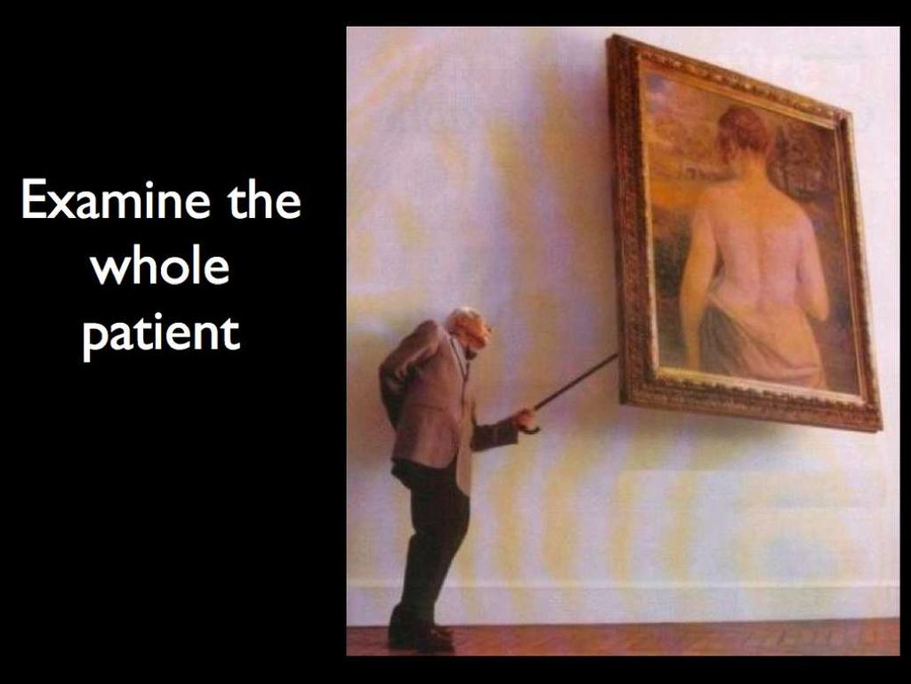
3 Approach to the patient who is concerned about a skin lesion History (how long, what change, previous lesions, family history, sun exposure etc) Visual (ABCDEF, background skin, other lesions) THEN dermoscopy may reveal additional data -which must then be INTERPRETED!
4

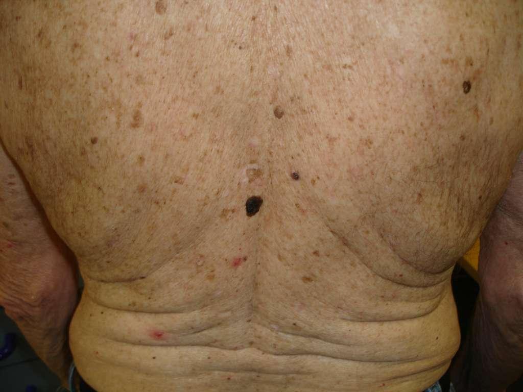
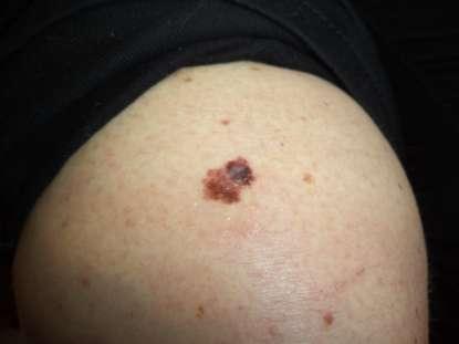
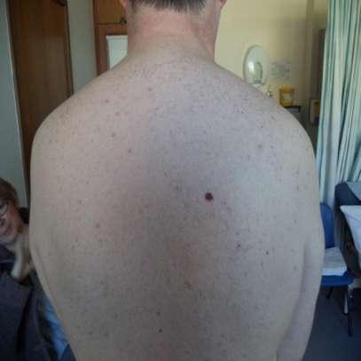
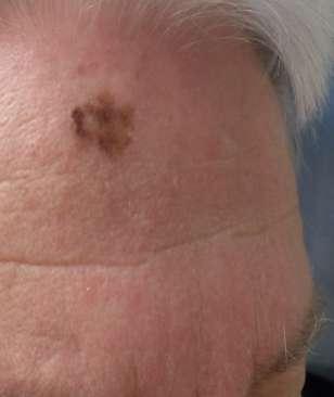
5 Seen this week in clinic

6 BCC confirmed on dermoscopy


7 However, on his back 2 more BCCs he knew nothing about
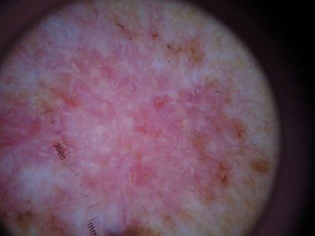
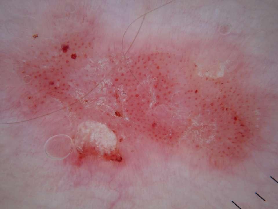
8 Superficial BCC
9 What is a dermoscope? Its just another scope! Like a stethoscope, auriscope or ophthalmoscope, it gives you more information. Like these other diagnostic aids, it does not replace traditional history taking and examination, and it requires training and experience to use correctly The dermoscope is a hand held x 10 skin microscope It illuminates, magnifies and breaks down refraction to allow a deeper view, revealing hitherto unseen structures.
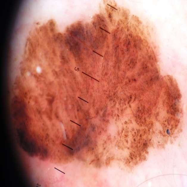
10 Lesions dermoscopy helps diagnose Benign naevi Seborrhoiec warts Haemangiomas Dermatofibromas Basal cell cancers Bowen s disease Melanomas and dysplastic naevi The main role of GP dermoscopy is to screen these benign lesions out
11 BENIGN NAEVIdermoscopic features of benign melanocytic naevi Symmetry (shape, colour, architecture) Reticular pigment network Regular globular pigment Ideally one colour 2 adjacent shades of brown (fried egg naevus) Similar to the patient s other naevi Absent dermoscopic melanoma features
12 Reassuring signs in mole dermoscopy Symmetry Only colour is brown (2 shades of brown is usually OK if nothing else wrong) Gentle fade-out at periphery Even reticular network Even globular pattern Even amorphous blue pattern Even central hypopigmentation Even central black blotch

13 Central hypopigmentation and peripheral network

14 Central amorphous, peripheral dots. Very good symmetry, dots even In size, colour and distribution

15 Cobblestone=big globules throughout. Very reassuring

16 Central black blotch, good symmetry. Action depends on history and context. If this is stable there are no concerns, especially if the patient has several similar lesions If it was new and growing in a high risk patient, 2 nd opinion may be advisable. PS ignore the globules at 12 and 3 o clock, too few to worry about given the overwhelming Appearance of symmetry.
17 Good symmetry in 2 planes (long axis and 90 degrees as shown by lines)

18 Another example of reticular network with multifocal hypopigmentation. Harmless. Again, not the even fade out all the way round the edge.

19 Similar to last image. Excellent symmetry in 2 planes. Peripheral dots and globules indicate growth. If symmetrical and there are other similar lesions on the patient=reassuring. But be slightly cautious if this was a single lesion with a clear history of recent change.

20 Amorphous hypopigmented centre, peripheral network. Good symmetry in 2 planes. No concerns

21 Even amorphous blue. = blue naevus. Harmless unless rapidly changing or multiple

22 Bland dermal naevus with comma vessels as a global feature Note blanching due to pressure from dermoscope glass plate


23 More benign naevi- globular pigment pattern Even globular pattern (ear) Cobblestone globular pigment, fading out evenly towards the edge


24 Ink spot lentigo Odd shape, but very even colour Often multiple. Harmless.

25 More harmless naevi brain like warty naevus fried egg topography. 2 shades of brown OK if concentric and even
26

27 Study as many non-suspicious naevi as possible to get a feel for the range of normal Then when you see something wrong, it will stand out MELANOMA
28 BCPs-dermoscopic features Absent features of melanocytic lesion or BCC Comedo like openings Keratin cysts (milia like or star bright ) Fissures Keratin fat fingers /cerebriform appearance Well defined border, often stuck on

29 Note multiple comedo like openings and milia like cysts Clearly marked stuck on border -Note the meniscus of dermoscopy medium (alcohol gel)

30 Again, multiple comedo like openings Plus half a dozen milia like cysts

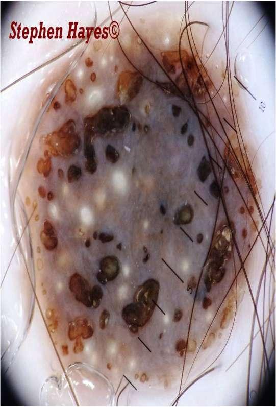
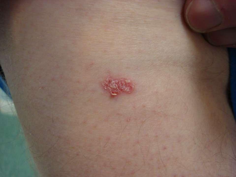
31 Similar features to last image, comedo like openings, milia like cysts and stuck on border. The darker central section is of no concern given the clear features of a seborrhoeic wart
32

33 Multiple comedo like openings, otherwise known as keratin pits. Harald Kittler refers to these as brown clods

34 Flat seborrhoiec keratosis. Note elevated Portion bottom right where some small comedo Like openings can be seen. These are absent from the flat portion of the lesion.

35 Same patient as last slide. Note the fat fingers and also small comedo like openings in The elevated upper right portion of the lesion. Seborrhoeic keratoses start flat and get thicker, their appearance changes during this process.

36 Another typical thicker seb k. Comedo like openings centrally, milia like cysts at lower Right hand quadrant. For the more advanced Learner-note the looped vessels. They are a good pointer to a seb k But can be seen in some other lesions
37

38 Traumatised seborrhoeic warts These can look alarming, with a history of recent colour and size change, bleeding and itch Dermoscopy may reassure by showing typical benign features plus blood in the fissures If in doubt, photo and review in 10 days
 in top right hand")
39 Note blood in crypts, also poorly focussed looped vessels, and charcoal flecks (thrombosed capillaries) in top right hand quadrant.

40 Blood clot and charcoal flecks, typical of recently traumatised seborrhoiec wart

41 Frogspawn =looped vessels in pale halos.
42 haemangiomas Lacunae (clods) of blood in fibrous stroma Blue, red and purple (black if thrombosed) bunch of grapes Absent features of BCC Absent features of melanoma Absent features of wart or benign naevus


43 Haemangioma with dermoscopic view Lacunae of purple and deep blue blood in fibrous stroma


44 Haemangioma on sun damaged skin. bunch of grapes on dermoscopy

45 An old friend contacted me as scared and GP unable to confidently reassure
46
47 Dermatofibroma clinical features Typically 5-8mm hard dermal structures Most often found on legs and shoulders? Insect bite chronic reaction BIDIGITAL PALPATION is the key to diagnosis...however, due to pigmentation these are sometimes referred as? Melanomas Dermoscopy may offer Decision support


48 Central white scar and peripheral pigment network

49 Note the pseudolattice with brown circles and ovals around the irregular central white scar. PALPATION is the main key to diagnosis of dermatofibroma. Dermoscopy is icing on the cake
50 Basal cell carcinoma naked eye features magnified Absence of benignity features Small ulcers revealed Arborising telangiectases Ovoid nests/blue grey structures Cart wheel/maple leaf structures orange and raspberry trifle (SH)

51 BCC pigmented structures

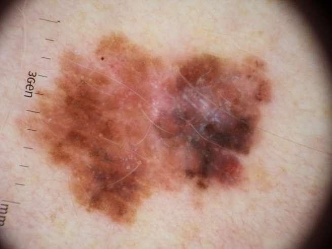
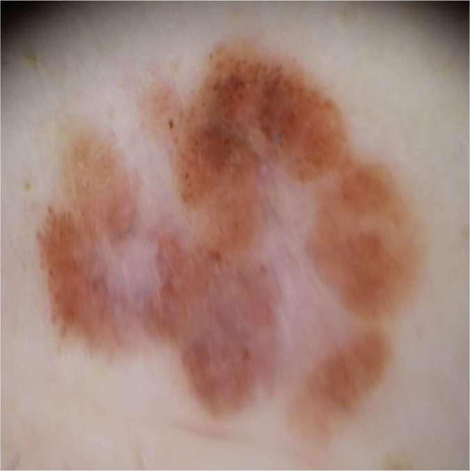
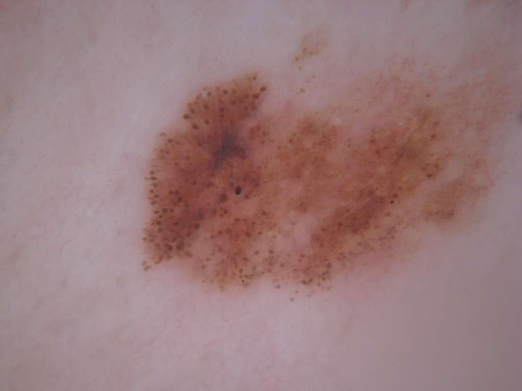
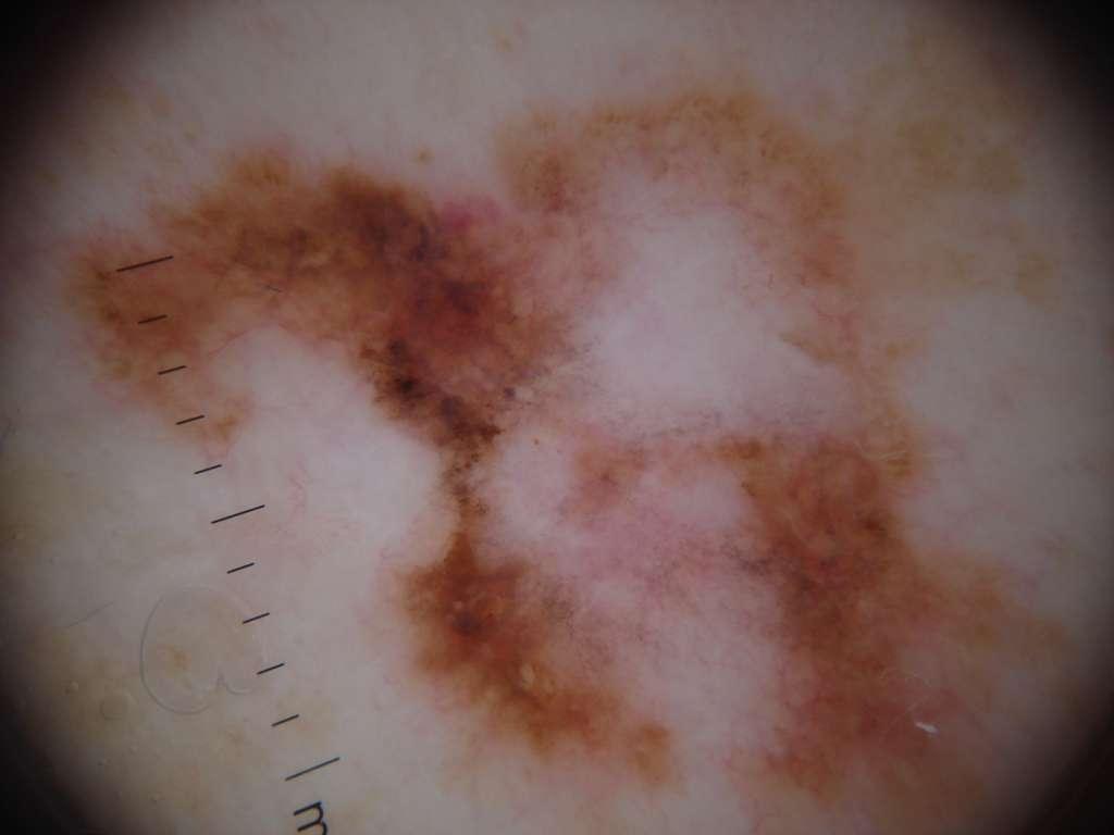
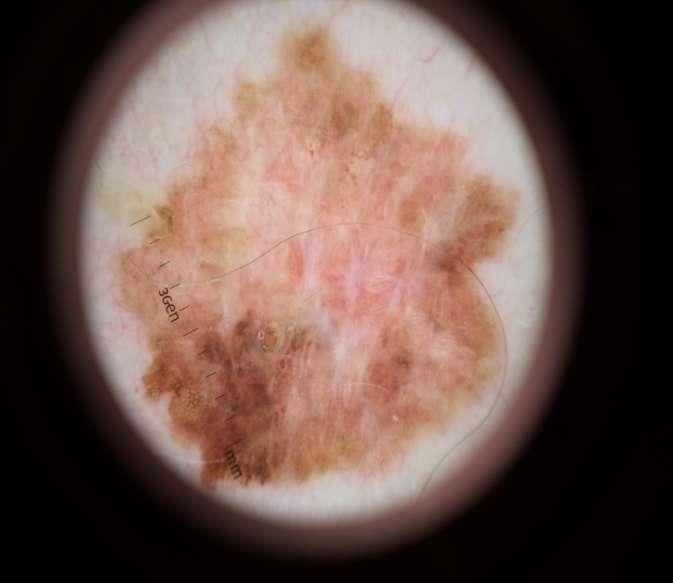
52 BCC the 4 classic dermoscopic features -micro ulcers -pink background -sharply focused branching vessels -irregular pigmented structures Don t look too hard for maple leaves or cart wheels!!!

53 Classical arborising vessels. Note also some blue-grey ovoid nests

54 Classic arborising BCC vessels, note also the pink background and irregular pigmented structures between 6 and8 o clock Probable micro ulcer (yellow)

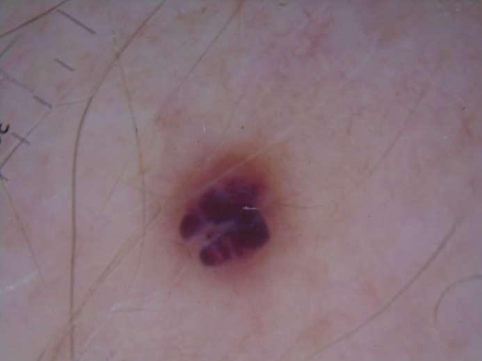
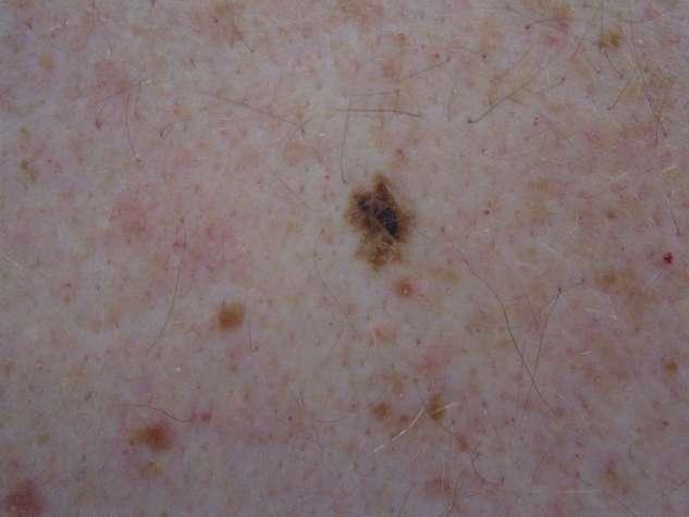
55 Full house! Typical blue grey ovoid nests Pink background, arborising vessels, micro ulcers, irregular pigmented structures

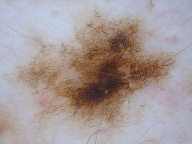
56 BCC and blue tattoo ink
57 Bowen s disease History (typically elderly white female, slow growing solitary plaque on lower leg) Rare under age of cm red scaly plaque DERMOSCOPY Confirms keratin scale Glomerular capillaries Absent dermoscopic features of BCC
58
59
60 MELANOMA dermoscopic features Asymmetry Multiple colours Multiple patterns Irregular dots and globules Streaks Pseudopods Blue/grey (blue/white) veil Amorphous/featureless
61 Algorithms and scoring systems Several exist, all work if used by Experienced dermoscopists The most basic is CHAOS AND CLUES
62 CHAOS AND CLUES chaos is defined as MULTIPLE COLOURS and/or MULTIPLE PATTERNS in a flat suspicious lesion (different rules apply to nodular lesions) If you see chaos, look for clues If chaotic and not a seborrhoiec keratosis, consider excision


63 Irregular globules 1cm pigmented lesion on jaw, history or recent change, female 75 Irregular dots and globules-also broadened irregular network. histology = melanoma

64 Note peripheral globules between 2 and 6 o clock Asymmetrical black blotch (? Blue grey veil?)
65
66
67
68
69
70
71 Irregular/asymmetrical globules
72 CHAOS!!!

73 GREY GLOBULES

74 ERYTHEMA Atypical network BLUE GREY VEIL
75
76

77 Incidental finding, this week


78 Multiple colours and patterns
79 Worried?
80
81
82 Dermoscopy -SUMMARY Useful adjunct to history and examination Limited use for nodular lesions Proven ability to reduce needless referral and excision of benign lesions Requires training and experience CAUTION-no 2 lesions are identical IF IN DOUBT YOU MUST REFER


83 3 good dermoscopy books about 50 each from Amazon. Jonathan Bowling Soyer, Argnziano et al Menzies et al
84 Dermoscopy learning resources My blog at has links to other resources as well as some of my pictures Plus a PDF of this presentation Try YouTube!!! Stephen Hayes
50 interactive dermoscopic case discussions Dr Stephen Hayes
 50 interactive dermoscopic case discussions Dr Stephen Hayes Annotations will be found on your memory drive, as will 100 case discussions and other learning material Melanoma 2mm thick Ugly duckling-one
It can be helpful in some cases of actinic keratosis, Bowen s disease and squamous cell carcinoma
 Dermoscopy Introduction, Terminology and Structures (to be read in conjunction with the Diagnostic Dermoscopic Algorithm) Copyright to Cunliffe TP (Jan. 2017) All rights reserved Introduction Dermoscopy
22/04/2015. Dermoscopy of Melanoma. Ilsphi Browne. Overview
 Dermoscopy of Melanoma Ilsphi Browne Overview The device Dermoscopic criteria (terminology) Colour Patterns Global features Local features Approach to diagnosing pigmented lesions Other uses in general
Fundamentals of dermoscopy
 Fundamentals of dermoscopy Learning objectives Upon completion of this session, participants should be able to: describe the basic principles of dermoscopy identify features associated with pigmented and
comedo-like openings (clods, brown or orange & circles) milia-like cysts (dots or clods, white) 1/29/18 Dotted vessels are also commonly seen in SCC
 milia-like cysts (dots or clods, white) 1/29/18 Dotted vessels are also commonly seen in SCC") Brown circles Dotted vessels are also commonly seen in SCC Step1 1. Nevus (unequivocal) 2. DF/IDN 3. BCC 4. SCC Network Patchy network Peripheral network & central hypopigmentation DF: network with central
Brown circles Dotted vessels are also commonly seen in SCC Step1 1. Nevus (unequivocal) 2. DF/IDN 3. BCC 4. SCC Network Patchy network Peripheral network & central hypopigmentation DF: network with central
Disclosure. Objectives. PAFP CME Conference Lou Mancano MD, FAAFP Reading Health System November 18, 2016
 PAFP CME Conference Lou Mancano MD, FAAFP Reading Health System November 18, 2016 1 Disclosure The speaker has no conflict of interest, financial agreement, or working affiliation with any group or organization.
Dermoscopy: Recognizing Top Five Common In- Office Diagnoses
 Dermoscopy: Recognizing Top Five Common In- Office Diagnoses Vu A. Ngo, DO Department of Family Medicine and Dermatology Choctaw Nation Health Services Authority Learning Objectives Introduction to dermoscopy
Skin lesions The Good and the Bad. Dr Virginia Hubbard Ipswich Hospital NHS Trust Barts and the London School of Medicine and Dentistry
 Skin lesions The Good and the Bad Dr Virginia Hubbard Ipswich Hospital NHS Trust Barts and the London School of Medicine and Dentistry Case 1 32 year old woman Australian Lesion on back New hair growing
Dermoscopy in everyday practice. What and Why? When in doubt cut it out? Trilokraj Tejasvi MD
 Dermoscopy in everyday practice Trilokraj Tejasvi MD Assistant Professor, Department of Dermatology, Director Teledermatology services, University of Michigan, Faculty Associate, GLOBAL REACH, Michigan
Mole mapping and monitoring. Dr Stephen Hayes. Associate Specialist in Dermatology, University Hospital Southampton
 Mole mapping and monitoring Dr Stephen Hayes Associate Specialist in Dermatology, University Hospital Southampton Outline of presentation The melanoma epidemic Benefits of early detection Risks of the
Common Benign Lesions and Skin Cancers. 22nd May 2015 Dr Mark Foley
 Common Benign Lesions and Skin Cancers 22nd May 2015 Dr Mark Foley Thank you for downloading this file. This intended to supplement the presentation given at the NZ Wound Care Conference, it is not intended
Dermoscopy Quiz 3-Point Checklist Algorithm
 Dermoscopy Quiz 3-Point Checklist Algorithm GLOBAL PATTERN Globular LOCAL CRITERIA Aggregated globules Milia-like cysts 3 POINT CHECK LIST Symmetrical No abnormal net Slight Blue-white veil BENIGN MELANOCYTIC
Introduction to Dermoscopy. Nicholas Compton, MD June 16, 2010
 Introduction to Dermoscopy Nicholas Compton, MD June 16, 2010 Overview What is dermoscopy Brief history Types of dermoscopy General approach to lesion of interest 2 step algorithm 3-point checklist Practice
Dr Stephen Hayes Associate Specialist in Dermatology University Hospital Southampton
 South East Dermatology Transformation and Sustainability Network Guildford, 19 th April 2018 Dermoscopy as an effective skin lesion triage tool in GP surgeries Dr Stephen Hayes Associate Specialist in
Basics in Dermoscopy
 Basics in Dermoscopy Manal Bosseila Professor of Dermatology, Cairo University Member of European Academy Dermatology & Venereology EADV Member of International Dermoscopy Society IDS Member of Aesthetic
MODULE 1. LOCAL AND GENERAL CRITERIA IN PIGMENTED MELANOCYTIC LESIONS.
 DERMOSCOPY TEACHING PROGRAMME Dermoscopy Teaching Programme Module 1 MODULE 1. LOCAL AND GENERAL CRITERIA IN PIGMENTED MELANOCYTIC LESIONS. Dermoscopy is a non-invasive in vivo technique that provides
INTRODUCTION HOUSEKEEPING June 11 th Dr John Adams Dermatologist/Dermoscopist MOLEMAP NZ/Australia MOLESAFE USA
 INTRODUCTION HOUSEKEEPING June 11 th 2015 Dr John Adams Dermatologist/Dermoscopist MOLEMAP NZ/Australia MOLESAFE USA Program Skin cancer statistics. Dermoscopy description and usefulness. Patient /lesion
Non-melanocytic Patterns
 Non-melanocytic Lesions Non-melanocytic Patterns Michelle Tarbox, MD Assistant Professor of Dermatology and Dermatopathology Texas Tech University Health Sciences Center 2018 Seborrheic keratoses Acanthotic
Skin Cancer A Personal Approach. Dr Matthew Strack Dunedin New Zealand
 Skin Cancer A Personal Approach Dr Matthew Strack Dunedin New Zealand Outline Dermoscopy Instruments and setup Photochemosurgery Clinical Aim: Leave with 2-3 ideas JLE Benign Junctional Nevus Management
6/17/2018. Breaking Bad (Part 1) Dermoscopy of Brown(ish) Things. Bad?
 Dermoscopy of Brown(ish) Things. Bad?") Breaking Bad (Part 1) Dermoscopy of Brown(ish) Things Jennie T. Clarke, MD ssociate Professor of Dermatology University of Utah School of Medicine Bad? 1 Brown(ish) Things Bad Melanoma Pigmented basal
Breaking Bad (Part 1) Dermoscopy of Brown(ish) Things Jennie T. Clarke, MD ssociate Professor of Dermatology University of Utah School of Medicine Bad? 1 Brown(ish) Things Bad Melanoma Pigmented basal
Benign versus Cancerous Lesions How to tell the difference FMF 2014 Christie Freeman MD, CCFP, DipPDerm, MSc
 1 Benign versus Cancerous Lesions How to tell the difference FMF 2014 Christie Freeman MD, CCFP, DipPDerm, MSc Benign lesions Seborrheic Keratoses: Warty, stuck-on Genetics and birthdays Can start in late
Appendix : Dermoscopy
 Go Back to the Top To Order, Visit the Purchasing Page for Details APP Appendix : Dermoscopy Dermoscopy, also known as dermatoscopy, epiluminoscopy and epiluminescent microscopy, is an effective non-invasive
Clinical and Dermoscopic Features of Thin Nodular Melanoma
 Clinical and Dermoscopic Features of Thin Nodular Melanoma A study of the International Dermoscopy Society Coordinator: Dr. Alexander J. Stratigos and colleagues, alstrat2@gmail.com ** Extended to May
Key factors in successfully integrating dermoscopy into your clinical practice
 Key factors in successfully integrating dermoscopy into your clinical practice S051 Dilemmas and challenges in skin cancer therapies and management Monday, March 4 th 2019 (9AM-12PM) Room 209A 10:56-11:09AM
The impact of GP sub-specialisation and dermatoscopy use on diagnostic accuracy for melanomas in Australia
 The impact of GP sub-specialisation and dermatoscopy use on diagnostic accuracy for melanomas in Australia Cliff Rosendahl, Gail Williams, Diann Eley, Tobias Wilson, Greg Canning, Jeffrey Keir, Ian McColl,
Malignant non-melanocytic lesions
 Malignant non-melanocytic lesions Course C023: Fundamentals of Dermoscopy March 4, 2019, 11:20 AM - 11:50 PM Room: 146B Jason B. Lee, MD Professor & Vice Chair Director of Dermatopathology & Pigmented
The GP s approach to the patient who is worried about sun, skin and moles. Dr Stephen Hayes
 The GP s approach to the patient who is worried about sun, skin and moles Dr Stephen Hayes Associate Specialist in Dermatology, University Hospital Southampton Dr Stephen Hayes DECLARATION OF INTERESTS
10/3/2018. Dermoscopy: Looking beneath the surface of the skin. Dermoscopy for Family Medicine 10/11/2018
 Dermoscopy for Family Medicine 10/11/2018 Jane M. Grant-Kels, MD, FAAD Founding Chair Emeritus, Dept of Dermatology Professor of Dermatology, Pathology & Pediatrics Director of the Cut Oncology Ctr & Melanoma
Dermoscopy. Synonyms. Dermoscopy. Definition. Dermoscopy opens up a world of colour and structure that can t be seen with the naked eye
 Synonyms Dermoscopy Australasian College of Dermatologists G.P Training Module Dermoscopy Dermatoscopy Epiluminescence microscopy Skin surface microscopy Incident light microscopy Oil immersion microscopy
Non-Melanocytic Pattern Dermoscopy
 Non-Melanocytic Pattern Dermoscopy I have no conflicts of interest to disclose Except that I LOVE dermoscopy Michelle Tarbox, MD Assistant Professor of Dermatology and Dermatopathology Texas Tech University
Dermoscopy. Enhanced Diagnostic Ability: Pigmented Lesions. Ted Rosen, MD Baylor College of Medicine Houston, Texas
 Dermoscopy Enhanced Diagnostic Ability: Pigmented Lesions Ted Rosen, MD Baylor College of Medicine Houston, Texas Faculty Disclosure Statement No conflicts relevant to this workshop! Sir William Osler
Dermoscopy. Sir William Osler. Dermoscopy. Dermoscopy. Melanoma USA Primary Care Update Faculty Disclosure Statement
 Diagnostic Ability: Pigmented Lesions Ted Rosen, MD Baylor College of Medicine Houston, Texas Enhanced 2010 Primary Care Update Faculty Disclosure Statement Ted Rosen, MD Speakers Bureau: Abbott, Amgen,
Revised Pattern Analysis: a method for the accurate diagnosis of pigmented skin lesions
 Dermatoscopy for Students A concise outline of: Revised Pattern Analysis: a method for the accurate diagnosis of pigmented skin lesions And Chaos and Clues: a decision algorithm for routine practice to
BJD British Journal of Dermatology. Summary. What s already known about this topic? CLINICAL AND LABORATORY INVESTIGATIONS
 CLINICAL AND LABORATORY INVESTIGATIONS BJD British Journal of Dermatology Pigmented nodular melanoma: the predictive value of dermoscopic features using multivariate analysis M.A. Pizzichetta, 1 H. Kittler,
Dermoscopic Features of Non-Pigmented Eccrine Poromas in. Department of Dermatology, Shinshu University School of Medicine,
 Original article Dermoscopic Features of Non-Pigmented Eccrine Poromas in Association with their Histopathological Features Akane Minagawa, Hiroshi Koga,* Masaomi Takahashi, + Kenji Sano, + Ryuhei Okuyama,
VACAVILLE DERMATOLOGY
 Connecting the Dots on those Spots NANDAN V. KAMATH, M.D. VACAVILLE DERMATOLOGY Sources All of the photos were taken with permission from the Dermnet NZ website - Dermnet New Zealand after communicating
Dermoscopy of non-pigmented skin lesions: a literature review
 Hong Kong J. Dermatol. Venereol. (2017) 25, 13-21 Review Article Dermoscopy of non-pigmented skin lesions: a literature review S Thomas, X Li, HP Soyer In this article, we will review benchmark dermoscopic
Dermoscopy STFM Richard Usatine, MD 5/2/16. Disclosure Statement: Some Dermatoscopes. Dermoscopy Video. Thanks to Dr.
 Disclosure Statement: Dermoscopy STFM 2016 Richard P. Usatine, MD, FAAFP Professor, Family and Community Medicine Professor, Dermatology and Cutaneous Surgery Medical Director, Clinic University of Texas
Dermoscopy, the use of a handheld
 ONLINE EXCLUSIVE Dermoscopy in family medicine: A primer Dermoscopy allows you to see deeper into the skin than with the naked eye. Here s how you can make use of it to spot malignant conditions sooner.
Histopathological and SIAscopic Correlation of Pigmented Skin Lesions
 Histopathological and SIAscopic Correlation of Pigmented Skin Lesions Professor Sujatha Fernando MBBS(Hon), MSc(London, Distinction), FRSTM&H, FRCPA, FIAC, FACTM Senior Consultant in Anatomical Pathology,
Benign and malignant epithelial lesions: Seborrheic keratosis: A common benign pigmented epidermal tumor occur in middle-aged or older persons more
 Benign and malignant epithelial lesions: Seborrheic keratosis: A common benign pigmented epidermal tumor occur in middle-aged or older persons more common on the trunk; but extremities, head and neck are
Reports on Scientific Meetings
 Hong Kong J. Dermatol. Venereol. (2016) 24, 146-153 The Hong Kong Society of Dermatology and Venereology Annual Scientific Meeting 2016 Reported by BTH Chan, CT Chau, CW Chow, CC Koh, WYK Lam, BS Tong,
Yes. Breaking Bad II: Dermoscopy of Pink-ish Things. Does it Fit? Yes 6/17/2018. Yes. Joslyn Kirby, MD, MS, MEd
 Breaking Bad II: Dermoscopy of Pink-ish Things Joslyn Kirby, MD, MS, MEd Yes Observe Yes Step 2. Fit a Benign Nevus Pattern? Does it Fit? Step 1: Melanocytic? pigment network, globules, homogeneous? No
Thursday 21 st August Skin Problems
 Thursday 21 st August 2014 Skin Problems Skin Problems The Sun and the Skin Sun Damage Recognising the early signs of skin cancer The Big 3 inflammatory condi=ons Acne & Rosacea Eczema (Including Seborrhoeic
Dr Amanda Oakley. Dermatologist Dept of Dermatology, Health Waikato Adjunct Associate Professor, Waikato Clinical Campus
 Dr Amanda Oakley Dermatologist Dept of Dermatology, Health Waikato Adjunct Associate Professor, Waikato Clinical Campus 14:00-16:00 WS #14: Dermoscopy Part 1 Skin Lesions and Dermatoscopy 16 August 2018
Skin Cancer. Dr Elizabeth Ogden Associate Specialist in Dermatology East and North Herts Dr Elizabeth Ogden
 Skin Cancer Dr Elizabeth Ogden Associate Specialist in Dermatology East and North Herts 13.10.16 Skin Cancer Melanoma mole cancer - is a true cancer which can metastasize and kill Non Melanoma skin cancer
Histopathology: skin pathology
 Histopathology: skin pathology These presentations are to help you identify, and to test yourself on identifying, basic histopathological features. They do not contain the additional factual information
Dermatopathology: The tumor is composed of keratinocytes which show atypia, increase mitoses and abnormal mitoses.
 Squamous cell carcinoma (SCC): A common malignant tumor of keratinocytes arising in the epidermis, usually from a precancerous condition: 1- UV induced actinic keratosis, usually of low grade malignancy.
Finding Melanoma. Is not easy!
 Finding Melanoma Is not easy! Finding Melanoma Victoria mean depth at diagnosis is 1.5 mm. Melanoma 1.5mm Has Stage 1B Mortality 10% Melanoma Spotting a killer! Spotting a killer Visual Clues What are
Regression 2/3/18. Histologically regression is characterized: melanosis fibrosis combination of both. Distribution: partial or focal!
 Regression Margaret Oliviero MSN, ARNP Harold S. Rabinovitz MD Histologically regression is characterized: melanosis fibrosis combination of both Distribution: partial or focal! Dermatoscopic terminology
Clinical characteristics
 Skin Cancer Fernando Vega, MD Seattle Healing Arts Clinical characteristics Precancerous lesions Common skin cancers ACTINIC KERATOSIS Precancerous skin lesions Actinic keratoses Dysplastic melanocytic
Derm quiz. Go to this link: goo.gl/forms/kchrhmtzl3vfnlv52. bit.ly/2a8asoy. Scan the QR code with your phone
 Dermatology quiz Derm quiz Go to this link: goo.gl/forms/kchrhmtzl3vfnlv52 OR bit.ly/2a8asoy OR Scan the QR code with your phone Contents Childhood rashes Pigmented lesions Sun damage Pityriasis References
Review of vasculature visualized on dermoscopy
 doi: 10.1111/1346-8138.13686 Journal of Dermatology 2017; 44: 525 532 REVIEW ARTICLE Review of vasculature visualized on dermoscopy Yaei TOGAWA Department of Dermatology, Chiba University Graduate School
Prediction without Pigment: a decision algorithm for non-pigmented skin malignancy
 DERMATOLOGY PRACTICAL & CONCEPTUAL www.derm101.com Prediction without Pigment: a decision algorithm for non-pigmented skin malignancy Cliff Rosendahl 1, Alan Cameron 1, Philipp Tschandl 2, Agata Bulinska
Features Causing Confusion between Basal Cell Carcinoma and Squamous Cell Carcinoma in Clinical Diagnosis
 TH Ryu, et al pissn 1013-9087ㆍeISSN 2005-3894 Ann Dermatol Vol. 30, No. 1, 2018 https://doi.org/10.5021/ad.2018.30.1.64 ORIGINAL ARTICLE Features Causing Confusion between Basal Cell Carcinoma and Squamous
Apps and Telemedicine H. Peter Soyer Dermatology Research Centre
 Apps and Telemedicine H. Peter Soyer Dermatology Research Centre p.soyer@uq.edu.au https://twitter.com/hpsoyer William Gibson The future is already here it's just not very evenly distributed Vision 3D
Hauora Maori. Map. Non-Melanoma Skin Lesion. Clinically Seborrhoeic Keratosis. Management Options. Punch Biopsy. Refer to Public Skin Lesion Service
 Care map information Information resources for patients and carers Updates to this Care Map Hauora Maori Pacific Skin lesion history Clinical examination and diagnostic test Suspicious of Melanoma Non-Melanoma
What is Dermoscopy? Early Dermoscopes. Deciphering Dermoscopy: Terminology, Features & Algorithms 6/17/2018
 Deciphering Dermoscopy: Terminology, Features & Algorithms Where did it come from and why do we use it? Jennie T. Clarke, MD Associate Professor of Dermatology University of Utah School of Medicine What
MELANOMA. 4 Fitzroy Square, London W1T 5HQ Tel: Fax: Registered Charity No.
 MELANOMA This leaflet had been written to help you understand more about melanoma. It tells you what it is, what causes it, what can be done about it, how it can be prevented, and where you can find out
MELANOMA. Some people are more likely to get a m Melanoma than others:
 MELANOMA This leaflet has been written to help you understand more about Melanoma. It tells you what is it, what causes it, what can be done about it, how it can be prevented, and where you can find out
Aspects on in vivo imaging techniques for diagnostics of pigmented skin lesions
 Thesis for the Degree of Doctor of Philosophy Aspects on in vivo imaging techniques for diagnostics of pigmented skin lesions Karin Terstappen (Westerhoff) Department of Dermatology and Venereology Institure
STUDY. Identification of Clinically Featureless Incipient Melanoma Using Sequential Dermoscopy Imaging
 STUDY Identification of Clinically Featureless Incipient Melanoma Using Sequential Dermoscopy Imaging Harald Kittler, MD; Pascale Guitera, MD; Elisabeth Riedl, MD; Michelle Avramidis, MD; Ligia Teban,
Management of patients with melanocytic and non-melanocytic neoplasms
 Management of patients with melanocytic and non-melanocytic neoplasms Ashfaq Marghoob MD Harold Rabinovitz MD Margaret Oliviero ARNP Harald Kittler MD Jupiter Cancer Centrer Characteristic Dermoscopic
Learning Objectives. Tanning. The Skin. Classic Features. Sun Reactive Skin Type Classification. Skin Cancers: Preventing, Screening and Treating
 Learning Objectives Skin Cancers: Preventing, Screening and Treating Robert A. Baldor, MD, FAAFP Professor, Family Medicine & Community Health University of Massachusetts Medical School Distinguish the
Oral and Maxillofacial Surgery Department
 Oral and Maxillofacial Surgery Department This leaflet explains: Lentigo Maligna What are the aims of this leaflet? This leaflet has been written to help you understand more about lentigo maligna and melanoma
Pathology of the skin. 2nd Department of Pathology, Semmelweis University
 Pathology of the skin 2nd Department of Pathology, Semmelweis University Histology of the skin Epidermis: Stratum corneum Stratum granulosum Stratum spinosum Stratum basale Dermis: papillary and reticular
BLINCK A diagnostic algorithm for skin cancer diagnosis combining clinical features with dermatoscopy findings
 DERMATOLOGY PRACTICAL & CONCEPTUAL www.derm101.com BLINCK A diagnostic algorithm for skin cancer diagnosis combining clinical features with dermatoscopy findings Peter Bourne, MBBS 1, Cliff Rosendahl,
Service Specification: CPO Skin Lesions
 Service Specification: CPO Skin Lesions This pathway description needs to be read in conjunction with the CPO Admin Service Specifications. 1. Outcomes Framework The outcomes sought from this service are
DIFFERENCES IN DERMOSCOPIC IMAGES FROM NON-POLARIZED DERMOSCOPE AND POLARIZED DERMOSCOPE INFLUENCE THE DIAGNOSTIC ACCURACY AND CONFIDENCE LEVEL.
 DIFFERENCES IN DERMOSCOPIC IMAGES FROM NON-POLARIZED DERMOSCOPE AND POLARIZED DERMOSCOPE INFLUENCE THE DIAGNOSTIC ACCURACY AND CONFIDENCE LEVEL. 1. Steven Q. Wang MD 1 (wangs@mskcc.org) 2. Stephen W. Dusza
Associate Professor Amanda Oakley. Professor H. Peter Soyer. Academic Dermatologist The University of Queensland Brisbane. Dermatologist Hamilton
 Associate Professor Amanda Oakley Dermatologist Hamilton Professor H. Peter Soyer Academic Dermatologist The University of Queensland Brisbane 8:30-10:30 WS #3: Dermoscopy Workshop Part 1 11:00-13:00 WS
Diagnosis of Lentigo Maligna Melanoma. Steven Q. Wang, M.D. Memorial Sloan-Kettering Cancer Center Basking Ridge, NJ
 Diagnosis of Lentigo Maligna Melanoma Steven Q. Wang, M.D. Memorial Sloan-Kettering Cancer Center Basking Ridge, NJ Conflict of Interest: None Topics Epidemiology and Natural History Clinical and Histologic
MELANOCYTIC LESIONS: EFFECTIVE MANAGEMENT IN PRIMARY CARE: Part 2
 MELANOCYTIC LESIONS: EFFECTIVE MANAGEMENT IN PRIMARY CARE: Part 2 In the second part of our feature on pigmented skin lesions, dermatology specialist Dr Sweta Rai describes the steps to rational decision-making
CANCER INSIGHT. FOR GPs. Summer 2018 WHAT YOU NEED TO KNOW ABOUT SKIN CANCER VISIT. our Skin Cancer Recognition Toolkit at
 CANCER INSIGHT FOR GPs Summer 2018 WHAT YOU NEED TO KNOW ABOUT SKIN CANCER VISIT our Skin Cancer Recognition Toolkit at www.doctors.net. uk/sct Cancer Research UK, Angel Building, 407 St John Street, London
I have a skin lump doc! What s next? 12 th August 2017 Dr. Sue-Ann Ho Ju Ee
 I have a skin lump doc! What s next? 12 th August 2017 Dr. Sue-Ann Ho Ju Ee Some thoughts Is this skin cancer? How common is this? How likely is this in this patient? What happens next if it s something
Melanoma. Consultation on draft guideline - stakeholder comments. Comments to be submitted before 5pm on Friday 13 March 2015
 Please note: Please fill in both the stakeholder organisation and name of commentator fields. We cannot accept forms with attachments such as research articles, letters or leaflets. Stakeholder organisation(s)
Abrupt Intralesional Color Change on Dermoscopy as a New Indicator of Early Superficial Spreading Melanoma in a Japanese Woman
 Published online: June 24, 2015 1662 6567/15/0072 0123$39.50/0 This is an Open Access article licensed under the terms of the Creative Commons Attribution-NonCommercial 3.0 Unported license (CC BY-NC)
This is a repository copy of Easily missed? Amelanotic melanoma. White Rose Research Online URL for this paper:
 This is a repository copy of Easily missed? Amelanotic melanoma. White Rose Research Online URL for this paper: http://eprints.whiterose.ac.uk/127789/ Version: Accepted Version Article: Muinonen-Martin,
Wellness Along the Cancer Journey: Cancer Types Revised October 2015 Chapter 7: Skin Cancer
 Wellness Along the Cancer Journey: Cancer Types Revised October 2015 Chapter 7: Skin Cancer Cancer Types Rev. 10.20.15 Page 56 Skin Cancer Group Discussion True False Not Sure 1. People with darker skin
Introduction to Dermoscopy. Disclosure. Introduction
 Introduction to Dermoscopy 1 Disclosure Dr. Deborah Bren has no conflict of interest, financial agreement, or working affiliation with any group or organization. 2 Introduction Deborah A. Bren, DO Family
The prevention, diagnosis, referral and management of melanoma of the skin: concise guidelines
 CLINICAL GUIDANCE The prevention, diagnosis, referral and management of melanoma of the skin: concise guidelines Julia Newton Bishop, Veronique Bataille, Alice Gavin, Marko Lens, Jerry Marsden, Tania Mathews
IT S FUNDAMENTAL MY DEAR WATSON! A SHERLOCKIAN APPROACH TO DERMATOLOGY
 IT S FUNDAMENTAL MY DEAR WATSON! A SHERLOCKIAN APPROACH TO DERMATOLOGY Skin, Bones, and other Private Parts Symposium Dermatology Lectures by Debra Shelby, PhD, DNP, FNP-BC, FADNP, FAANP Debra Shelby,
STUDY. Dermoscopy of Squamous Cell Carcinoma and Keratoacanthoma
 ONLINE FIRST STUDY Dermoscopy of Squamous Cell Carcinoma and Keratoacanthoma Cliff Rosendahl, MBBS; Alan Cameron, MBBS; Giuseppe Argenziano, MD; Iris Zalaudek, MD; Philipp Tschandl, MD; Harald Kittler,
LUMPS AND BUMPS: AN ORGANIZED APPROACH TO DIAGNOSIS AND MANAGEMENT
 LUMPS AND BUMPS: AN ORGANIZED APPROACH TO DIAGNOSIS AND MANAGEMENT Tammy P. Than, M.S., O.D., F.A.A.O. The University of Alabama at Birmingham / School of Optometry 1716 University Blvd. Birmingham, AL
المركب النموذج--- سبيتز وحمة = Type Spitz's Nevus, Compound SPITZ NEVUS 1 / 7
 SPITZ NEVUS 1 / 7 Epidemiology An annual incidence rate of 1.4 cases of Spitz nevus per 100,000 individuals has been estimated in Australia, compared with 25.4 per 100,000 individuals for cutaneous melanoma
Case Report Dermoscopy Clues in Pigmented Bowen s Disease
 Dermatology Research and Practice Volume 2010, Article ID 464821, 9 pages doi:10.1155/2010/464821 Case Report Dermoscopy Clues in Pigmented Bowen s Disease Daniela Gutiérrez-Mendoza, 1 Roberto Narro-Llorente,
Malignant tumors of melanocytes: Part 1. Deba P Sarma, MD., Omaha
 Malignant tumors of melanocytes: Part 1 Deba P Sarma, MD., Omaha The melanocytic tumor is one of the most difficult and confusing areas in Dematopathology. It is true that most (95%) of such lesions are
Graph-based Pigment Network Detection in Skin Images
 Graph-based Pigment Network Detection in Skin Images M. Sadeghi a,b, M. Razmara a, M. Ester a, T. K. Lee a,b,c, M. S. Atkins a a Simon Fraser University, 8888 University Drive, Burnaby, BC, Canada, V5A1S6;
Periocular skin cancer
 Periocular skin cancer Information for patients Skin cancer involving the skin of the eyelid or around the eye is called a periocular skin cancer. Eyelid skin cancers occur most often on the lower eyelid,
Desmoplastic Melanoma R/O BCC. Clinical Information. 74 y.o. man with lesion on left side of neck r/o BCC
 R/O BCC Sabine Kohler, M.D. Professor of Pathology and Dermatology Dermatopathology Service Stanford University School of Medicine Clinical Information 74 y.o. man with lesion on left side of neck r/o
Pathology. Skin Tumor. Bayan N. Mohammad 15/10/2015. Mohammad al-orjani. Page 0 of 23
 #7 35 Pathology Skin Tumor Bayan N. Mohammad 15/10/2015 Mohammad al-orjani Page 0 of 23 بسم هللا الرحمن الرحيم GREETINGS This lecture is about skin tumors, all the slides are included and every slide will
Malignant Melanoma Early Stage. A guide for patients
 This melanoma patient brochure is designed to help educate melanoma patients and their caregivers. It was developed under the guidance of Dr. Michael Smylie, Professor, Department of Oncology, University
Living Beyond Cancer Skin Cancer Detection and Prevention
 Living Beyond Cancer Skin Cancer Detection and Prevention Cutaneous Skin Cancers Identification Diagnosis Treatment options Prevention What is the most common cancer in people? What is the most common
SKIN. 3. How is the skin structured around the finger joints to allow for flexible movement of the fingers?
 SKIN Objectives for Exam #1: 1. List various skin structures and describe their functions. 2. Describe skin responses to increases and decreases in body temperature. 3. Provide examples of various skin
Skin Care in Renal Transplant Patients
 Skin Care in Renal Transplant Patients Introduction Skin Care in Renal Transplant Patients Skin care is very important for everyone, but particularly for renal patients who have received transplants. Because
Accepted Article. Dermoscopic diagnosis of amelanotic/hypomelanotic melanoma
 Received Date : 19-May-2016 Revised Date : 01-Sep-2016 Accepted Date : 20-Sep-2016 Article type : Research Letter Dermoscopic diagnosis of amelanotic/hypomelanotic melanoma M.A. Pizzichetta, 1 H. Kittler,
MECHANISMS OF HUMAN DISEASE: LABORATORY SESSION PATHOLOGY OF THE SKIN LAB. Friday, February 13, :30 am 11:00 am
 MECHANISMS OF HUMAN DISEASE: LABORATORY SESSION PATHOLOGY OF THE SKIN LAB Friday, February 13, 2009 9:30 am 11:00 am FACULTY COPY GOALS: Describe the basic clinical and morphologic features of various
Identifying Skin Cancer. Mary S. Stone MD Professor of Dermatology and Pathology University of Iowa Carver College of Medicine March, 2018
 Identifying Skin Cancer Mary S. Stone MD Professor of Dermatology and Pathology University of Iowa Carver College of Medicine March, 2018 American Cancer Society web site Skin Cancer Melanoma Non-Melanoma
MECHANISMS OF HUMAN DISEASE: LABORATORY SESSION PATHOLOGY OF THE SKIN LAB. Friday, February 12, :30 am 11:00 am
 MECHANISMS OF HUMAN DISEASE: LABORATORY SESSION PATHOLOGY OF THE SKIN LAB Friday, February 12, 2012 9:30 am 11:00 am FACULTY COPY GOALS: Describe the basic clinical and morphologic features of various