Title. CitationBritish Journal of Dermatology, 155(5): Issue Date Doc URL. Rights. Type. File Information
|
|
|
- Domenic Chapman
- 6 years ago
- Views:
Transcription
1 Title Childhood epidermolysis bullosa acquisita with autoa collagen : case report and review of the literature Author(s)Mayuzumi, M.; Akiyama, M.; Nishie, W.; Ukae, S.; Abe CitationBritish Journal of Dermatology, 155(5): Issue Date Doc URL Rights The definitive version is available at Type article (author version) File Information BJD155-5.pdf Instructions for use Hokkaido University Collection of Scholarly and Aca
2 British Journal of Dermatology Manuscript No. BJD Revised Version Case Report Childhood epidermolysis bullosa acquisita with autoantibodies against the non-collagenous 1 and 2 domains of type VII collagen: a case report and a review of the literature M. MAYUZUMI 1, 2, M. AKIYAMA 1, W. NISHIE 1, 2, S. UKAE 3, M. ABE 1, D. SAWAMURA 1, T. HASHIMOTO 4 and H. SHIMIZU 1 1 Department of Dermatology, Hokkaido University Graduate School of Medicine, Sapporo, Japan; Departments of 2 Dermatology and 3 Pediatrics, Sapporo Social Insurance General Hospital, Sapporo, Japan; 4 Department of Dermatology, Kurume University School of Medicine, Kurume, Japan Short title: Childhood EBA Key words: EBA, epitope, immunoblot, immunofluorescence, inflammatory phenotype Corespondence Masashi Akiyama Department of Dermatology, Hokkaido University Graduate School of Medicine, N15 W7, Sapporo , Japan akiyama@med.hokudai.ac.jp Telephone: ext Fax:
3 Summary Epidermolysis bullosa acquisita (EBA) is an acquired subepidermal bullous disease characterized by IgG autoantibodies to type VII collagen, a major component of anchoring fibrils. Most patients with EBA are adult and develop autoantibodies to the non-collagenous 1 (NC-1) domain of type VII collagen.in this report, we describe a 4-year-old Japanese boy presenting pruritic vesicles and tense blisters over his whole body. Immunofluorescence studies revealed linear IgG/C3 deposits along the dermal-epidermal junction of the patient s skin, and circulating IgG autoantibodies mapping to the dermal side of 1M NaCl-split skin. By immunoblotting analysis using dermal extracts as a substrate, the patient s IgG antibodies labeled a 290 kd protein corresponding to type VII collagen. Immunoblotting studies using recombinant proteins demonstrated that the patient s circulating autoantibodies recognized not only the NC-1 but also the non-collagenous 2 (NC-2) domain of type VII procollagen. Review of the previously reported cases and the present case suggested that EBA cases with autoantibodies to regions other than the NC-1 domain are all children younger than 10 years of age with clinical features of an inflammatory phenotype. 2
4 Introduction Epidermolysis bullosa acquisita (EBA) is an autoimmune blistering skin disease which has circulating IgG autoantibodies to type VII collagen, a major component of anchoring fibrils 1, composed of three identical alpha chains. Each chain consists of a long central triple-helical collagenous domain flanked by a large amino-terminal 145 kd non-collagenous 1 (or NC1) domain and a smaller carboxyl-terminal 20 kd non-collagenous 2 (NC2) domain. Type VII procollagen molecules form anti-parallel dimers that are stabilized by disulfide bonding at the carboxyl terminus, and a portion of the NC2 domain is removed by specific proteolytic cleavage to yield the mature type VII collagen. Several dimers aggregate laterally to form the unique cross-banded structure, i.e. anchoring fibrils, which comprise anti-parallel dimers and contain NC1 domains at both ends, locating in the lamina densa and forming semi-circular loops visible by the electron microscope 2. Two distinct phenotypes of EBA have been described; the classical non-inflammatory type and the inflammatory type. In a large number of the adults with clinically classical type of EBA, it has previously been recognized that autoantibodies are directed against epitopes within the NC1 domain, whereas no immunoreactivity with the NC2 or the triple helical domain was detected 3. However, recently, in five children with the inflammatory subtype of EBA, novel variants with reactivity to the NC2 or/and the triple helical domains have been reported 4-6. These findings indicated that some distinct immunopathological differences between childhood EBA and those occurred in adulthood could be present. In this report, we describe a case of EBA in a 4-year-old Japanese boy and have reviewed previous reported cases of childhood EBA. 3
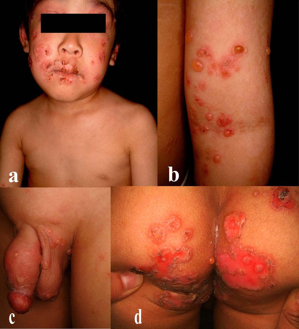
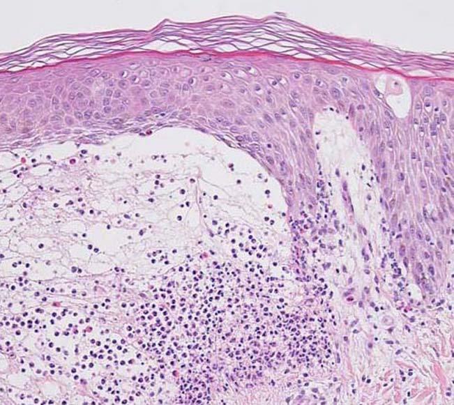
5 Case Report A 4-year-old Japanese boy, generally in good health, developed painful oral blisters and erosions. A few weeks later, widespread pruritic vesicles and tense blisters appeared over almost his entire body, but especially on his scrotum and buccal mucosa. The blisters were seen both on erythematous bases and on normal skin, and showed an annular arrangement in some legions (Fig.1). His nails were not affected. He had no family history of any blistering disorders. General laboratory examinations including full blood count, hepatic and renal function tests, urinalysis and C-reactive protein were within normal limits except for an increased number of eosinophils (13.5 %) in his peripheral blood. Anti-nuclear antibodies were negative. Anti-herpes simplex virus antibodies were not detected, either. Viral culture of vesicular fluid and a Tzanck smear test on a vesicular base were all negative. Histopathological examination of skin biopsy specimens from the dorsal side of his foot revealed subepidermal blisters with an inflammatory infiltrate of lymphocytes, neutrophils, and eosinophils in the papillary and superficial reticular dermis. Especially, neutrophils and nuclear dusts were seen at the tips of the edematous dermal papillae (Fig.2). On direct immunofluorescence (IF) studies, linear deposits of IgG and C3, but no IgA were seen along the dermal-epidermal junction of the patient s skin biopsy specimen. Indirect IF studies using a 1M NaCl-split normal human skin as a substrate showed IgG deposits on the dermal side of the artificial split at a titer of 1:160 (Fig.3). Circulating autoantibodies to the NC16a domain of 180 kd bullous pemphigoid antigen (BP180) were not detected using a BP 180 enzyme-linked immunosorbent assay (ELISA) 4
6 kit (MBL, Naka-ku, Nagoya, Japan). Immunoblot analysis demonstrated that the patient s sera reacted both with a 290 kd protein in dermal extracts and recombinant type VII collagen (Figs. 4a, b). In addition to the 290 kd band, many additional bands were seen in the immunoblot analysis with patient sera using dermal extract as a substrate. These additional bands were thought to reflect reactivity of patient s sera to background, degradation products because, in this immunoblot analysis, we used high concentration sera in order not to overlook 200 kd band in p200 pemphigoid. Epitope analyses with type VII pro-collagen recombinant fragments revealed that the patient s sera recognized both of the NC1 and NC2 domains (Fig.4c). The boy was treated with oral prednisolone (1.1mg/kg per day) for 2 weeks, which inhibited new blister formation. When the dose of prednisolone was reduced to 0.4mg/kg per day, new blister formation was observed. Oral dapsone 1.5mg/kg per day was added, but on the next day, erythematous macules appeared on his upper trunk. Thus, dapsone was discontinued and the dose of prednisolone was increased to 0.7mg/kg per day. During the next 2 months, the patient kept taking oral prednisolone 0.5mg/kg per day, and the blisters steadily healed, leaving hyper- and hypo-pigmented macules and milia. Discussion EBA is a relatively uncommon disease with an incidence about 10 times less than that of bullous pemphigoid (BP). EBA was first defined with following characteristic features: trauma-induced bullae that heal with milia and scar mainly over the joints of the hands, elbows, feet and knees; nail dystrophy; typically adult onset; a negative family history of 5
7 epidermolysis bullosa; and exclusion of other bullous diseases on the basis of clinical and laboratory evidence. The definitive features now include histological and immunopathological findings, such as subepidermal blisters; circulating autoantibodies against skin basement membrane zone antigens, binding to the dermal side of NaCl-split skin; IgG/C3 deposits in the anchoring fibril zone; and the presence of anti-type VII collagen antibodies that are identified as a 290 kd band by immunoblot analysis on dermal extracts 1. Among clinically diagnosed EBA patients, antibodies can be detected by immunoblotting with dermal extracts in approximately 30% of the cases 7, and by indirect IF using NaCl-split skin as a substrate in approximately 50% of the cases 8. Two distinct phenotypes of EBA have been described, i.e. the classical (non-inflammatory)type and inflammatory type. The classical type presents with marked skin fragility, blisters and erosions at sites of trauma, and healing with scarring and milia. The inflammatory type can mimic almost all other chronic bullous diseases, and its clinical differentiation from BP, cicatricial pemphigoid and linear IgA bullous dermatosis may be difficult 4-6, In some patients, characteristics of both classical and inflammatory phenotypes of EBA have been observed 9, but most patients with EBA in adulthood appear to suffer from the classical type of EBA. Conversely, patients with childhood EBA (16-years-old or younger) are very rare. To our knowledge, there are 33 cases of childhood EBA including the present case reported in the literature 4-6, 9-26 Among them, six cases were Japanese and the others were non-japanese including at least five Caucasian and five African. These patients presented mainly with the inflammatory subtype. Some differences exist between EBA in adults and children. In the adult, the occurrence of mucosal involvement was seen in 6
8 approximately 50 % of patients, while in the children, mucosal lesions were seen in the vast majority of the reported cases 9, 18. The adult form of EBA is known to be difficult to treat, requiring high doses of prednisolone, dapsone, immunosuppressive agents, plasmapheresis, etc. In contrast, dapsone and low dose prednisolone are usually effective in treatments for childhood EBA. The prognosis in childhood EBA seems to be much better than adult cases 4-6, The major epitopes of circulating autoantibodies in adult patients with the classical type of EBA are known to be located within the NC1 domain, but neither in the NC2 nor in the triple helical domains 3. However in five cases of childhood EBA (four Japanese children and one European child) reported in the literature, autoantibodies have been found to recognize epitopes on the NC2 and/or the triple helical domain over the last years (Table 1) 4-6. These five patients and the present case were all young children less than 10 years of age, who all showed similar clinical features of the inflammatory subtype of EBA, and complete clearing or a relatively good response to treatments. Interestingly, two of these children also demonstrated autoantibody reactivity towards the 230kD BP antigen (BP230) and the NC16A domain of BP Schmidt et al. 5 mentioned this phenomenon and referred it as epitope spreading. Epitope spreading sometimes occurs in other adult human autoimmune skin disorders, and has been observed in a few cases of EBA, specifically in young children 5. The molecular events in epitope spreading remain to be elucidated, but it is supposed that the skin damage from autoimmune or inflammatory processes subsequently could induce autoimmunity to a sequestered or closely related antigen or epitope. Five out of the six EBA patients with autoantibodies to the NC2 and/or 7
9 the triple helical domains are Japanese children. Tanaka et al. 4 suggested the phenomenon may be unique to a particular ethnic group. To confirm this hypothesis, detailed epitope mappings in bullous dermatosis cases with similar clinical and pathological features will be required, especially in children from different ethnic groups. Further studies on autoantibodies in larger series of patients with EBA are needed to clarify the correlation between the presence of certain autoantibodies against specific subdomains of type VII collagen and their clinical features, such as classical or inflammatory phenotype, the patient s age of onset, clinical courses, and the prognosis. 8
10 References 1) Woodley DT, Briggaman RA, O Keefe EJ et al. Identification of the skin basement-membrane autoantigen in epidermolysis bullosa acquisita. N Engl J Med 1984; 310: ) Shimizu H, Ishiko A, Masunaga T et al. Most anchoring fibrils in human skin originate and terminate in the lamina densa. Lab Invest 1997; 76: ) Lapiere J-C, Woodley DT, Parente MG et al. Epitope mapping of type VII collagen: identification of discrete peptide sequences recognized by sera from patients with acquired epidermolysis bullsa. J Clin Invest 1993; 92: ) Tanaka H, Ishida-Yamamoto A, Hashimoto T et al. A novel variant of acquired epidermolysis bullosa with autoantibodies against the central triple-helical domain of type VII collagen. Lab Invest 1997; 77: ) Schmidt E, Hopfner B, Chen C et al. Childhood epidermolysis bullosa acquisita: a novel variant with reactivity to all three structural domains of type VII collagen. Br J Dermatol 2002; 147: ) FukumotoT, Umemura T, Higuchi M. et al. Childhood epidermolysis bullosa acquisita with autoantibodies against all 3 structural domains of type VII collagen. J Am Acad Dermatol 2004; 50(3): ) Chen M, Chan LS, Cai X et al. Development of an ELISA for rapid detection of anti-type VII collagen autoantibodies in epidermolysis bullosa acquisita. J Invest Dermatol 1997; 108: ) Gammon WR, Fine JD, Forbes M, Briggaman RA. Immunofluorescence on split skin for the detection and differentiation of basement membrane zone autoantibodies. J Am Acad Dermatol 1992; 27: ) Stewart MI, Woodley DT, Briggaman RA. Epidermolysis bullosa 9
11 acquisita and associated symptomatic esophageal webs. Arch Dermatol 1991; 127: ) Borok M, Heng MCY, Ahmed AR et al. Epidermolysis bullosa acquisita in an 8-year-old girl. Pediatric Dermatol 1986; 3: ) Rubenstein R, Esterly NB, Fine JD. Childhood epidermolysis bullosa acquisita. Arch Dermatol 1987; 123: ) McCuaig CC, Chan LS, Woodley DT et al. Epidermolysis bullosa acquisita in childhood. Arch Dermatol 1989; 125: ) Espagne E, Prost C, Janssen F et al. Epidermolyse bulleuse acquise de l enfant. A propos d un cas. Ann Dermatol Venereol 1990; 117: ) Arpey CJ, Elewski BE, Moritz DK, Gammon WR. Childhood epidermolysis bullosa acquisita.report of three cases and review of literature. J Am Acad Dermatol 1991; 24: ) Roger H, Machado P, Nicolas JF et al. Epidermolysis bullosa acquisita in a 3 1 / 2 -year-old girl. J Am Acad Dermatol 1992; 27: ) Kirtschig G, Wojnarowska F, Marsden RA et al. Acquired bullous disease of childhood: re-evaluation of diagnosis by indirect immunofluorescence examination on 1M NaCl split skin and immunoblotting. Br J Dermatol 1994; 130: ) Lacour JP, Bernard P, Rostain G et al. Childhood acquired epidermolysis bullosa. Pediatric Dermatol 1995; 12: ) Caux F, Kirtschig G, Lemarchand-Venencie et al. IgA-epidermolysis bullosa acquisita in a child resulting in blindness. Br J Dermatol 1997; 137: ) Callot-Mellot C, Bodemer C, Caux F et al. Epidermolysis bullosa acquisita in childhood. Arch Dermatol 1997; 133: ) Feliciani C, Fasciocco D, Franchi A, Amerio P. Epidermolysis bullosa 0
12 acquisita in a 10-year-old Alabian boy. Br J Dermatol 1997; 137: ) Park SB, Cho KH, Youn JI et al. Epidermolysis bullosa acquisita in childhood- a case mimicking chronic bullous dermatosis of childhood. Clin Exp Dermatol 1997; 22: ) Hayashi K, Ueda M, Sakai M et al. Epidermolysis bullosa acquisita in a 6-year-old Japanese boy. Int J Dermatol 1998; 37: ) Edwards S, Wakelin SH, Wojnarowska F et al. Bullous pemphigoid and epidermolysis bullosa acquisita. Pediatric Dermatol 1998; 15: ) Chorzelski T, Karczewska K, Dyduch A et al. Epidermolysis bullosa acquisita in a 4-year-old boy. Pediatric Dermatol 2000; 17: ) Trigo-Guzman FX, Conti A, Aoki V et al. Epidermolysis bullosa acquisita in childhood. J Dermatol 2003; 30: ) Parodi A, Cozzani C, Verrini A et al. Epidermolysis bullosa acquisita in a child:an additional case. Int J Dermatol 2005; 44:
13 Figure legends Fig 1. Clinical features. Disseminated, tense vesicles, bullae and erosions with crusts were seen over erythematous plaques on the face (a), arm (b), genital area (c) and the buttocks (d). Fig 2. Histopathology. A subepidermal blister with an inflmmatory infiltrate of lymphocytes together with eosinophils and neutrophils in the papillary dermis. (Original magnification 125). Fig 3. (a) By direct IF staining, linear IgG deposits were seen along the dermal-epidermal junction in the patient s skin. (b) Indirect IF using 1M NaCl-split human skin as a substrate revealed linear IgG deposits along the dermal side of the artificial split. ( dots: Roof side of the split skin). Fig 4. (a) Immunoblot analysis of patient s circulating autoantibodies on normal human dermal extracts. Arrows indicate the 290kD EBA antigen and 200kD anti-p200 pemphigoid antigen. Control EBA serum (lane 1) and patient's serum (lane 3) reacted with EBA antigen, whereas control anti-p200 serum (lane 2) showed no 290kD band. (b) Immunoblot analysis of patient s circulating autoantibodies on recombinant type VII collagen. Arrow indicates the 290kD EBA antigen. Control EBA serum (lane 1) and patient's serum (lane 3) reacted with EBA antigen, whereas normal control serum (lane 2) showed no reactivity. (c) Immunoblot analyses using recombinant fusion proteins of NC1 and NC2 domains of type VII collagen. Arrows indicate respective positions of fusion proteins. Both control EBA serum (lane 1) and patient's serum (lane 2) reacted with NC1 and with 2
14 NC2. 3
15 Table 1. Cases with childhood EBA in which epitopes for autoantibodies were detected by immunoblot analysis IgG deposits in Immunoblot on Immunoblot on Recombinant protein immunoblot analysis Case Age/Race/Sex DIF (BMZ) NaCl split skin IIF dermal extracts epidermal extracts NC1 TH NC2 rbp180 NC16a ELISA Therapy Reference 1 8/Japanese/F IgG, IgA, C3 Dermal side 290kD (+) 230kD(+) ND - PSL+DDS 4 2 1/Japanese/M IgG, C3 Dermal side 290kD (+) no reactivity ND + Methylprednisolone+DDS 4 3 2/Japanese/F IgG, C3 Dermal side 290kD (+) no reactivity ND - PSL 4 4 4/European/F IgG, C3 Dermal side 290kD (+) no reactivity ND PSL+DDS 5 5 5/Japanese/M IgG, C3 Dermal side 290kD (+) no reactivity ND - Betamethasone 6 6 4/Japanese/M IgG, C3 Dermal side 290kD (+) ND + ND + ND - PSL Present case DIF: direct IF, IIF: indirect IF, BMZ: basement membrane zone, TH: triple helical collagenous domain, NC16a: NC16a domain of BP180, rbp180: reconbinant BP180, ND: not done PSL: prednisolone, DDS: diaminodiphenylsulfone
16
17

18
19
Epidermolysis Bullosa Acquisita
 Introduction Epidermolysis Bullosa Acquisita Pages with reference to book, From 192 To 194 Nasser Rashid Dar ( Departments of Dermatology, Combined Military Hospital, Peshawar. ) Ahsan Hameed, Ashfaq Ahmad
Erythema gyratumrepens-like eruption in a patient with epidermolysisbullosaacquisita associated with ulcerative colitis
 Erythema gyratumrepens-like eruption in a patient with epidermolysisbullosaacquisita associated with ulcerative colitis A. España C. Sitaru* M. Pretel L. Aguado J. Jimenez# Department of Dermatology, University
Background information of DIF
 Napa Dermatopathology Meeting 2018: Immunobullous Disease Whitney A. High, MD, JD, MEng whitney.high@ucdenver.edu Professor of Dermatology & Pathology Vice-Chairman, Dermatology Director of Dermatopathology
A case of bullous pemphigoid following pemphigus foliaceus
 #2228 A case of bullous pemphigoid following pemphigus foliaceus Priyanka Vedak MD 1, Danielle Levine MD 1,3, Lyn Duncan MD 2,3, Hensin Tsao 1,3, Daniela Kroshinsky MD MPH 1,3 1. Department of Dermatology,
Bullous Pemphigoid with Lymphocytic Colitis: A Case Report and Short Literature Review
 Dermatol Ther (Heidelb) (2016) 6:437 441 DOI 10.1007/s13555-016-0135-4 CASE REPORT Bullous Pemphigoid with Lymphocytic Colitis: A Case Report and Short Literature Review Alexandra Sperl. Johann W. Bauer.
Epidermolysis Bullosa Acquisita: A Retrospective Clinical Analysis of 30 Cases
 Acta Derm Venereol 2011 Epub ahead of print CLINICAL REPORT Epidermolysis Bullosa Acquisita: A Retrospective Clinical Analysis of 30 Cases Jong Hoon Kim 1, Yeon Hee Kim 2 and Soo-Chan Kim 1 1 Department
Autoimmune Diseases with Oral Manifestations
 Autoimmune Diseases with Oral Manifestations Martin S. Greenberg DDS, FDS RCSEd Professor Emeritus Department of Oral Medicine University of Pennsylvania Disclosure Statement I have no actual or potential
Interesting Case Series. Linear IgA Bullous Dermatosis
 Interesting Case Series Linear IgA Bullous Dermatosis Sean Chen, BA, a Peter Mattei, MD, a Max Fischer, MD, MPH, a Joshua D. Gay, PA-C, b Stephen M. Milner, MBBS, BDS, FRCS (Ed), FACS, b and Leigh Ann
Acquired and Inherited Bullous Diseases
 Acquired and Inherited Bullous Diseases Erin Wei MD Brigham and Women s Hospital, Department of Dermatology Instructor, Harvard Medical School Director, Bullous Disease Clinic No disclosures Conflict of
A cross-sectional study of clinical, histopathological and direct immmunofluorescence diagnosis in autoimmune bullous diseases
 Original Article A cross-sectional study of clinical, histopathological and direct immmunofluorescence diagnosis in autoimmune bullous diseases Anchal Jindal, MD 1 Rushikesh Shah, MBBS 2 Neela Patel, MD
Introduction to Pemphigoid: Spectrum of Disease & Treatment
 Introduction to Pemphigoid: Spectrum of Disease & Treatment A Razzaque Ahmed, MD Center for Blistering Diseases Boston, MA A.Razzaque.Ahmed@tufts.edu centerforblisteringdiseases.com SPECTRUM OF PEMPHIGOID
BULLOUS SYSTEMIC lupus erythematosus
 OBSERVATION Bullous Systemic Lupus Erythematosus With Autoantibodies Recognizing Multiple Skin Basement Membrane Components, Bullous Pemphigoid Antigen 1, Laminin-5, Laminin-6, and Type VII Collagen Lawrence
Department of Dermatology, Nippon Medical School, 1-1-5, Sendagi, Bunkyo-ku, Tokyo , Japan 2
 Dermatology Research and Practice Volume 2010, Article ID 931340, 5 pages doi:10.1155/2010/931340 Case Report Paraneoplastic Pemphigus Presenting as Mild Cutaneous Features of Pemphigus Foliaceus and Lichenoid
Autoimmune bullous dermatoses
 Autoimmune bullous dermatoses Overview of serological diagnostics in autoimmune blister-forming diseases of the skin Pemphigoid diseases Pemphigus diseases Epidermolysis bullosa acquisita Dermatitis herpetiformis
Classification: 1. Infective: 2. Traumatic: 3. Idiopathic: Recurrent Aphthous Stomatitis (RAS) 4. Associated with systemic disease:
 4. Associated with systemic disease:") Classification: 1. Infective: 2. Traumatic: 3. Idiopathic: Recurrent Aphthous Stomatitis (RAS) 4. Associated with systemic disease: Hematological GIT Behcet s HIV 5. Associated with dermatological diseases:
Classification: 1. Infective: 2. Traumatic: 3. Idiopathic: Recurrent Aphthous Stomatitis (RAS) 4. Associated with systemic disease: Hematological GIT Behcet s HIV 5. Associated with dermatological diseases:
B. Autoimmune blistering diseases
 Go Back to the Top To Order, Visit the Purchasing Page for Details formation immediately above the basal layer. The dermal papillae, which are covered by basal cells in the single layer that is left in
Dr Saleem Taibjee. Consultant Dermatologist & Dermatopathologist
 Dr Saleem Taibjee saleem.taibjee@dchft.nhs.uk Consultant Dermatologist & Dermatopathologist Case S14-10797 and S15-4023 F50. Previous blistering, now marked milia on dorsum of hands. 4mm punch biopsy The
Egyptian Dermatology Online Journal Vol. 8 No 2: 6, December Yasmeen J Bhat*, Iffat Hasan*, Atiya Yaseen*, Hina Altaf*, Shylla Mir**
 Pemphigoid gestationis in a multigravida Yasmeen J Bhat*, Iffat Hasan*, Atiya Yaseen*, Hina Altaf*, Shylla Mir** * Department of Dermatology, STD & Leprosy; Government Medical College, Srinagar ** Department
HEMORRHAGIC BULLOUS HENOCH- SCHONLEIN PURPURA: A CASE REPORT
 HEMORRHAGIC BULLOUS HENOCH- SCHONLEIN PURPURA: A CASE REPORT Nirmala Ponnuthurai, Sabeera Begum, Lee Bang Rom Paediatric Dermatology Unit, Institute of Paediatric, Hospital Kuala Lumpur, Malaysia Abstract
Clinicopathological correlation of blistering diseases of skin
 Bangladesh Med Res Counc Bull 2008; 34: 48-53 Copyright 2008 by Bangladesh Medical Research Council Clinicopathological correlation of blistering diseases of skin A.K.M. Nurul Kabir 1, Mohammed Kamal 1
EPIDERMOLYSIS BULLOSA
 EPIDERMOLYSIS BULLOSA Definition Epidermolysis bullosa (EB) is a term used to describe a group of rare mainly hereditary, chronic, non-inflammatory diseases of skin and mucous membranes. EB is characterized
Department of Dermatology, Christian Medical College and Hospital, Ludhiana, Punjab, India.
 Bullous pemphigoid mimicking granulomatous inflammation Abhilasha Williams, Emy Abi Thomas. Department of Dermatology, Christian Medical College and Hospital, Ludhiana, Punjab, India. Egyptian Dermatology
Sarolta Kárpáti. Technology Transfer in Diagnostic Pathology, 5th Central European Regional Meeting May 1, 2010, Siófok
 Blistering diseases Sarolta Kárpáti SEMMELWEIS UNIVERSITY, BUDAPEST Technology Transfer in Diagnostic Pathology, 5th Central European Regional Meeting May 1, 2010, Siófok Blistering diseases Autoimmune
CLINCOPATHOLOGICAL CASE
 CLINCOPATHOLOGICAL CASE Generalized vesiculo-bullous and pustular eruption in an adult man Hassab El-Naby H, MD, El-Khalawany M, MD Department of Dermatology, Al-Azhar University, Cairo, Egypt CLINICAL
Original Article ABSTRACT
 Original Article doi: 10.5146/tjpath.2015.01345 Utility of Direct Immunofluorescence Studies in Subclassification of Autoimmune Sub-Epidermal Bullous Diseases: A 2-Year Study in a Tertiary Care Hospital
Index. derm.theclinics.com. Note: Page numbers of article titles are in boldface type.
 Note: Page numbers of article titles are in boldface type. A Adhesion and migration, the diverse functions of the laminin a3 subunit, 79 87 Alopecia in epidermolysis bullosa, 165 169 Amblyopia and inherited
A RARE CASE OF LICHEN PLANUS PEMPHIGOIDES Ashok Jain 1, Anjali Dalal 2
 A RARE CASE OF LICHEN PLANUS PEMPHIGOIDES Ashok Jain 1, Anjali Dalal 2 HOW TO CITE THIS ARTICLE: Ashok Jain, Anjali Dalal. A Rare Case of Lichen Planus Pemphigoides. Journal of Evolution of Medical and
Citation The Journal of Dermatology, 37(8), available at
, available at") NAOSITE: Nagasaki University's Ac Title Two cases of blaschkitis with promi Author(s) Utani, Atsushi Citation The Journal of Dermatology, 37(8), Issue Date 2010-08 URL Right http://hdl.handle.net/10069/25634
NAOSITE: Nagasaki University's Ac Title Two cases of blaschkitis with promi Author(s) Utani, Atsushi Citation The Journal of Dermatology, 37(8), Issue Date 2010-08 URL Right http://hdl.handle.net/10069/25634
Pemphigus in younger age group in Bangladeshi population
 ORIGINAL ARTICLE in younger age group in Bangladeshi population Abdul Wahab 1, MD, Lubna Khondker 1, MD, Jamal Uddin 1, MD, Ishrat Bhuiyan 2, MD Shirajul Islam Khan 3, MD, Zafrul Islam 1, MD, Rahmat Ali
Citation for published version (APA): Buijsrogge, J. J. A. (2011). Unusual variants of subepidermal autoimmune bullous diseases. Groningen: s.n.
: Buijsrogge, J. J. A. (2011). Unusual variants of subepidermal autoimmune bullous diseases. Groningen: s.n.") University of Groningen Unusual variants of subepidermal autoimmune bullous diseases Buijsrogge, Jacqueline Johanna Angela IMPORTANT NOTE: You are advised to consult the publisher's version (publisher's
University of Groningen Unusual variants of subepidermal autoimmune bullous diseases Buijsrogge, Jacqueline Johanna Angela IMPORTANT NOTE: You are advised to consult the publisher's version (publisher's
Immunobullous Diseases: Review and Update. May P. Chan, MD Associate Professor of Pathology and Dermatology University of Michigan
 Immunobullous Diseases: Review and Update May P. Chan, MD Associate Professor of Pathology and Dermatology University of Michigan Diagnosis of Immunobullous Diseases Clinical H&E DIF DIAGNOSIS IIF ELISA
Paul K. Shitabata, M.D. Dermatopathology Institute
 Paul K. Shitabata, M.D. Dermatopathology Institute Technical Considerations Storage of slides at room temperature
atorvastatin 10mg, amlodipine 5mg and dilitazem 60mg. He had unexplained iron deficiency anaemia (hemoglobin-8.4gm/dl, ferritin- 4.73ng/ml, total iron
 Pemphigus herpetiformis : A rare clinical variant of pemphigus Shrestha P 1, Tajhya RB 2, Pokharel A 3 1,2 Consultant Dermatologist, Department of Dermatology, Vayodha Hospital Pvt. Ltd, Balkhu, Kathmandu,
Current concepts of autoimmune bullous diseases Advances in pathogenesis. Luca Borradori
 Current concepts of autoimmune bullous diseases Advances in pathogenesis Luca Borradori Dept. of Dermatology Inselspital, University Hospital of Berne Switzerland Luca.Borradori@insel.ch Autoimmune bullous
Comment on Association of bullous pemphigoid with malignancy: A systematic review and meta-analysis
 Accepted Manuscript Comment on Association of bullous pemphigoid with malignancy: A systematic review and meta-analysis Maglie Roberto, MD, Antiga Emiliano, MD, PhD, Caproni Marzia, MD, PhD PII: S0190-9622(17)32812-8
Importance of serological tests in diagnosis of autoimmune blistering diseases
 doi: 10.1111/1346-8138.12703 Journal of Dermatology 2015; 42: 3 10 REVIEW ARTICLE Importance of serological tests in diagnosis of autoimmune blistering diseases Ken ISHII Department of Dermatology, Toho
السكري للداء مرافقة فقاعات diabeticorum= Bullosis
 1 / 6 Bullosis diabeticorum Bullous disease of diabetes (bullosis diabeticorum) is a distinct, spontaneous, noninflammatory, blistering condition of acral skin unique to patients with diabetes mellitus.
Original Contribution
 Direct Immunofluorescence Test of Skin Biopsy Samples Results of 204 Cases Kabir AN, 1 Das RK, 2 Kamal M 3 Direct immunofluorescence (DIF) test of skin and renal biopsy specimens is being done on regular
Mucous membrane pemphigoid in a patient with hypertension treated with atenolol: a case report
 Kanjanabuch et al. Journal of Medical Case Reports 2012, 6:373 JOURNAL OF MEDICAL CASE REPORTS CASE REPORT Open Access Mucous membrane pemphigoid in a patient with hypertension treated with atenolol: a
Bullous Eruption: A Manifestation of Lupus Erythematosus
 CONTINUING MEDICAL EDUCATION : A Manifestation of Lupus Erythematosus CPT Ronea Harris-Stith, USAF, MC; CPT Quenby L. Erickson, USAF, MC; Dirk M. Elston, MD; COL Kathleen David-Bajar, MC, USA GOAL To gain
Pharmacologyonline 1: 1-6 (2010) Case Report Ravishankar and Hiremath CIPROFLOXACIN INDUCED BULLOUS PEMPHIGOID: A CASE REPORT
 Case Report Ravishankar and Hiremath CIPROFLOXACIN INDUCED BULLOUS PEMPHIGOID: A CASE REPORT") CIPROFLOXACIN INDUCED BULLOUS PEMPHIGOID: A CASE REPORT Ravishankar AC 1*, Hiremath SV 1 1 Dept of Pharmacology and Pharmacotherapeutics, JN Medical College, Belgaum, India. Summary Bullous pemphigoid
CIPROFLOXACIN INDUCED BULLOUS PEMPHIGOID: A CASE REPORT Ravishankar AC 1*, Hiremath SV 1 1 Dept of Pharmacology and Pharmacotherapeutics, JN Medical College, Belgaum, India. Summary Bullous pemphigoid
and Isolation of Antibody in Linear Immunoglobulin A Bullous Dermatosis
 Identification of the Cutaneous Basement Membrane Zone Antigen and Isolation of Antibody in Linear Immunoglobulin A Bullous Dermatosis John J. Zone, Ted B. Taylor, Donald P. Kadunce, and Laurence J. Meyer
BJD British Journal of Dermatology. Summary REVIEW ARTICLE
 REVIEW ARTICLE BJD British Journal of Dermatology International Bullous Diseases Group: consensus on diagnostic criteria for epidermolysis bullosa acquisita C. Prost-Squarcioni id, 1,2,3 F. Caux, 1 E.
To Correlate Clinical Diagnosis with Histopathology and DIF Pattern of Autoimmune Based Vesiculobullous Disorders In A Tertiary Teaching Hospital
 IOSR Journal of Dental and Medical Sciences (IOSR-JDMS) e-issn: 2279-0853, p-issn: 2279-0861.Volume 17, Issue 7 Ver. 2 (July. 2018), PP 01-06 www.iosrjournals.org To Correlate Clinical Diagnosis with Histopathology
Indian Journal of Dermatopathology and Diagnostic Dermatology
 Volume 1 Issue 1 Jan-Jun 2014 Online full text at www.ijdpdd.com Indian Journal of Dermatopathology and Diagnostic Dermatology DSI IADVL Karnataka Branch Official Publication of Dermatopathology Society
Recent Advances in the Molecular Pathology of Bullous Skin Disorders
 1 Bahrain Medical Bulletin, Vol. 27, No. 2, June 2005 Recent Advances in the Molecular Pathology of Bullous Skin Disorders John A McGrath* Maintenance of an intact epidermis depends on secure adhesion
Diagnostic performance of the MESACUP anti-skin profile TEST
 Investigative report Eur J Dermatol 216; 26(1): 56-63 Orsolya N. HORVÁTH 1,2 Rita VARGA 1 Makoto KANEDA 3 Enno SCHMIDT 4 Thomas RUZICKA 1 Miklós SÁRDY 1 1 Department of Dermatology, Allergology, Ludwig
DERMATOLOGY VOLUME 40 NUMBER 5 PART 1 MAY 1999
 CONTINUING MEDICAL EDUCATION The new pemphigus variants Journal of the American Academy of DERMATOLOGY VOLUME 40 NUMBER 5 PART 1 MAY 1999 Neha D. Robinson, MD, a Takashi Hashimoto, MD, b Masayuki Amagai,
A. Erythema multiforme and related diseases
 Go Back to the Top To Order, Visit the Purchasing Page for Details Chapter Erythema, Erythroderma (Exfoliative Dermatitis) Erythema is caused by telangiectasia or hyperemia in the papillary and reticular
Novel and recurrent COL7A1 mutations in Chinese patients with dystrophic epidermolysis bullosa pruriginosa
 Case Report Novel and recurrent COL7A1 mutations in Chinese patients with dystrophic epidermolysis bullosa pruriginosa K.J. Zhu*, C.Y. Zhu*, Y. Zhou and Y.M. Fan Department of Dermatology, Affiliated Hospital
Ann Dermatol Vol. 24, No 1,
 Ann Dermatol Vol. 24, No 1, 2012 http://dx.doi.org/10.5021/ad.2012.24.1.45 ORIGINAL ARTICAL Usefulness of Enzyme-linked Immunosorbent Assay Using Recombinant BP180 and BP230 for Serodiagnosis and Monitoring
Bullous colon lesions in a patient with bullous pemphigoid
 Bullous colon lesions in a patient with bullous pemphigoid Evelyn Maria Sachsenberg-Studer, MD, Ulf Runne, MD, Till Wehrmann, MD, Manfred Wolter, MD, Susanne Kriener, MD, Knut Engels, MD, Thomas Elshorst-
Dermatitis Herpetiformis (DH) in Association with H. pylori Infection: Description of a Case Report
 in Association with H. pylori Infection: Description of a Case Report") British Journal of Medicine & Medical Research 1(3): 163-169, 2011 SCIENCEDOMAIN international www.sciencedomain.org Dermatitis Herpetiformis (DH) in Association with H. pylori Infection: Description of
British Journal of Medicine & Medical Research 1(3): 163-169, 2011 SCIENCEDOMAIN international www.sciencedomain.org Dermatitis Herpetiformis (DH) in Association with H. pylori Infection: Description of
Title: Erythema annulare centrifugum associated with chronic lymphocytic leukaemia. Authors: Helbling I, Walewska R, Dyer MJS, Bamford M, Harman KE
 Title: Erythema annulare centrifugum associated with chronic lymphocytic leukaemia Authors: Helbling I, Walewska R, Dyer MJS, Bamford M, Harman KE Sir, A wide range of conditions have been described as
Immunofluorescence in Oral Dermatological Disorders- No Shiny Matter
 Journal of Academy of Dental Education Journal of Academy of Dental Education, 24-28, DOI: 10.18311/jade/2015-2016/15951 ISSN (Print): 2348-1595 ISSN (Online) : 2348-2621 Immunofluorescence in Oral Dermatological
MESACUP BP180 ELISA Kit
 Semi-quantitative test kit for anti-bp180 antibodies ELISA Kit for measuring anti BP180 antibodies MESACUP BP180 ELISA Kit CONTENTS INTENDED USE... 1 SUMMARY AND EXPLANATION... 1 PRINCIPLE... 1 BRIEF ASSAY
University of Groningen. Acantholysis in pemphigus van der Wier, Gerda
 University of Groningen Acantholysis in pemphigus van der Wier, Gerda IMPORTANT NOTE: You are advised to consult the publisher's version (publisher's PDF) if you wish to cite from it. Please check the
Autoimmune Blistering Disease
 life. science. discovery. life. science. discovery. Autoimmune Blistering Disease - Diagnostic Methodology for Pemphigus and Pemphigoid - Pemphigus Epidermal cell-cell junction EBA Epidermal side Epidermal
Rameshwar Gutte and Uday Khopkar
 Extragenital unilateral lichen sclerosus et atrophicus in a child: a case report Rameshwar Gutte and Uday Khopkar Department of Dermatolgy, Seth GSMC and KEM Hospital, Parel, Mumbai-400012, India Egyptian
Autoimmune bullous disorders 1)
") Clin Chem Lab Med 2006;44(2):144 149 2006 by Walter de Gruyter Berlin New York. DOI 10.1515/CCLM.2006.027 2006/39 Review Autoimmune bullous disorders 1) Rüdiger Eming* and Michael Hertl for the members
Clin Chem Lab Med 2006;44(2):144 149 2006 by Walter de Gruyter Berlin New York. DOI 10.1515/CCLM.2006.027 2006/39 Review Autoimmune bullous disorders 1) Rüdiger Eming* and Michael Hertl for the members
The incidence of internal malignancies in autoimmune bullous diseases
 Tokai J Exp Clin Med., Vol. 32, No. 1, pp. 42-47, 2007 The incidence of internal malignancies in autoimmune bullous diseases Kenichi IWASHITA, Takashi MATSUYAMA, Emiko AKASAKA, Kimihiko MIZUTANI, Kozo
Paraneoplastic Pemphigus in A Patient with Chronic Lymphocytic Leukemia: A Case Report
 Case Report Paraneoplastic Pemphigus in A Patient with Chronic Lymphocytic Leukemia: A Case Report Arif Kuş 1, Abdulkerim Yıldız 2*, Betül Erdem 3, Murat Albayrak 2, Çiğdem Pala Öztürk 2, Müzeyyen Gönül
If a drug trigger is suspected, stop the offending drug as this may reduce the risk of relapse.
 There is so much we don't know in medicine that could make a difference, and often we focus on the big things, and the little things get forgotten. To highlight some smaller but important issues, we've
Figure 25.1 Figure 25.2
 CASE 25 Patient: A 75-year-old Thai man from Lamphun Chief Complaint: 6-month-history of itchy vesicles at both thighs and elbows, upper back and sacral area Present Illness: The patient presented with
Detection of Type VII Collagen Autoantibodies Before the Onset of Bullous Systemic Lupus Erythematosus
 Research Case Report/Case Series Detection of Type VII Collagen utoantibodies efore the Onset of ullous Systemic Lupus Erythematosus Daniel. Grabell, M; Loderick. Matthews, MS; Kim. Yancey, MD; enjamin
What's New in Oncodermatopathology: Immunotherapy Reactions
 What's New in Oncodermatopathology: Immunotherapy Reactions Emily Y. Chu, M.D., Ph.D. Assistant Professor of Dermatology & Pathology and Laboratory Medicine Hospital of the University of Pennsylvania March
S003 CPC Self-Assessment
 S003 CPC Self-Assessment Alina G. Bridges, D.O. Associate Professor Program Director, Dermatopathology Fellowship Department of Dermatology, Division of Dermatopathology and Cutaneous Immunopathology Mayo
CIC Edizioni Internazionali. Herpes gestationis associated with normal pregnancy or complete hydatiform mole: report of three cases.
 Case-based review Herpes gestationis associated with normal pregnancy or complete hydatiform mole: report of three cases Marta Fusano 1 Luisa Lorenzi 2 Cristina Zane 1 Valentina Zizioli 3 PierGiacomo Calzavara-Pinton
Citation for published version (APA): Meijer, J. M. (2018). Diagnosis of pemphigoid diseases. [Groningen]: Rijksuniversiteit Groningen.
![Citation for published version (APA): Meijer, J. M. (2018). Diagnosis of pemphigoid diseases. [Groningen]: Rijksuniversiteit Groningen.](/thumbs/94/122280961.jpg "Citation for published version (APA): Meijer, J. M. (2018). Diagnosis of pemphigoid diseases. [Groningen]: Rijksuniversiteit Groningen.") University of Groningen Diagnosis of pemphigoid diseases Meijer, Joost Martien IMPORTANT NOTE: You are advised to consult the publisher's version (publisher's PDF) if you wish to cite from it. Please check
University of Groningen Diagnosis of pemphigoid diseases Meijer, Joost Martien IMPORTANT NOTE: You are advised to consult the publisher's version (publisher's PDF) if you wish to cite from it. Please check
Autoimmune retinopathy associated with colonic adeno. The original publication is available at Instructions for use
 Title Autoimmune retinopathy associated with colonic adeno Author(s)Saito, Wataru; Kase, Satoru; Ohguro, Hiroshi; Ishida CitationGraefe's Archive for Clinical and Experimental Ophth Issue Date 2013-05
Case Report Ocular Involvement and Blindness Secondary to Linear IgA Dermatosis
 Ophthalmology Volume 2010, Article ID 280396, 4 pages doi:10.1155/2010/280396 Case Report Ocular Involvement and Blindness Secondary to Linear IgA Dermatosis Cinthya Ramos-Castellón, 1 Gabriela Ortiz-Nieva,
SWISS SOCIETY OF NEONATOLOGY. Neonatal blistering - a butterfly child
 SWISS SOCIETY OF NEONATOLOGY Neonatal blistering - a butterfly child August 2010 2 Kaelin S, Weibel L, Arlettaz Mieth R, Neonatal Intensive Care Unit (KS, AMR), Department of Dermatology (WL), University
Comparative study of indirect immunofluorescence, enzyme-linked immunosorbent assay, and the Tzanck smear test for the diagnosis of pemphigus
 (2016) 45: 786 790 2016 John Wiley & Sons A/S. Published by John Wiley & Sons Ltd wileyonlinelibrary.com/journal/jop doi: 10.1111/jop.12439 Comparative study of indirect immunofluorescence, enzyme-linked
Discovering potential drug-targets for personalized treatment of autoimmune disorders - what we learn from epidermolysis bullosa acquisita
 Expert Opinion on Therapeutic Targets ISSN: 1472-8222 (Print) 1744-7631 (Online) Journal homepage: http://www.tandfonline.com/loi/iett20 Discovering potential drug-targets for personalized treatment of
The Utility Of Congo Red Stain And Cytokeratin Immunostain In The Detection Of Primary Cutaneous Amyloidosis
 ISPUB.COM The Internet Journal of Pathology Volume 17 Number 1 The Utility Of Congo Red Stain And Cytokeratin Immunostain In The Detection Of Primary Cutaneous A,A Citation A, A.. The Internet Journal
Citation The Journal of dermatology, 37(1), available at
, available at") NAOSITE: Nagasaki University's Ac Title Author(s) Case of localized scleroderma assoc Muroi, Eiji; Ogawa, Fumihide; Yamao Sato, Shinichi Citation The Journal of dermatology, 37(1), Issue Date 2010-01 URL
NAOSITE: Nagasaki University's Ac Title Author(s) Case of localized scleroderma assoc Muroi, Eiji; Ogawa, Fumihide; Yamao Sato, Shinichi Citation The Journal of dermatology, 37(1), Issue Date 2010-01 URL
AUTOIMMUNE BLISTERING DISEASES; WINDOW TO SYSTEMIC DISEASE
 AUTOIMMUNE BLISTERING DISEASES; WINDOW TO SYSTEMIC DISEASE Ron Feldman, MD, PhD Assistant Professor of Dermatology Emory University ron.j.feldman@emory.edu Disclosures I have no conflict of interest to
Egyptian Dermatology Online Journal Vol. 6 No 1: 14, June 2010
 Wells Syndrome H. Gammaz, H. Amer, A. Adly and S. Mahmoud Egyptian Dermatology Online Journal 6 (1): 14 Al-Haud Al-Marsoud Hospital, Cairo, Egypt e-mail: hananderma@hotmail.com Submitted: April 15, 2010
Review Article Clinical Relevance of Autoantibodies in Patients with Autoimmune Bullous Dermatosis
 Clinical and Developmental Immunology Volume 2012, Article ID 369546, 9 pages doi:10.1155/2012/369546 Review Article Clinical Relevance of Autoantibodies in Patients with Autoimmune Bullous Dermatosis
Prognostic factors in pemphigus vulgaris and pemphigus foliaceus M. Saha, 1 B. Bhogal, 1 M.M. Black, 1 D. Cooper, 2 R.W. Vaughan 3 and R.W.
 CLINICAL AND LABORATORY INVESTIGATIONS BJD British Journal of Dermatology Prognostic factors in pemphigus vulgaris and pemphigus foliaceus M. Saha, 1 B. Bhogal, 1 M.M. Black, 1 D. Cooper, 2 R.W. Vaughan
2018 Oregon Dental Conference Course Handout Denis Lynch, DDS, PhD
 2018 Oregon Dental Conference Course Handout Denis Lynch, DDS, PhD Course 9148: Diagnosis and Treatment of Recurrent Oral Ulcers Friday, April 6 9 am - 12 pm Diagnosis and Treatment of Recurrent Oral Ulcers
A critical role for neutrophil elastase in experimental bullous pemphigoid
 A critical role for neutrophil elastase in experimental bullous pemphigoid Zhi Liu, 1 Steven D. Shapiro, 2,3 Xiaoye Zhou, 1 Sally S. Twining, 4 Robert M. Senior, 2 George J. Giudice, 1,4 Janet A. Fairley,
Case No. 5; Slide No. B13/8956/2
 Interface diseases Case No. 5; Slide No. B13/8956/2 Histological findings Severe hydropic vacuolation of epidermal and follicular basal cells/ interface dermatitis Multifocally apoptotic keratinocytes
Grover s disease: A case report.
 320 Case report Thai J Dermatol, October-December 2011 ABSTRACT: Grover s disease: A case report. Supicha Chavanich MD, Praneet Sajjachareonpong MD. CHAVANICH C, SAJJACHAREONPONG P. GROVER S DISEASE: A
CONFLICT OF INTEREST DISCLOSURE
 WHEN CHILDHOOD HURTS: EPIDERMOLYSIS BULLOSA JENNY STYERS, RN-BC, MSN, CPNP-AC/PC LYNN CLARK, RN, MS, CPNP-PC, AP-PMN CONFLICT OF INTEREST DISCLOSURE JENNY STYERS, RN-BC, MSN, CPNP-AC/PC NOTHING TO DISCLOSE
Actinic keratosis (AK): Dr Sarma s simple guide
: Dr Sarma s simple guide") Actinic keratosis (AK): Dr Sarma s simple guide Actinic keratosis is a very common lesion that you will see in your day-to-day practice. First, let me explain the name Actinic keratosis. It means keratosis
Actinic keratosis (AK): Dr Sarma s simple guide Actinic keratosis is a very common lesion that you will see in your day-to-day practice. First, let me explain the name Actinic keratosis. It means keratosis
Elsevier B.V.; この論文は出版社版でありま Right 引用の際には出版社版をご確認ご利用ください This is
 Title Refractory cutaneous lichenoid sarc tranilast. Author(s) Nakahigashi, Kyoko; Kabashima, Kenj Utani, Atsushi; Miyachi, Yoshiki Citation Journal of the American Academy of 63(1): 171-172 Issue Date
Junctional adhesion molecule overexpression in Kaposi varicelliform eruption skin lesions - as a possible herpes virus entry site
 Case Report OPEN ACCESS Junctional adhesion molecule overexpression in Kaposi varicelliform eruption skin lesions - as a possible herpes virus entry site Ana Maria Abreu-Velez 1, MD., PhD, A. Deo Klein
Gentamicin induces functional type VII collagen in recessive dystrophic epidermolysis bullosa patients
 Gentamicin induces functional type VII collagen in recessive dystrophic epidermolysis bullosa patients David T. Woodley,, Douglas Keene, Mei Chen J Clin Invest. 2017;127(8):3028-3038. https://doi.org/10.1172/jci92707.
Scholars Journal of Medical Case Reports
 Scholars Journal of Medical Case Reports Sch J Med Case Rep 2015; 3(9B):896-900 Scholars Academic and Scientific Publishers (SAS Publishers) (An International Publisher for Academic and Scientific Resources)
CASE REPORT. doi: /iwj.12670
 International Wound Journal ISSN 1742-4801 CASE REPORT Development of bullous pemphigoid during the haemodialysis of a young man: case report and literature survey Katarzyna Osipowicz, Agnieszka Kalinska-Bienias,
Abrupt Intralesional Color Change on Dermoscopy as a New Indicator of Early Superficial Spreading Melanoma in a Japanese Woman
 Published online: June 24, 2015 1662 6567/15/0072 0123$39.50/0 This is an Open Access article licensed under the terms of the Creative Commons Attribution-NonCommercial 3.0 Unported license (CC BY-NC)
Role of direct immunofluorescence on Tzanck smear and plucked hair in the diagnosis of pemphigus vulgaris
 Original Article Role of direct immunofluorescence on Tzanck smear and plucked hair in the diagnosis of pemphigus vulgaris Kehkshan Tahir, Saelah Batool, Muhammad Shahid, Shahbaz Aman Department of Dermatology,
STUDY. High Risk of Death in Elderly Patients With Extensive Bullous Pemphigoid
 STUDY High Risk of Death in Elderly Patients With Extensive Bullous Pemphigoid Jean-Claude Roujeau, MD; Catherine Lok, MD; Sylvie Bastuji-Garin, MD; Sami Mhalla, MD; Véronique Enginger, MD; Philippe Bernard,
IMMUNOELECTRONMICROSCOPIC LOCALIZATION OF IgA IN SKIN OF PATIENTS WITH DERMATITIS HERPETIFORMIS HIDEO YAOITA, M.D., AND STEPHEN 1. KATZ, M.D., PH.D.
 THE JOURNAL OF INVESTIGATIVE DERMATOLOGY. 67:502-506. 1976 Copyright 1976 by The Williams & Wilkins Co. Vol. 67. :-':0. 4 Printed in U.S.A. REPORTS IMMUNOELECTRONMICROSCOPIC LOCALIZATION OF IgA IN SKIN
MUCOCUTANEOUS LESIONS Normal structures in epithelium cell adhesion to each other and to underlying connective tissue:
 ORAL DERMATOSES AND MUCOSAL/GINGIVAL LESIONS MUCOCUTANEOUS LESIONS Normal structures in epithelium cell adhesion to each other and to underlying connective tissue: Diagram taken from: Oral and Maxillofacial
Proceedings of the Southern European Veterinary Conference - SEVC -
 Close this window to return to IVIS www.ivis.org Proceedings of the Southern European Veterinary Conference - SEVC - Sep. 30-Oct. 3, 2010, Barcelona, Spain Next SEVC Conference: Sep. 30-Oct. 2, 2011 -
Treatment with steroids and immunosuppressants
 Treatment with steroids and immunosuppressants Donna Culton, MD, PhD University of North Carolina IPPF 2017 Annual Patient Conference Newport Beach, CA September 16, 2017 Factors that will influence therapy
LESIONS OF THE ORAL CAVITY ORAL CAVITY. Oral Cavity Subsites 4/10/2013 LIPS TEETH GINGIVA ORAL MUCOUS MEMBRANES PALATE TONGUE ORAL LYMPHOID TISSUES
 LESIONS OF THE ORAL CAVITY David I. Kutler, MD, FACS Associate Professor Division of Head and Neck Surgery Department of Otolaryngology HNS Weill Cornell Medical Center ORAL CAVITY LIPS TEETH GINGIVA ORAL
INFLAMMATORY DISEASES PART I. Immunopathology Part I
 INFLAMMATORY DISEASES PART I Immunopathology Part I Nonspecific & T Cell Mediated Mucosal Inflammatory Lesions Nonspecific and Idiopathic Mucositis Hypersensitivity and Autoimmune T cell mediated Immunoglobulin