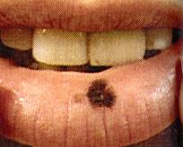
11/21/2016. Introduction. Diagnosis: Invasive mucosal melanoma. Mucosal melanoma diagnosis. Diagnosis: Invasive mucosal melanoma
|
|
|
- Marshall Weaver
- 5 years ago
- Views:
Transcription
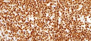
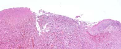
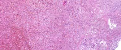
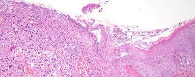
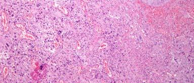
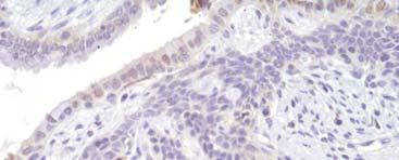
1 Introduction MUCOSAL MELANOMA AND PIGMENTED LESIONS OF MUCOSAL SURFACES Adriano Piris, M.D. Co-Director Mihm Cutaneous Pathology Consultative Service (MCPCS) Brigham and Women s Hospital, Harvard Medical School Mucosal melanoma as different biological entity than cutaneous melanoma Rarity of disease and timing of diagnosis precludes significant studies to standardize staging and management Stewart Rahr-MRA Young Investigator: Melanoma Research Alliance, Washington, DC Mucosal melanoma diagnosis Diagnosis: Invasive mucosal melanoma Mucosal lentigo (melanosis) Benign mucosal melanocytic proliferation: junctional or compound nevus, blue nevus Atypical melanocytic hyperplasia Melanoma in situ Invasive Melanoma Malignant pigmented nodule Rule out metastatic disease: Clinical history and identification of an in situ component Small samples: no obvious in situ component Examination of adjacent uninvolved mucosa is crucial Diagnosis: Invasive mucosal melanoma Mucosal melanoma: in situ component Amelanotic nodule: Undifferentiated or sarcomatoid proliferation First: establish the melanocytic nature of the tumor (Immunohistochemistry)- S100, HMB45, Melan-A, Mart-1, and MitF Second: Identify in situ component Confluent growth of lentiginous and nested intraepidermal melanocytes with atypical features Emphasis s of lentiginous growth with insidious and multifocal pattern, extending along underlying native glandular units These precursor lesions may be subtle and not readily visualized with H&E only. Melanocytic markers are crucial to identify in situ/precursor lesions and extent of disease 1
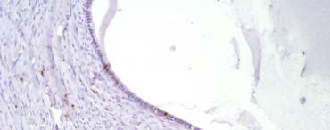
2 Pathological Evaluation of the Tumor Evaluation of Local Disease Tumor thickness correlates with survival 2.0 mm or less better prognosis Due to late diagnosis most lesions are usually thicker than 2.0 mm AJCC histomorphological criteria (from cutaneous melanoma) have not been validated A: Established primary mucosal melanoma B: Extent of local disease: CT or MRI C: Basic metastatic workup: serum lactate dehydrogenase, chest x-ray, combined PET/CT scanning of chest, abdomen, and pelvis (Clinical) Evaluation of Extent of Disease Sites of origin Clinical staging system for cutaneous disease applied to mucosal melanomas Stage I: Localized disease Stage II: Regional lymph node disease Stage III: Disseminated disease Respiratory mucosa Oral cavity Esophagus Genital mucosal surfaces Gastrointestinal mucosa Urinary tract Auditory canal Conjunctiva Primary mucosal melanoma of the sinonasal tract: a clinicopathologic and immunohistochemical study of thirty-two cases. Mark C. Mochel, MD, Lyn M. Duncan, MD, Adriano Piris, MD, and Stefan Kraft, MD* Pathology Service, Massachusetts General Hospital, Harvard Medical School, Boston, MA, USA, 02114, * now at Institute of Pathology, University Medical Center Hamburg- Eppendorf, Hamburg, Germany Sinonasal mucosal melanoma Rare disease with poor survival Poorly characterized early/precursor lesions Retrospective analysis found 31 of 32 patients with associated intraepidermal melanocytic proliferations Head and Neck Pathology, in print 2
: 67% of cases, confirmed by MITF")

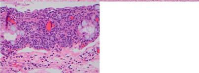
3 Sinonasal mucosal melanoma Sinonasal mucosal melanoma Age: years (median 71) M-F ratio 3:2 Follow up p( (31 pts): 5 to 211 months (mean 42) 58% died of melanoma associated conditions 19% died of unknown causes 6% alive with metastatic disease 19% alive without melanoma MMIS (confluent intraepithelial proliferation of cytologically atypical melanocytes): 67% of cases, confirmed by MITF Melanocytic hyperplasia (intraepithelial melanocytic proliferation without confluent growth or atypia): 16% (5 cases) Overall incidence of associated intraepidermal melanocytic proliferations: 83% Characteristics of the invasive component Determination of accurate tumor thickness was not possible in the majority of cases due to fragmentation of the specimen In 7 specimens: 0.3 to 15.0 mm Morphology: epithelioid, spindled, and small cell morphology Presence of >3 mitoses/mm2 and necrosis correlated with tumor progression and overall survival Single atypical melanocytes H&E Atypical melanocytic hyperplasia 3


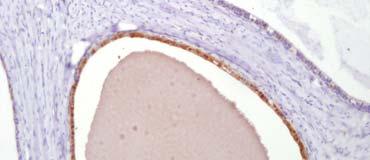
4 Hyperplasia and atypia, not MMIS Mochel et al: Melanocytes within sinonasal mucosa Negative control: sinonasal mucosa removed for rhinosinusitis No intraepithelial melanocytes found in these negative controls Identification of melanocytic hyperplasia within the context of melanosis in sinonasal mucosa should raise the concern of a precursor lesion. MMIS, resp. epithelium Easy to miss MMIS 4


5 MITF extensive MMIS MMIS in resp epithelium MITF Extensive gland involvement Invasive component Epithelioid cell with central necrosis 5


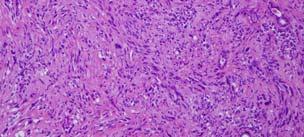
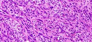
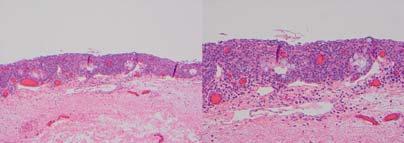
6 Small round cell Spindle cell fascicular Melanoma mimics in the mucosal surface Freckles, Lentigines and Melanoses 6


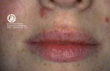
7 The Freckles, lentigines, and melanoses Not classified as a form of precancerous melanocytic proliferation Their recognition is important because : clinical i l appearance resembes melanoma Multiple lentigines/ melanoses may be a sign of a systemic disease associated with non-melanocytic cancers. Peutz-Jeghers Syndrome: macules on lips + buccal mucosa Labial melanotic macule CLINICAL FEATURES Classically lower lip on or just off midline Similar lesions anywhere in oropharynx Identical lesions in the genital mucosae are termed vulvar and penile melanosis HISTOLOGY Slight epithelial hyperplasia; parakeratosis Increased numbers of banal melanocytes with dendritic morphology; basilar hyperpigmentation Melanophages in stroma 7


; similar phenomenon on penis is")

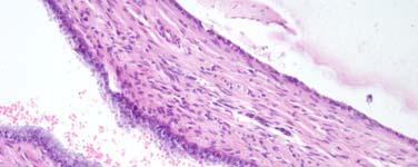
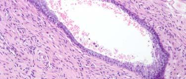
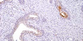
8 Atypical melanocytes in squamous mucosa: NOT melanosis The genital melanocytic proliferations: main forms Vulvar Melanosis Mucosal lentigo Vulvar melanosis Common acquired nevus Dysplastic nevus Malignant melanoma Clinically presents as a solitary (up to or several centimeters in size) or multiple intensely pigmented macule(s); similar phenomenon on penis is called penile melanosis. Clinical differential diagnosis: radial growth phase mucosal LMM, patch type bowenoid papulosis, and pigmented Bowen s disease 8


9 Vulvar Melanosis : Histomorphology Hypermelanosis Mild increase in number of basilar melanocytes Acanthosis No melanocytic atypia Differential diagnosis: atypical lentiginous melanocytic hyperplasia (precursor lesion to melanoma) Mucosal Lentigo From Simulators of Malignant Melanoma Drs. Kerl and Cerroni, University of Graz Atypical melanocytes in squamous mucosa: NOT melanosis Cervico-vaginal invasive melanoma: Case 1 9

10 10


11 Vulvo-urethral invasive melanoma: Case 2 Malignant melanoma of anorectal region: a clinicopathologic study of 61 cases Muhammad Usman Tariq, Nasir Ud Din, Nausheen Feroz Ud Din, Saira Fatima, and Zubair Ahmad Annals of Diagnostic Pathology, , Volume 18, Issue 5, Pages Copyright 2014 Elsevier Inc. 11

 in 20% of cases in")

 Nodular Melanoma BRAF/NRAS CKIT CKIT?")

12 Ano-rectal Mucosal Melanoma Ano-rectal Mucosal Melanoma Anorectal melanoma Expression of vimentin, S-100, HMB-45, and Melan A in 100%, 100%, 94.4%, and 93.3% cases, respectively. Cytokeratins were positive in 9% and CD117 (c-kit) in 20% of cases in which they were performed. All cases were BRAF negative Cutaneous Malignant Melanoma Superficial Spreading Melanoma Acral Lentiginous Melanoma Lentigo Maligna Melanoma Chronic Sun Damage (CSD) Nodular Melanoma BRAF/NRAS CKIT CKIT?BRAF/NRAS Mucosal Malignant Melanoma: CKIT Pigmented Lesions of the Conjunctiva Types of Non-Melanocytic Ocular Pigmentation Scleral Diseases Blue Sclera Staphyloma Scleromalacia Senile hyaline plaque Metabolic Disorders Ochronosis Gaucher s disease Jaundice 12


13 Benign Epithelial Pigmented Tumors Ophthalmic Pathology Describes Melanocytic Lesions to Arise from 3 Types of Melanocytes Pigmented seborrheic keratosis Benign squamous papilloma Verruca vulgaris Pigmented eccrine poroma 1. Intraepithelial melanocytes that lie among the basal epithelial cells and may show dendritic processes between keratinocytes 2. Nevus cells- oval cells that form nests and sheets at the epidermaldermal or epithelial-subepithelial junction 3. Fusiform dendritic melanocytes that lie in the deeper mesenchymal or subepithelial tissue Congenital Melanosis Benign Epithelial Melanosis - Clinical 1. Patchy flat brown pigmentation of conjunctival epithelium 2. Associated with skin color 3. Usually bilateral l non-inflamed non-vascularized and stationary ti 4. Most common in limbal area and may advance to caruncle 5. May advance onto cornea after surgery or trauma- streaks and swirls 6. Usually congenital may be acquired in african-americans Conjunctival Nevi General Clinical i l Considerations Single most common site- juxta-limbal followed by epibulbar, the plica, and caruncle May be focal or diffuse but not multifocal 13


14 Pigmented Lesions of the Conjunctiva Nevi most unusual in palpebral or forniceal conjunctiva, suspect melanoma All bulbar nevi freely movable with Q tip traction unless hinged at limbus -if hinged and immovable, suspect melanoma All palpebral nevus-like lesions should be biopsied Conjunctival nevi do not extend onto the cornea-if observed probably melanoma Common Acquired Nevi 1. Junctional- Type A cells in nests 2. Compound Intraepithelial and substantia propria proliferation - subepithelial cells have a lymphocytoid appearance and are not melanized in deeper component 3. Subepithelial May show type C cell proliferation 4. Blue nevi Characteristic dendritic cells % of conjunctival nevi have a combined element, often very focal Common Acquired Nevi 6. Downward protrusion of small solid pegs of epithelium is typical for sub epithelial nevi 7. Epithelium- lined cysts multiple and diffuse are characteristic for benign sub epithelial nevi 8. Balloon cells and spindle cells may be found in conjunctival nevi 9. Blue nevi Characteristic dendritic cells. 14
15 Junctional Nevus in a 7 year old boy Courtesy of Dr. Frederick A. Jakobiec


 The")


16 Combined Nevi 40% of a series of 95 conjunctival nevi had a combined element (personal series) The combined component varied from a blue nevus to a deep penetrating nevus In most instances, the combined aspect was very small but rarely accounted for most of the nevus These lesions are benign 16


17 COMPOUND NEVUS OF SPITZ Sub-epithelial Melanocytes and Associated Lesions- Scleral Dendritic 1. Blue nevus 2. Cellular blue nevus 3. Occulodermal melanocytosis Blue Nevus 17


18 Oculodermal Melanocytes (Ota) Clinical- 1. Ipsilateral pigmentation of periocular skin along with melanosis oculi 2. Periorbital skin may be brown, slate or bluish 3. Pigment deep to conjunctiva and doesn t move with Q tip traction in contrast to benign epithelial melanosis 4. Most common in Blacks and Asians 5. Low risk of uveal or orbital melanoma Acquired Melanosis of the Conjunctiva 1. Benign epithelial melanosis of conjunctiva - congenital 2. Primary acquired melanosis of conjunctiva 3. Secondary acquired melanosis of conjunctiva Jakobiec, F.A et al. Clinicopathologic Characteristics of Premalignant and Malignant Melanocytic Lesions of the Conjunctiva. Ophthalmology 96: ;


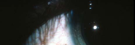
19 PRIMARY ACQUIRED MELANOSIS Definition accepted by World Health Organization (WHO) 1. Lesion is primary because not the result of racial, metabolic or local topical factors 2. Acquired not congenital 3. Melanosis due to melanin production Primary Acquired Melanosis Clinical Flat Brown (golden brown to chocolate) May involve cornea May involve any aspect of conjunctiva including tarsal conjunctiva, fornix and caruncle May be multiple Almost always unilateral. May extend across lid margin into epidermis. May shrink, progress or remain stable for prolonged periods. Occurs in middle-aged or elderly, usually white patients (rare in blacks). 19


20 PAM Histology PAM 1 without atypia (overproduction of Melanin with hyperplasia) Normal Conjunctiva Hyperplasia of benign melanocytes confined to basilar epithelium ( not considered premalignant) PAM 2 with atypia Examine for epithelioid cells and pattern of growth Courtesy of Dr. Frederick A. Jakobiec Primary Acquired Melanosis with Mild Atypia PAM without atypia there is an increased number of melanocytes Courtesy of Dr. Frederick A. Jakobiec PAM with mild atypia there is an increased number of basal melanocytes and some scattered higher level dendritic melanocytes Courtesy of Dr. Frederick A. Jakobiec Primary Acquired Melanosis with Moderate Atypia Primary Acquired Melanosis with Severe Atypia PAM with severe atypia there are numerous large dendritic melanocytes PAM with moderate-to-severe atypia there are large dendritic melanocytes Courtesy of Dr. Frederick A. Jakobiec Courtesy of Dr. Frederick A. Jakobiec
 Pre-existing existing Primary Acquired Melanosis")

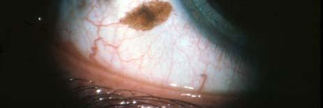
21 Malignant melanoma of the Conjunctiva 1. Melanoma with PAM (75%) 2. Melanoma without PAM (25%) Note: 25% of all lesions have evidence of preexisting nevus 3. Overall mortality 25% of all types Malignant Melanoma of the Conjunctiva Risk Factors Predominantly caucasians Older age (average age years) Pre-existing existing Primary Acquired Melanosis (PAM) give rise to 60% of conjunctival melanoma Rarely associated with a pre-existing nevus History of extensive sunlight exposure Jovanovic P. et al. Int J Clin Exp Pathol Malignant Melanoma of the Conjunctiva Symptoms and Clinical Features 5-10% of all ocular melanomas Average age years Equal sex incidence Most common complaint is a pigmented spot or nodule; irritation and pain less common Location: Bulbar conjunctiva (92%) Temporal quadrant (63%) Touching limbus (61%) Pigmented lesion in the palpebral, forniceal conjunctiva, plica semilunaris, and caruncula are prima facie melanoma Farber M. et al. JAAD Griewank KG et al. Clin Cancer Res Jovanovic P. et al. Int J Clin Exp Pathol Malignant Melanoma of the Conjunctiva Symptoms and Clinical Features Melanoma without PAM is solitary nodule Early invasion in PAM may be associated with a plaque Any ypg pigmented lesion surrounded by numerous vessels should be biopsied Rarely the lesion is non-pigmented and the multiple vessels are a clue Multiple lesions common 33% Local recurrence 26% at five years; 51% at 10 years Mortality 38% at 10 years Griewank KG et al. Clin Cancer Res Jovanovic P. et al. Int J Clin Exp Pathol
 Invasive component can be")


22 Malignant Melanoma of the Conjunctiva Histopathology Most lesions exhibit radial growth extending beyond the invasive component. Pattern most commonly pagetoid but lentiginous variants occur (lentigo maligna can extend into the epithelium) Invasive component can be spindled, small cell, epithelioid or mixed. Mitoses frequent; greater than 5 per 10 hpf associated with high risk of metastases. Thickness > 2mm. High risk of metastasis. Lymphatic invasion. High risk of metastases. Tils: absence retlated to high risk of metasases. Zembowicz,A et al. Arch Pathol Lab Med Griewank KG et al. Clin Cancer Res Jovanovic P. et al. Int J Clin Exp Pathol Clinical Pathologic Correlations of Mutations Location: Caruncle BRAF 66% ; NRAS 0% ; Wild Type 33% Pathology: Pre-existing nevus BRAF 65% ; NRAS 27% ; Wild Type 8% No other clinical pathologic correlations found, including relationship to mitoses, metastases, and disease free or overall survival. Griewank KG et al. Clin Cancer Res Jovanovic P. et al. Int J Clin Exp Pathol Malignant Melanoma of the Conjunctiva Prognosis Palpebral conjunctiva, fornices, plica, caruncula, and lid margins: Higher mortality than epibulbar Higher risk for local recurrence Higher risk for distant metastases Thickness > 2 mm. associated with high risk of metastasis and death Cell type: Mixed type 3x higher mortality than spindle cells Pure spindle cells excellent survival Positive margins predict higher risk of recurrence Lymphatic invasion: 4x higher mortality De novo melanoma 35% mortality at 10 years Melanoma in PAM 9% mortality at 10 years Jovanovic P. et al. Int J Clin Exp Pathol
23 Activating Mutations in Conjunctival Melanoma BRAF - 29% (V600E 21%) NRAS - 18% Wild Type - 53% c-kit - 0% (15% showed non-activating i mutations) PTEN (Chromosome 10) loss commonly found in association with BRAF and usually with NRAS Activation of AKT pathway Griewank KG et al. Clin Cancer Res Treatment of Conjunctival Melanoma Melanoma with PAM easily confused with nevus. Any lesion that changes should be removed Any pigmented lesion extending on to the cornea should be excised Primary: Wide local excision Adjuvant Therapy: Brachytherapy, cryotherapy, mitomysin Multiple lesions and/or multiple recurrences: Mitomysin or brachytherapy primarily Exenteration: Reserved for extensive wide-spread recurrences or invasion of sclera Sentinel Lymph Node Biopsy: (Preliminary Results) Performed for tumors > 2mm. In thickness or with ulceration or mitoses >5 per 10 hpf Griewank KG et al. Clin Cancer Res Jovanovic P. et al. Int J Clin Exp Pathol Cohen VML. Br J Ophthalmol Acknowledgments Martin C. Mihm Jr., MD Frederick A. Jakobiec, MD Cynthia Magro, MD 23
Dermatopathology. Dr. Rafael Botella Estrada. Hospital La Fe de Valencia
 Dermatopathology Dr. Rafael Botella Estrada. Hospital La Fe de Valencia Melanoma and mimics Dr. Martin Mihm Malignant lesions result from the accumulation of mutations Class I lesions (benign) Class II
Benign and malignant epithelial lesions: Seborrheic keratosis: A common benign pigmented epidermal tumor occur in middle-aged or older persons more
 Benign and malignant epithelial lesions: Seborrheic keratosis: A common benign pigmented epidermal tumor occur in middle-aged or older persons more common on the trunk; but extremities, head and neck are
Pathology of the skin. 2nd Department of Pathology, Semmelweis University
 Pathology of the skin 2nd Department of Pathology, Semmelweis University Histology of the skin Epidermis: Stratum corneum Stratum granulosum Stratum spinosum Stratum basale Dermis: papillary and reticular
Diagnoses of Cases 1. Lentigo, other melanosis and the acquired nevus 2. Variations on the acquired nevus 3. Dermal melanocytosis
 Diagnoses of Cases 1. Lentigo, other melanosis and the acquired nevus 1 1A. Lentigo simplex 4 1B. Psoralens and ultraviolet A (PUVA) lentigo 6 1C. Solar lentigo 8 1D. Café au lait macule 10 1E. Ink-spot
Nasal mucosal melanosis may act as a harbinger of melanoma: A case report
 Nasal mucosal melanosis may act as a harbinger of melanoma: A case report The Harvard community has made this article openly available. Please share how this access benefits you. Your story matters. Citation
Desmoplastic Melanoma R/O BCC. Clinical Information. 74 y.o. man with lesion on left side of neck r/o BCC
 R/O BCC Sabine Kohler, M.D. Professor of Pathology and Dermatology Dermatopathology Service Stanford University School of Medicine Clinical Information 74 y.o. man with lesion on left side of neck r/o
Malignant tumors of melanocytes: Part 1. Deba P Sarma, MD., Omaha
 Malignant tumors of melanocytes: Part 1 Deba P Sarma, MD., Omaha The melanocytic tumor is one of the most difficult and confusing areas in Dematopathology. It is true that most (95%) of such lesions are
NEOPLASMS OF THE SURFACE EPITHELIUM (KERATINOCYTES)
") NEOPLASMS OF THE SURFACE EPITHELIUM (KERATINOCYTES) Papillary Lesions Precancerous Lesions Keratinocyte Proliferations Carcinomas Melanotic Lesions Melanomas Normal Mucosa Keratin layer Spinous layer Basal
NEOPLASMS OF THE SURFACE EPITHELIUM (KERATINOCYTES) Papillary Lesions Precancerous Lesions Keratinocyte Proliferations Carcinomas Melanotic Lesions Melanomas Normal Mucosa Keratin layer Spinous layer Basal
Dermatopathology: The tumor is composed of keratinocytes which show atypia, increase mitoses and abnormal mitoses.
 Squamous cell carcinoma (SCC): A common malignant tumor of keratinocytes arising in the epidermis, usually from a precancerous condition: 1- UV induced actinic keratosis, usually of low grade malignancy.
Springer Healthcare. Staging and Diagnosing Cutaneous Melanoma. Concise Reference. Dirk Schadendorf, Corinna Kochs, Elisabeth Livingstone
 Concise Reference Staging and Diagnosing Cutaneous Melanoma Dirk Schadendorf, Corinna Kochs, Elisabeth Livingstone Extracted from Handbook of Cutaneous Melanoma: A Guide to Diagnosis and Treatment Published
LENTIGO SIMPLEX. Epidemiology
 LENTIGO SIMPLEX Epidemiology The frequency of lentigo simplex in children and adults has not been determined. There does not appear to be a racial or gender predilection. Lentigo simplex is the most common
Ocular Neoplasia What s Common? What s New? Richard R Dubielzig
 Ocular Neoplasia What s Common? What s New? Richard R Dubielzig Orbit 288 6% Tumors of the globe make up 3225 out of 6110 total neoplasms = 53%. Tumors of the conjunctiva make up 1192 out of 6110 total
Conjunctival Melanoma: A New Clinical and Therapeutical Approach
 149 Conjunctival Melanoma: A New Clinical and Therapeutical Approach M. Rodríguez-Martín a J. Rodríguez-Martín c N. Merino de Paz a P. Contreras Ferrer a P. Rocha Cabrera c B. Rodríguez Martín d G. Gordillo
David B. Troxel, MD. Common Medicolegal Situations: Misdiagnosis of Melanoma
 Common Medicolegal Situations: Misdiagnosis of Melanoma David B. Troxel, MD Medical Director, The Doctors Company, Napa, California Clinical Professor Emeritus, University of California at Berkeley Past
Malignant Melanoma Early Stage. A guide for patients
 This melanoma patient brochure is designed to help educate melanoma patients and their caregivers. It was developed under the guidance of Dr. Michael Smylie, Professor, Department of Oncology, University
Conjunctival malignant melanoma: A rare variant and review of important diagnostic and therapeutic considerations
 Saudi Journal of Ophthalmology (2012) 26, 151 156 Ophthalmic Pathology Update Conjunctival malignant melanoma: A rare variant and review of important diagnostic and therapeutic considerations Danah H.
A PRACTICAL APPROACH TO ATYPICAL MELANOCYTIC LESIONS BIJAN HAGHIGHI M.D, DIRECTOR OF DERMATOPATHOLOGY, ST. JOSEPH HOSPITAL
 A PRACTICAL APPROACH TO ATYPICAL MELANOCYTIC LESIONS BIJAN HAGHIGHI M.D, DIRECTOR OF DERMATOPATHOLOGY, ST. JOSEPH HOSPITAL OBJECTIVES Discuss current trends and changing concepts in our understanding of
Benign versus Cancerous Lesions How to tell the difference FMF 2014 Christie Freeman MD, CCFP, DipPDerm, MSc
 1 Benign versus Cancerous Lesions How to tell the difference FMF 2014 Christie Freeman MD, CCFP, DipPDerm, MSc Benign lesions Seborrheic Keratoses: Warty, stuck-on Genetics and birthdays Can start in late
Melanocytic Lesions: Use of Immunohistochemistry and Special Studies Napa Valley 2018
 Melanocytic Lesions: Use of Immunohistochemistry and Special Studies Napa Valley 2018 Victor G. Prieto, MD, PhD Professor Depts. of Pathology and Dermatology University of Texas - MD Anderson Cancer Center
Clinical characteristics
 Skin Cancer Fernando Vega, MD Seattle Healing Arts Clinical characteristics Precancerous lesions Common skin cancers ACTINIC KERATOSIS Precancerous skin lesions Actinic keratoses Dysplastic melanocytic
Simulators of melanoma
 Simulators of melanoma Philip E. LeBoit, M.D. Depts. of Pathology and Dermatology University of California, San Francisco Simulators of melanoma Simulators of melanoma in situ Melanocytic Non-melanocytic
COMBINED NEVI are composed
 CLINICAL SCIENCES Combined Nevi of the Conjunctiva J. rooks Crawford, MD; Edward L. Howes, Jr, MD; Devron H. Char, MD Objective: To report the clinical and histologic features of combined nevi of the conjunctiva,
Conflict of Interest 9/2/2014. Pathogenesis and Comparison of Atypical Spitz Nevi vs Benign Spitz, and Childhood Melanoma
 Pathogenesis and Comparison of Atypical Spitz Nevi vs Benign Spitz, and Childhood Melanoma Martin C. Mihm Jr., M.D., F.A.C.P. Harvard Medical School Brigham and Women s Hospital Dana Farber Cancer Center
Periocular Malignancies
 Periocular Malignancies Andrew Gurwood, O.D., F.A.A.O., Dipl. Marc Myers, O.D., F.A.A.O. Drs. Myers and Gurwood have no financial interests to disclose. Course Description Discussion of the most common
Pigmented lesions of the Oral cavity
 Oral medicine أ.م.د احسان عبد هللا كميل Pigmented lesions of the Oral cavity Pigmented oral lesions are a large group of disorders in which the dark or brown color is the essential clinical characteristic.
Protocol applies to melanoma of cutaneous surfaces only.
 Melanoma of the Skin Protocol applies to melanoma of cutaneous surfaces only. Procedures Biopsy (No Accompanying Checklist) Excision Re-excision Protocol revision date: January 2005 Based on AJCC/UICC
Therapy of melanocytic conjunctival tumors
 DOI: 10.4149/BLL_2013_093 Bratisl Lek Listy 2013; 114 (8) CLINICAL STUDY Therapy of melanocytic conjunctival tumors Halas M Jr 1, Svetlosakova Z 1, Babal P 2 Department of Ophthalmology, Comenius University,
An Overview of Melanoma. Harriet Kluger, M.D. Associate Professor Section of Medical Oncology Yale Cancer Center
 An Overview of Melanoma Harriet Kluger, M.D. Associate Professor Section of Medical Oncology Yale Cancer Center Melanoma Statistics Median age at presentation 45-55 55 years Incidence: 2003 54,200 cases
Blue Melanocytic Proliferations
 Blue Melanocytic Proliferations Labib R. Zakka M.D., M.A. Research Fellow Melanoma Program Department of Dermatology Brigham and Women s Hospital Harvard Medical School Conflicts of Interest No conflicts
ORAL MELANOMA Definition Epidemiology Clinical Presentation
 ORAL MELANOMA Definition Melanoma is a highly malignant neoplasia, arising from melanocytes, the cells that produce the brownish pigment melanin. Melanin is the determinant in skin colour and protects
IT S FUNDAMENTAL MY DEAR WATSON! A SHERLOCKIAN APPROACH TO DERMATOLOGY
 IT S FUNDAMENTAL MY DEAR WATSON! A SHERLOCKIAN APPROACH TO DERMATOLOGY Skin, Bones, and other Private Parts Symposium Dermatology Lectures by Debra Shelby, PhD, DNP, FNP-BC, FADNP, FAANP Debra Shelby,
المركب النموذج--- سبيتز وحمة = Type Spitz's Nevus, Compound SPITZ NEVUS 1 / 7
 SPITZ NEVUS 1 / 7 Epidemiology An annual incidence rate of 1.4 cases of Spitz nevus per 100,000 individuals has been estimated in Australia, compared with 25.4 per 100,000 individuals for cutaneous melanoma
Among the benign intraepithelial melanocytic proliferations, Inflamed Conjunctival Nevi. Histopathological Criteria. Resident Short Reviews
 Resident Short Reviews Inflamed conjunctival nevi (ICN) may suggest malignancy because of their rapid growth and atypical histology. The objective of this study was to characterize the diagnostic features
Female 18. Deeply pigmented lesion on trunk.?warty naevus?seborrhoeic keratosis?malignant melanoma. The best diagnosis is:
 Female 18. Deeply pigmented lesion on trunk.?warty naevus?seborrhoeic keratosis?malignant melanoma. The best diagnosis is: A. deep penetrating naevus B. naevoid malignant melanoma C. pigment synthesising
Dispelling Rumors about Tumors. Case
 Dispelling Rumors about Tumors Jesse L. Berry, MD Arizona Ophthalmology Society 2017 Associate Director, Ocular Oncology Service Associate Program Director USC/CHLA, Keck School of Medicine Case 65 year
Toby Maurer, MD University of California, San Francisco. Lifetime risk of an American developing melanoma
 Distinguishing Pigmented Skin Lesions and Melanoma Toby Maurer, MD University of California, San Francisco Epidemiology of Melanoma Lifetime risk of an American developing melanoma 1935: 1 in 1500 1980:
Atypical Nevi When to Re-excise. Catherine Barry, DO Dermatopathologist
 Atypical Nevi When to Re-excise Catherine Barry, DO Dermatopathologist Why talk about skin cancer? Because it s the most common type of cancer! Non-melanoma Skin Cancers Basal Cell Carcinoma Squamous Cell
Patricia Chevez-Barrrios AAOOP-USCAP /12/2016
 Biomarkers in Ocular Melanoma Patricia Chévez-Barrios, MD Pathology and Genomic Medicine, Houston Methodist Hospital Professor of Pathology and Laboratory Medicine and Ophthalmology, Weill Cornell Medical
Ocular Neoplasia CL Davis 9/08. Richard R Dubielzig
 Ocular Neoplasia CL Davis 9/08 Richard R Dubielzig 2135/5722 Canine Melanocytic Tumors Outside the Globe: 264 Conjunctival: 159 Eye Lid: 72 Skin: 33 Affecting the Globe: 1871 Anterior Uveal Melanocytoma:
Original Articles Primary orbital melanoma in association with cellular blue nevus
 Original Articles Primary orbital melanoma in association with cellular blue nevus Tarek El-Sawy, MD, PhD, a Mathieu F. Bakhoum, PhD, a Michael Tetzlaff, MD, PhD, b Qasiem J. Nasser, MD, a Victor G. Prieto,
ARTICLE INFO ABSTRACT
 Melanocytic Pigmentation: A Single Manifestation of Myriad of Pathologies [PP: 05-09] Dr. Swapna Honwad Department of Oral Pathology dr.swapnahonwad@gmail.com Dr. Elsy P. Simon Department of Endodontics
Basal cell carcinoma 5/28/2011
 Goal of this Presentation A practical approach to the diagnosis of cutaneous carcinomas and their mimics Thaddeus Mully, MD University of California San Francisco To review common non-melanoma skin cancers
CLINICAL PEARLS IN OCULAR ONCOLOGY
 CLINICAL PEARLS IN OCULAR ONCOLOGY IRIS NEVUS - Two kinds circumscribed and diffuse - Photodocumentation important to monitor growth - Risk Factors for iris nevus growth to melanoma (ABCDEF) A Age (young),
LUMPS AND BUMPS: AN ORGANIZED APPROACH TO DIAGNOSIS AND MANAGEMENT
 LUMPS AND BUMPS: AN ORGANIZED APPROACH TO DIAGNOSIS AND MANAGEMENT Tammy P. Than, M.S., O.D., F.A.A.O. The University of Alabama at Birmingham / School of Optometry 1716 University Blvd. Birmingham, AL
Anatomic Divisions. Ocular Surface. Intraocular. Orbital. Lacrimal. Eyelid
 Anatomic Divisions Ocular Surface Intraocular Orbital Lacrimal Eyelid Ocular Surface Melanocytic Squamous Neoplasia Lymphoid Melanocytic Nevi PAM (Primary Acquired Melanosis) Ocular Melanocytosis Melanoma
Toby Maurer, MD University of California, San Francisco. Lifetime risk of an American developing melanoma
 Distinguishing Pigmented Skin Lesions and Melanoma Toby Maurer, MD University of California, San Francisco Epidemiology of Melanoma Lifetime risk of an American developing melanoma 1935: 1 in 1500 1980:
World Articles of Ear, Nose and Throat Page 1
 World Articles of Ear, Nose and Throat ---------------------Page 1 Primary Malignant Melanoma of the Tongue: A Case Report Authors: Nanayakkara PR*, Arudchelvam JD** Ariyaratne JC*, Mendis K*, Jayasekera
Chapter 6 Squamous Cell Carcinoma: Variants and Challenges
 Chapter 6 Squamous Cell Carcinoma: Variants and Challenges Michael B. Morgan EPIDEMIOLOGY: Second most common skin cancer, rare in the dark-skinned races. ETIOLOGY: Ultraviolet light, HPV infection. PATHOGENESIS:
Dataset for the histopathological reporting of conjunctival melanoma and. October 2007
 The Royal College of Pathologists Pathology: the science behind the cure Standards and datasets for reporting cancers Dataset for the histopathological reporting of conjunctival melanoma and melanosis
Premalignant skin tumours
 Chapter 14: Premalignant skin tumours page: 434 Premalignant skin tumours page: 435 Solar keratoses (senile keratoses) Raised red and well-defined plaques with a rough surface covered in scales of varying
Acral Melanoma in Japan
 Acral Melanoma in Japan MAKOTO SEUI, M.D., HIDEAKI TAKEMATSU, M.D., MICHIKO HOSOKAWA, M.D., MASAAKI OBATA, M.D., YASUSHI TOMITA, M.D., TAIZO KATO, M.D., MASAAKI TAKAHASHI, M.D., AND MARTIN C. MIHM, JR.,
Maligna Melanoma and Atypical Fibroxanthoma: An Unusual Collision Tumour G Türkcü 1, A Keleş 1, U Alabalık 1, D Uçmak 2, H Büyükbayram 1 ABSTRACT
 Maligna Melanoma and Atypical Fibroxanthoma: An Unusual Collision Tumour G Türkcü 1, A Keleş 1, U Alabalık 1, D Uçmak 2, H Büyükbayram 1 ABSTRACT Two different neoplasia in the same biopsy material called
Enterprise Interest Nothing to declare
 Enterprise Interest Nothing to declare Diagnoses one would not like to miss in soft tissue pathology early in your career Marta Sbaraglia, MD Department of Pathology Hospital of Treviso University of Padua
Multiple Primary Melanoma in a Thai Male: A Case Report
 Case Report Multiple Primary Melanoma in a Thai Male: A Case Report J Med Assoc Thai 2014; 97 (Suppl. 2): S234-S238 Full text. e-journal: http://www.jmatonline.com Kittisak Payapvipapong MD*, Pinyapat
NAEVUS OF OTA* naevus" occurring in the skin areas supplied by the ophthalmic and maxillary
 Brit. J. Ophthal. (1965) 49, 364 NAEVUS OF OTA* BY G. P. GUPTA AND D. N. GANGWAR From the Muslim University Institute of Ophthalmology and Gandhi Fye Hospital, Aligarh, India THE naevus of Ota is characterized
The Dermal Melanocytoses. Conflicts of Interest 5/22/2018. The Nevi of Ota and Ito. Martin C. Mihm M.D.
 The Dermal Melanocytoses Martin C. Mihm M.D. Director Mihm Cutaneous Pathology Consultative Service (MCPCS) Brigham and Women s Hospital Director Melanoma Program Brigham and Women s Hospital and Harvard
Retina Center of Oklahoma Sam S. Dahr, M.D. Adult Intraocular Tumors
 Adult Intraocular Tumors Sam S. Dahr, M.D. Retina Center of Oklahoma www.retinacenteroklahoma.com www.rcoklahoma.com Table of Contents Posterior uveal malignant melanoma Uveal metastasis Uveal melanoma
Melanoma Update: 8th Edition of AJCC Staging System
 Melanoma Update: 8th Edition of AJCC Staging System Rosalie Elenitsas, M.D. Professor of Dermatology Director, Dermatopathology University of Pennsylvania DISCLOSURE OF RELATIONSHIPS WITH INDUSTRY None
أملس عضلي غرن = Leiomyosarcoma. Leiomyosarcoma 1 / 5
 Leiomyosarcoma 1 / 5 EPIDEMIOLOGY Exact incidence is unknown, but older studies suggest that leiomyosarcomas comprise approximately 3 percent of soft-tissue sarcomas. Superficial leiomyosarcoma occurs
1/10/2018. Soft Tissue Tumors Showing Melanocytic Differentiation. Overview. Desmoplastic/ Spindle Cell Melanoma
 2016 MFMER slide-1 2016 MFMER slide-2 2016 MFMER slide-3 Soft Tissue Tumors Showing Melanocytic Differentiation Andrew L. Folpe, M.D. Professor of Laboratory Medicine and Pathology Mayo Clinic, Rochester,
Pathology. Skin Tumor. Bayan N. Mohammad 15/10/2015. Mohammad al-orjani. Page 0 of 23
 #7 35 Pathology Skin Tumor Bayan N. Mohammad 15/10/2015 Mohammad al-orjani Page 0 of 23 بسم هللا الرحمن الرحيم GREETINGS This lecture is about skin tumors, all the slides are included and every slide will
Primary Cutaneous Melanoma Pathology Reporting Proforma DD MM YYYY. *Tumour site. *Specimen laterality. *Specimen type
 Primary Cutaneous Melanoma Pathology Reporting Proforma Includes the International Collaboration on Cancer reporting dataset denoted by * Family name Given name(s) Date of birth DD MM YYYY Sex Male Female
SKIN CANCER. Most common cancer diagnosis 40% of all cancers
 SKIN CANCER Most common cancer diagnosis 40% of all cancers OBJECTIVES Review common and uncommon cancers of the skin. Special emphasis on melanoma and dysplastic nevus Review pathology/tnm/staging, which
Histopathology of Melanoma
 THE YALE JOURNAL OF BIOLOGY AND MEDICINE 48, 409-416 (1975) Histopathology of Melanoma G. J. WALKER SMITH Department ofpathology, Yale University School ofmedicine, 333 Cedar Street, New Haven, Connecticut
BAP-oma & BEYOND MICHAEL A NOWAK, MD
 BAP-oma & BEYOND MICHAEL A NOWAK, MD CONFLICTS No conflicts with the content of this lecture BAP-oma Wiesner 2011: Families with multiple tan dome-shaped papules of head, neck, trunk, and extremities.
Update on Cutaneous Mesenchymal Tumors. Thomas Brenn
 Update on Cutaneous Mesenchymal Tumors Thomas Brenn Cutaneous Mesenchymal Tumours Wide morphological and biological spectrum Myofibroblastic, smooth muscle, neural, vascular, apidocytic, undifferentiated;
Cutaneous Malignancies: A Primer COPYRIGHT. Marissa Heller, M.D.
 Cutaneous Malignancies: A Primer Marissa Heller, M.D. Associate Director of Dermatologic Surgery Department of Dermatology Beth Israel Deaconess Medical Center December 10, 2016 Skin Cancer Non-melanoma
Learning Objectives. Tanning. The Skin. Classic Features. Sun Reactive Skin Type Classification. Skin Cancers: Preventing, Screening and Treating
 Learning Objectives Skin Cancers: Preventing, Screening and Treating Robert A. Baldor, MD, FAAFP Professor, Family Medicine & Community Health University of Massachusetts Medical School Distinguish the
Ocul Oncol Pathol 2015;1:
 Received: November 19, 2014 Accepted after revision: January 9, 2015 Published online: April 1, 2015 2296 4681/15/0014 0231$39.50/0 Case Series and Brief Reports Unilateral Conjunctival in situ Squamous
We are IntechOpen, the world s leading publisher of Open Access books Built by scientists, for scientists. International authors and editors
 We are IntechOpen, the world s leading publisher of Open Access books Built by scientists, for scientists 3,500 108,000 1.7 M Open access books available International authors and editors Downloads Our
Management of pediatric melanocytic lesions
 Open Journal of Clinical & Medical Case Reports Management of pediatric melanocytic lesions Volume 3 (2017) Issue 8 ISSN 2379-1039 Jin Kim, BS; Emmanuel Gabriel MD, PhD; Weiguo Liu MD, PhD; Lin Lin MD,
Living Beyond Cancer Skin Cancer Detection and Prevention
 Living Beyond Cancer Skin Cancer Detection and Prevention Cutaneous Skin Cancers Identification Diagnosis Treatment options Prevention What is the most common cancer in people? What is the most common
Lichenoid Tissue Reaction in Malignant Melanoma A Potential Diagnostic Pitfall
 natomic Pathology / LICHENOID TISSUE RECTION IN MLIGNNT MELNOM Lichenoid Tissue Reaction in Malignant Melanoma Potential Diagnostic Pitfall CPT Scott R. Dalton, MC, US, 1,3 Capt Matt. aptista, USF, MC,
THE CLASSIFICATION of melanocytic
 CLINICAL SCIENCES Indeterminate Melanocytic Proliferations of the Conjunctiva Hans E. Grossniklaus, MD; Curtis E. Margo, MD, MPH; Alvin R. Solomon, MD Objective: To test the hypothesis that a subset of
Financial disclosures
 Mesenchymal Neoplasms with Melanocytic Differentiation By Konstantinos Linos MD, FCAP, FASDP Bone, Soft Tissue and Dermatopathology Assistant Professor of Pathology Dartmouth-Hitchcock Medical Center Geisel
Index. Springer-Verlag Berlin Heidelberg 2017 J.A. Plaza, V.G. Prieto, Pathology of Pigmented Skin Lesions, DOI /
 A Acral lentiginous (mucosal lentiginous) melanoma, 483 Acral lentiginous melanoma (ALM) asymmetric and irregular lentiginous junctional growth, 431 clinical features, 427 428 differential diagnosis, 428
CLINICAL SCIENCES. is a potentially fatal tumor that arises from melanocytes, most often
 Conjunctival Melanoma CLINICAL SCIENCES Risk Factors for Recurrence, Exenteration, Metastasis, and Death in 150 Consecutive Patients Carol L. Shields, MD; Jerry A. Shields, MD; Kaan Gündüz, MD; Jacqueline
Cancer Reporting for Dermatologists. Florida Department of Health Florida Cancer Data System. March 9, Agenda
 Cancer Reporting for Dermatologists Florida Department of Health Florida Cancer Data System March 9, 2011 Agenda Welcome Introductions Cancer Reporting in Florida BETA Participation Expectations Review
Michael T. Tetzlaff MD, PhD
 Update on American Joint Cancer Committee (AJCC) staging system for primary cutaneous melanoma Emphasis on concise and accurate reporting of primary and metastatic melanoma for effective risk stratification
Associate Clinical Professor of Dermatology MUSC
 Re-excision of Moderately Dysplastic Nevi: Should we or shouldn t we? John C. Maize, Jr, M.D. Dermatologist and Dermatopathologist Trident Dermatology, Charleston SC Associate Clinical Professor of Dermatology
VULVAR CARCINOMA. Page 1 of 5
 VULVAR CARCINOMA EXAMPLE OF A VULVAR CARCINOMA USING PROPOSED TEMPLATE Case: Invasive squamous cell carcinoma arising in D-VIN Tumor in left labia major Left partial vaginectomy and sentinel lymph node
Case year old female presented with asymmetric enlargement of the left lobe of the thyroid
 Case 4 22 year old female presented with asymmetric enlargement of the left lobe of the thyroid gland. No information available relative to a prior fine needle aspiration biopsy. A left lobectomy was performed.
Case 4 Diagnosis 2/21/2011 TGB
 Case 4 22 year old female presented with asymmetric enlargement of the left lobe of the thyroid gland. No information available relative to a prior fine needle aspiration biopsy. A left lobectomy was performed.
MECHANISMS OF HUMAN DISEASE: LABORATORY SESSION PATHOLOGY OF THE SKIN LAB. Friday, February 12, :30 am 11:00 am
 MECHANISMS OF HUMAN DISEASE: LABORATORY SESSION PATHOLOGY OF THE SKIN LAB Friday, February 12, 2012 9:30 am 11:00 am FACULTY COPY GOALS: Describe the basic clinical and morphologic features of various
Squamous papilloma Squamous acanthoma Keratoacanthoma Verruca vulgaris Condyloma acuminatum Focal epithelial hyperplasia Sino nasal papilloma
 Benign tumors Epithelial origin Squamous papilloma Squamous acanthoma Keratoacanthoma Verruca vulgaris Condyloma acuminatum Focal epithelial hyperplasia Sino nasal papilloma Squamous papilloma Exophytic
COMMUNICATIONS. NAEVI AND MELANOMATA OF THE CONJLTNCTIVA*t. diagnosis of naevi and melanomata is not difficult, but many cases are not typical, and
 Brit. J. Ophthal. (1965) 49, 169 COMMUNICATIONS NAEVI AND MELANOMATA OF THE CONJLTNCTIVA*t BY BARRIE JAY Department ofpathology, Institute of Ophthalmology, University oflondon NAEvi are the most common
Subject Index. Dry desquamation, see Skin reactions, radiotherapy
 Subject Index Actinic keratosis disseminated disease 42 surgical excision 42 AIDS, see Kaposi s sarcoma Amifostine, skin reaction prophylaxis 111 Basal cell carcinoma, superficial X-ray therapy Bowen s
Metastatic Melanoma. Cynthia Kwong February 16, 2017 SUNY Downstate Medical Center Department of Surgery Grand Rounds
 Metastatic Melanoma Cynthia Kwong February 16, 2017 SUNY Downstate Medical Center Department of Surgery Grand Rounds Case Presentation 77 year old male with previous history of scalp melanoma and thyroid
Guy Perrot (Ги Перро)
") НАУЧНО-ПРАКТИЧЕСКАЯ КОНФЕРЕНЦИЯ (МАСТЕР-КЛАСС) «ПРАКТИЧЕСКИЕ АСПЕКТЫ ДИАГНОСТИКИ И ЛЕЧЕНИЯ МЕЛАНОМЫ КОЖИ» DIAGNOSTIC AND PITFALLS IN MELANOMA Guy Perrot (Ги Перро) MD PHD pathologist, University Hospital
НАУЧНО-ПРАКТИЧЕСКАЯ КОНФЕРЕНЦИЯ (МАСТЕР-КЛАСС) «ПРАКТИЧЕСКИЕ АСПЕКТЫ ДИАГНОСТИКИ И ЛЕЧЕНИЯ МЕЛАНОМЫ КОЖИ» DIAGNOSTIC AND PITFALLS IN MELANOMA Guy Perrot (Ги Перро) MD PHD pathologist, University Hospital
Lumps and Bumps: The Dermatology of Lid Lesions
 Lumps and Bumps: The Dermatology of Lid Lesions Thomas J. Joly, MD, PhD Assistant Professor of Ophthalmology Eastern Virginia Medical School Ophthalmic Plastic Surgery Service Virginia Eye Consultants
Gross Appearance & Histology of Skin Cancer. Kyle Mannion M.D. January 21, 2005
 Gross Appearance & Histology of Skin Cancer Kyle Mannion M.D. January 21, 2005 Actinic Keratosis 5-20% will develop squamous/basal cell ca Almost solely from solar damage Usually develop during 4 th decade
Major Topic. Malignant Melanoma Plastic and Reconstructive Surgery R3 陸尊惠 /VS 吳瑞星
 Major Topic Malignant Melanoma Plastic and Reconstructive Surgery R3 陸尊惠 /VS 吳瑞星 Patient Data Name: OOO Age: 70 Gender: Male Date of admission: Day 1 Chief Complaint Black skin tumor at the back of the
Malignant Peripheral Nerve Sheath Tumor
 C H A P T E R 120 Malignant Peripheral Nerve Sheath Tumor Currently, malignant peripheral nerve sheath tumor (MPNST) is the most commonly used generic name for the neoplasms known in the past as neurosarcoma,
Histopathology: skin pathology
 Histopathology: skin pathology These presentations are to help you identify, and to test yourself on identifying, basic histopathological features. They do not contain the additional factual information
Malignant melanoma of the eyelid skin:
 British Journal of Ophthalmology, 1985, 69, 180-186 Malignant melanoma of the eyelid skin: histopathology and behaviour A GARNER,' L KOORNNEEF,2 A LEVENE,3 AND J R 0 COLLIN4 From the 'Department of Pathology,
Melanoma. Kaushik Mukherjee MD A. Scott Pearson MD
 Melanoma Kaushik Mukherjee MD A. Scott Pearson MD Disclosures You still have to study Not all inclusive No Western blots Extensive use of Google Image Search and Sabiston Melanoma Basics 8 th most common
NAACCR Webinar Series 1
 Collecting Cancer Data: Melanoma 2013 2014 NAACCR Webinar Series April 3, 2014 Q&A Please submit all questions concerning webinar content through the Q&A panel. Reminder: If you have participants watching
Case Report A Rare Cutaneous Adnexal Tumor: Malignant Proliferating Trichilemmal Tumor
 Case Reports in Medicine Volume 2015, Article ID 742920, 4 pages http://dx.doi.org/10.1155/2015/742920 Case Report A Rare Cutaneous Adnexal Tumor: Malignant Proliferating Trichilemmal Tumor Omer Alici,