Dual Wavelength Phototherapy System
|
|
|
- Lewis Cox
- 5 years ago
- Views:
Transcription


1 Dual Wavelength Phototherapy System The AKLARUS Blue and Red Combination System is an effective, drugfree alternative for treating acne & photodamaged skin. The non-invasive Aklarus treatment has been shown to significantly reduce inflammatory acne lesions. The narrow band blue light destroys the P. Acne bacteria while the Red light penetrates deeper stimulating cellular mechanisms responsible for tissue repair and regeneration. Aklarus Anti-Aging Infrared and Red Combination System is used to treat wrinkles & photodamaged skin by stimulating collagen synthesis and increasing the amount of dermal elastic fibers. Aklarus Wavelength Specifications: Blue, Red and Infrared Blue 420nm 10W+/ - 3W Total Output Standard dose 26J/ cm 2 in 20 Minutes Red 628nm 20W +/ - 3W Standard dose 52J/cm 2 in 20 Minutes Infrared 880nm 16W +/ - 3W Standard dose 42 J/cm 2 in 20 Minutes 3 N. Bacton Hill Road, Frazer, PA Fax
2 Hill HA90D Dermatology Chair
3 Photodynamic Therapy for non-pigmented skin malignancies In spite of the successful use of Photodynamic Therapy for actinic keratosis using aminolevulinic acid (ALA) and blue light (410 nm) in our practice, we recognized the limitations of this treatment for more advanced lesions. The topical photosensitizer methyl aminolevulinic acid (MAL) and the Aklarus Red Light System (628 nm) were chosen based on international phototherapy research studies suggesting this combination as more efficacious for treating thicker non-pigmented skin malignancies. Patient selection for treatment was based on factors, which made surgical treatment of their disease difficult. These factors included poor patient health, advanced age, tumor size and number of tumors, location and patient preference. A summary of the MAL/PDT treatment protocol: 1) Infiltration of long acting local anesthetic block in treated area. 2) Shave, debulking of thick portions of the tumor. 3) Treatment of shaved area with a CO2 laser set at 6W superpulse intermittent for hemostasis. 4) MAL application to area with a 3 hour incubation period or a 90 minute incubation period based on absorption due to debrided area. 5) Exposure to red light (628 nm) total dose of 75 J/cm2 for 29 minutes. 6) Local wound care started in 48 hours after exposure with topical antibiotic. 7) Three day course of oral antibiotic post treatment. A treated area was allowed to heal to early epithelialization. The need for additional treatment was based on clinical response and in some cases biopsy information. Residual atypical areas were selectively treated. Delaware Plastic Surgery, PA
4 Subject 1 50 year old man with multiple basal cell carcinomas involving the torso and extremities. He also has multiple areas of erythematous, keratotic and nodular changes. The left anterior shin showed a keratotic nodular lesion measuring approximately 2.5 cm. This area had three treatments. Eight days after the first treatment, the central area approximately 1 cm has some granular changes and bleeding. The surrounding area showed pink epidermis. Eight days after the second treatment there was still an area of erythema and slight induration. My impression is that there were some response but not as vigorous as the first. Examination 43 days after the second treatment, showed a residual area of hyperpigmentation, slight keratotic changes and no significant nodularity. A third treatment was performed, which produced a better result. 26 days after third treatment, the area shows hyperpigmentation, pink epidermis without nodularity. A total of three treatments ere performed with a 3 hour incubation period. Treatment timeline of left anterior leg: Txt. # Time interval between treatment days days days Delaware Plastic Surgery, PA


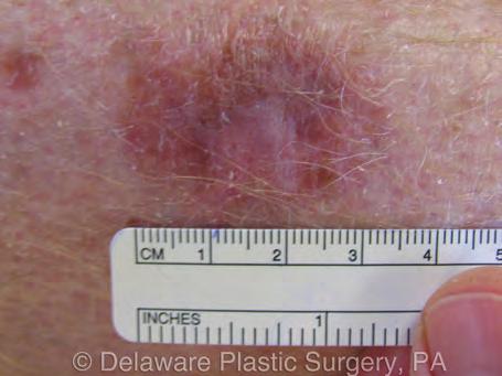
5 Left leg before 1st treatment Left leg before 1st treatment Left leg MAL application Left leg after laser 1 treatment 8 days after 1st tx L leg 210 day after 3rd tx
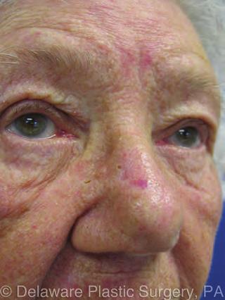
6 Subject 2 78 year old woman who has a past medical history of moderate-to-severe cardiovascular disease. In 2009 a stent was placed secondary to an MI. She is currently on anticoagulation and also on antihypertensive medications. Examination of the mid right nosed showed a ill-defined, pearly plaque with an area of central eschar. The lesion measures approximately 1.5 x 1 cm. A biopsy revealed and infiltrative basal cell carcinoma. 7 days following her first treatment, a moderate eschar was present with mild to moderate edema. After 20 days, an exam showed the eschar was gone and significant improvement with a new epidermis. A second treatment was performed with a 3 hour incubation period. Txt.# Time interval between treatment days days Examination after 17 days, following her second treatment, showed new pink epidermis with subtle nodular changes. 31 days after her last exam, showed the area progressively more pale, less nodular with slight edema. Overall, she had a very good result. Four months, 22 days after the second treatment revealed a pale scar with a 3 mm superficial vascular papule. The papule will be treated with CO2 laser. Delaware Plastic Surgery, PA


7 Nasal basal cell carcinoma at first txt 51 days after first txt 4 months, 22 days after 1st txt
8 Subject 3 85 year old woman with history of Alzheimer disease, hypertension and osteoporosis. The right upper back shows a 7 x 10.5 cm diffuse nodular papular patch. A biopsy revealed superficial nodular squamous cell carcinoma. There were superficial areas admixed with nodular areas and there was mild to moderate skin laxity in this area A total of three treatments were performed with a 3 hour incubation period. Treatment timeline: Txt. # Time interval between treatment days days days Eight days after her first treatment the central area of nodularity had an eschar that was cleaned and there was a small area of granulation tissue. Overall, the erythematous patch appeared less keratotic and smaller. A second treatment was performed focusing on the nodular areas. Following her second treatment exam shows an area of erythema and clearing of the areas of superficial involvement. Overall this area appeared to contract. Following the third treatment, a residual central nodular lesion remained surrounded by healed epidermis. A biopsy of the residual lesion of right upper back showed nodular infiltrating basal cell carcinoma approximately 1.5 x 2 cm. A Mohs micrographic excision was preformed followed by a primary closure. Delaware Plastic Surgery, PA


9 Exam at 1st txt Before 2nd treatment Debriedment 2nd txt 22 days after 1st txt Treated area after 3 txts and Mohs excision of residual central lession
10 Subject 4 80 year old woman with long history of sun exposure. Patient is on long-term anticoagulation for a mechanical heart valve. The right nasal dorsum showed a nodular ill-defined mass and associated subcutaneous component measuring 1 x 1.5 cm. Biopsy revealed squamous cell carcinoma. A total of two treatments performed with a three hour incubation period. Treatment Timeline: Txt. # Time interval between treatment days #1-2 20days Eight days after first treatment noted a small residual eschar and slight irregularity and erythema to the base of the lesion. Twenty days after her first treatment showed a residual, erythematous, nodular margin anteriorly and on the right side of the previous margin base. Therefore, the second treatment was performed. Ten days after her second treatment, exam revealed new pink epidermis without evidence of nodularity. Delaware Plastic Surgery, PA


11 Pretreatment of nasal squamous cell carcinoma Pretreatment of nasal squamous cell carcinoma MAL application First treatment of debridement following CO2 laser 9 days after 1st treatment Exam before 2nd txt Debridement of 2nd txt 176 days after 2nd txt
12 Subject 5 71 year old man with a past medical history significant for severe COPD. He is oxygen dependant with long term oral and inhaled steroids. Past history shows many years of treatments for pre-malignant and malignant face and scalp lesions. Treatments included cryotherapy, laser treatment, topical 5-FU, ALA/PDT and Mohs surgery with reconstruction. Scalp exam with first treatment revealed three keratotic/nodular patches, each approximately 2 cm in diameter. A single treatment with a 90 minute incubation period, which resulted in clearing of all three lesions. 50 days after treatment, previously treated areas show no evidence of nodularity with new pink epidermis. Delaware Plastic Surgery, PA


13 Before 1st txt 50 days after 1st txt
14 Subject 6 87 year old man who has had a history of multiple nasal skin malignancies, which required treatments, including Mohs surgery with reconstruction. His medical history is significant for severe cardiovascular disease, requiring anticoagulation. Nasal skin exam at first treatment sowed diffuse keratotic changes with superficial ulcerations on right inferior and superior nasal tip. After the first treatment, most of the diffuse keratotic erythematous changes were cleared, however a second treatment was performed to treat residual nodular areas on the mid-nasal tip. A total of two treatments were performed with a three hour incubation period. Treatment Timeline: Txt.# Timeline interval between treatment days days One month after the second treatment, all areas cleared with pink epidermis at the mid and right nasal tip. Seven months after the second treatment, a new keratotic shallow ulcerative lesion developed on the left superior aspect of the previously treated area. This area will receive addition treatment. Delaware Plastic Surgery, PA


15 7 months after 2nd tx lesion developed adjacent to previously treated area Before first tx 36 days after second tx
16 Subject 7 70 year old man who has had a history of sun exposure and prior superficial keratotic lesion removals. The right temple shows a 1.7 cm erythematous keratotic patch that is ulcerative. A biopsy revealed a nodular basal cell carcinoma. 8 days after treatment shows new pink epidermis without nodularity in the treated area. Delaware Plastic Surgery, PA


17 Ulcerative nodular basal cell carcinoma at time of txt Right temple lesion at time of txt 8 days after first txt

18 Subject 8 76 year old man with a long history of sun exposure. He does have a history of cardiovascular disease and possible abdominal aortic aneurysm, had a Greenfield filter placed in 2005 and is currently on Coumadin. The vertex scalp shows a keratotic patch measuring approximately 1.5 cm with an associated subcutaneous component. One treatment was performed with a 90 minute incubation period. 7 days after first treatment, there is a slight depression from the eschar and some granulation tissue. No evidence of nodularity. A biopsy revealed actinic keratosis with ulceration. 55 days after treatment, the scalp exam shows new pink epidermis with minimal depression, no nodularity. Delaware Plastic Surgery, PA


19 Before first treatment 17 days after first treatment 55 days after txt

20 Hill Laboratories Company 3 N. Bacton Hill Road Frazer, PA Phone: Fax:
I have a skin lump doc! What s next? 12 th August 2017 Dr. Sue-Ann Ho Ju Ee
 I have a skin lump doc! What s next? 12 th August 2017 Dr. Sue-Ann Ho Ju Ee Some thoughts Is this skin cancer? How common is this? How likely is this in this patient? What happens next if it s something
Dermatopathology: The tumor is composed of keratinocytes which show atypia, increase mitoses and abnormal mitoses.
 Squamous cell carcinoma (SCC): A common malignant tumor of keratinocytes arising in the epidermis, usually from a precancerous condition: 1- UV induced actinic keratosis, usually of low grade malignancy.
Clinical characteristics
 Skin Cancer Fernando Vega, MD Seattle Healing Arts Clinical characteristics Precancerous lesions Common skin cancers ACTINIC KERATOSIS Precancerous skin lesions Actinic keratoses Dysplastic melanocytic
Periocular skin cancer
 Periocular skin cancer Information for patients Skin cancer involving the skin of the eyelid or around the eye is called a periocular skin cancer. Eyelid skin cancers occur most often on the lower eyelid,
Learning Objectives. Tanning. The Skin. Classic Features. Sun Reactive Skin Type Classification. Skin Cancers: Preventing, Screening and Treating
 Learning Objectives Skin Cancers: Preventing, Screening and Treating Robert A. Baldor, MD, FAAFP Professor, Family Medicine & Community Health University of Massachusetts Medical School Distinguish the
Living Beyond Cancer Skin Cancer Detection and Prevention
 Living Beyond Cancer Skin Cancer Detection and Prevention Cutaneous Skin Cancers Identification Diagnosis Treatment options Prevention What is the most common cancer in people? What is the most common
Dermoscopy: Recognizing Top Five Common In- Office Diagnoses
 Dermoscopy: Recognizing Top Five Common In- Office Diagnoses Vu A. Ngo, DO Department of Family Medicine and Dermatology Choctaw Nation Health Services Authority Learning Objectives Introduction to dermoscopy
Skin lesions The Good and the Bad. Dr Virginia Hubbard Ipswich Hospital NHS Trust Barts and the London School of Medicine and Dentistry
 Skin lesions The Good and the Bad Dr Virginia Hubbard Ipswich Hospital NHS Trust Barts and the London School of Medicine and Dentistry Case 1 32 year old woman Australian Lesion on back New hair growing
Nonmelanoma skin cancers
 Skin cancer Philip Clarke Nonmelanoma skin cancers Treatment options Background Australia has one of the highest skin cancer rates in the world. Early detection and treatment of skin cancer is vital to
Glenn D. Goldman, MD. University of Vermont Medical Center. University of Vermont College of Medicine
 Glenn D. Goldman, MD University of Vermont Medical Center University of Vermont College of Medicine Recognize and identify the main types of skin cancer and their precursors Identify and understand new
Fractional CO 2 Laser Skin Resurfacing for the Treatment of Sun-Damaged Skin and Actinic Keratoses COS DERM
 Case Report Fractional CO 2 Laser Skin Resurfacing for the Treatment of Sun-Damaged Skin and Actinic Keratoses LindaSusan Marcus, MD; Neal Carlin, BS; Robert Carlin, BS, MA It is important to realize that
Cutaneous Malignancies: A Primer COPYRIGHT. Marissa Heller, M.D.
 Cutaneous Malignancies: A Primer Marissa Heller, M.D. Associate Director of Dermatologic Surgery Department of Dermatology Beth Israel Deaconess Medical Center December 10, 2016 Skin Cancer Non-melanoma
Histopathology: skin pathology
 Histopathology: skin pathology These presentations are to help you identify, and to test yourself on identifying, basic histopathological features. They do not contain the additional factual information
Interesting Case Series. Aggressive Tumor of the Midface
 Interesting Case Series Aggressive Tumor of the Midface Adrian Frunza, MD, Dragos Slavescu, MD, and Ioan Lascar, MD, PhD Bucharest Emergency Clinical Hospital, Bucharest University School of Medicine,
General information about skin cancer
 Skin Cancer General information about skin cancer Key points Skin cancer is a disease in which malignant (cancer) cells form in the tissues of the skin. There are different types of cancer that start in
Corporate Medical Policy
 Corporate Medical Policy Dermatologic Applications of Photodynamic Therapy File Name: Origination: Last CAP Review: Next CAP Review: Last Review: dermatologic_applications_of_photodynamic_therapy 10/2003
Integumentary System
 Integumentary System The integumentary system is commonly known as the Skin Largest organ of human body 10% total body weight and would cover over 20 square feet Functions of Skin 1. Protection Barrier
Identifying Skin Cancer. Mary S. Stone MD Professor of Dermatology and Pathology University of Iowa Carver College of Medicine March, 2018
 Identifying Skin Cancer Mary S. Stone MD Professor of Dermatology and Pathology University of Iowa Carver College of Medicine March, 2018 American Cancer Society web site Skin Cancer Melanoma Non-Melanoma
Hands-on: Lasers. NonAblative Rejuvenation
 Hands-on: Lasers NonAblative Rejuvenation Anthony M. Rossi, MD Assistant Attending Memorial Sloan Kettering Cancer Center Assistant Professor Weill Cornell Medical College DISCLOSURE OF RELEVANT RELATIONSHIPS
MOHS MICROGRAPHIC SURGERY: AN OVERVIEW
 MOHS MICROGRAPHIC SURGERY: AN OVERVIEW SKIN CANCER: Skin cancer is far and away the most common malignant tumor found in humans. The most frequent types of skin cancer are basal cell carcinoma, squamous
B. Incorrect! The ectoderm does not produce the dermis. C. Incorrect! The dermis is derived from the mesoderm.
 Human Anatomy - Problem Drill 04: The Integumentary System Question No. 1 of 10 Instructions: (1) Read the problem and answer choices carefully, (2) Work the problems on paper as 1. From the inner cell
Benign versus Cancerous Lesions How to tell the difference FMF 2014 Christie Freeman MD, CCFP, DipPDerm, MSc
 1 Benign versus Cancerous Lesions How to tell the difference FMF 2014 Christie Freeman MD, CCFP, DipPDerm, MSc Benign lesions Seborrheic Keratoses: Warty, stuck-on Genetics and birthdays Can start in late
Photodynamic Therapy (PDT) Basics and clinical applications
 Basics and clinical applications") Photodynamic Therapy () Basics and clinical applications D. Roseeuw, S. T kint Department of Dermatology UZBrussel - VUB GOAL of : selective destruction of targeted abnormal cells Light O 2 Photosensitiser
Photodynamic Therapy () Basics and clinical applications D. Roseeuw, S. T kint Department of Dermatology UZBrussel - VUB GOAL of : selective destruction of targeted abnormal cells Light O 2 Photosensitiser
Skin Malignancies Non - Melanoma & Melanoma Marilyn Ng, MD Dept. of Surgery M&M Conference Downstate Medical Center July 19, 2012
 Skin Malignancies Non - Melanoma & Melanoma Marilyn Ng, MD Dept. of Surgery M&M Conference Downstate Medical Center July 19, 2012 Case Presentation 57 yo man with 3 month hx of a nonhealing < 1 cm right
Periocular Malignancies
 Periocular Malignancies Andrew Gurwood, O.D., F.A.A.O., Dipl. Marc Myers, O.D., F.A.A.O. Drs. Myers and Gurwood have no financial interests to disclose. Course Description Discussion of the most common
Diagnosis and Management of Actinic Keratosis (AKs)
") Diagnosis and Management of Actinic Keratosis (AKs) Andrei Metelitsa, MD, FRCPC, FAAD Co-Director, Institute for Skin Advancement Clinical Associate Professor, Dermatology University of Calgary, Canada
Diagnosis and Management of Actinic Keratosis (AKs) Andrei Metelitsa, MD, FRCPC, FAAD Co-Director, Institute for Skin Advancement Clinical Associate Professor, Dermatology University of Calgary, Canada
Know who is at risk: LOOK! for ABCDs, rapidly changing lesions, do a biopsy when indicated
 Lindy P. Fox, MD Assistant Professor Director, Hospital Consultation Service Department of Dermatology University of California, San Francisco Applies to adults without history of malignancy or premalignant
Dual wavelength (1540nm nm) mixed technology for fractional resurfacing in skin rejuvenation Background
 mixed technology for fractional resurfacing in skin rejuvenation Background") Dual wavelength (1540nm + 10600 nm) mixed technology for fractional resurfacing in skin rejuvenation Dr. Paolo Sbano, M.D. Specialist in Dermatology and Venereology Department of Dermatology of Belcolle
Dual wavelength (1540nm + 10600 nm) mixed technology for fractional resurfacing in skin rejuvenation Dr. Paolo Sbano, M.D. Specialist in Dermatology and Venereology Department of Dermatology of Belcolle
Skin Cancer. 5 Warning Signs. American Osteopathic College of Occupational and Preventive Medicine OMED 2012, San Diego, Monday, October 8, 2012 C-1
 Skin Cancer AMERICAN OSTEOPATHIC COLLEGE OF OCCUPATIONAL & PREVENTIVE MEDICINE OMED 2012 October 8, 2012 E. Robert Wanat II, D.O., M.P.H. Learning Objectives: Identify the 3 Basic Types of Skin Cancer
Know who is at risk: LOOK! for ABCDs, rapidly changing lesions, do a biopsy when indicated
 Lindy P. Fox, MD Associate Professor Director, Hospital Consultation Service Department of Dermatology University of California, San Francisco Applies to adults without history of malignancy or premalignant
Benign and malignant epithelial lesions: Seborrheic keratosis: A common benign pigmented epidermal tumor occur in middle-aged or older persons more
 Benign and malignant epithelial lesions: Seborrheic keratosis: A common benign pigmented epidermal tumor occur in middle-aged or older persons more common on the trunk; but extremities, head and neck are
Skin disorders. Basal cell carcinoma December 2009 Anthony Ormerod, Sanjay Rajpara, and Fiona Craig ...
 December 29 Anthony Ormerod, Sanjay Rajpara, and Fiona Craig.................................................. ABSTRACT INTRODUCTION: (BCC) is the most common form of skin cancer, predominantly affecting
PHOTOTHERAPY: WHAT DO WE KNOW AND HOW DOES IT WORK? Suite 3/36 O'Riordan Street Alexandria, Sydney NSW Australia
 PHOTOTHERAPY: WHAT DO WE KNOW AND HOW DOES IT WORK? Suite 3/36 O'Riordan Street Alexandria, Sydney NSW Australia 2015 02 8331 8933 info@infinity-led.com.au DOCTORATE IN HEALTH SCIENCE DEAKIN UNIVERSITY
Eyelid basal cell carcinoma Patient information
 Eyelid basal cell carcinoma Patient information Your procedure relates to the face, eyelids, orbit or tear drainage system that together are treated by specialist surgeons in the field of oculoplastic
Clinical Policy: Benign Skin Lesion Removal Reference Number: CP.MP.HN150
 Clinical Policy: Reference Number: CP.MP.HN150 Effective Date: 6/04 Last Review Date: 8/17 Coding Implications Revision Log See Important Reminder at the end of this policy for important regulatory and
Non-Ablative Rejuvenation
 Non-Ablative Rejuvenation Denise Baker, MD Non-Ablative Skin Rejuvenation Denise Baker, MD The following potential conflict of interest relationships are germane to my presentation. Intrinsic Aging Inevitably
Large majority caused by sun exposure Often sun exposure before age 20 Persons who burn easily and tan poorly are at greatest risk.
 Basics of Skin Cancer Detection and Treatment of Non- Melanoma Skin Cancers Large majority caused by sun exposure Often sun exposure before age 20 Persons who burn easily and tan poorly are at greatest
Skin lesions & Abrasions
 Skin lesions & Abrasions What Are Skin Lesions? A skin lesion is a part of the skin that has an abnormal growth or appearance compared to the skin around it Types of Skin Lesions Two types of skin lesions
Glenn D. Goldman, MD. Fletcher Allen Health Care. University of Vermont College of Medicine
 Glenn D. Goldman, MD Fletcher Allen Health Care University of Vermont College of Medicine Recognize and identify the main types of skin cancer Understand how and why Mohs surgery is utilized for the treatment
Thursday 21 st August Skin Problems
 Thursday 21 st August 2014 Skin Problems Skin Problems The Sun and the Skin Sun Damage Recognising the early signs of skin cancer The Big 3 inflammatory condi=ons Acne & Rosacea Eczema (Including Seborrhoeic
Common Benign Lesions and Skin Cancers. 22nd May 2015 Dr Mark Foley
 Common Benign Lesions and Skin Cancers 22nd May 2015 Dr Mark Foley Thank you for downloading this file. This intended to supplement the presentation given at the NZ Wound Care Conference, it is not intended
Topical Photodynamic Therapy Using Intense Pulsed Light for Treatment of Actinic Keratosis: Clinical and Histopathologic Evaluation
 Topical Photodynamic Therapy Using Intense Pulsed Light for Treatment of Actinic Keratosis: Clinical and Histopathologic Evaluation HYUNG SU KIM, MD,JONG YEOP YOO, MD,KWANG HYUN CHO, MD,OH SANG KWON, MD,
Technicians & Nurses Program
 ASCRS ASOA Symposium & Congress Technicians & Nurses Program May 6-10, 2016 New Orleans Evaluation and Treatment of Eyelid Malignancies Richard C. Allen MD PhD FACS Professor Section of Ophthalmology Dept.
 Page 1 of 15 Title Authored By Course No Contact Hours 2 Skin Cancer the Real Picture for Early Detection and Treatment Cheryl Sommer RN, MSN, ARNP SC120604 Purpose The purpose of this course is to provide
EXPERIMENTAL THERMAL BURNS I. A study of the immediate and delayed histopathological changes of the skin.
 EXPERIMENTAL THERMAL BURNS I A study of the immediate and delayed histopathological changes of the skin. RJ Brennan, M.D. and B. Rovatti M.D. The purpose of this study was to determine the progressive
Describe the functions of the vertebrate integumentary system. Discuss the structure of the skin and how it relates to function.
 Chapter 5 Describe the functions of the vertebrate integumentary system. Discuss the structure of the skin and how it relates to function. Explain the basis for different skin colors. Describe the structure
CLINICAL EVALUATION REPORT ON THE EFFICACY AND SAFETY OF THE CORE SYSTEM FOR FACIAL ENHANCEMENT TREATMENTS
 CLINICAL EVALUATION REPORT ON THE EFFICACY AND SAFETY OF THE CORE SYSTEM FOR FACIAL ENHANCEMENT TREATMENTS BACKGROUND Efforts to improve fractional ablative laser systems have led to the development of
NONABLATIVE RESURFACING
 `AESTHETIC LASER SURGERY 0094-1298/00 $15.00 +.00 NONABLATIVE RESURFACING David J. Goldberg, MD TYPES OF LASERS AND THEIR DIFFERENCES Pulsed char-free CO2 laser skin resurfacing has provided a method of
ICD 10 Codes. L82.1 Seborrheic Keratosis L82.0 Irritated Seborrheic Keratosis
 Leon H. Kircik M.D. Clinical Associate Professor of Dermatology Indiana University School of Medicine Mount Sinai Medical Center, New York, NY Physicians Skin Care, PLLC Louisville, KY 1 ICD 10 Codes L82.1
NO MORE NEWS AN EVIDENCE BASED APPROACH ON LASERS IN SKIN OF COLOR PATIENTS DR. EDUARDO WEISS, M.D., FAAD
 NO MORE NEWS AN EVIDENCE BASED APPROACH ON LASERS IN SKIN OF COLOR PATIENTS DR. EDUARDO WEISS, M.D., FAAD KEY POINTS Hispanics/Latinos are the fastest growing segment of the skin of color population Use
Appendix D: Authorization Guidelines for Dermatology Services
 Appendix D: Authorization Guidelines for Dermatology Services Revised June 2011 1 Appendix D: Authorization Guidelines for Dermatology Dermatologists are limited to the CPT codes referenced in this Section.
Skin Cancer 101: Diagnosis and Management of the Most Common Cancer
 Skin Cancer 101: Diagnosis and Management of the Most Common Cancer Sarah Patton, PA-C, MSHS Skin Surgery Center www.skinsurgerycenter.com Seattle/Bellevue, WA Skin cancer Skin cancer is by far the most
Clinical Efficacy and Statistical Evaluation of INTRAcel Treatment
 Unique technology of Fractional RF Micro-needles TM 1 Clinical Efficacy and Statistical Evaluation of INTRAcel Treatment Takashi Takahashi, M.D. Dermatologist Takahashi Clinic Shibuya, Tokyo, Japan BACKGROUND:
Richard Turner Consultant Dermatologist
 Old Problems & New Treatments Photo Album by Administrator Richard Turner Consultant Dermatologist Plan for tonight? Refresher on SCC and solar keratosis How to distinguish the two Classic therapy than
Knowledge-Powered Medicine
 mohs surgery A patient guide to specialized treatment for skin cancer Knowledge-Powered Medicine upgdocs.org/dermatology upgdocs.org/dermatology A patient guide Preparing for Mohs Surgery > What is Mohs
Skin Cancer - Non-Melanoma
 Skin Cancer - Non-Melanoma Introduction Each year, millions of people find out that they have skin cancer. Skin cancer is almost 100% curable if found early and treated right away. It is possible to prevent
Principles of Anatomy and Physiology
 Principles of Anatomy and Physiology 14 th Edition CHAPTER 5 The Integumentary System Introduction The organs of the integumentary system include the skin and its accessory structures including hair, nails,
A PATIENT S GUIDE to MOHS MICROGRAPHIC SURGERY
 A PATIENT S GUIDE to MOHS MICROGRAPHIC SURGERY Microscopically Controlled Surgery for Skin Cancer ALBUQUERQUE DERMATOLOGY ASSOCIATES, P.A. Sky B. Connolly, M.D. F.A.A.D A Checklist Before Surgery Please
Dermatology Associates Mohs Micrographic Surgery
 Dermatology Associates Johnson City 2885 Boones Creek Road 423-928-9014 Kingsport 2300 W Stone Drive 423-246-4961 1-800-445-7274 Bristol 3183 W State Street 423-764-7131 In preparation for your upcoming
LUMPS AND BUMPS: AN ORGANIZED APPROACH TO DIAGNOSIS AND MANAGEMENT
 LUMPS AND BUMPS: AN ORGANIZED APPROACH TO DIAGNOSIS AND MANAGEMENT Tammy P. Than, M.S., O.D., F.A.A.O. The University of Alabama at Birmingham / School of Optometry 1716 University Blvd. Birmingham, AL
fluorouracil 0.5% / salicylic acid 10% cutaneous solution (Actikerall ) SMC No. (728/11) Almirall S.A.
 SMC No. (728/11) Almirall S.A.") fluorouracil 0.5% / salicylic acid 10% cutaneous solution (Actikerall ) SMC No. (728/11) Almirall S.A. 09 September 2011 The Scottish Medicines Consortium (SMC) has completed its assessment of the above
fluorouracil 0.5% / salicylic acid 10% cutaneous solution (Actikerall ) SMC No. (728/11) Almirall S.A. 09 September 2011 The Scottish Medicines Consortium (SMC) has completed its assessment of the above
WOCN Document:
 WOCN Document: www.cms.hhs.gov/medicaid/surveycert/080601.pdf OASIS Training Internet site: www.oasistraining.org M0440 Does this patient have a Skin Lesion or an Open Wound? This excludes "OSTOMIES."
Case Report No-Needle Jet Intradermal Aminolevulinic Acid Photodynamic Therapy for Recurrent Nodular Basal Cell Carcinoma of the Nose: A Case Report
 Skin Cancer Volume 0, Article ID 790509, 5 pages doi:0.55/0/790509 Case Report No-Needle Jet Intradermal Aminolevulinic Acid Photodynamic Therapy for Recurrent Nodular Basal Cell Carcinoma of the Nose:
The Reverse Galeal Hinge Flap: Another Valuable Technique in the Repair of Scalp
 TITLE PAGE TITLE: The Reverse Galeal Hinge Flap: Another Valuable Technique in the Repair of Scalp Defects Extending to the Calvarium AUTHORS: Lam, Thomas, BA; Indiana University School of Medicine Miletta,
A Retrospective Study of Treatment of Squamous Cell Carcinoma In situ. Övermark, Meri.
 https://helda.helsinki.fi A Retrospective Study of Treatment of Squamous Cell Carcinoma In situ Övermark, Meri 2016 Övermark, M, Koskenmies, S & Pitkanen, S 2016, ' A Retrospective Study of Treatment of
Patient Guide. The precise answer for tackling skin cancer. Brachytherapy: Because life is for living
 Patient Guide Brachytherapy: The precise answer for tackling skin cancer Because life is for living Overview of skin cancer Skin cancer is the most common cancer worldwide. In fact more people are diagnosed
Skin Cancers Emerging Trends and Treatment Approaches
 Skin Cancers Emerging Trends and Treatment Approaches Andrei Metelitsa, MD, FRCPC, FAAD Clinical Associate Professor, Dermatology, U of C Co-Director, Institute for Skin Advancement Copyright 2017 by Sea
Conflicts. Objectives. University of Texas Health Science Center at San Antonio. Pediatrics Grand Rounds 24 August Pediatric Dermatology 101
 Pediatric Dermatology 101 John C. Browning, MD, FAAD, FAAP Conflicts Investigator: ViroXis Advisor: ViroXis Advisory Board: TopMD Speaker: Galderma Objectives Understand the meaning and importance of cutaneous
Chapter 11 Worksheet Code It
 Class: Date: Chapter 11 Worksheet 3 2 1 Code It True/False Indicate whether the statement is true or false. 1. Surgical destruction is considered part of the surgical procedure description. 2. Prepping
Wound Jeopardy: Name That Wound Session 142 Saturday, September 10 th 2011
 Initial Wound Care Consult History Physical Examination Detailed examination of the wound Photographs Cultures Procedures TCOM ABI Debridement Management Decisions A Detailed History and Physical (wound)
MOHS MICROGRAPHIC SURGERY
 MOHS MICROGRAPHIC SKIN CANCER Skin cancer is by far the most common malignant tumor in humans. The most common types of skin cancer are basal cell carcinoma, squamous cell carcinoma, and melanoma. Both
Management of patients with melanocytic and non-melanocytic neoplasms
 Management of patients with melanocytic and non-melanocytic neoplasms Ashfaq Marghoob MD Harold Rabinovitz MD Margaret Oliviero ARNP Harald Kittler MD Jupiter Cancer Centrer Characteristic Dermoscopic
30 Actinic Keratosis (Solar Keratosis)
") 30 Actinic Keratosis (Solar Keratosis) CLINICAL APPLICATION QUESTIONS A 65-year-old white man is seen at your office for multiple scaling lesions over his face, ears, neck, and the V of the chest. These
30 Actinic Keratosis (Solar Keratosis) CLINICAL APPLICATION QUESTIONS A 65-year-old white man is seen at your office for multiple scaling lesions over his face, ears, neck, and the V of the chest. These
GentleFamily. GentleLASE, GentleYAG, GentleMAX & Pro Series. Banubeautylaser.com.au
 GentleFamily GentleLASE, GentleYAG, GentleMAX & Pro Series Banubeautylaser.com.au Alexandrite 755nm Laser Hair Reduction Pigmented Lesions Vascular Lesions Wrinkle Reduction Nd:YAG 1064nm Laser Hair Reduction
Evaluation of the wound healing response post deep dermal heating by fractional RF: INTRAcel
 12th symposium of the Association of Korean Dermatologists (2009) 1 Evaluation of the wound healing response post deep dermal heating by fractional RF: INTRAcel Un-Cheol.Yeo, M.D. S&U Dermatologic Clinic,
Interesting Case Series. Rhinophyma
 Interesting Case Series Rhinophyma Jake Laun, BS, Jared Gopman, BS, Joshua B. Elston, MD, and Michael A. Harrington, MD Department of Surgery, Division of Plastic Surgery, University of South Florida Morsani
Dermatology for the PCP Deanna G. Brown, MD, FAAD Susong Dermatology Consulting Staff at CHI Memorial
 Dermatology for the PCP Deanna G. Brown, MD, FAAD Susong Dermatology Consulting Staff at CHI Memorial Cutaneous Oncology for the PCP Deanna G. Brown, MD, FAAD Susong Dermatology Consulting Staff at CHI
Malignant Melanoma Early Stage. A guide for patients
 This melanoma patient brochure is designed to help educate melanoma patients and their caregivers. It was developed under the guidance of Dr. Michael Smylie, Professor, Department of Oncology, University
Lid Lesions: Relax or Refer
 Lid Lesions: Relax or Refer Blair Lonsberry, MS, OD, MEd., FAAO Professor of Optometry Pacific University College of Optometry blonsberry@pacificu.edu Agenda Benign vs. Malignant lesions Benign Eyelid
Actinic keratosis (AK): Dr Sarma s simple guide
: Dr Sarma s simple guide") Actinic keratosis (AK): Dr Sarma s simple guide Actinic keratosis is a very common lesion that you will see in your day-to-day practice. First, let me explain the name Actinic keratosis. It means keratosis
Actinic keratosis (AK): Dr Sarma s simple guide Actinic keratosis is a very common lesion that you will see in your day-to-day practice. First, let me explain the name Actinic keratosis. It means keratosis
Field vs Lesional Therapies for AKs 3/2/2019, 9:00-12 AM
 Dilemmas and Challenges in Skin Cancer Therapies and Management Field vs Lesional Therapies for AKs 3/2/2019, 9:00-12 AM Roger I. Ceilley, M.D. Clinical Professor of Dermatology The University of Iowa
Skin Integrity and Wound Care
 Skin Integrity and Wound Care By Dr. Amer Hasanien & Dr. Ali Saleh Skin Integrity and Wound Care Skin integrity: the presence of normal Skin & Uninterrupted skin layers by wounds. Factors affecting appearance
Dermatology Procedure Coding
 Dermatology Procedure Coding Anatomy Two layers that make up human skin Epidermis most superficial layer Composed of four to five layers called stratum Anyone remember the mnemonic? Thickness varies based
Skin Cancer in Organ Transplant Recipients Challenges and Opportunities
 Disclosure Skin Cancer in Organ Transplant Recipients Challenges and Opportunities Investigator: DUSA Pharmaceuticals, Inc. Investigator: Genentech Consultant: Gerson Lehrman Group Sarah Tuttleton Arron,
LARYNGEAL DYSPLASIA. Tomas Fernandez M; 3 rd year ENT resident, Son Espases University Hospital
 LARYNGEAL DYSPLASIA Tomas Fernandez M; 3 rd year ENT resident, Son Espases University Hospital INTRODUCTION Laryngeal cancer constitutes 1-2% of all malignancies diagnosed worldwide Survival is related
Non-melanoma Skin Cancer
 Non-melanoma Skin Cancer Understanding your diagnosis 1 888 939-3333 cancer.ca Non-melanoma Skin Cancer Understanding your diagnosis When you first hear that you have cancer, you may feel alone and afraid.
STUDY. Laser-Mediated Photodynamic Therapy of Actinic Keratoses
 STUDY Laser-Mediated Photodynamic Therapy of Actinic Keratoses Macrene R. Alexiades-Armenakas, MD, PhD; Roy G. Geronemus, MD Objective: To assess the safety and efficacy of the longpulsed pulsed dye laser
SKIN. 3. How is the skin structured around the finger joints to allow for flexible movement of the fingers?
 SKIN Objectives for Exam #1: 1. List various skin structures and describe their functions. 2. Describe skin responses to increases and decreases in body temperature. 3. Provide examples of various skin
Treatments used Topical including cleansers and moisturizer Oral medications:
 Discipline: Dermatology Extended Topic: Acne & Rosacea : Onset: Location: Face Chest Back Menses if female: Regular Irregular PCOS Treatments used Topical including cleansers and moisturizer Oral medications:
Glistening, Skin-Colored Nodule
 To Print: Click your browser's PRINT button. NOTE: To view the article with Web enhancements, go to: http://www.medscape.com/viewarticle/436334 Medscape Dermatology Clinic Glistening, Skin-Colored Nodule
Lasers in Gastroenterology, Otorhinolaryngology & Pulmonology
 Lasers in Gastroenterology, Otorhinolaryngology & Pulmonology Eloise Anguluan Laser-Tissue Interactions Fall Semester 2016 Gastroenterology the branch of medicine which deals with disorders of the stomach
SKIN CANCER. Most common cancer diagnosis 40% of all cancers
 SKIN CANCER Most common cancer diagnosis 40% of all cancers OBJECTIVES Review common and uncommon cancers of the skin. Special emphasis on melanoma and dysplastic nevus Review pathology/tnm/staging, which
DERMATOLOGIC APPLICATIONS OF PHOTODYNAMIC THERAPY
 DERMATOLOGIC APPLICATIONS OF PHOTODYNAMIC THERAPY Non-Discrimination Statement and Multi-Language Interpreter Services information are located at the end of this document. Coverage for services, procedures,
1) Photodynamic therapy with topical 5 aminolevulinic acid is considered medically necessary and is covered for the treatment of:
 Photodynamic therapy with topical 5 aminolevulinic acid is considered medically necessary and is covered for the treatment of:") Medical Policy Title: Photodynamic Therapy ARBenefits Approval: 10/26/2011 for Dermatologic Conditions Effective Date: 01/01/2012 Document: ARB0282:02 Revision Date: 03/20/2013 Code(s): 96567 Photodynamic
Medical Policy Title: Photodynamic Therapy ARBenefits Approval: 10/26/2011 for Dermatologic Conditions Effective Date: 01/01/2012 Document: ARB0282:02 Revision Date: 03/20/2013 Code(s): 96567 Photodynamic
Maligna Melanoma and Atypical Fibroxanthoma: An Unusual Collision Tumour G Türkcü 1, A Keleş 1, U Alabalık 1, D Uçmak 2, H Büyükbayram 1 ABSTRACT
 Maligna Melanoma and Atypical Fibroxanthoma: An Unusual Collision Tumour G Türkcü 1, A Keleş 1, U Alabalık 1, D Uçmak 2, H Büyükbayram 1 ABSTRACT Two different neoplasia in the same biopsy material called
Using the Mohs Technique for Thin Melanomas
 Dermatology Associates Mohs Micrographic Surgery 2300 W Stone Drive Kingsport TN 37660 Telephone 423-246-4961 1-800-445-7274 (VA Toll Free) Fax 423-245-1200 Using the Mohs Technique for Thin Melanomas
MULTIDIODE PDT 630 TM
 MULTIDIODE PDT 630 TM DERMA Photodynamic therapy with 630nm laser Intralesional and percutaneous photodynamic therapy with PDT 630nm laser for benign and precancerous skin lesions MULTIDIODE PDT 630 TM
Award Winning. Award Winning. FDA-Cleared. Light Therapy! LED Therapy. Illuminating Vitality
 Award Winning Award Winning FDA-Cleared Light Therapy! LED Therapy Illuminating Vitality What is Light Therapy? The application of specific wavelengths of light to tissue to obtain therapeutic benefits
Case-Based Approach to Common Dermatologic Neoplasms
 Case-Based Approach to Common Dermatologic Neoplasms Patrick Retterbush, MD, FAAD Mohs Surgery & Dermatologic Oncology Associate Member of the American College of Mohs Surgery Private Practice: Lockman