Premalignant lesion is a morphologically altered tissue in which cancer is more likely to occur, than its apparently normal counter parts.
|
|
|
- Anabel Ferguson
- 5 years ago
- Views:
Transcription
1 Oral Premalignancy
2 Premalignant lesion is a morphologically altered tissue in which cancer is more likely to occur, than its apparently normal counter parts. Premalignant condition is a generalized state or condition associated with significantly increased risk for cancer development. Various oral mucosal lesions, particularly red lesions (erythroplakias) and some white lesions (leukoplakias), have a potential for malignant change

3 Risk factors for malignant change in white lesions
4 Classification: Premalignant lesions: Leukoplakia Erythroplakia Pipe smokers keratosis Snuff-dippers keratosis Carcinoma in situ Bowen disease Actinic keratosis Contd.
5 Premalignant Conditions: Oral submucous fibrosis Tertiary syphilis Lichen planus Discoid lupus erythematosus Dyskeratosis congenita Sideropenic dysplasia

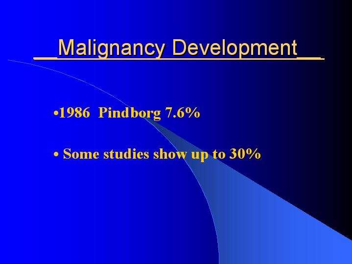
6 Lesions / Conditions with potential for malignant change Risks of malignant change are difficult to determine accurately and vary with many factors discussed below. If more than 25% of lesions become malignant in 5 years this is considered an exceptionally high risk. Malignant change in 2 3% of lesions in 5 years is considered a relatively low risk.note that the highest risk lesions are uncommon. Common lesions appear in between 1% and 5% of the population.
7 The best predictor of the potential for malignant transformation is the degree of dysplasia seen histologically. For this reason, and because a few lesions will already be malignant, biopsy of red and white patches is mandatory. The term dysplasia (literally, abnormal growth) is given to the cytological abnormalities seen in both malignant and premalignant cells. Premalignancy is distinguished from malignancy only by the latter s invasiveness and release of metastases.
8 Epithelial dysplasia: histological features Drop-shaped rete ridges Nuclear hyperchromatism Nuclear pleomorphism Altered nuclear/cytoplasmic ratio Excess mitotic activity Loss of polarity of cells Deep cell keratinisation Disordered or loss of differentiation Loss of intercellular adherence


9 Mild dysplasia. In this lesion there is a thin layer of parakeratin and the structure, maturation and orderly differentiation of the epithelial cells is largely unaffected. However, there is a degree of irregularity of basal cells with variation in size and hyperchromatism. Mild dysplasia. In this lesion there is prominent orthokeratosis and a keratohyaline layer immediately below it. Dysplasia is more prominent than in the previous figure, with enlarged hyperchromatic and bizarre cells in the basal and lower prickle cell layers.

10 Moderate dysplasia. The dermal papillae extend close to the surface and there are elongated rete processes, some of which are broader deeply. Enlarged and hyperchromatic cells are visible at this low power in rete processes and in most of the prickle cell layer.

11 Severe dysplasia. This rete process is composed almost entirely of cells with dark and irregularly shaped nuclei. Only the most superficial layers of cells show maturation to squamous cells and the orderly maturation and differentiation of epithelial cells has been lost.
12 LEUKOPLAKIA ( Leuko-white, Plakia-patch) Definition: A white hyperkeratotic,non scrapable patch in the oral cavity which cannot be characterized as any other clinical entity and is always associated with tobacco intake. Definition does not carry any histological connotation with it.
13 Classification Clinical Homogenous -Flat -Corrugated -Pumice like -Wrinkled Non Homogenous -Nodular / Speckled -Verrucous -Ulcerated -Erythroleukoplakia According to extent -Localized -Diffuse According to Banoczy -Leukoplakia simplex -Leukoplakia erosiva -Leukoplakia verrucosa
14 Staging of Leukoplakia: Size- It is denoted by L -L1 size< 2cm -L2 size 2-4cm -L3 size >4cm -Lx size not specified Clinical aspect- Denoted by C -C1 Homogenous -C2 Non-homogenous -Cx Not specified Pathological aspect- Denoted by P -P1 No dysplasia -P2 Mild dysplasia -P3 Moderate dysplasia -P4 Severe dysplasia -Px Not specified
15 Etiopathogenesis LOCAL FACTORS Tobacco Alcohol Chronic irritation Candidiasis Electromagnetic reaction or Galvanism REGIONAL AND SYSTEMIC FACTORS Syphilis Vitamin deficiency Sideropenic anemia Nutritional deficiency Xerostomia Hormonal disorders Drugs Virus Actinic Radiation
16 Clinical features Sex and distribution Common sites Appearance Surface Color Symptoms Sharp staging Ebbing tide type

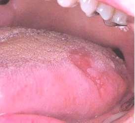
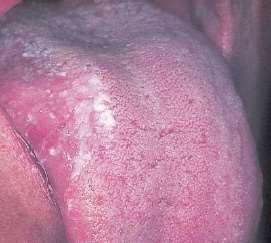
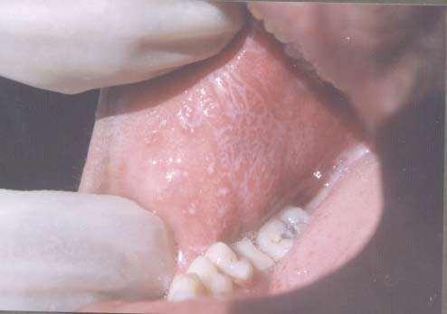
17 Homogeneous leukoplakia.

18 Sublingual Homogeneous leukoplakia

19 Sublingual Homogeneous Leukoplakia

20 Commissural Leukoplakia
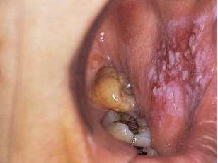
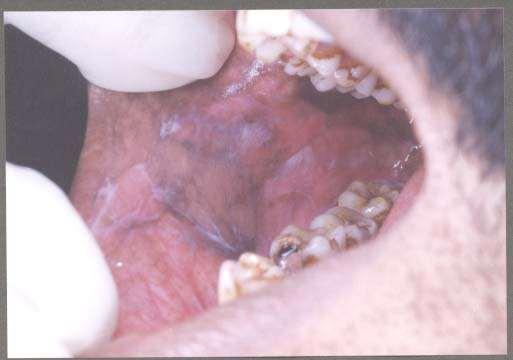
21 Speckled leukoplakia.
22 Speckled leukoplakia

23 Nodular or speckled leukoplakia

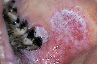
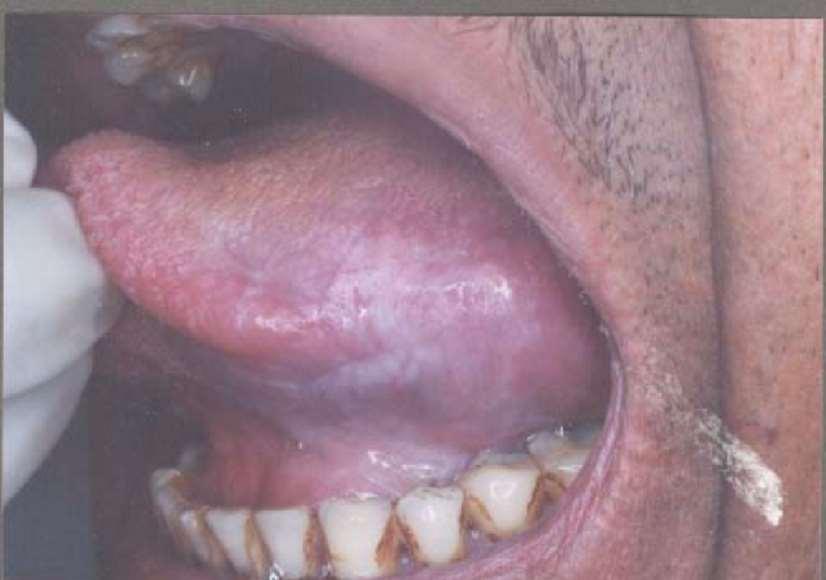
24 Thick white plaque on the lateral border of tongue represents verrucous leukoplakia.
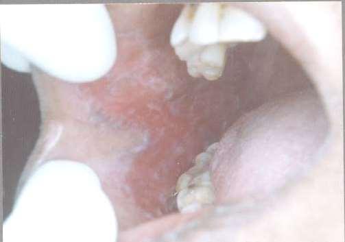
25 Erythroplakia.
26 Erythroplakia
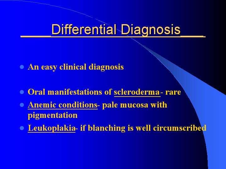
27 Differential Diagnosis Lichen planus Chemical burns Syphilitic mucus patches White spongy nevus DLE Psoriasis Leukoedema Hairy leukoplakia Verruca vulgaris Verrucous carcinoma Electro galvanic lesion Chronic cheek biting
28 Management: Limitation of etiological factor Conservative treatment -Vitamin therapy (Vit.A ,00,000 IU ) -Vit.A +Vit.E -13-cis-retinoic acid -Antioxidant therapy -Lycopene 2-8mg/ day for 2 months -Beta-carotene -Vit.A Palmitate -Vit.B complex -Antimycotic Tt. (Nystatin 5 lac IU bid +20% borax or 1% gentian violet) -Panthenol lingual tab -Estrogen
29 Surgical Management -Conventional surgery -Excision -Mucosal flap -Cryosurgery -Fulguration (electrosurgery or electrocautry) -LASER -Biopsy -Laser peel -Ablation -Surface vaporization Miscellaneous -Radiation therapy -Topical chemotherapy (Bleomycin,Azathiprine etc)
30 LICHEN PLANUS Descirbed in 1869 by Erasmus Wilson. Various mucosal surfaces involved. Common inflammatory disease of skin with characteristic violaceous, polygonal, pruritic papules. It also involves mucosa,nails & hair
31 Definition Relatively common dermatological disorder occurring on skin and oral mucus membrane and refers to lace like pattern produced by symbolic algae and fungal colonies on the surface of rocks in nature (lichens).
32 Reticular Papular Atrophic Erosive Bullous Annular Actinic Linear Hypertrophic Types:
33 Etiology: Immunological i)cell mediated immune response ii)autoimmunity iii)immunodeficiency Genetic factors Infections Drugs and chemicals Psychogenic factor Habits
34 Clinical Features: Age & Sex: Yrs ; F > M Incidence & Prevalence : 0.9 % % Site : Skin, oral & other mucus membranes. About 50% of skin cases have oral lesions, 25% only oral lesions Oral and genital mucosal lesions: Vaginogingival syndrome Symptoms: Intense pruritis (skin),burning mouth Signs: Characteristic Six P ; Planar, polygonal, purple, pruritic, papules & plaques.
35 Oral lichen planus Sites: Buccal mucosa (84%) Burning mouth Wickham s striae Typical lacy, reticular patterns, rings over the buccal mucosa. Associated hyper melanosis Usually bilateral. Stress induced
36
37
38
39

40
41 Treatment: Medicinal therapy -Steroids -Antifungal agents -Vit.-A -Cyclosporine -Dapsone Surgical Therapy Psychotherapy PUVA Therapy Symptomatic treatment
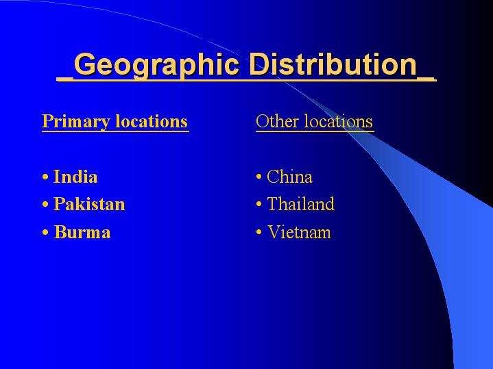
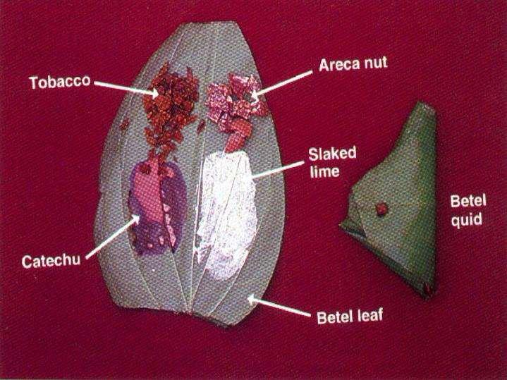
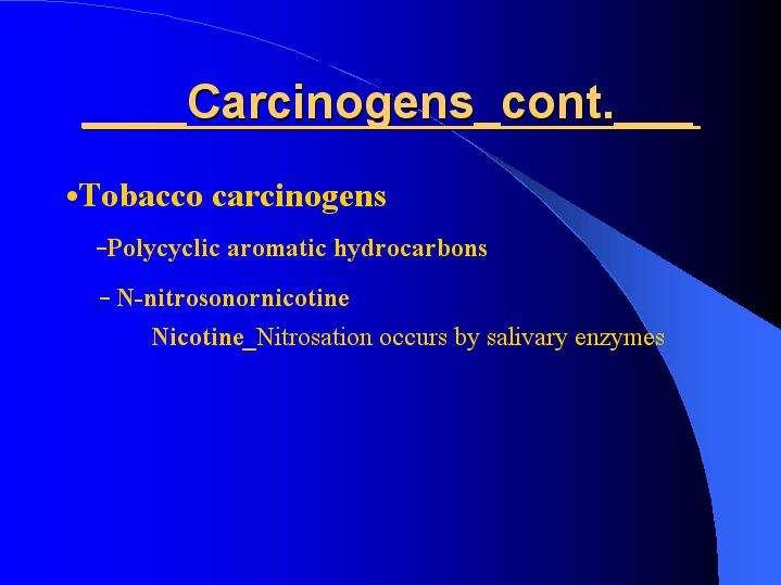
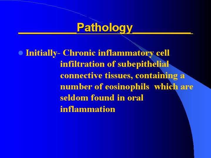
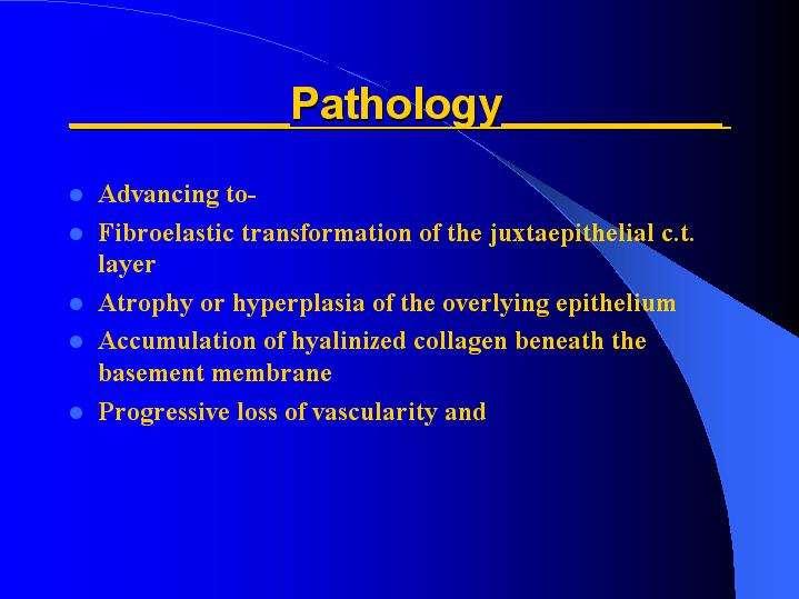
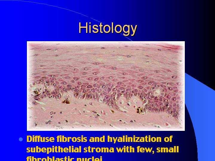
42 Oral Submucous Fibrosis Synonyms:Asian sideropenic dysphagia, oral mucosal disease, oral fibrosis, OSMF, oral soft tissue disease.
43
44

45
46
47
48
49
50

51
52
53
54
55
56
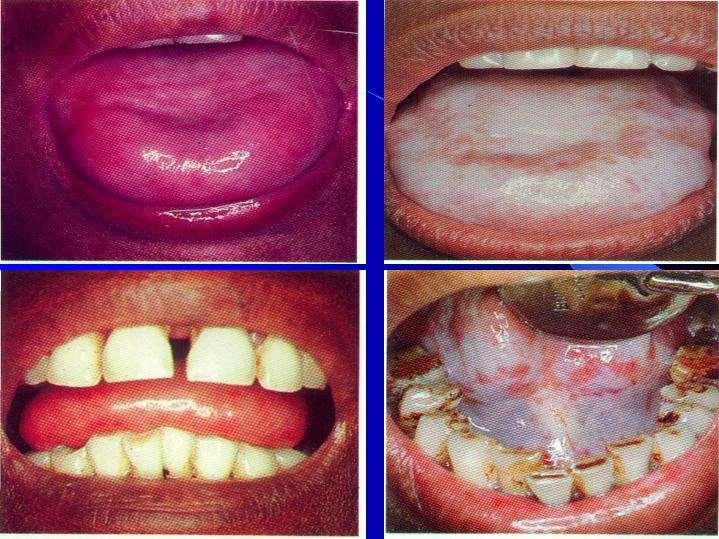
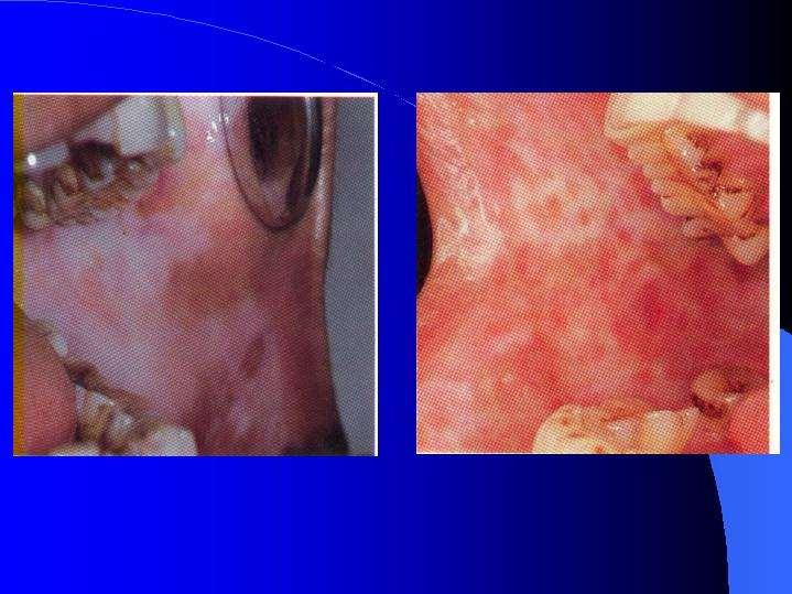
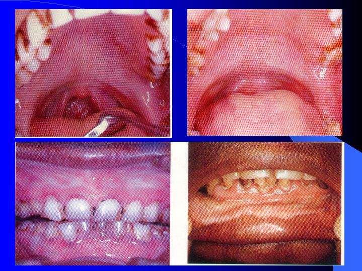
57 Symptoms of OSF Progressive inability to open the mouth (trismus) due to oral fibrosis and scarring Oral pain and burning sensation upon consumption of spicy foodstuffs Other symptoms -- Change of gustatory sensation - Hearing loss due to stenosis of the eustachian tubes - Dryness of the mouth - Nasal tonality to the voice - Dysphagia to solids (if the esophagus is involved) - Impaired mouth movements (eg, eating, whistling, blowing, sucking)
58 Staging of OSF Stage 1: Stomatitis includes erythematous mucosa, vesicles, mucosal ulcers, melanotic mucosal pigmentation, and mucosal petechia. Stage 2: Fibrosis occurs in ruptured vesicles and ulcers when they heal, which is the hallmark of this stage. Early lesions demonstrate blanching of the oral mucosa. Stage 3: Sequelae of OSF Speech and hearing deficits Leukoplakia
59
60
61
62
63
64
65
66
67 Lab Studies: No specific diagnostic tests are available for OSF. Some OSF studies report the following laboratory findings: Decreased hemoglobin levels Decreased iron levels Decreased protein levels Increased erythrocyte sedimentation rate (ESR) Decreased vitamin B complex levels Other Tests: Cytologic smears may be performed.
68 Treatment Medical Care Steroids Placental extracts Hyaluronidase IFN-g Antioxidants
69 Surgical Care: Simple excision of the fibrous bands Split-thickness skin grafting following bilateral temporalis myotomy or coronoidectomy Nasolabial flaps and lingual pedicle flaps
70 Patient Education Discontinue the habit Eliminate tobacco Avoid spicy foodstuffs Eat a complete and healthy diet Maintain proper oral hygiene Schedule regular oral examinations
71 Special Concerns An unhealing ulcer in the lesion Lesion undergoing red changes (erythroplakia) A burning sensation in the mouth An exophytic mass A lump in the neck Difficulty in chewing, swallowing, or speaking
72
Premalignant lesions may expose to a promoting. factor & may be induced to undergo malignant. Carcinoma in situ displays the cytologic features of
 بسم رلاهللا Def. Premalignant lesions may expose to a promoting factor & may be induced to undergo malignant transformation. Carcinoma in situ displays the cytologic features of malignancy without invasion
Oral cavity cancer accounts for approximately 3% of all malignancies and is a significant worldwide health problem.
 Oral cavity cancer accounts for approximately 3% of all malignancies and is a significant worldwide health problem. Majority are SCC ( 5-year survival rate only about 50-60% ) Many SCC arrive from premalignant
الطلاوة = Leukoplakia LEUKOPLAKIA
 LEUKOPLAKIA Leukoplakia is a clinical term that refers to a predominantly white lesion of the oral mucosa that cannot be rubbed off or characterized by any other definable lesion or known disease. 130
04/09/2018. Squamous Cell Neoplasia and Precursor Lesions. Agenda. Squamous Dysplasia. Squamo-proliferative lesions. Architectural features
 Squamous Cell Neoplasia and Precursor Lesions Jennifer L. Hunt, MD, MEd Aubrey J. Hough Jr, MD, Endowed Professor of Pathology Chair of Pathology and Laboratory Medicine University of Arkansas for Medical
Lesions & Lifestyles
 Lesions & Lifestyles attended a 3 hour Continuing Education Seminar on Oral Pathology presented by Nancy Dewhirst, RDH,BS on (date) at (location):. Course material is directly related patient care. Notes:
Squamous Cell Neoplasia and Precursor Lesions
 Squamous Cell Neoplasia and Precursor Lesions Jennifer L. Hunt, MD, MEd Aubrey J. Hough Jr, MD, Endowed Professor of Pathology Chair of Pathology and Laboratory Medicine University of Arkansas for Medical
LEUKOPLAKIA Definition Epidemiology Clinical presentation
 LEUKOPLAKIA Definition Leukoplakia is the most common premalignant or "potentially malignant" lesion of the oral mucosa. Leukoplakia is a predominantly white lesion of the oral mucosa than cannot be clinicopathologically
OROPHYRENGEAL CANCERS
 OROPHYRENGEAL CANCERS INTRODUCTION 2 % 4 % of all malignant Tumors in west Asia India 40% Men ^ Age :Over 60 yrs 90% of all oral cancers results from Tobacco and Alcohol Pan (Betel Leaf,Nut, Lime), Reverse
NEOPLASMS OF THE SURFACE EPITHELIUM (KERATINOCYTES)
") NEOPLASMS OF THE SURFACE EPITHELIUM (KERATINOCYTES) Papillary Lesions Precancerous Lesions Keratinocyte Proliferations Carcinomas Melanotic Lesions Melanomas Normal Mucosa Keratin layer Spinous layer Basal
NEOPLASMS OF THE SURFACE EPITHELIUM (KERATINOCYTES) Papillary Lesions Precancerous Lesions Keratinocyte Proliferations Carcinomas Melanotic Lesions Melanomas Normal Mucosa Keratin layer Spinous layer Basal
That. Name QUIZ. 60 SEPTEMBER 2017 // dentaltown.com
 QUIZ Name That General dentists are first in the line of practitioners that patients see for an oral lesion evaluation; therefore, a sound understanding of oral mucosal diseases and their clinical presentation
LESIONS OF THE ORAL CAVITY ORAL CAVITY. Oral Cavity Subsites 4/10/2013 LIPS TEETH GINGIVA ORAL MUCOUS MEMBRANES PALATE TONGUE ORAL LYMPHOID TISSUES
 LESIONS OF THE ORAL CAVITY David I. Kutler, MD, FACS Associate Professor Division of Head and Neck Surgery Department of Otolaryngology HNS Weill Cornell Medical Center ORAL CAVITY LIPS TEETH GINGIVA ORAL
Benign and malignant epithelial lesions: Seborrheic keratosis: A common benign pigmented epidermal tumor occur in middle-aged or older persons more
 Benign and malignant epithelial lesions: Seborrheic keratosis: A common benign pigmented epidermal tumor occur in middle-aged or older persons more common on the trunk; but extremities, head and neck are
Contents. 3 Diagnostic Tests and Studies Introduction Examination... 27
 Contents 1 Normal Anatomy... 1 1.1 Introduction... 1 1.2 Surface Landmarks... 1 1.3 Oral Mucosa... 3 1.4 Tongue... 5 1.5 Floor of Mouth... 6 1.6 Palate... 6 1.7 Dentition... 7 1.8 Temporomandibular Joint...
Contents. 1 Normal Anatomy Introduction... 17
 Contents 1 Normal Anatomy... 1 Introduction... 1 Surface Landmarks... 1 Oral Mucosa... 1 Tongue... 4 Floor of Mouth... 6 Palate... 7 Dentition... 7 Temporomandibular Joint... 9 Innervation... 10 Jaws and
Oral Cancer Dr Christine Goodall Consultant Oral Surgeon University of Glasgow Dental School
 Oral Cancer Dr Christine Goodall Consultant Oral Surgeon University of Glasgow Dental School christine.goodall@glasgow.ac.uk Locations Lip, mouth, oropharynx Tongue, floor of mouth, buccal mucosa, palate,
Dysplasia, Mimics and Other Controversies
 Dysplasia, Mimics and Other Controversies Mary S. Richardson, MD Dept. of Pathology Medical University of South Carolina Charleston, SC Notice of Faculty Disclosure In accordance with ACGME guidelines,
Leukoplakia is a white patch on the oral mucous membrane, which is undeliable and can not diagnose neither clinically nor pathologically as an other
 Leukoplakia Leukoplakia is a white patch on the oral mucous membrane, which is undeliable and can not diagnose neither clinically nor pathologically as an other disease. (Pindborg. 1978) Precancerous lesion
Diagnostic difficulties with lesions of the oral mucosa
 BDIAP London, November 2010 School of Clinical Dentistry University of Sheffield Diagnostic difficulties with lesions of the oral mucosa Paul M Speight Dept Oral & Maxillofacial Pathology University of
Dermatopathology: The tumor is composed of keratinocytes which show atypia, increase mitoses and abnormal mitoses.
 Squamous cell carcinoma (SCC): A common malignant tumor of keratinocytes arising in the epidermis, usually from a precancerous condition: 1- UV induced actinic keratosis, usually of low grade malignancy.
Oral Medicine Update for the dental practitioner Oral white patches
 IN BRIEF Most white lesions in the mouth are inconsequential and caused by friction or trauma. However, cancer and some systemic diseases such as lichen planus and candidosis may present in this way. Biopsy
LARYNGEAL DYSPLASIA. Tomas Fernandez M; 3 rd year ENT resident, Son Espases University Hospital
 LARYNGEAL DYSPLASIA Tomas Fernandez M; 3 rd year ENT resident, Son Espases University Hospital INTRODUCTION Laryngeal cancer constitutes 1-2% of all malignancies diagnosed worldwide Survival is related
Oral Health & HIV. Professor Sudeshni Naidoo Department of Community Dentistry University of the Western Cape
 Oral Health & HIV Professor Sudeshni Naidoo Department of Community Dentistry University of the Western Cape Importance & relevance of Oral HIV Lesions >70% of HIV+ve patients present with oral manifestations
Oral Epithelial Tumors, Melanocytic Nevi, and Melanoma (I)
") Introduction: Oral Epithelial Tumors, Melanocytic Nevi, and Melanoma (I) Oral Epithelial Tumors may be: Benign tumors Sequamous cell Papilloma Malignant tumors Sequamous cell carcinoma, Basal cell carcinoma
Introduction: Oral Epithelial Tumors, Melanocytic Nevi, and Melanoma (I) Oral Epithelial Tumors may be: Benign tumors Sequamous cell Papilloma Malignant tumors Sequamous cell carcinoma, Basal cell carcinoma
WHITE LESIONS OF THE UPPER AIRWAY
 WHITE LESIONS OF THE UPPER AIRWAY WHITE LESION CONFIGURATIONS Solitary vrs Multifocal Flat Plaque Verrucous/rippled Lacey White with red component Papular (curdled milk plaques) Pseudomembranous PLAQUES
Pattern of oral lesions Cytohistopathological study in tertiary care centre.
 International Journal of Current Research in Medical Sciences ISSN: 2454-5716 P-ISJN: A4372-3064, E -ISJN: A4372-3061 www.ijcrims.com Original Research Article Volume 3, Issue 10-2017 Pattern of oral lesions
Pigmented lesions of the Oral cavity
 Oral medicine أ.م.د احسان عبد هللا كميل Pigmented lesions of the Oral cavity Pigmented oral lesions are a large group of disorders in which the dark or brown color is the essential clinical characteristic.
The Oral Cavity. Image source:
 The Oral Cavity Anatomy Image source: http://anatomyforlayla.blogspot.co.za/2007/04/blog-post.html The major structures of the oral cavity are the lips, the teeth, the alveolar ridges (bony areas that
Role of the Dental Hygienist in Oral Pathology. Role of the Dental Hygienist in Oral Pathology. Cancers of the Oral Cavity.
 Gum Gardeners Study Club April 25, 2016 Early Detection of Oral Cancer Cindy Kleinegger, DDS, MS NW Oral Pathology Tigard, OR nworalpathology.com Role of the Dental Hygienist in Oral Pathology Work closely
Pathology of the skin. 2nd Department of Pathology, Semmelweis University
 Pathology of the skin 2nd Department of Pathology, Semmelweis University Histology of the skin Epidermis: Stratum corneum Stratum granulosum Stratum spinosum Stratum basale Dermis: papillary and reticular
PREVENTION OF ORAL CANCER
 PREVENTION OF ORAL CANCER Oral cancer is increasing in incidence worldwide. Throughout the world, malignant neoplasms of the mouth and pharynx rate as the fifth most common cancer in men and the seventh
TANYA A. WRIGHT, DDS OBJECTIVES
 TANYA A. WRIGHT, DDS OBJECTIVES One will be able to recognize pathological entities One will be able to establish a reasonable differential diagnosis One will be able to identify various types of lesions
Squamous Cell Carcinoma of the Head and Neck (SCCHN)
") Squamous Cell Carcinoma of the Head and Neck (SCCHN) Part 1 Bruce M. Wenig, M.D. Dept. of Pathology & Laboratory Medicine Continuum Health Partners New York, NY College of American Pathologists 2004. Materials
Squamous Cell Carcinoma of the Head and Neck (SCCHN) Part 1 Bruce M. Wenig, M.D. Dept. of Pathology & Laboratory Medicine Continuum Health Partners New York, NY College of American Pathologists 2004. Materials
Dr Rodney Itaki Lecturer Division of Pathology Anatomical Pathology Discipline
 Oral Lesions & Oral Cancer Dr Rodney Itaki Lecturer Division of Pathology Anatomical Pathology Discipline University of Papua New Guinea School of Medicine & Health Sciences Division of Pathology Overview
Original Article- A CYTOLOGICAL STUDY OF LEUKOPLASTIC LESIONS IN ORAL CAVITY
 Original Article- A CYTOLOGICAL STUDY OF LEUKOPLASTIC LESIONS IN ORAL CAVITY I. GUJRAL*, P. SINGH**, S. SHARMA***, N. GANGANE*** ABSTRACT Oral white lesions that cannot be clinically or pathologically
The Prevalence of Oral Leukoplakia: Results From a Romanian Medical Center
 The Prevalence of Oral Leukoplakia: Results From a Romanian Medical Center Ramona Vlad, DMD Department of Odontology and Oral Pathology, Faculty of Dental Medicine, University of Medicine and Pharmacy
Diseases of oral cavity
 Diseases of oral cavity Diseases of Teeth and Supporting Structures Inflammatory/Reactive Lesions Infections Oral Manifestations of Systemic Disease Precancerous and Cancerous Lesions Odontogenic Cysts
A clinical diagnosis of oral leukoplakia; A guide for dentists
 Journal section: Oral Medicine and Pathology Publication Types: Review doi:10.4317/medoral.22292 http://dx.doi.org/doi:10.4317/medoral.22292 ; A guide for dentists Vinicius C. Carrard 1, Isaäc van der
A Speckled Lesion. Angela C. Chi, DMD; Michele Carter Ravenel, DMD
 A Speckled Lesion Angela C. Chi, DMD; Michele Carter Ravenel, DMD The following Case Challenge is provided in conjunction with the American Academy of Oral and Maxillofacial Pathology. Case Summary This
WHITE LESIONS OF THE ORAL CAVITY - diagnostic appraisal & management strategies
 WHITE LESIONS OF THE ORAL CAVITY - diagnostic appraisal & management strategies * Joshy V.R ** Hari.S * Reader, Dept of Oral Pathology, Yenepoya Dental College, Yenepoya University, Mangalore 575 018.
Clinically Microscopically Pathogenesis: autoimmune not lifetime
 Vulvar Diseases: Can be divided to non-neoplastic and neoplastic diseases. The neoplastic diseases are much less common. Of those, squamous cell carcinoma is the most common. most common in postmenopausal
Red and White Tissue Reactions: A white appearance of the oral mucosa may be caused by: An increased production of keratin (hyperkeratosis).
.") Burket, chapter 4 Red and White Tissue Reactions: A white appearance of the oral mucosa may be caused by: An increased production of keratin (hyperkeratosis). An abnormal but benign thickening o stratum
Burket, chapter 4 Red and White Tissue Reactions: A white appearance of the oral mucosa may be caused by: An increased production of keratin (hyperkeratosis). An abnormal but benign thickening o stratum
Oral Manifestations of Dermatologic Disease: A Focus on Lichenoid Lesions. Proceedings of the NASHNP Companion Meeting, March, 2011, San Antonio, TX
 1 Oral Manifestations of Dermatologic Disease: A Focus on Lichenoid Lesions Proceedings of the NASHNP Companion Meeting, March, 2011, San Antonio, TX Susan Müller, DMD, MS Professor Department of Pathology
Epidemiological and clinicopathological study of oral leukoplakia
 Study Epidemiological and clinicopathological study of oral Minati Mishra, Janardan Mohanty*, Sujata Sengupta, Satyabrata Tripathy Department of Dermatology & Venereology, S.C.B. Medical College, *Department
Review Article- Leukoplakia: A mysterious white patch.
 International Journal Of Scientific Research And Education Volume 2 Issue 9 Pages 1824-1830 September-2014 ISSN (e): 2321-7545 Website: http://ijsae.in Review Article- Leukoplakia: A mysterious white patch.
Medical History. Oral Medicine and General Medicine
 Medical History Oral Medicine and General Medicine Gingivitis herpetica acuta NECROTIZÁLÓ SIALOMETAPLASIA SOOR Medical History The life expectancy has recently increased and increasing By dental prevention
IN THE NAME OF GOD. Dr.kheirandish DDS,MSC Oral and maxillofacial pathology
 IN THE NAME OF GOD Dr.kheirandish DDS,MSC Oral and maxillofacial pathology Dermatologic Diseases Chapter 16 ECTODERMAL DYSPLASIA o Two or more ectodermally derived anatomic structures fail to develop o
PACIFIC JOURNAL OF MEDICAL SCIENCES {Formerly: Medical Sciences Bulletin} ISSN:
 PACIFIC JOURNAL OF MEDICAL SCIENCES {Formerly: Medical Sciences Bulletin} ISSN: 2072 1625 Pac. J. Med. Sci. (PJMS) www.pacjmedsci.com. Email: pacjmedsci@gmail.com. EROSIVE LICHEN PLANUS A CASE REPORT *Prathima
ORAL LEUKOPLAKIA IN A SOUTH AFRICAN SAMPLE: A CLINICOPATHOLOGICAL STUDY
 ORAL LEUKOPLAKIA IN A SOUTH AFRICAN SAMPLE: A CLINICOPATHOLOGICAL STUDY Rakesh Chandran A research report submitted to the Faculty of Health Sciences, University of Witwatersrand, Johannesburg, in partial
Histopathology: skin pathology
 Histopathology: skin pathology These presentations are to help you identify, and to test yourself on identifying, basic histopathological features. They do not contain the additional factual information
Actinic keratosis (AK): Dr Sarma s simple guide
: Dr Sarma s simple guide") Actinic keratosis (AK): Dr Sarma s simple guide Actinic keratosis is a very common lesion that you will see in your day-to-day practice. First, let me explain the name Actinic keratosis. It means keratosis
Actinic keratosis (AK): Dr Sarma s simple guide Actinic keratosis is a very common lesion that you will see in your day-to-day practice. First, let me explain the name Actinic keratosis. It means keratosis
POTENTIALLY MALIGNANT DISORDERS OF ORAL CAVITY
 95 POTENTIALLY MALIGNANT DISORDERS OF ORAL CAVITY 1 Antony George, 1 Sreenivasan BS, 1 Sunil S, 1 Soma Susan Varghese, 1 Jubin Thomas, 1 Devi Gopakumar, 2 Varghese Mani. 1 Dept. of Oral & Maxillofacial
Index. Dent Clin N Am 49 (2005) Note: Page numbers of article titles are in boldface type.
 Note: Page numbers of article titles are in boldface type.") Dent Clin N Am 49 (2005) 273 278 Index Note: Page numbers of article titles are in boldface type. A Acanthosis nigricans, familial, 251 Amalgam tattoo, 197 198 Amphotericin B, 62 Ankyloglossia, 11 Anti-inflammatory
Dent Clin N Am 49 (2005) 273 278 Index Note: Page numbers of article titles are in boldface type. A Acanthosis nigricans, familial, 251 Amalgam tattoo, 197 198 Amphotericin B, 62 Ankyloglossia, 11 Anti-inflammatory
VIN/VAIN O C T O B E R 3 RD J M O R G A N
 VIN/VAIN O C T O B E R 3 RD 2 0 1 8 J M O R G A N Vaginal Intraepithelial Neoplasia VAIN I, II, III Incidence 0.1/100,000 women in US Mean age 50s (J Womens Health (Larchmt) 2009:18:1731) (J Obstet Gynaecol
JMSCR Vol 05 Issue 10 Page October 2017
 www.jmscr.igmpublication.org Impact Factor 5.84 Index Copernicus Value: 71.58 ISSN (e)-2347-176x ISSN (p) 2455-0450 DOI: https://dx.doi.org/10.18535/jmscr/v5i10.125 Histomorphological Study of Lichen Planus
Premalignant skin tumours
 Chapter 14: Premalignant skin tumours page: 434 Premalignant skin tumours page: 435 Solar keratoses (senile keratoses) Raised red and well-defined plaques with a rough surface covered in scales of varying
INFLAMMATORY DISEASES PART I. Immunopathology Part I
 INFLAMMATORY DISEASES PART I Immunopathology Part I Nonspecific & T Cell Mediated Mucosal Inflammatory Lesions Nonspecific and Idiopathic Mucositis Hypersensitivity and Autoimmune T cell mediated Immunoglobulin
DR.SHERIN.A.KHALAM,MSc(PSY),MDS,FICOI Associate Professor, PMS College of Dental Science & Research, Kerala University of Health Sciences; Consultant
,MDS,FICOI Associate Professor, PMS College of Dental Science & Research, Kerala University of Health Sciences; Consultant") DR.SHERIN.A.KHALAM,MSc(PSY),MDS,FICOI Associate Professor, PMS College of Dental Science & Research, Kerala University of Health Sciences; Consultant Maxillofacial Surgeon & Surgical Head, SUT Royal Hospitals,
DR.SHERIN.A.KHALAM,MSc(PSY),MDS,FICOI Associate Professor, PMS College of Dental Science & Research, Kerala University of Health Sciences; Consultant Maxillofacial Surgeon & Surgical Head, SUT Royal Hospitals,
Oral Leukoplakia: An Insight
 Oral Leukoplakia: An Insight Gigi Roy 1, Anu Vijayan 2, Shamji Shajahan 3, Anuja S 4, Rashmi Elizabeth Mathen 5 1,3,4,5-Post Graduate, Department of Oral Medicine and Radiology, Mar Baselios Dental College,
DENIS P. LYNCH, DDS, PHD
 140 TH ANNUAL MEETING MAY 6 MAY 7, 2010 JEWEL OF THE GREAT LAKES DENIS P. LYNCH, DDS, PHD FRIDAY, MAY 7, 2010 9:00 A.M. TO 12:00 NOON ORAL CANCER AND RELATED PREMALIGNANCY Oral Cancer and Premalignancy
Orofacial Disease: Update For The Dental Clinical Team: 3. White Lesions
 ORAL MEDICINE Orofacial Disease: Update For The Dental Clinical Team: 3. White Lesions Crispian Scully and Stephen Porter Abstract: White lesions usually contain an increased amount of keratin. Some are
Histopathology: Cervical HPV and neoplasia
 Histopathology: Cervical HPV and neoplasia These presentations are to help you identify basic histopathological features. They do not contain the additional factual information that you need to learn about
Head and Neck Cancer How to recognize it in your office
 Head and Neck Cancer How to recognize it in your office Peter M Hunt, MD, FACS Associates in ENT/Head & Neck Surgery Director CHI Memorial Head & Neck and Melanoma Centers of Excellence September 8, 2018
Oral Cancer FAQs. What is oral cancer? How many people are diagnosed with oral cancer each year?
 Oral Cancer FAQs What is oral cancer? Oral cancer or oral cavity cancer, is cancer that starts in the mouth. Areas affected by this type of cancer are the lips, the inside lining of the lips and cheeks
Benign versus Cancerous Lesions How to tell the difference FMF 2014 Christie Freeman MD, CCFP, DipPDerm, MSc
 1 Benign versus Cancerous Lesions How to tell the difference FMF 2014 Christie Freeman MD, CCFP, DipPDerm, MSc Benign lesions Seborrheic Keratoses: Warty, stuck-on Genetics and birthdays Can start in late
Autoimmune Diseases with Oral Manifestations
 Autoimmune Diseases with Oral Manifestations Martin S. Greenberg DDS, FDS RCSEd Professor Emeritus Department of Oral Medicine University of Pennsylvania Disclosure Statement I have no actual or potential
IT S FUNDAMENTAL MY DEAR WATSON! A SHERLOCKIAN APPROACH TO DERMATOLOGY
 IT S FUNDAMENTAL MY DEAR WATSON! A SHERLOCKIAN APPROACH TO DERMATOLOGY Skin, Bones, and other Private Parts Symposium Dermatology Lectures by Debra Shelby, PhD, DNP, FNP-BC, FADNP, FAANP Debra Shelby,
Large majority caused by sun exposure Often sun exposure before age 20 Persons who burn easily and tan poorly are at greatest risk.
 Basics of Skin Cancer Detection and Treatment of Non- Melanoma Skin Cancers Large majority caused by sun exposure Often sun exposure before age 20 Persons who burn easily and tan poorly are at greatest
Benign Oral cavity lesions. Mohammed ALESSA MBBS,FRCSC Assistant Professor Consultant Otolaryngology, Head & Neck Surgery
 Benign Oral cavity lesions Mohammed ALESSA MBBS,FRCSC Assistant Professor Consultant Otolaryngology, Head & Neck Surgery Anatomy Histology Physiology Pathology Clinical cases Introduction The oral cavity
Tongue cancer. Patient information
 What is cancer? The human body is made up of billions of cells. In healthy people, cells grow, divide and die. New cells constantly replace old ones in an orderly way. This process ensures each part of
Differential Diagnosis of Oral Lesions. An Interactive Lecture Using Audience Response Polling. John L. Alonge, MS, DDS
 Differential Diagnosis of Oral Lesions An Interactive Lecture Using Audience Response Polling John L. Alonge, MS, DDS Goals 1. Review the diagnostic process needed to formulate a differential diagnosis
Periocular Malignancies
 Periocular Malignancies Andrew Gurwood, O.D., F.A.A.O., Dipl. Marc Myers, O.D., F.A.A.O. Drs. Myers and Gurwood have no financial interests to disclose. Course Description Discussion of the most common
أملس عضلي غرن = Leiomyosarcoma. Leiomyosarcoma 1 / 5
 Leiomyosarcoma 1 / 5 EPIDEMIOLOGY Exact incidence is unknown, but older studies suggest that leiomyosarcomas comprise approximately 3 percent of soft-tissue sarcomas. Superficial leiomyosarcoma occurs
Evaluation and Management of Head and Neck Cancer in Patients with Fanconi anemia David I. Kutler, M.D., F.A.C.S.
 Evaluation and Management of Head and Neck Cancer in Patients with Fanconi anemia David I. Kutler, M.D., F.A.C.S. Residency Site Director Weill Cornell Medical Center Associate Professor Division of Head
Vascular. Extravasated blood. Melanocytic. Tattoo. Epidermolysis bullosa. Lichen planus. Pemphigoid Pemphigus Lupus. Candidosis. Surface Epithelial
 Oral Soft Tissue Pathology Epithelial Thickening (white) Combination Erythema migrans Epithelial atrophy (red) Surface Lesions Clinical Impression Enlargements Surface Debris Pigmented Vesicular Ulcerated
Clinical characteristics
 Skin Cancer Fernando Vega, MD Seattle Healing Arts Clinical characteristics Precancerous lesions Common skin cancers ACTINIC KERATOSIS Precancerous skin lesions Actinic keratoses Dysplastic melanocytic
Review Article. Oral leukoplakia Tsvetanov TS1 ABSTRACT
 Oral leukoplakia Tsvetanov TS1 1 Dr Tsvetan Borisov Tsvetanov Chief Assistant Professor PhD, DDS, DMD Department of Oral Surgery Dental Faculty Medical University Plovdiv, Bulgaria Received: 19-06-2016
MECHANISMS OF HUMAN DISEASE: LABORATORY SESSION PATHOLOGY OF THE SKIN LAB. Friday, February 12, :30 am 11:00 am
 MECHANISMS OF HUMAN DISEASE: LABORATORY SESSION PATHOLOGY OF THE SKIN LAB Friday, February 12, 2012 9:30 am 11:00 am FACULTY COPY GOALS: Describe the basic clinical and morphologic features of various
White Oral Lesions: How to Distinguish the Benign From the Deadly
 ABSTRACT: Chronic irritation from smoking is the most common cause of white mucosal lesions. Because benign leukoplakic growths are virtually impossible to distinguish from carcinoma, biopsy is essential.
Oral Medicine. Dr. Qianming Ian CHEN
 Oral Medicine Dr. Qianming Ian CHEN ORAL MEDICINE Oral medicine is the specialty of dentistry that is concerned with the oral health care of medically compromised patients and with the diagnosis and nonsurgical
30 Actinic Keratosis (Solar Keratosis)
") 30 Actinic Keratosis (Solar Keratosis) CLINICAL APPLICATION QUESTIONS A 65-year-old white man is seen at your office for multiple scaling lesions over his face, ears, neck, and the V of the chest. These
30 Actinic Keratosis (Solar Keratosis) CLINICAL APPLICATION QUESTIONS A 65-year-old white man is seen at your office for multiple scaling lesions over his face, ears, neck, and the V of the chest. These
Case Report II Sri Lanka Dental Journal 2018; 48(01) 41-45
 41-45") Case Report II Sri Lanka Dental Journal 2018; 48(01) 41-45 White sponge nevus in the oral cavity: case report and P.V.K.S. Hettiarachchi, J.C.M. Jayasinghe, B.S.M.S. Siriwardena, R.D. Jayasinghe Abstract
Case Report II Sri Lanka Dental Journal 2018; 48(01) 41-45 White sponge nevus in the oral cavity: case report and P.V.K.S. Hettiarachchi, J.C.M. Jayasinghe, B.S.M.S. Siriwardena, R.D. Jayasinghe Abstract
Oral Cancer- Improving Early Detection
 Oral Cancer- Improving Early Detection GDC Recommended Subject Aims: To give an overview of the dental team's role in detecting the early signs of oral cancer; to give an overview of the risk factors associated
Chapter 6 Squamous Cell Carcinoma: Variants and Challenges
 Chapter 6 Squamous Cell Carcinoma: Variants and Challenges Michael B. Morgan EPIDEMIOLOGY: Second most common skin cancer, rare in the dark-skinned races. ETIOLOGY: Ultraviolet light, HPV infection. PATHOGENESIS:
Smoking Habits Among Patients Diagnosed with Oral Lichen Planus
 TOBACCO INDUCED DISEASES Vol. 2, No. 2: 103-108 (2004) PTID Society Smoking Habits Among Patients Diagnosed with Oral Lichen Planus Meir Gorsky, 1 Joel B. Epstein, 2 Haya Hasson-Kanfi, 1 Eliezer Kaufman
MECHANISMS OF HUMAN DISEASE: LABORATORY SESSION PATHOLOGY OF THE SKIN LAB. Friday, February 13, :30 am 11:00 am
 MECHANISMS OF HUMAN DISEASE: LABORATORY SESSION PATHOLOGY OF THE SKIN LAB Friday, February 13, 2009 9:30 am 11:00 am FACULTY COPY GOALS: Describe the basic clinical and morphologic features of various
Integumentary system pertains to the skin, subcutaneous tissue and areolar tissue.
 TRICARE/CHAMPUS POLICY MANUAL 6010.47-M DEC 1998 Surgery And Related Services CHAPTER 3 SECTION 2.1 Issue Date: August 26, 1985 Authority: 32 CFR 199.4(c)(2) and (c)(3) I. PROCEDURE CODE RANGE 10040-19499
Gross Appearance & Histology of Skin Cancer. Kyle Mannion M.D. January 21, 2005
 Gross Appearance & Histology of Skin Cancer Kyle Mannion M.D. January 21, 2005 Actinic Keratosis 5-20% will develop squamous/basal cell ca Almost solely from solar damage Usually develop during 4 th decade
Principles of Management of Head & Neck Cancer. Jinka Sathya Associate professor of Oncology
 Principles of Management of Head & Neck Cancer Jinka Sathya Associate professor of Oncology Oral cavity Oro-pharynx Larynx Hypopharynx Nasophaynx Major sites of Mucosal H&N Cancers Head & Neck Cancer Oral
From the Cradle to the Grave: Oral pathology through the life span
 From the Cradle to the Grave: Oral pathology through the life span Conditions Exhibited in Infants and Children: Dental Lamina Cysts Etiology: Developmental Clinical appearance: Cystic nodules on alveolar
4Ps LUMPS AND BUMPS B.L.&T. BUMPS, LUMPS, AND TATTOOS. Most Common BUMP in the oral cavity Fibroma INTERDENTAL PAPILLAE LESIONS
 B.L.&T. BUMPS, LUMPS, AND TATTOOS LUMPS AND BUMPS DIFFERENTIAL DIAGNOSIS FOR LUMPS AND BUMPS Traumatic Fibroma Papilloma Epulis Fissuratum Inflammatory Papillary Hyperplasia Lesions of Attached Gingiva
Dual Wavelength Phototherapy System
 Dual Wavelength Phototherapy System The AKLARUS Blue and Red Combination System is an effective, drugfree alternative for treating acne & photodamaged skin. The non-invasive Aklarus treatment has been
Proliferative Verrucous Leukoplakia of the Gingiva, Report of two Cases with Malignant Transformation
 Journal of Clinical and Anatomic Pathology Case Report Open Access Proliferative Verrucous Leukoplakia of the Gingiva, Report of two Cases with Malignant Transformation Nadereh Ghanee DMD, Selene Saraf
Health Effects of Smokeless Tobacco
 Health Effects of Smokeless Tobacco Smokeless tobacco use is a significant health risk and cause of death & disease globally. Despite what the tobacco companies may claim, it is NOT a safe alternative
An Approach to Common and not so Common Rashes in the Office FMF 2014 Christie Freeman MD, CCFP, DipPDerm, MSc
 An Approach to Common and not so Common Rashes in the Office FMF 2014 Christie Freeman MD, CCFP, DipPDerm, MSc 1 Common Rashes Tinea Corporis: Annular- this is not the only criteria Advancing erythematous
Histopathology of Melanoma
 THE YALE JOURNAL OF BIOLOGY AND MEDICINE 48, 409-416 (1975) Histopathology of Melanoma G. J. WALKER SMITH Department ofpathology, Yale University School ofmedicine, 333 Cedar Street, New Haven, Connecticut
When Immunostains Can Get You in Trouble: Gynecologic Pathology p16: Panacea or Pandora s Box?
 When Immunostains Can Get You in Trouble: Gynecologic Pathology p16: Panacea or Pandora s Box? Teri A. Longacre, MD Stanford Medicine Stanford California pi6 in Gynecologic Pathology: Panacea or Pandora
General and Oral Pathology Topic Outlines. Copyright Fehrenbach and rdhedu.com. All rights reserved.
 General and Oral Pathology Topic Outlines Are you ready? Are you Ready? ARE YOU READY? TO LEARN AND EXPERIENCE real ORAL BIOLOGY??? BOOTCAMP STYLE Oral Biology BOOTCAMP through rdhedu.com For Oral Biology
Morsicatio Mucosae Oris A Chronic Oral Frictional Keratosis, Not a Leukoplakia
 J Oral Maxillofac Surg 67:140-146, 2009 Morsicatio Mucosae Oris A Chronic Oral Frictional Keratosis, Not a Leukoplakia Sook-Bin Woo, DMD,* and Dorothy Lin Purpose: Morsicatio mucosae oris (MMO) presents