Common Benign Lesions and Skin Cancers. 22nd May 2015 Dr Mark Foley
|
|
|
- Lorena Shelton
- 5 years ago
- Views:
Transcription
1 Common Benign Lesions and Skin Cancers 22nd May 2015 Dr Mark Foley
2 Thank you for downloading this file. This intended to supplement the presentation given at the NZ Wound Care Conference, it is not intended to be a standalone resource. The Skin Clinic Marlborough
3 Overview Reminder of skin structure Role of dermoscopy Common benign lesions Common skin cancers The Skin Clinic Marlborough

4 Reminder of skin structure The Skin Clinic Marlborough

5 Reminder of skin structure The Skin Clinic Marlborough

6 Epidermis Dermis Fat The Skin Clinic Marlborough
7 The Skin Clinic Marlborough
8 Reproduction The Skin Clinic Marlborough
9 Reproduction Keratin production The Skin Clinic Marlborough
10 Lipid and enzyme production Reproduction Keratin production The Skin Clinic Marlborough

11 Physical Barrie Lipid and enzyme production Reproduction Keratin production The Skin Clinic Marlborough
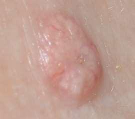
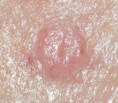
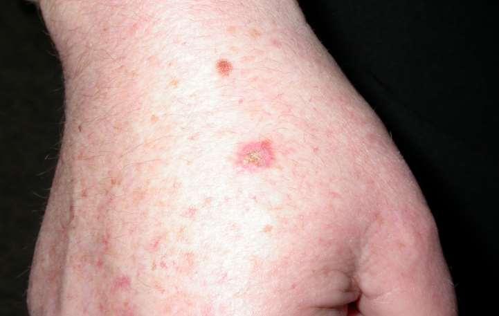
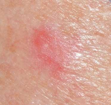
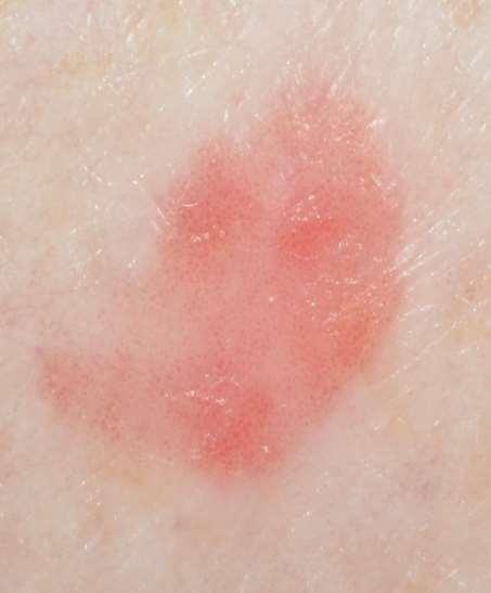
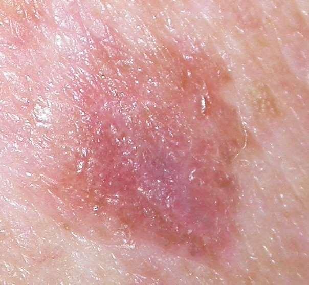
12 BCC The Skin Clinic Marlborough
13 AK/SCCi/SCC BCC The Skin Clinic Marlborough
14 AK/SCCi/SCC BCC Melanoma The Skin Clinic Marlborough
15 AK/SCCi/SCC Aldara BCC Melanoma The Skin Clinic Marlborough
16 Epidermis: Melanoma insitu Squamous cell carcinoma insitu Wart virus infection Seborrhoeic keratosis Junctional Naevus Psoriasis Eczema Merkel Cell Tumour Dermis: Compound Naevus Fungal infections (superficial) Intradermal Naevus Blue Naevus Dermatofibroma Basal cell carcinoma Squamous Cell Carcinoma
17 Skin Cancers and Benign Lesions can mimic each other The Skin Clinic Marlborough
18 How do we differentiate benign from malignant? The Skin Clinic Marlborough
19 Dermoscopy / Dermatoscopy The Skin Clinic Marlborough
20 Dermoscopy Allows visualisation of skin architecture not able to be seen with the naked eye. Dramatically improves ability to diagnose early skin cancer and benign lesions The Skin Clinic Marlborough

21 The Skin Clinic Marlborough

22 The Skin Clinic Marlborough

23 The Skin Clinic Marlborough
24 The Skin Clinic Marlborough
25 Coiled vessels The Skin Clinic Marlborough
26

27 Branching or arborising vessels
28 Two important dermoscopic signs for non-pigmented lesions These will improve your pick up rate dramatically The Skin Clinic Marlborough
29 Branching /Arborising vessels The Skin Clinic Marlborough

30 Coiled Vessels Coiled vessels The Skin Clinic Marlborough

31 BCC SCC insitu The Skin Clinic Marlborough

32 Common Benign Lesions The Skin Clinic Marlborough
33 Common Benign Lesions Epidermis: Wart virus infection Seborrhoeic keratosis Junctional Naevus Psoriasis Eczema Fungal infections (superficial) Dermis: Compound Naevus Intradermal Naevus Sebaceous Hyperplasia Blue Naevus Dermatofibroma The Skin Clinic Marlborough
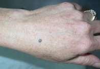
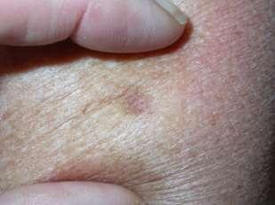
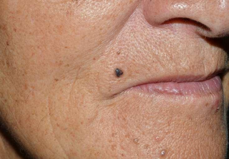
34 Solar Lentigo Sun induced freckle Associated with sun damage Harmless Can mimic melanoma The Skin Clinic Marlborough
35 Epidermis: Lentigo The Skin Clinic Marlborough

36 The Skin Clinic Marlborough

37 The Skin Clinic Marlborough

38 The Skin Clinic Marlborough

39 The Skin Clinic Marlborough
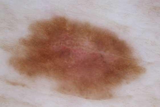
40 Seborrhoeic Keratoses Often referred to as senile warts But they occur in younger people They are not a viral infection Are not sun related Harmless They can mimic SCC s and Melanoma The Skin Clinic Marlborough
41 Epidermis: Seborrhoeic keratosis Senile wart, barnacle The Skin Clinic Marlborough

42 The Skin Clinic Marlborough

43 The Skin Clinic Marlborough

44 The Skin Clinic Marlborough

45 The Skin Clinic Marlborough

46 The Skin Clinic Marlborough
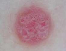
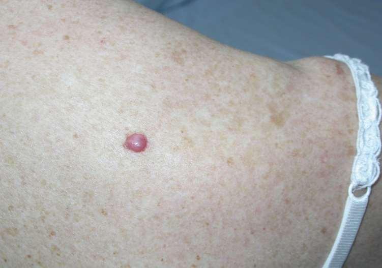
47 Haemangioma Benign growth of vasculature- forming locules. Harmless The Skin Clinic Marlborough
48 Dermis: Haemangioma The Skin Clinic Marlborough

49 The Skin Clinic Marlborough

50 The Skin Clinic Marlborough

51 The Skin Clinic Marlborough

52 The Skin Clinic Marlborough

53 The Skin Clinic Marlborough

54 The Skin Clinic Marlborough
55 Sebaceous Hyperplasia Overgrowth of sebaceous gland Can mimic BCC The Skin Clinic Marlborough
56 Sebaceous Hyperplasia Dermis: Mid to deep dermis as involves piloseb unit The Skin Clinic Marlborough

57 The Skin Clinic Marlborough

58 The Skin Clinic Marlborough

59 The Skin Clinic Marlborough

60 The Skin Clinic Marlborough

61 The Skin Clinic Marlborough

62 The Skin Clinic Marlborough

63 The Skin Clinic Marlborough

64 The Skin Clinic Marlborough

65 The Skin Clinic Marlborough

66 The Skin Clinic Marlborough

67 The Skin Clinic Marlborough

68 The Skin Clinic Marlborough
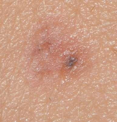
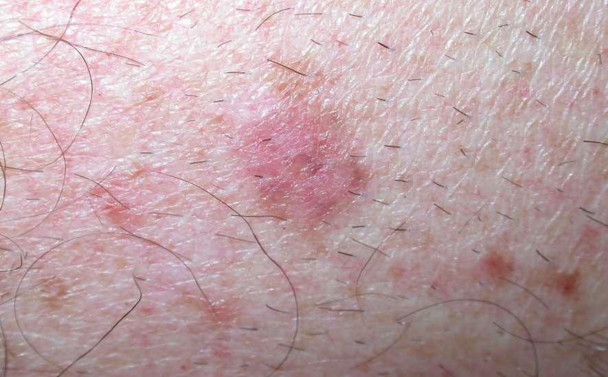
69 Moles or Naevi Multiple classification systems for moles Most commonly used is based on histopathology. Accurate classification can be problematic clinically. The Skin Clinic Marlborough
70 Junctional - confined to epidermis Compound - epidermal and dermal Intradermal - confined to dermis Blue - blue grey colour, dermis Dysplastic - can mimic melanoma but harmless Spitz.and others The Skin Clinic Marlborough
71 Epidermis: Junctional Naevus The Skin Clinic Marlborough
72 Epidermis and Dermis: Compound Naevus The Skin Clinic Marlborough
73 Dermis: Intradermal Naevus Blue Naevus The Skin Clinic Marlborough

74 Junctional Naevus Compound Naevus Intradermal Naevus Blue Naevus The Skin Clinic Marlborough

75 Spitz Naevus Dysplastic Naevus The Skin Clinic Marlborough
76 Dermatofibroma Fibrous lump deep in the dermis May have +ve pinch sign May be related to micro-trauma (eg insect bite or vaccination) The Skin Clinic Marlborough
77 Dermis: Dermatofibroma The Skin Clinic Marlborough
78 +ve Pinch Sign The Skin Clinic Marlborough


79 +ve Pinch Sign The Skin Clinic Marlborough


80 +ve Pinch Sign The Skin Clinic Marlborough
81 Psoriasis Epidermal Multiple red scaly plaques Extensor surfaces, scalp, trunk and limbs Face is usually spared The Skin Clinic Marlborough

82 The Skin Clinic Marlborough

83 The Skin Clinic Marlborough

84 The Skin Clinic Marlborough

85 The Skin Clinic Marlborough

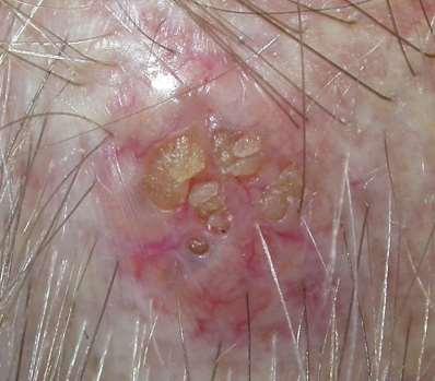
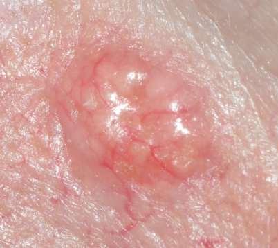
86 The Skin Clinic Marlborough

87 The Skin Clinic Marlborough
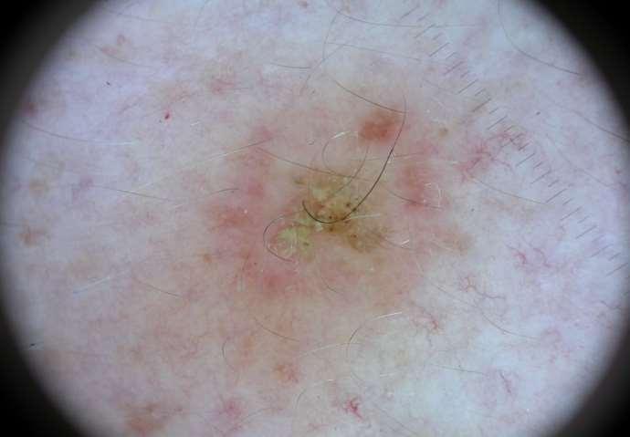
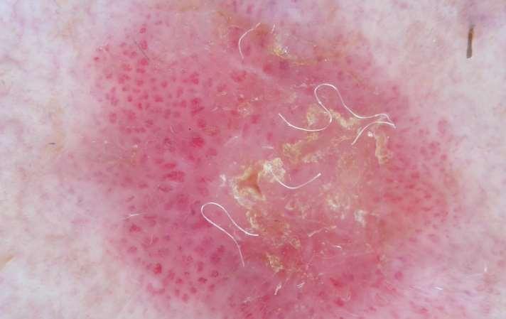
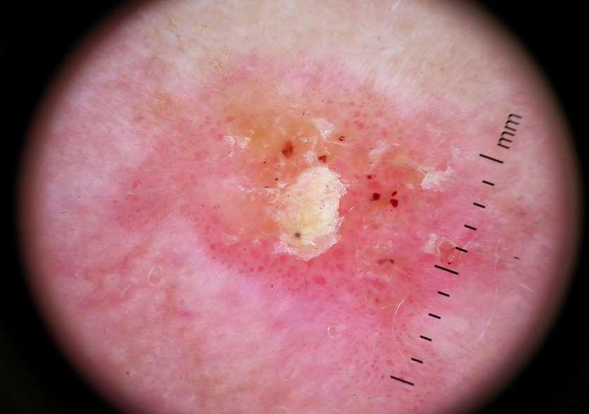
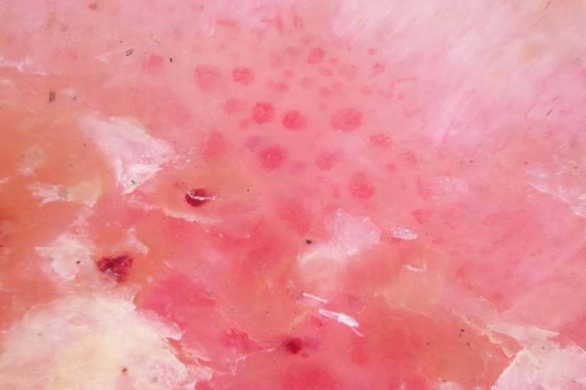
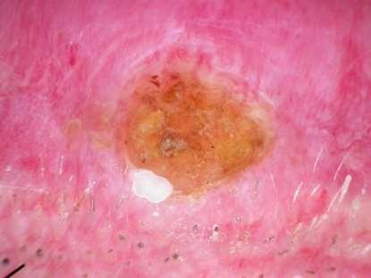
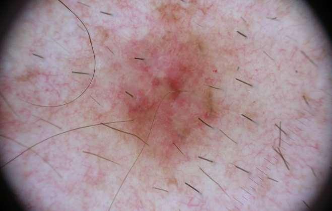
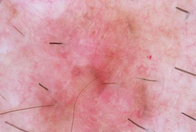
88 BCC s Common Skin Cancers SCC s Melanoma The Skin Clinic Marlborough

89 Common Skin Cancers Basal Cell Carcinoma - Superficial - Nodular - Aggressive - Micronodular - Sclerosing - Infiltrating The Skin Clinic Marlborough
90 Common Skin Cancers Squamous Cell Carcinoma Insitu Invasive The Skin Clinic Marlborough

91 Common Skin Cancers Melanoma - Insitu - Invasive - Metastatic The Skin Clinic Marlborough
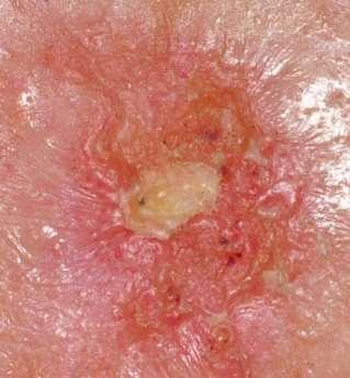
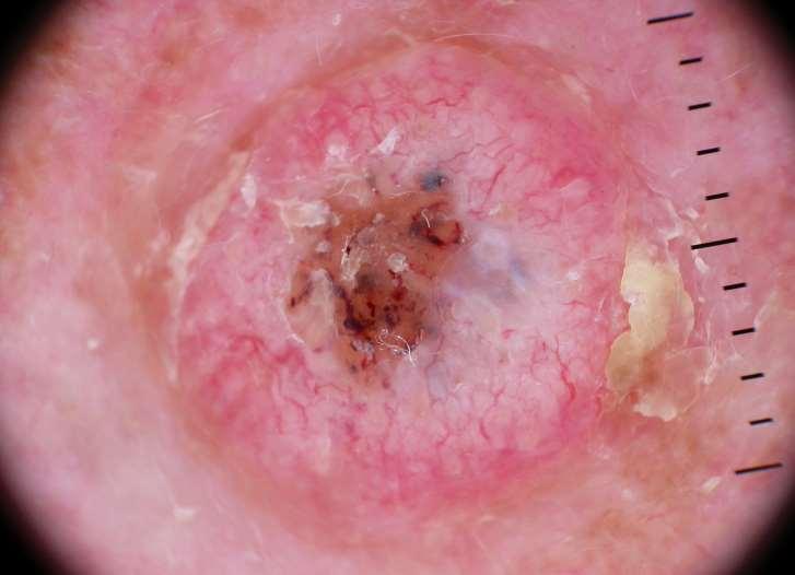
92 Common Skin Cancers The Skin Clinic Marlborough
93 Solar Keratosis / Actinic Keratosis Considered Pre-malignant AK vs SCCi
94 Epidermis AK lower 1/3 SCCi full thickness Dermis Invasive SCC SCC AK SCCi
95 Actinic Keratosis and SCC insitu AK and SCCi therefore have similar naked eye features Dermoscopically key differences Dot vessels, if any with AK Coiled vessels +/- dot vessels with SCCi
96
97
98

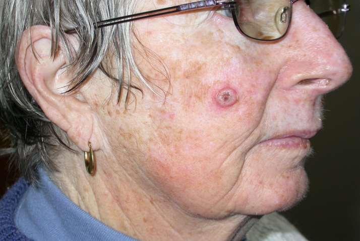
99 SCC insitu
100 Epidermis AK lower 1/3 SCCi full thickness SCCi AK

101 The Skin Clinic Marlborough

102 The Skin Clinic Marlborough

103 The Skin Clinic Marlborough

104 The Skin Clinic Marlborough

105 The Skin Clinic Marlborough
106 SCC insitu The Skin Clinic Marlborough
107
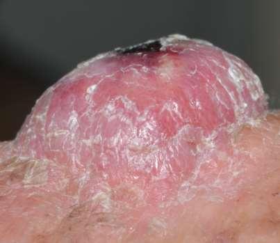
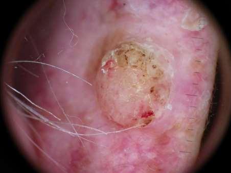
108
109 SCCi
110
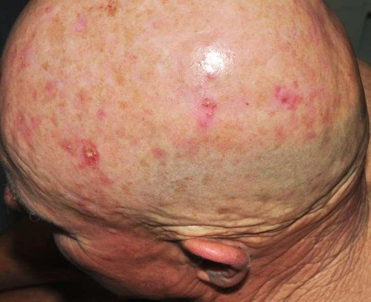
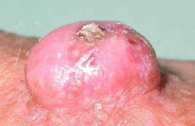
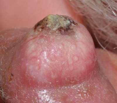
111
112
113 SCCi
114 Epidermis AK lower 1/3 SCCi full thickness Dermis Invasive SCC SCC AK SCCi
115 Well differentiated SCC SCC s are usually a naked eye diagnosis as there are very few useful dermoscopic signs. The Skin Clinic Marlborough

116 The Skin Clinic Marlborough

117 The Skin Clinic Marlborough
118 SCC can grow rapidly in the elderly immunocompromised patients Differential includes Keratoacanthoma Excision is indicated rather than observation The Skin Clinic Marlborough
119
120 wdscc

121 The Skin Clinic Marlborough

122 The Skin Clinic Marlborough
123
124

125 The Skin Clinic Marlborough

126 The Skin Clinic Marlborough


127 The Skin Clinic Marlborough

128 The Skin Clinic Marlborough

129 The Skin Clinic Marlborough

130 The Skin Clinic Marlborough

131 The Skin Clinic Marlborough

132 The Skin Clinic Marlborough

133 The Skin Clinic Marlborough
134 Common Skin Cancers The Skin Clinic Marlborough

135 The Skin Clinic Marlborough

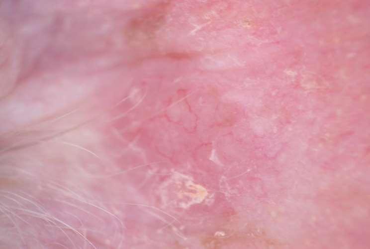
136 The Skin Clinic Marlborough
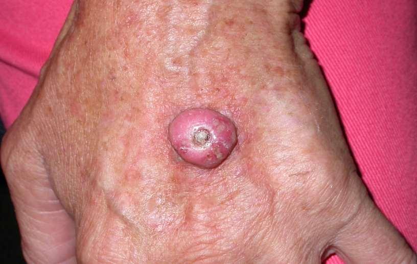
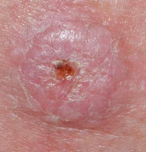
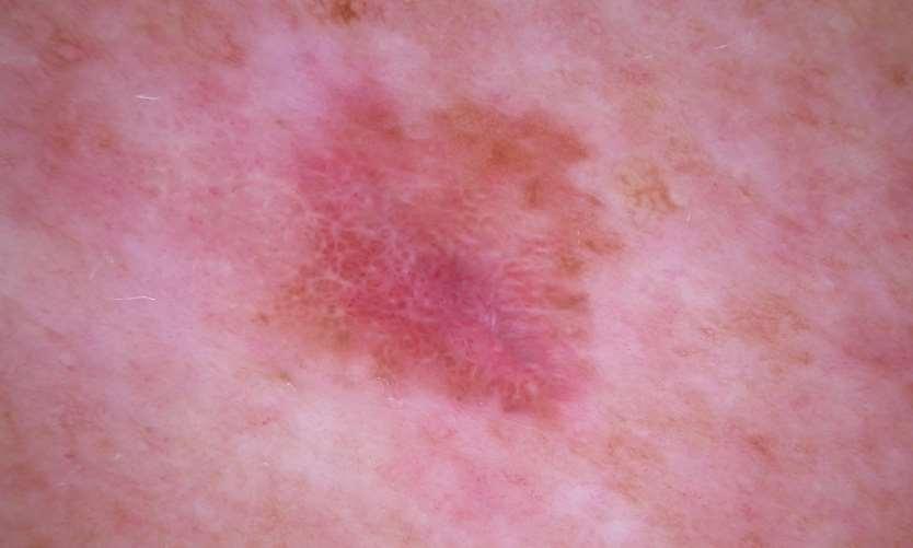
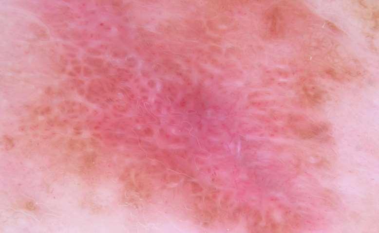
137
138 Dermis: Superficial BCC The Skin Clinic Marlborough

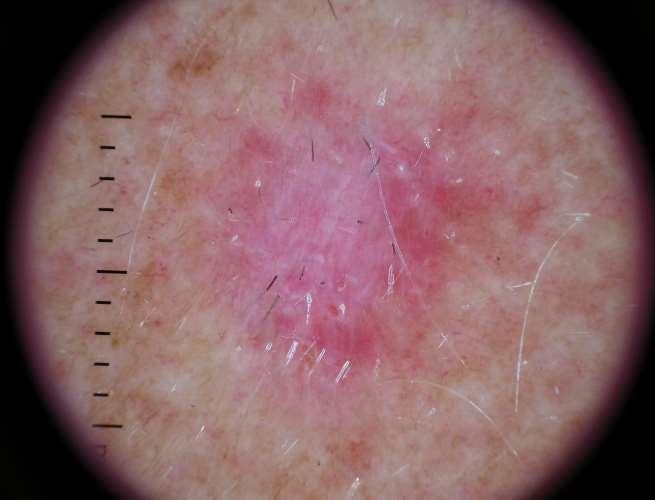
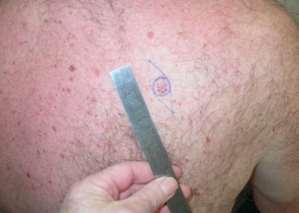
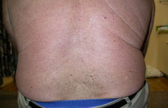
139 The Skin Clinic Marlborough

140 The Skin Clinic Marlborough

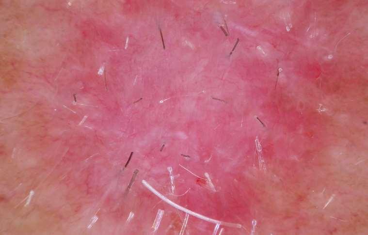
141 The Skin Clinic Marlborough
142 The Skin Clinic Marlborough

143 sbcc The Skin Clinic Marlborough


144 Nodular BCC The Skin Clinic Marlborough
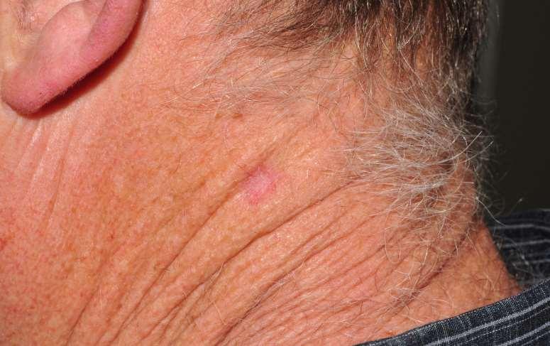
145 Dermis: Nodular BCC The Skin Clinic Marlborough

146 48 yr old woman, new lesion The Skin Clinic Marlborough

147 The Skin Clinic Marlborough

148 60 yr old woman, slow growing lesion, past 3 yrs. The Skin Clinic Marlborough

149 The Skin Clinic Marlborough
150
151
152 nbcc
153
154
155 mnbcc
156 The Skin Clinic Marlborough

157 The Skin Clinic Marlborough

158 The Skin Clinic Marlborough

159 The Skin Clinic Marlborough
160 Dermis: Infiltrating BCC The Skin Clinic Marlborough


161 The Skin Clinic Marlborough

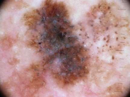
162 The Skin Clinic Marlborough

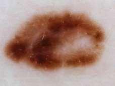
163 The Skin Clinic Marlborough

164 The Skin Clinic Marlborough

165 The Skin Clinic Marlborough
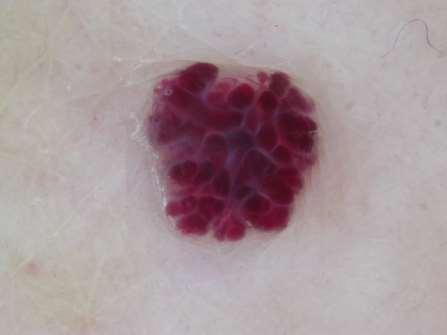
166
167

168 Pigmented BCC
169 Common Skin Cancers The Skin Clinic Marlborough
170 Epidermis: Melanoma insitu The Skin Clinic Marlborough

171 Melanoma Insitu The Skin Clinic Marlborough

172 The Skin Clinic Marlborough


173 2 Melanoma Insitu The Skin Clinic Marlborough

174 The Skin Clinic Marlborough

175 Melanoma Insitu The Skin Clinic Marlborough
176
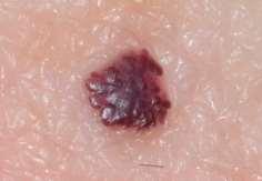
177
178 MIS
179
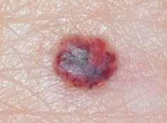
180
181 MIS

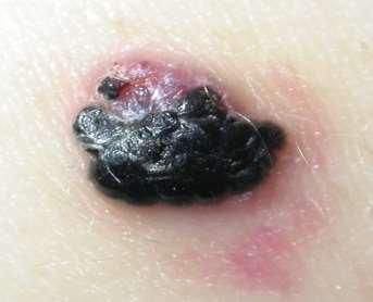
182 The Skin Clinic Marlborough

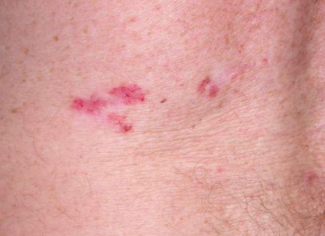
183 The Skin Clinic Marlborough
184 MELANOMA Breslow 0.5 mm The Skin Clinic Marlborough

185 The Skin Clinic Marlborough
186 The Skin Clinic Marlborough

187 Melanoma, Breslow 2.5 mm The Skin Clinic Marlborough
188

189 Nodular Melanoma 3.5 mm
190 Chronic ulceration Dermoscopy may not be helpful Biopsy to exclude malignancy Some chronic ulceration -> SCC (Marjolin ulcer) The Skin Clinic Marlborough

191 SCC The Skin Clinic Marlborough
192 Summary Common Skin Cancers have epidermal origin They can be difficult to differentiate from benign lesions Dermoscopy can help Biopsy chronic ulcers The Skin Clinic Marlborough

193 Thank you Dr. Mark Foley 28 Alfred St Blenheim Tel theskinclinic.co.nz
Skin lesions The Good and the Bad. Dr Virginia Hubbard Ipswich Hospital NHS Trust Barts and the London School of Medicine and Dentistry
 Skin lesions The Good and the Bad Dr Virginia Hubbard Ipswich Hospital NHS Trust Barts and the London School of Medicine and Dentistry Case 1 32 year old woman Australian Lesion on back New hair growing
Work Place Carcinogens Solar Radiation and Skin Cancer. November 2013 Dr Mark Foley
 Work Place Carcinogens Solar Radiation and Skin Cancer November 2013 Dr Mark Foley Overview Work place carcinogens and skin cancer Who is a risk? Screening and Self skin exam Common skin cancers Many work
IT S FUNDAMENTAL MY DEAR WATSON! A SHERLOCKIAN APPROACH TO DERMATOLOGY
 IT S FUNDAMENTAL MY DEAR WATSON! A SHERLOCKIAN APPROACH TO DERMATOLOGY Skin, Bones, and other Private Parts Symposium Dermatology Lectures by Debra Shelby, PhD, DNP, FNP-BC, FADNP, FAANP Debra Shelby,
Dermoscopy: Recognizing Top Five Common In- Office Diagnoses
 Dermoscopy: Recognizing Top Five Common In- Office Diagnoses Vu A. Ngo, DO Department of Family Medicine and Dermatology Choctaw Nation Health Services Authority Learning Objectives Introduction to dermoscopy
Basal cell carcinoma 5/28/2011
 Goal of this Presentation A practical approach to the diagnosis of cutaneous carcinomas and their mimics Thaddeus Mully, MD University of California San Francisco To review common non-melanoma skin cancers
Clinical characteristics
 Skin Cancer Fernando Vega, MD Seattle Healing Arts Clinical characteristics Precancerous lesions Common skin cancers ACTINIC KERATOSIS Precancerous skin lesions Actinic keratoses Dysplastic melanocytic
Dermoscopy-a BRIEF introduction
 Dermoscopy-a BRIEF introduction Aim of presentation -to tell you what dermoscopy is -to show some of what it can do -point the interested learner to further resources Overview of dermoscopy Dermoscopy
22/04/2015. Dermoscopy of Melanoma. Ilsphi Browne. Overview
 Dermoscopy of Melanoma Ilsphi Browne Overview The device Dermoscopic criteria (terminology) Colour Patterns Global features Local features Approach to diagnosing pigmented lesions Other uses in general
It can be helpful in some cases of actinic keratosis, Bowen s disease and squamous cell carcinoma
 Dermoscopy Introduction, Terminology and Structures (to be read in conjunction with the Diagnostic Dermoscopic Algorithm) Copyright to Cunliffe TP (Jan. 2017) All rights reserved Introduction Dermoscopy
Fundamentals of dermoscopy
 Fundamentals of dermoscopy Learning objectives Upon completion of this session, participants should be able to: describe the basic principles of dermoscopy identify features associated with pigmented and
Benign versus Cancerous Lesions How to tell the difference FMF 2014 Christie Freeman MD, CCFP, DipPDerm, MSc
 1 Benign versus Cancerous Lesions How to tell the difference FMF 2014 Christie Freeman MD, CCFP, DipPDerm, MSc Benign lesions Seborrheic Keratoses: Warty, stuck-on Genetics and birthdays Can start in late
Histopathology: skin pathology
 Histopathology: skin pathology These presentations are to help you identify, and to test yourself on identifying, basic histopathological features. They do not contain the additional factual information
Dermatopathology: The tumor is composed of keratinocytes which show atypia, increase mitoses and abnormal mitoses.
 Squamous cell carcinoma (SCC): A common malignant tumor of keratinocytes arising in the epidermis, usually from a precancerous condition: 1- UV induced actinic keratosis, usually of low grade malignancy.
Cutaneous Malignancies: A Primer COPYRIGHT. Marissa Heller, M.D.
 Cutaneous Malignancies: A Primer Marissa Heller, M.D. Associate Director of Dermatologic Surgery Department of Dermatology Beth Israel Deaconess Medical Center December 10, 2016 Skin Cancer Non-melanoma
Benign and malignant epithelial lesions: Seborrheic keratosis: A common benign pigmented epidermal tumor occur in middle-aged or older persons more
 Benign and malignant epithelial lesions: Seborrheic keratosis: A common benign pigmented epidermal tumor occur in middle-aged or older persons more common on the trunk; but extremities, head and neck are
LUMPS AND BUMPS: AN ORGANIZED APPROACH TO DIAGNOSIS AND MANAGEMENT
 LUMPS AND BUMPS: AN ORGANIZED APPROACH TO DIAGNOSIS AND MANAGEMENT Tammy P. Than, M.S., O.D., F.A.A.O. The University of Alabama at Birmingham / School of Optometry 1716 University Blvd. Birmingham, AL
Malignant non-melanocytic lesions
 Malignant non-melanocytic lesions Course C023: Fundamentals of Dermoscopy March 4, 2019, 11:20 AM - 11:50 PM Room: 146B Jason B. Lee, MD Professor & Vice Chair Director of Dermatopathology & Pigmented
July 2012 SKIN SURGERY SERVICE BRIEFING NOTES
 SKIN SURGERY SERVICE BRIEFING NOTES Introduction The WBoP PHO has an agreement with the BoP District Health Board to deliver the Skin Surgery Service. The current period will expire on 30 June 2012 and
Histopathological and SIAscopic Correlation of Pigmented Skin Lesions
 Histopathological and SIAscopic Correlation of Pigmented Skin Lesions Professor Sujatha Fernando MBBS(Hon), MSc(London, Distinction), FRSTM&H, FRCPA, FIAC, FACTM Senior Consultant in Anatomical Pathology,
50 interactive dermoscopic case discussions Dr Stephen Hayes
 50 interactive dermoscopic case discussions Dr Stephen Hayes Annotations will be found on your memory drive, as will 100 case discussions and other learning material Melanoma 2mm thick Ugly duckling-one
VACAVILLE DERMATOLOGY
 Connecting the Dots on those Spots NANDAN V. KAMATH, M.D. VACAVILLE DERMATOLOGY Sources All of the photos were taken with permission from the Dermnet NZ website - Dermnet New Zealand after communicating
Lumps and Bumps: An Organized Approach to Diagnosis and Management. Disclosure. Introduction. References. Structure of Skin.
 Lumps and Bumps: An Organized Approach to Diagnosis and Management Nothing to disclose Disclosure Tammy Pifer Than, MS, OD, FAAO Carl Vinson VAMC tammythan@bellsouth.net References Fitzpatrick's Color
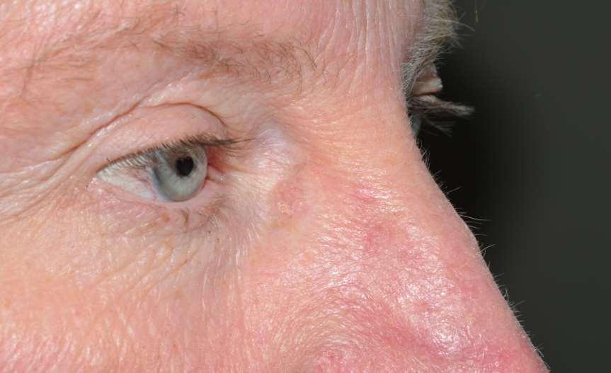
Periocular Malignancies
 Periocular Malignancies Andrew Gurwood, O.D., F.A.A.O., Dipl. Marc Myers, O.D., F.A.A.O. Drs. Myers and Gurwood have no financial interests to disclose. Course Description Discussion of the most common
Pathology of the skin. 2nd Department of Pathology, Semmelweis University
 Pathology of the skin 2nd Department of Pathology, Semmelweis University Histology of the skin Epidermis: Stratum corneum Stratum granulosum Stratum spinosum Stratum basale Dermis: papillary and reticular
Living Beyond Cancer Skin Cancer Detection and Prevention
 Living Beyond Cancer Skin Cancer Detection and Prevention Cutaneous Skin Cancers Identification Diagnosis Treatment options Prevention What is the most common cancer in people? What is the most common
Skin Cancer. Dr Elizabeth Ogden Associate Specialist in Dermatology East and North Herts Dr Elizabeth Ogden
 Skin Cancer Dr Elizabeth Ogden Associate Specialist in Dermatology East and North Herts 13.10.16 Skin Cancer Melanoma mole cancer - is a true cancer which can metastasize and kill Non Melanoma skin cancer
Mole mapping and monitoring. Dr Stephen Hayes. Associate Specialist in Dermatology, University Hospital Southampton
 Mole mapping and monitoring Dr Stephen Hayes Associate Specialist in Dermatology, University Hospital Southampton Outline of presentation The melanoma epidemic Benefits of early detection Risks of the
SKIN CANCER. Most common cancer diagnosis 40% of all cancers
 SKIN CANCER Most common cancer diagnosis 40% of all cancers OBJECTIVES Review common and uncommon cancers of the skin. Special emphasis on melanoma and dysplastic nevus Review pathology/tnm/staging, which
Know who is at risk: LOOK! for ABCDs, rapidly changing lesions, do a biopsy when indicated
 Lindy P. Fox, MD Assistant Professor Director, Hospital Consultation Service Department of Dermatology University of California, San Francisco Applies to adults without history of malignancy or premalignant
Know who is at risk: LOOK! for ABCDs, rapidly changing lesions, do a biopsy when indicated
 Lindy P. Fox, MD Associate Professor Director, Hospital Consultation Service Department of Dermatology University of California, San Francisco Applies to adults without history of malignancy or premalignant
Large majority caused by sun exposure Often sun exposure before age 20 Persons who burn easily and tan poorly are at greatest risk.
 Basics of Skin Cancer Detection and Treatment of Non- Melanoma Skin Cancers Large majority caused by sun exposure Often sun exposure before age 20 Persons who burn easily and tan poorly are at greatest
Hauora Maori. Map. Non-Melanoma Skin Lesion. Clinically Seborrhoeic Keratosis. Management Options. Punch Biopsy. Refer to Public Skin Lesion Service
 Care map information Information resources for patients and carers Updates to this Care Map Hauora Maori Pacific Skin lesion history Clinical examination and diagnostic test Suspicious of Melanoma Non-Melanoma
Dermoscopy in everyday practice. What and Why? When in doubt cut it out? Trilokraj Tejasvi MD
 Dermoscopy in everyday practice Trilokraj Tejasvi MD Assistant Professor, Department of Dermatology, Director Teledermatology services, University of Michigan, Faculty Associate, GLOBAL REACH, Michigan
Dr Stephen Hayes Associate Specialist in Dermatology University Hospital Southampton
 South East Dermatology Transformation and Sustainability Network Guildford, 19 th April 2018 Dermoscopy as an effective skin lesion triage tool in GP surgeries Dr Stephen Hayes Associate Specialist in
Dermatological Manifestations in the Elderly. Sanjay Siddha Staff Dermatologist UHN & MSH
 Dermatological Manifestations in the Elderly Sanjay Siddha Staff Dermatologist UHN & MSH Disclosure No actual or potential conflicts of interest or commercial relationships to declare Objectives Recognize
Richard Turner Consultant Dermatologist
 Old Problems & New Treatments Photo Album by Administrator Richard Turner Consultant Dermatologist Plan for tonight? Refresher on SCC and solar keratosis How to distinguish the two Classic therapy than
Regression 2/3/18. Histologically regression is characterized: melanosis fibrosis combination of both. Distribution: partial or focal!
 Regression Margaret Oliviero MSN, ARNP Harold S. Rabinovitz MD Histologically regression is characterized: melanosis fibrosis combination of both Distribution: partial or focal! Dermatoscopic terminology
Thursday 21 st August Skin Problems
 Thursday 21 st August 2014 Skin Problems Skin Problems The Sun and the Skin Sun Damage Recognising the early signs of skin cancer The Big 3 inflammatory condi=ons Acne & Rosacea Eczema (Including Seborrhoeic
Dr Amanda Oakley. Dermatologist Dept of Dermatology, Health Waikato Adjunct Associate Professor, Waikato Clinical Campus
 Dr Amanda Oakley Dermatologist Dept of Dermatology, Health Waikato Adjunct Associate Professor, Waikato Clinical Campus 14:00-16:00 WS #14: Dermoscopy Part 1 Skin Lesions and Dermatoscopy 16 August 2018
Disclosure. Objectives. PAFP CME Conference Lou Mancano MD, FAAFP Reading Health System November 18, 2016
 PAFP CME Conference Lou Mancano MD, FAAFP Reading Health System November 18, 2016 1 Disclosure The speaker has no conflict of interest, financial agreement, or working affiliation with any group or organization.
MECHANISMS OF HUMAN DISEASE: LABORATORY SESSION PATHOLOGY OF THE SKIN LAB. Friday, February 12, :30 am 11:00 am
 MECHANISMS OF HUMAN DISEASE: LABORATORY SESSION PATHOLOGY OF THE SKIN LAB Friday, February 12, 2012 9:30 am 11:00 am FACULTY COPY GOALS: Describe the basic clinical and morphologic features of various
Skin Malignancies. Presented by Dr. Douglas Paauw
 Skin Malignancies Presented by Dr. Douglas Paauw Disclosure: Dr. Paauw has no significant financial interest in any of the products or manufacturers mentioned. How Common Is Skin Cancer? *½ of all White
Desmoplastic Melanoma R/O BCC. Clinical Information. 74 y.o. man with lesion on left side of neck r/o BCC
 R/O BCC Sabine Kohler, M.D. Professor of Pathology and Dermatology Dermatopathology Service Stanford University School of Medicine Clinical Information 74 y.o. man with lesion on left side of neck r/o
INTRODUCTION HOUSEKEEPING June 11 th Dr John Adams Dermatologist/Dermoscopist MOLEMAP NZ/Australia MOLESAFE USA
 INTRODUCTION HOUSEKEEPING June 11 th 2015 Dr John Adams Dermatologist/Dermoscopist MOLEMAP NZ/Australia MOLESAFE USA Program Skin cancer statistics. Dermoscopy description and usefulness. Patient /lesion
Lid Lesions: Relax or Refer
 Lid Lesions: Relax or Refer Blair Lonsberry, MS, OD, MEd., FAAO Professor of Optometry Pacific University College of Optometry blonsberry@pacificu.edu Agenda Benign vs. Malignant lesions Benign Eyelid
Diagnostics guidance Published: 11 November 2015 nice.org.uk/guidance/dg19
 VivaScope 1500 and 3000 imaging systems for detecting skin cancer lesions Diagnostics guidance Published: 11 November 2015 nice.org.uk/guidance/dg19 NICE 2018. All rights reserved. Subject to Notice of
Cutaneous Adnexal Tumors
 Cutaneous Adnexal Tumors Lesions with Predominant Follicular Differentiation Special Emphasis on Basal Cell Carcinoma 2014-04-01 Prof. Dr. med. Katharina Glatz Pathologie Cutaneous Adnexal Tumors Hair
Aspects of skin cancer diagnosis in clinical practice
 Aspects of skin cancer diagnosis in clinical practice Ahnlide, Ingela 2015 Document Version: Publisher's PDF, also known as Version of record Link to publication Citation for published version (APA): Ahnlide,
Pathology. Skin Tumor. Bayan N. Mohammad 15/10/2015. Mohammad al-orjani. Page 0 of 23
 #7 35 Pathology Skin Tumor Bayan N. Mohammad 15/10/2015 Mohammad al-orjani Page 0 of 23 بسم هللا الرحمن الرحيم GREETINGS This lecture is about skin tumors, all the slides are included and every slide will
Premalignant skin tumours
 Chapter 14: Premalignant skin tumours page: 434 Premalignant skin tumours page: 435 Solar keratoses (senile keratoses) Raised red and well-defined plaques with a rough surface covered in scales of varying
Technicians & Nurses Program
 ASCRS ASOA Symposium & Congress Technicians & Nurses Program May 6-10, 2016 New Orleans Evaluation and Treatment of Eyelid Malignancies Richard C. Allen MD PhD FACS Professor Section of Ophthalmology Dept.
Prediction without Pigment: a decision algorithm for non-pigmented skin malignancy
 DERMATOLOGY PRACTICAL & CONCEPTUAL www.derm101.com Prediction without Pigment: a decision algorithm for non-pigmented skin malignancy Cliff Rosendahl 1, Alan Cameron 1, Philipp Tschandl 2, Agata Bulinska
Epithelial Cancer- NMSC & Melanoma
 Epithelial Cancer- NMSC & Melanoma David Chin MB, BCh, BAO, LRCP, LRCS (Ireland) MCh(MD), PhD (UQ), FRCS, FRACS (Plast) Plastic & Reconstructive Surgeon Visiting Scientist Melanoma Genomic Group & Drug
Accuracy of Clinical Skin Tumour Diagnosis in a Dermatological Setting.
 Accuracy of Clinical Skin Tumour Diagnosis in a Dermatological Setting. Ahnlide, Ingela; Bjellerup, Mats Published in: Acta Dermato-Venereologica DOI: 10.2340/00015555-1560 2013 Link to publication Citation
Glenn D. Goldman, MD. University of Vermont Medical Center. University of Vermont College of Medicine
 Glenn D. Goldman, MD University of Vermont Medical Center University of Vermont College of Medicine Recognize and identify the main types of skin cancer and their precursors Identify and understand new
Fluorescence spectroscopy and microscopy of cutaneous tumours correlation between micro- and macro- spectral measurements
 Fluorescence spectroscopy and microscopy of cutaneous tumours correlation between micro- and macro- spectral measurements E. Borisova 1, L. Avramov 1, M. Lomova 2, O. Semyachkina-Glushkovskaya 2, D. Gorin
Skin Cancer A Personal Approach. Dr Matthew Strack Dunedin New Zealand
 Skin Cancer A Personal Approach Dr Matthew Strack Dunedin New Zealand Outline Dermoscopy Instruments and setup Photochemosurgery Clinical Aim: Leave with 2-3 ideas JLE Benign Junctional Nevus Management
David B. Troxel, MD. Common Medicolegal Situations: Misdiagnosis of Melanoma
 Common Medicolegal Situations: Misdiagnosis of Melanoma David B. Troxel, MD Medical Director, The Doctors Company, Napa, California Clinical Professor Emeritus, University of California at Berkeley Past
Nonmelanoma skin cancers
 Skin cancer Philip Clarke Nonmelanoma skin cancers Treatment options Background Australia has one of the highest skin cancer rates in the world. Early detection and treatment of skin cancer is vital to
Periocular skin cancer
 Periocular skin cancer Information for patients Skin cancer involving the skin of the eyelid or around the eye is called a periocular skin cancer. Eyelid skin cancers occur most often on the lower eyelid,
Clinical Pathological Conference. Malignant Melanoma of the Vulva
 Clinical Pathological Conference Malignant Melanoma of the Vulva History F/48 Chinese Married Para 1 Presented in September 2004 Vulval mass for 2 months Associated with watery and blood stained discharge
Skin & Subcutaneous Tissues Dr. Tarek Said
 Skin & Subcutaneous Tissues Dr. Tarek Said Professor of Plastic Surgery Cairo University Lipoma -Solitary / Multiple / Diffuse -Subcutaneous -Subfacial (Palmar / Plantar) -Intermuscular -Submucous -Subserous
Describe the functions of the vertebrate integumentary system. Discuss the structure of the skin and how it relates to function.
 Chapter 5 Describe the functions of the vertebrate integumentary system. Discuss the structure of the skin and how it relates to function. Explain the basis for different skin colors. Describe the structure
Glenn D. Goldman, MD. Fletcher Allen Health Care. University of Vermont College of Medicine
 Glenn D. Goldman, MD Fletcher Allen Health Care University of Vermont College of Medicine Recognize and identify the main types of skin cancer Understand how and why Mohs surgery is utilized for the treatment
Identifying Skin Cancer. Mary S. Stone MD Professor of Dermatology and Pathology University of Iowa Carver College of Medicine March, 2018
 Identifying Skin Cancer Mary S. Stone MD Professor of Dermatology and Pathology University of Iowa Carver College of Medicine March, 2018 American Cancer Society web site Skin Cancer Melanoma Non-Melanoma
المركب النموذج--- سبيتز وحمة = Type Spitz's Nevus, Compound SPITZ NEVUS 1 / 7
 SPITZ NEVUS 1 / 7 Epidemiology An annual incidence rate of 1.4 cases of Spitz nevus per 100,000 individuals has been estimated in Australia, compared with 25.4 per 100,000 individuals for cutaneous melanoma
I have a skin lump doc! What s next? 12 th August 2017 Dr. Sue-Ann Ho Ju Ee
 I have a skin lump doc! What s next? 12 th August 2017 Dr. Sue-Ann Ho Ju Ee Some thoughts Is this skin cancer? How common is this? How likely is this in this patient? What happens next if it s something
Service Specification: CPO Skin Lesions
 Service Specification: CPO Skin Lesions This pathway description needs to be read in conjunction with the CPO Admin Service Specifications. 1. Outcomes Framework The outcomes sought from this service are
Non-Melanocytic Pattern Dermoscopy
 Non-Melanocytic Pattern Dermoscopy I have no conflicts of interest to disclose Except that I LOVE dermoscopy Michelle Tarbox, MD Assistant Professor of Dermatology and Dermatopathology Texas Tech University
Malignant tumors of melanocytes: Part 1. Deba P Sarma, MD., Omaha
 Malignant tumors of melanocytes: Part 1 Deba P Sarma, MD., Omaha The melanocytic tumor is one of the most difficult and confusing areas in Dematopathology. It is true that most (95%) of such lesions are
Doctors of Optometry Course Notes
 Doctors of Optometry Course Notes OD19 1CE COPE: 43871-AS Eyelid Lumps and Bumps Sunday, February 26, 2017 2:40 pm 3:30 pm Regency C 3 rd Floor Presenter: Blair Lonsberry, OD, FAAO Dr. Lonsberry is a Full
PATHOLOGY OF THE SKIN 2. Tumours of the skin
 PATHOLOGY OF THE SKIN 2. Tumours of the skin Máirín E. McMenamin MB MRCPI FRCPath Dip (Dermatopathol) RCPath St. James s Hospital and University of Dublin, Trinity College Tumour (Neoplasia) Benign or
Your Skin. Section 14.2 Your Skin, Hair, and Nails
 Your Skin The skin covers and protects the body from injury, infection, and water loss. The skin also helps to regulate body temperature and gathers information from the environment. Protection The skin
Minor Surgery. Using the Limbs & Things skin pads and jig. Skin Pad Jig Mk 3. Skin Pad Jig Strap. 44 Minor Surgery
 Using the Limbs & Things skin pads and jig The Skin Pad Jig can be used initially on a work surface for ease of learning, and can then be attached to the body of a fellow trainee to practise these skills,
Non-melanocytic Patterns
 Non-melanocytic Lesions Non-melanocytic Patterns Michelle Tarbox, MD Assistant Professor of Dermatology and Dermatopathology Texas Tech University Health Sciences Center 2018 Seborrheic keratoses Acanthotic
Skin cancer and benign lesions
 How to Treat PULL-OUT SECTION www.australiandoctor.com.au Complete How to Treat quizzes online (www.australiandoctor.com.au/cpd) to earn CPD or PDP points. Keratoacanthoma. inside Epidemiology and clinical
Identifying Benign and Malignant Skin Lesions. No Disclosures. Common Benign Lesions. Benign Lesions 2/25/2018. Stucco Keratoses.
 Dermatology in Primary Care Identifying Benign and Malignant Skin Lesions Christy Quire Baker, APRN, FNP-BC, DCNP Dermatology Certified Nurse Practitioner No Disclosures Common Benign Lesions Seborrheic
This is a repository copy of Easily missed? Amelanotic melanoma. White Rose Research Online URL for this paper:
 This is a repository copy of Easily missed? Amelanotic melanoma. White Rose Research Online URL for this paper: http://eprints.whiterose.ac.uk/127789/ Version: Accepted Version Article: Muinonen-Martin,
Diagnosis of Lentigo Maligna Melanoma. Steven Q. Wang, M.D. Memorial Sloan-Kettering Cancer Center Basking Ridge, NJ
 Diagnosis of Lentigo Maligna Melanoma Steven Q. Wang, M.D. Memorial Sloan-Kettering Cancer Center Basking Ridge, NJ Conflict of Interest: None Topics Epidemiology and Natural History Clinical and Histologic
Associate Professor Amanda Oakley. Professor H. Peter Soyer. Academic Dermatologist The University of Queensland Brisbane. Dermatologist Hamilton
 Associate Professor Amanda Oakley Dermatologist Hamilton Professor H. Peter Soyer Academic Dermatologist The University of Queensland Brisbane 8:30-10:30 WS #3: Dermoscopy Workshop Part 1 11:00-13:00 WS
The GP s approach to the patient who is worried about sun, skin and moles. Dr Stephen Hayes
 The GP s approach to the patient who is worried about sun, skin and moles Dr Stephen Hayes Associate Specialist in Dermatology, University Hospital Southampton Dr Stephen Hayes DECLARATION OF INTERESTS
Dermoscopy of non-pigmented skin lesions: a literature review
 Hong Kong J. Dermatol. Venereol. (2017) 25, 13-21 Review Article Dermoscopy of non-pigmented skin lesions: a literature review S Thomas, X Li, HP Soyer In this article, we will review benchmark dermoscopic
MECHANISMS OF HUMAN DISEASE: LABORATORY SESSION PATHOLOGY OF THE SKIN LAB. Friday, February 13, :30 am 11:00 am
 MECHANISMS OF HUMAN DISEASE: LABORATORY SESSION PATHOLOGY OF THE SKIN LAB Friday, February 13, 2009 9:30 am 11:00 am FACULTY COPY GOALS: Describe the basic clinical and morphologic features of various
Revised Pattern Analysis: a method for the accurate diagnosis of pigmented skin lesions
 Dermatoscopy for Students A concise outline of: Revised Pattern Analysis: a method for the accurate diagnosis of pigmented skin lesions And Chaos and Clues: a decision algorithm for routine practice to
6/17/2018. Breaking Bad (Part 1) Dermoscopy of Brown(ish) Things. Bad?
 Dermoscopy of Brown(ish) Things. Bad?") Breaking Bad (Part 1) Dermoscopy of Brown(ish) Things Jennie T. Clarke, MD ssociate Professor of Dermatology University of Utah School of Medicine Bad? 1 Brown(ish) Things Bad Melanoma Pigmented basal
Breaking Bad (Part 1) Dermoscopy of Brown(ish) Things Jennie T. Clarke, MD ssociate Professor of Dermatology University of Utah School of Medicine Bad? 1 Brown(ish) Things Bad Melanoma Pigmented basal
Learning Objectives. Tanning. The Skin. Classic Features. Sun Reactive Skin Type Classification. Skin Cancers: Preventing, Screening and Treating
 Learning Objectives Skin Cancers: Preventing, Screening and Treating Robert A. Baldor, MD, FAAFP Professor, Family Medicine & Community Health University of Massachusetts Medical School Distinguish the
They can develop anywhere on the skin and also inside the mouth. They can develop in normal skin or where there is an existing skin mole.
 What are malignant melanomas? Malignant melanomas are one type of skin cancer. They can develop anywhere on the skin and also inside the mouth. They can develop in normal skin or where there is an existing
Exenteration. Introduction. The skin. Epidermal malignancies 8/3/2017. Neglected basal cell carcinoma
 Jeremiah Tao, MD, FACS Director, Oculoplastic and Orbital Surgery Associate Professor, UC Irvine Neglected basal cell carcinoma Exenteration Introduction Chief question with any eyelid lesion: Suspicious
Appendix : Dermoscopy
 Go Back to the Top To Order, Visit the Purchasing Page for Details APP Appendix : Dermoscopy Dermoscopy, also known as dermatoscopy, epiluminoscopy and epiluminescent microscopy, is an effective non-invasive
Integumentary System (Skin) Unit 6.3 (6 th Edition) Chapter 7.3 (7 th Edition)
 Unit 6.3 (6 th Edition) Chapter 7.3 (7 th Edition)") Integumentary System (Skin) Unit 6.3 (6 th Edition) Chapter 7.3 (7 th Edition) 1 Learning Objectives Identify the major components (anatomy) of skin Differentiate between the two types of skin glands Explain
Integumentary System (Skin) Unit 6.3 (6 th Edition) Chapter 7.3 (7 th Edition) 1 Learning Objectives Identify the major components (anatomy) of skin Differentiate between the two types of skin glands Explain
Skin Malignancies Non - Melanoma & Melanoma Marilyn Ng, MD Dept. of Surgery M&M Conference Downstate Medical Center July 19, 2012
 Skin Malignancies Non - Melanoma & Melanoma Marilyn Ng, MD Dept. of Surgery M&M Conference Downstate Medical Center July 19, 2012 Case Presentation 57 yo man with 3 month hx of a nonhealing < 1 cm right
Skin Cancer of the Nose: Common and Uncommon
 Skin Cancer of the Nose: Common and Uncommon Mark Russell, M.D. Associate Professor of Dermatology, Otolaryngology, and Pathology University of Virginia Objectives Review clinical presentations of select
Cover Page. The handle holds various files of this Leiden University dissertation.
 Cover Page The handle http://hdl.handle.net/1887/22172 holds various files of this Leiden University dissertation. Author: Rhee, Jasper Immanuel van der Title: Clinical characteristics and management of
Malignant Melanoma Early Stage. A guide for patients
 This melanoma patient brochure is designed to help educate melanoma patients and their caregivers. It was developed under the guidance of Dr. Michael Smylie, Professor, Department of Oncology, University
Dermatology for the PCP Deanna G. Brown, MD, FAAD Susong Dermatology Consulting Staff at CHI Memorial
 Dermatology for the PCP Deanna G. Brown, MD, FAAD Susong Dermatology Consulting Staff at CHI Memorial Cutaneous Oncology for the PCP Deanna G. Brown, MD, FAAD Susong Dermatology Consulting Staff at CHI
Disclosures. I have no conflicts of interest to disclose
 Disclosures I have no conflicts of interest to disclose Lindy P. Fox, MD Associate Professor Director, Hospital Consultation Service Department of Dermatology University of California, San Francisco 2
Dermoscopy. Synonyms. Dermoscopy. Definition. Dermoscopy opens up a world of colour and structure that can t be seen with the naked eye
 Synonyms Dermoscopy Australasian College of Dermatologists G.P Training Module Dermoscopy Dermatoscopy Epiluminescence microscopy Skin surface microscopy Incident light microscopy Oil immersion microscopy
JAM ACAD DERMATOL VOLUME 76, NUMBER 2. Research Letters 351
 JAM ACAD DERMATOL Research Letters 351 Standard step sectioning of skin biopsy specimens diagnosed as superficial basal cell carcinoma frequently yields deeper and more aggressive subtypes To the Editor:
Disclosures. Melanoma and Non melanoma Skin Cancer: What You Need to Know. I have no conflicts of interest to disclose
 Disclosures Melanoma and Non melanoma Skin Cancer: What You Need to Know I have no conflicts of interest to disclose Lindy P. Fox, MD Associate Professor Director, Hospital Consultation Service Department