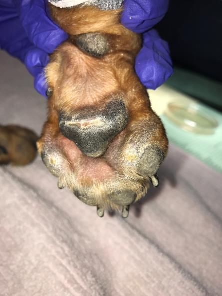
Signalment: 2-year-old male castrated Chihuahua beagle mix. Presenting complaint: Areas of alopecia and ulcers/erosions. Figure 1. Figure 2.
|
|
|
- Anne Wade
- 5 years ago
- Views:
Transcription


1 Signalment: 2-year-old male castrated Chihuahua beagle mix. Presenting complaint: Areas of alopecia and ulcers/erosions Figure 1. Figure 2.

2 Figure 3. Figure 4. Figure 5.

 for flea/tick control and ivermectin (Heartgard ) for heartworm prevention.")
3 Figure 6. Figure 7. Clinical history: The patient presented for evaluation of sudden worsening of skin lesions over a one week period. An initial area of alopecia was noted on the top of the patient s head and right hind limb, and mild crust on his ear tips starting 2 months ago. At that time skin biopsies were submitted by the referring veterinarian and described a mild eosinophilic perivascular dermatitis. The patient was initially treated with cefpodoxime with no resolution. The patient receives fluralaner (Bravecto ) for flea/tick control and ivermectin (Heartgard ) for heartworm prevention. The patient eats beef flavored Purina small breed, although he was recently switched to this from Beneful one to two months ago. The patient lives with 1 other dog with no skin lesions. The patient s vaccination status [rabies and DA2PP (distemper virus, adenovirus, parainfluenza virus, and parvovirus) is current.
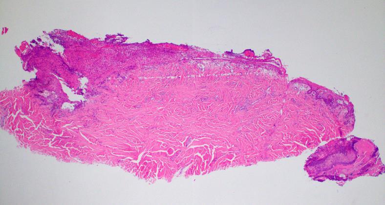
4 Question: What is the most likely cause/etiology? Answer: A) Food allergy B) Leishmania C) Vaccine reaction (Rabies, Distemper) D) Autoimmune E) Cold-agglutinin disease Clinical presentation: Over the dorsal aspect of the calvarium is an approximately 4-5 cm round area of alopecia and hyperpigmentation. Alopecia with mild erosion and crusting is present at mucocutaneous junctions (especially along lip commissures). Periocular regions are swollen, with hypotrichosis and mild moist erosion with crusting. Along the dorsal muzzle is hyperpigmentation with crust. Alar folds are slightly crusted with hypopigmentation. Ventrally along the external nares on the left side is an area of hypopigmentation that appears slightly swollen. Bilaterally at distal tip of concave pinna are approximately 2-3 cm symmetrical full thickness ulcers with surrounding hyperpigmentation. Pinnal margins have mild crust/scale. The dorsal pedal surface has patchy alopecia with mild scale and underlying hyperpigmentation. Hocks and elbows are alopecic hyperpigmented with fine scale. On the ventral aspect of paw pads are fine pinpoint to linear indurated areas of scale and hypopigmentation. Along the right lateral thigh there is an area of moderate scale and patchy hyperpigmentation. Histopathologic description: Four samples are examined with a serocellular crust of neutrophils, cell debris and serum. The samples show similar features with a varying degree of inflammation. The least inflamed haired skin sample has features typical of ischemic dermatopathy. Hair follicles are atrophied (some faded), and the dermal collagen and vascular tunics of small capillaries are slightly smudged. There is interstitial mucin deposition, small foci of perivascular hemorrhage, as well as scattered macrophages and neutrophils. The epidermis is mildly hyperplastic, hyperpigmented, and has a cell poor interface change. There is orthokeratotic hyperkeratosis. A second skin sample has these features, along with more prominent acanthosis and a dense neutrophilic infiltrate in the superficial dermis. In the smaller punch biopsies, there is dermoepidermal clefting with ulcers and fibrinosuppurative inflammation. Regionally, the epidermis shows thinning and squamatization with a more prominent hydropic change in the basal layer. The crust contains serum, necrotic granulocytes, bacteria, neutrophils, pigmented fungi, and a portion of an insect.


5 Figure 8. Figure 9. Morphologic diagnosis CELL-POOR INTERFACE DERMATITIS WITH FOLLICULAR ATROPHY (ISCHEMIC DERMATOPATHY) AND SUPERFICIAL NEUTROPHILIC DERMATITIS WITH ULCER AND CRUST Comments: In this case, vaccine-associated generalized ischemic dermatopathy was considered most likely given the history of vaccination 3 months prior and localized lesion at a known vaccine site.
6 Ischemic dermatopathy is a histologic diagnosis that typifies a number of different clinical syndromes with common features and a wide range of possible etiologies. Histologic features, including follicular atrophy, collagen smudging and cell poor interface pattern (with or without clefting of the basement membrane zone) are thought to represent a chronic oxygen deficient state of the affected tissue. A large number of cases demonstrate a mild perivascular to interstitial lymphoplasmacytic dermatitis and more inflamed lesions may be ulcerated with suppurative inflammation. Rarely, vasculitis may be noted, although some cases may have changes consistent with vascular atrophy. Muscle fibers, if present, may be atrophied. (Gross, 2005) Without a complete history and clinical signs, the various syndromes are indistinguishable histologically, thus some effort has been made to group the diseases based on clinical features. Primary ischemic diseases have previously been grouped into five subtypes; Canine familial dermatomyositis (FDM) {see November 2017 COM}, vaccine associated ischemic dermatopathy, generalized idiopathic ischemic dermatopathy, familial cutaneous vasculopathy of the German Shepherd dog, and pinnal margin vasculopathy. (Morris, 2013) The most recognized and studied subtype, canine familial dermatomyositis, has been described in rough collies, Shetland sheepdogs and a group of Beauceron shepherds. While the pathogenesis is not well described, an immune-mediated mechanism is thought to drive the development of disease in genetically susceptible individuals following an environmental trigger. Environmental triggers such as infections, vaccines, drugs, UV exposure and stressful events have been postulated in connection to individual cases. (Hargis, 1992, Wilcock, 1986, Vitale 1999) A few genetic studies have been able to identify susceptible MHC haplotypes in collie and Shetland sheepdogs. Recently two candidate genes were identified, which, along with the corresponding risk MHC haplotypes, confer increases susceptibility to disease. These findings have also shed light on the differences in age of onset, indicating that those individual with more risk alleles have a greater likelihood in developing the disease at a younger age. (Evans, 2017) Unlike humans, compelling evidence for cell mediated or humoral mechanisms has not been demonstrated. (Wahl, 2008) (Hargis, 1986) (Haupt, 1985) Less is known about the other subtypes of ischemic dermatopathy other than a few case reports and clinical anecdotal mantra. Generally, rabies associated ischemic dermatopathy may be focal or generalized. These cases are reported most commonly in young to middle aged small breed dogs, with miniature and toy poodles, Chihuahuas and Yorkshire terriers being the most commonly reported breeds. (Backel, unpublished dated) Alopecia, variably ulcerated or pigmented lesions are found most commonly over a vaccine sites and may generalize to include areas such as the ears, tip of the tail, head and face, pressure points and paw pads. The first case series was published in 1986, outlining focal alopecic and vasculitis in 13 dogs associated with rabies vaccine administration. The report described alopecic lesions that appeared 3-6 months after vaccination in those areas. Ten out of the thirteen cases were miniature poodles. In 3 out of 3 cases tested, rabies-specific fluorescence was identified in blood vessel walls and follicular epithelium indicating a possible involvement of the virus product itself. (Wilcock,
7 1986) A second case series in 1999, described generalized lesions in three small breed dogs who developed more generalized lesions 1-5 months after vaccination. Using an endothelial marker (factor VIII) and immunofluorescence for complement deposition in skeletal muscle, this study demonstrated reduced vascularity and presence of activated complement components in affected skeletal muscle. (Vitale, 1999) Thus, similar to human dermatomyositis a complementmediated mechanism of disease induction is considered most likely. Even less is known about idiopathic generalized ischemic dermatopathy in which an obvious vaccine trigger cannot be identified. Cases may present in a similar manner as described above, except without a history of recent vaccination or a localized vaccine-site lesion. The prognosis of these clinical syndromes is also relatively unknown. In one study the cases responded well to therapy with Pentoxifylline, Vitamin E and in two cases, short courses of prednisone.(vitale, 1999) Anecdotally, some cases are more difficult to manage, requiring many different immunosuppressive agents and sometimes progress despite treatment. A more recent case report described the management of vaccine associated generalized ischemic dermatopathy in which remission was achieved utilizing IVIG, and prednisone, following by a combination Chlorambucil and cyclosporine. (Kim, 2011) Cited works: Evans JM, Noorai RE, Tsai KL, Starr-Moss AN, Hill CM, et al. (2017) Beyond the MHC: A canine model of dermatomyositis shows a complex pattern of genetic risk involving novel loci. PLOS Genetics 13(2): e Wahl JM, Clark LA, Scalli O, et al. Analysis of gene transcript profiling and immunobiology in Shetland sheepdogs with dermatomyositis. Vet Dermatol 2008;19: Hargis AM, Prieur DJ, Haupt KH, et al. Prospective study of familial canine dermatomyositis. Correlation of the severity of dermatomyositis and circulating immune complex levels. Am J Pathol 1986;123: Haupt KH, Prieur DJ, Hargis AM, et al. Familial canine dermatomyositis: clinicopathologic, immunologic, and serologic studies. Am J Vet Res 1985;46: Morris, DO. Ischemic Dermatopathies. Vet Clin North Am Small Anim Pract Jan;43(1): Wilcock BP, Yager JA. Focal cutaneous vasculitis and alopecia at sites of rabies vaccination in dogs. J Am Vet Med Assoc 1986;188:
8 Vitale CB, Gross TL, Magro CM. Vaccine-induced ischemic dermatopathy in the dog. Vet Dermatol 1999;10: Kim HJ, Kang MH, Kim JW, et al. Long-term management of vaccine-induced refractory ischemic dermatopathy in a miniature pinscher puppy. J Vet Med Sci 2011;73: Contributors: Drs. Katherine Backel and Charles Bradley School of Veterinary Medicine, University of Pennsylvania (US)
Case No. 5; Slide No. B13/8956/2
 Interface diseases Case No. 5; Slide No. B13/8956/2 Histological findings Severe hydropic vacuolation of epidermal and follicular basal cells/ interface dermatitis Multifocally apoptotic keratinocytes
CUTANEOUS DRUG REACTIONS OR I WOULDN T HAVE SEEN IT, IF I HADN T BELIEVED IT Edmund J. Rosser Jr., DVM, DACVD
 CUTANEOUS DRUG REACTIONS OR I WOULDN T HAVE SEEN IT, IF I HADN T BELIEVED IT Edmund J. Rosser Jr., DVM, DACVD DERMATOLOGY Pathogenesis Immunologic: can involve Type I, II, III, IV hypersensitivity reactions.
Skin Disorders of the Nose in Dogs
 Customer Name, Street Address, City, State, Zip code Phone number, Alt. phone number, Fax number, e-mail address, web site Skin Disorders of the Nose in Dogs (Canine Nasal Dermatoses) Basics OVERVIEW Conditions
Discoid Lupus Erythematosus. DLE Treatment. Tacrolimus (Protopic ) DLE Treatment. Uses for Tacrolimus (Protopic ) 9/7/2016
 DLE Treatment. Uses for Tacrolimus (Protopic ) 9/7/2016") Common Immune mediated Dermatoses William H. Miller, Jr VMD Professor of Dermatology College of Veterinary Medicine Cornell University Ithaca, NY 14853 Discoid Lupus Erythematosus Most common autoimmune
Common Immune mediated Dermatoses William H. Miller, Jr VMD Professor of Dermatology College of Veterinary Medicine Cornell University Ithaca, NY 14853 Discoid Lupus Erythematosus Most common autoimmune
Name the condition: Canine sterile neutrophilic dermatosis (Sweet s syndrome)
") 5-year-old male miniature Schnauzer dog with acute onset of severe macular erythema and multiple tender violaceus plaques all over the body. Which of the following is the most likely diagnosis? 1. Canine
5-year-old male miniature Schnauzer dog with acute onset of severe macular erythema and multiple tender violaceus plaques all over the body. Which of the following is the most likely diagnosis? 1. Canine
Histiocytic Neoplasms of the Dog and Cat
 Histiocytic Neoplasms of the Dog and Cat V.E. Valli DVM Histiocytic and Dendritic Cell Populations Both lineages are bone marrow derived. Macrophages are part of the innate immune system that are phagocytic
Proceedings of the Southern European Veterinary Conference and Congreso Nacional de AVEPA
 www.ivis.org Proceedings of the Southern European Veterinary Conference and Congreso Nacional de AVEPA Oct. 18-21, 2012 - Barcelona, Spain Next Conference: Oct. 17-19, 2013 - Barcelona, Spain Reprinted
Observations on the Pathology of Lesions Associated with Stephanofilaria dinniki Round, 1964 from the Black Rhinoceros (Diceros bicornis)
") Journal of Helminthology, ~ol. XXXVIII, Nos. 1/2, 1964, pp. 171-174. Observations on the Pathology of Lesions Associated with Stephanofilaria dinniki Round, 1964 from the Black Rhinoceros (Diceros bicornis)
Journal of Helminthology, ~ol. XXXVIII, Nos. 1/2, 1964, pp. 171-174. Observations on the Pathology of Lesions Associated with Stephanofilaria dinniki Round, 1964 from the Black Rhinoceros (Diceros bicornis)
22 year old QH mare with regionally extensive alopecia and scaling on one front limb and ventral chest (Figure 1 and 2).
.") 22 year old QH mare with regionally extensive alopecia and scaling on one front limb and ventral chest (Figure 1 and 2). Which of the following is the most likely disease? a. Sterile granuloma complex
22 year old QH mare with regionally extensive alopecia and scaling on one front limb and ventral chest (Figure 1 and 2). Which of the following is the most likely disease? a. Sterile granuloma complex
HEMORRHAGIC BULLOUS HENOCH- SCHONLEIN PURPURA: A CASE REPORT
 HEMORRHAGIC BULLOUS HENOCH- SCHONLEIN PURPURA: A CASE REPORT Nirmala Ponnuthurai, Sabeera Begum, Lee Bang Rom Paediatric Dermatology Unit, Institute of Paediatric, Hospital Kuala Lumpur, Malaysia Abstract
Cutaneous Adverse Drug Reactions in Domestic Animals. Katherine Doerr, DVM, Dip. ACVD. Veterinary Dermatology Center
 Cutaneous Adverse Drug Reactions in Domestic Animals Katherine Doerr, DVM, Dip. ACVD Veterinary Dermatology Center Maitland, Rockledge, Waterford Lakes, FL Not highly studied in veterinary medicine Unknown
ESVD Conference Budapest Lucky s history. Lucky s history. Lucky s history. Lucky s history. MVDr. Lucia Panakova, DipECVD
 Lucky, Posavacki gonic, ca. 2y, castrated male ESVD Conference Budapest 2016 MVDr. Lucia Panakova, DipECVD Veterinärmedizinische Universität Wien 2 Lucky s history Lucky s history Referred to the dermatology
The Pathology and Pathogenesis of Acute Glaucoma in Dogs. Richard R Dubielzig
 The Pathology and Pathogenesis of Acute Glaucoma in Dogs Richard R Dubielzig Overview of Glaucoma Intraocular Pressure too High to Support a Healthy Optic Nerve Terminology used in the classification of
Cellular Pathology. Histopathology Lab #2 (web) Paul Hanna Jan 2018
 Paul Hanna Jan 2018") Cellular Pathology Histopathology Lab #2 (web) Paul Hanna Jan 2018 Slide #91 Clinical History: a necropsy was performed on an aged cat the gross pathological changes included: widespread subcutaneous edema
Cellular Pathology Histopathology Lab #2 (web) Paul Hanna Jan 2018 Slide #91 Clinical History: a necropsy was performed on an aged cat the gross pathological changes included: widespread subcutaneous edema
3/29/2015. Lucky, Posavacki gonic, 2y, castrated male. ESVD Conference Warsaw 2015 Cases. Main complaint. Lucky s history
 Lucky, Posavacki gonic, 2y, castrated male ESVD Conference Warsaw 2015 Cases MVDr. Lucia Panakova, DipECVD Veterinärmedizinische Universität Wien 2 Lucky s history Referred to the dermatology clinic for
1. Mucocutaneous Pyoderma
 Top 5 Lip Depigmentation Causes in Dogs Alexander Werner, VMD, DACVD Animal Dermatology Center Studio City, California Skin pigmentation in mammals is primarily produced by the transfer of eumelanin (ie,
Proceedings of the World Small Animal Veterinary Association Sydney, Australia 2007
 Proceedings of the World Small Animal Veterinary Association Sydney, Australia 2007 Hosted by: Australian Small Animal Veterinary Association (ASAVA) Australian Small Animal Veterinary Association (ASAVA)
BSD Self Assessment Workshop 7 th July 2013 CASE 27 RAC6123
 BSD Self Assessment Workshop 7 th July 2013 CASE 27 RAC6123 M55. 4/7 tender lesions on knee, legs and arms. Also iritis/ weight loss/headache, synovitis.?vasculitis. Sarcoidosis. Biopsy from left elbow
Proceedings of the Southern European Veterinary Conference - SEVC -
 Close this window to return to IVIS www.ivis.org Proceedings of the Southern European Veterinary Conference - SEVC - Sep. 30-Oct. 3, 2010, Barcelona, Spain Next SEVC Conference: Sep. 30-Oct. 2, 2011 -
Histopathology: skin pathology
 Histopathology: skin pathology These presentations are to help you identify, and to test yourself on identifying, basic histopathological features. They do not contain the additional factual information
Thermal Dermal Burn Modeling in Rats and Minipigs
 Thermal Dermal Burn Modeling in Rats and Minipigs Comparative Biosciences, Inc. 786 Lucerne Drive Sunnyvale, CA 94085 Telephone: 408.738.9261 www.compbio.com Premier Preclinical Contract Research Organization
WHEN IS PYODERMA NOT PYODERMA?
 WHEN IS PYODERMA NOT PYODERMA? Alison Diesel, DVM, Diplomate American College of Veterinary Dermatology College of Veterinary Medicine and Biomedical Sciences, Texas A&M University Introduction Staphylococcal
Keratoconjunctivitis Sicca (KCS) Dry Eye in Dogs
 Dry Eye in Dogs") Keratoconjunctivitis Sicca (KCS) Dry Eye in Dogs The eyes of dogs are beautiful orbs with intricate parts functioning together to enable each of us to see the incredible world surrounding us. Their eyes,
Keratoconjunctivitis Sicca (KCS) Dry Eye in Dogs The eyes of dogs are beautiful orbs with intricate parts functioning together to enable each of us to see the incredible world surrounding us. Their eyes,
Demodectic Mange. The initial increase in number of demodectic mites in the hair follicles may be the result of a genetic disorder
 Demodectic Mange (Demodicosis) Basics OVERVIEW An inflammatory parasitic skin disease of dogs and rarely cats, caused by a species of the mite genus, Demodex Skin disease is characterized by an increased
Sarcoptes, Otodectes & Demodex
 Sarcoptes, Otodectes & Demodex Dr Lee Strapp BVetMed MRCVS Veterinary Scientific Liaison Bayer Animal Health Overview Sarcoptes, Otodectes, Demodex Three different mites, all commonly encountered Obligate
Hypothyroidism (Low Levels of Thyroid Hormone) Basics
 Basics") Glendale Animal Hospital 623-934-7243 www.familyvet.com Hypothyroidism (Low Levels of Thyroid Hormone) Basics OVERVIEW Clinical condition that results from inadequate production and release of thyroid
Glendale Animal Hospital 623-934-7243 www.familyvet.com Hypothyroidism (Low Levels of Thyroid Hormone) Basics OVERVIEW Clinical condition that results from inadequate production and release of thyroid
External Neoplasms in Goats: A Clinicopathological Study on Five Types. Abu-Seida, A.M and Kawkab, A. Ahmed
 External Neoplasms in Goats: A Clinicopathological Study on Five Types By Abu-Seida, A.M and Kawkab, A. Ahmed Introduction Introduction Neoplasia is occasionally diagnosed in goats. A survey of 800000
Disorders of Cell Growth & Neoplasia. Histopathology Lab
 Disorders of Cell Growth & Neoplasia Histopathology Lab Paul Hanna April 2010 Case #84 Clinical History: 5 yr-old, West Highland White terrier. skin mass from axillary region. has been present for the
Actinic keratosis (AK): Dr Sarma s simple guide
: Dr Sarma s simple guide") Actinic keratosis (AK): Dr Sarma s simple guide Actinic keratosis is a very common lesion that you will see in your day-to-day practice. First, let me explain the name Actinic keratosis. It means keratosis
Actinic keratosis (AK): Dr Sarma s simple guide Actinic keratosis is a very common lesion that you will see in your day-to-day practice. First, let me explain the name Actinic keratosis. It means keratosis
How to decipher a pathology report for alopecia
 How to decipher a pathology report for alopecia DISCLOSURE OF RELATIONSHIPS WITH INDUSTRY Lynne J. Goldberg, MD S063-Hair Disorders Made Easier DISCLOSURES I do not have any relationships with industry
Proceedings of the 36th World Small Animal Veterinary Congress WSAVA
 www.ivis.org Proceedings of the 36th World Small Animal Veterinary Congress WSAVA Oct. 14-17, 2011 Jeju, Korea Next Congress: Reprinted in IVIS with the permission of WSAVA http://www.ivis.org 14(Fri)
Interstitial Granulomatous Dermatitis -A Case Report Associated with Rheumatoid Arthritis
 Interstitial Granulomatous Dermatitis -A Case Report Associated with Rheumatoid Arthritis Wen-Yu Chang Gwo-Shing Chen Interstitial granulomatous dermatitis is a rare entity first described by Ackerman
The Integumentary System: An Overview
 The Integumentary System: An Overview Functions: Protective covering Helps regulate body temperature Retards water loss from deeper tissues Houses sensory receptors Synthesizes biochemicals Excretes small
Encephalitis (Inflammation of the Brain) Basics
 Basics") Encephalitis (Inflammation of the Brain) Basics OVERVIEW Encephalitis is the medical term for inflammation of the brain May be accompanied by inflammation of the spinal cord (known as myelitis ) and/or
Encephalitis (Inflammation of the Brain) Basics OVERVIEW Encephalitis is the medical term for inflammation of the brain May be accompanied by inflammation of the spinal cord (known as myelitis ) and/or
Paraneoplastic diseases in cats and dogs in veterinary dermatology
 Zurich Open Repository and Archive University of Zurich Main Library Strickhofstrasse 39 CH-8057 Zurich www.zora.uzh.ch Year: 2012 Paraneoplastic diseases in cats and dogs in veterinary dermatology Wilhelm,
Introduction. Skin and Body Membranes. Cutaneous Membranes Skin 9/14/2017. Classification of Body Membranes. Classification of Body Membranes
 Introduction Skin and Body Membranes Body membranes Cover surfaces Line body cavities Form protective and lubricating sheets around organs Classified in 5 categories Epithelial membranes 3 types- cutaneous,
EXPERIMENTAL THERMAL BURNS I. A study of the immediate and delayed histopathological changes of the skin.
 EXPERIMENTAL THERMAL BURNS I A study of the immediate and delayed histopathological changes of the skin. RJ Brennan, M.D. and B. Rovatti M.D. The purpose of this study was to determine the progressive
Basal cell carcinoma 5/28/2011
 Goal of this Presentation A practical approach to the diagnosis of cutaneous carcinomas and their mimics Thaddeus Mully, MD University of California San Francisco To review common non-melanoma skin cancers
Selected Diseases of the Cornea Dick Dubielzig July 20 th, 2009
 Selected Diseases of the Cornea Dick Dubielzig July 20 th, 2009 Canine Collagenolytic Keratitis Canine Collagenolytic Keratitis 270 cases (1.5%) Usually associated with suppurative keratitis Collagenolytic
المركب النموذج--- سبيتز وحمة = Type Spitz's Nevus, Compound SPITZ NEVUS 1 / 7
 SPITZ NEVUS 1 / 7 Epidemiology An annual incidence rate of 1.4 cases of Spitz nevus per 100,000 individuals has been estimated in Australia, compared with 25.4 per 100,000 individuals for cutaneous melanoma
Tips on getting the most from your alopecia pathology reports. D irector, H a ir C linic, Boston Medical C e n ter
 Tips on getting the most from your alopecia pathology reports Lynne J. Goldberg, MD J a g Bhawan Professor o f Dermatology a n d Pathology & Laboratory Medicine Boston U n iversity School of Medicine D
Symmetric alopecia in the dog: not always an endocrine disorder. Lluis Ferrer Department of Clinical Sciences
 Symmetric alopecia in the dog: not always an endocrine disorder Lluis Ferrer Department of Clinical Sciences Alaskan Malamute Club of America Sturbridge Massachusetts, Nov. 2015 Alopecia: clinical presentations
POST-INJURY INTERVALS 1
 POST-INJURY INTERVALS 1 Introduction 1 Contusion dating 2 Skin 2 Brain 5 Hypoxic/ischemic injury and increased intracranial pressure 18 Brain incidentals (non-injurious) 21 Sexual violence 27 INTRODUCTION
A. Erythema multiforme and related diseases
 Go Back to the Top To Order, Visit the Purchasing Page for Details Chapter Erythema, Erythroderma (Exfoliative Dermatitis) Erythema is caused by telangiectasia or hyperemia in the papillary and reticular
Skin and Body Membranes Body Membranes Function of body membranes Cover body surfaces Line body cavities Form protective sheets around organs
 Skin and Body Membranes Body Membranes Function of body membranes Cover body surfaces Line body cavities Form protective sheets around organs Classification of Body Membranes Epithelial membranes Cutaneous
A clinical syndrome, composed mainly of:
 Nephritic syndrome We will discuss: 1)Nephritic syndrome: -Acute postinfectious (poststreptococcal) GN -IgA nephropathy -Hereditary nephritis 2)Rapidly progressive GN (RPGN) A clinical syndrome, composed
Eye Care for Animals Micki Armour VMD DACVO THE CORNEA
 Eye Care for Animals Micki Armour VMD DACVO THE CORNEA ANATOMY 0.5-0.6mm thick 4 primary layers Epithelium (5-7 cell layers) Stroma (90% total thickness) Descemet s membrane Endothelium (1 layer) ANATOMY-
Use of Histopathology and Microbiology in Veterinary Dermatology
 Use of Histopathology and Microbiology in Veterinary Dermatology Dr R D Last BVSc; M.Med.Vet (Pathology) Specialist Veterinary Pathologist Introduction The skin is the largest of the organ systems and
Diagnostic Value of Fluorescence Method on Melanoma in Dogs
 Jpn J Vet Dermatol 2003; 9 (4): 159 164 Diagnostic Value of Fluorescence Method on Melanoma in Dogs Masahiko Nagata 1), Atsuhiko Hasegawa 2) 1) Animal Dermatology Center, ASC 2) Department of Pathobiology,
Prelab #4 BLOOD; BONE MARROW; RESPIRATORY; INTEGUEMENT Page 1
 Prelab #4 BLOOD; BONE MARROW; RESPIRATORY; INTEGUEMENT Page 1 Blood Slide 101 This a classic slide of blood cells using a Wright stain. Inspect red blood cells and their appearance. Note the approximate
Histopathology: Glomerulonephritis and other renal pathology
 Histopathology: Glomerulonephritis and other renal pathology These presentations are to help you identify basic histopathological features. They do not contain the additional factual information that you
DERMATOLOGY SKIN DISEASE: APPROACH TO DIAGNOSIS
 DERMATOLOGY SKIN DISEASE: APPROACH TO DIAGNOSIS History Clinical Examination List and Prioritise Differentials Diagnostic Testing/Trials (eg Treatment Trial) Correlate All Findings History Signalment age,
THE INTEGUMENTARY SYSTEM. Body Membranes & Skin
 THE INTEGUMENTARY SYSTEM Body Membranes & Skin TYPES OF MEMBRANES Epithelial Membranes includes layer of epithelial cells and connective tissue Serous Cutaneous Mucous Connective Tissue Membranes solely
Histopathology Description:
 2013-2-1 CANINE HEART Ahmed M. Abubakar BOVINE PATHOLOGY CONTRIBUTING INSTITUTION : The Royal Veterinary college, Dept. of Pathology and Biology Signalment: 11-month-old male Border Collie dog (Canis familiaris)
Lymphomatoid Papulosis 3 Case Reports
 IOSR Journal of Dental and Medical Sciences (IOSR-JDMS) e-issn: 2279-0853, p-issn: 2279-0861.Volume 14, Issue 7 Ver. III (July. 2015), PP 31-35 www.iosrjournals.org Lymphomatoid Papulosis 3 Case Reports
(JPC ) Caprine lungs
 Caprine lungs") 2011-7-2 (JPC 3133973) Caprine lungs Bat Otgontugs Bovine Pathology Contributor: Natoinal Institute Animal Health, Tsukuba, Japan Signalment: 5-year 3-month old female Japanese native breed goat, (Capra
2011-7-2 (JPC 3133973) Caprine lungs Bat Otgontugs Bovine Pathology Contributor: Natoinal Institute Animal Health, Tsukuba, Japan Signalment: 5-year 3-month old female Japanese native breed goat, (Capra
FORELIMB SWEAT GLAND ADENOCARCINOMA IN A CAT
 I: 2047-2051 ISSN: 2277 4998 FORELIMB SWEAT GLAND ADENOCARCINOMA IN A CAT ABEDI G 1, HESARAKI S 2, ASGHARI A 1* 1: Department of Clinical Science, Science and Research branch, Islamic Azad University,
Histopathology of Melanoma
 THE YALE JOURNAL OF BIOLOGY AND MEDICINE 48, 409-416 (1975) Histopathology of Melanoma G. J. WALKER SMITH Department ofpathology, Yale University School ofmedicine, 333 Cedar Street, New Haven, Connecticut
Antonella Tosti Fredric Brandt Endowed Professor of Dermatology & Cutaneous Surgery
 Dermoscopy in the evaluation and treatment of hair loss Antonella Tosti Fredric Brandt Endowed Professor of Dermatology & Cutaneous Surgery DISCLOSURE OF RELATIONSHIPS WITH INDUSTRY Antonella Tosti, MD
Canine Skin Diseases: Streamlining Your Diagnosis from Top to Bottom and Nose to Tail
 Canine Skin Diseases: Streamlining Your Diagnosis from Top to Bottom and Nose to Tail Nasal planum Overview Pinnal margin Unusual Canine Skin Diseases: From Top to Bottom and Nose to Tail Perianal area
Cutaneous adverse food reactions in the canine patient
 Cutaneous adverse food reactions in the canine patient D R. M I C H A E L A. R O S S I, D V M, M N S D I P. A M E R I C A N C O L L E G E O F V E T E R I N A R Y D E R M A T O L O G Y C O A S T A L V E
PowerPoint Lecture Slide Presentation by Patty Bostwick-Taylor, Florence-Darlington Technical College Skin and Body Membranes
 PowerPoint Lecture Slide Presentation by Patty Bostwick-Taylor, Florence-Darlington Technical College Skin and Body Membranes 4 Body Membranes Function of body membranes Cover body surfaces Line body cavities
Conflicts. Objectives. University of Texas Health Science Center at San Antonio. Pediatrics Grand Rounds 24 August Pediatric Dermatology 101
 Pediatric Dermatology 101 John C. Browning, MD, FAAD, FAAP Conflicts Investigator: ViroXis Advisor: ViroXis Advisory Board: TopMD Speaker: Galderma Objectives Understand the meaning and importance of cutaneous
Case Study: Rabies Vaccine Adverse Reaction in a Dog
 Open Access Case Study: Rabies Vaccine Adverse Reaction in a Dog Case Report Dodds WJ * Hemopet, Santa Monica, USA A R T I C L E I N F O Article history: Received: 09 November 2016 Accepted: 16 June 2017
Canine Liver Eneku Wilfred Bovine Pathology
 2012-1-3 Canine Liver Eneku Wilfred Bovine Pathology Contributor: New Mexico Department of Agriculture Veterinary Diagnostic Services Signalment: 5 month old male Weimaraner dog (Canis familiaris) History:
Department of Dermatology, Christian Medical College and Hospital, Ludhiana, Punjab, India.
 Bullous pemphigoid mimicking granulomatous inflammation Abhilasha Williams, Emy Abi Thomas. Department of Dermatology, Christian Medical College and Hospital, Ludhiana, Punjab, India. Egyptian Dermatology
Oral Manifestations of Dermatologic Disease: A Focus on Lichenoid Lesions. Proceedings of the NASHNP Companion Meeting, March, 2011, San Antonio, TX
 1 Oral Manifestations of Dermatologic Disease: A Focus on Lichenoid Lesions Proceedings of the NASHNP Companion Meeting, March, 2011, San Antonio, TX Susan Müller, DMD, MS Professor Department of Pathology
Anatomy and Physiology I Student Outline The Integumentary System. Integumentary System. Page 1
 Anatomy and Physiology I Student Outline The Integumentary System Integumentary System Page 1 Have a very clear understanding of the each particular tissue and their unique functions in each layer of the
Medical History. Oral Medicine and General Medicine
 Medical History Oral Medicine and General Medicine Gingivitis herpetica acuta NECROTIZÁLÓ SIALOMETAPLASIA SOOR Medical History The life expectancy has recently increased and increasing By dental prevention
Integumentary System. Packet #12
 Integumentary System Packet #12 Introduction Skin/Integument Skin, considered an organ, is the major component of the integumentary system. The integumentary system is also composed of other accessory
Skin and Body Membranes
 4 Skin and Body Membranes PowerPoint Lecture Slide Presentation by Jerry L. Cook, Sam Houston University ESSENTIALS OF HUMAN ANATOMY & PHYSIOLOGY EIGHTH EDITION ELAINE N. MARIEB Skin and Body Membranes
Nonulcerative Keratitis (Type of Inflammation of the Cornea) Basics
 Basics") Nonulcerative Keratitis (Type of Inflammation of the Cornea) Basics OVERVIEW Keratitis is inflammation of the cornea; the cornea is the clear outer layer of the front of the eye The corneal epithelium
Nonulcerative Keratitis (Type of Inflammation of the Cornea) Basics OVERVIEW Keratitis is inflammation of the cornea; the cornea is the clear outer layer of the front of the eye The corneal epithelium
Canine Atopic Dermatitis (Atopy)
") Canine Atopic Dermatitis (Atopy) What is atopic dermatitis? Atopic dermatitis (atopy) is an inherited predisposition to develop allergic symptoms after repeated exposure to some otherwise harmless substance.
Canine Atopic Dermatitis (Atopy) What is atopic dermatitis? Atopic dermatitis (atopy) is an inherited predisposition to develop allergic symptoms after repeated exposure to some otherwise harmless substance.
1. Introduction (Open your text to the image of a cross section of skin) i. Organ of the Integument. Connective Tissues. Epithelial Tissues
 i. Organ of the Integument. Connective Tissues. Epithelial Tissues") Integumentary System 1. Introduction (Open your text to the image of a cross section of skin) A. Integumentary System i. Organ of the Integument a. Tissues Connective Tissues * Tissue / Location Relationships
Integumentary System 1. Introduction (Open your text to the image of a cross section of skin) A. Integumentary System i. Organ of the Integument a. Tissues Connective Tissues * Tissue / Location Relationships
Nonulcerative Keratitis (Type of Inflammation of the Cornea) Basics
 Basics") Nonulcerative Keratitis (Type of Inflammation of the Cornea) Basics OVERVIEW Keratitis is inflammation of the cornea; the cornea is the clear outer layer of the front of the eye The corneal epithelium
Nonulcerative Keratitis (Type of Inflammation of the Cornea) Basics OVERVIEW Keratitis is inflammation of the cornea; the cornea is the clear outer layer of the front of the eye The corneal epithelium
New Insights into Diagnosing Leishmaniasis
 New Insights into Diagnosing Leishmaniasis Eric Zini Snow meeting, 13 March 2009 Climate Variability and Visceral Leishmaniasis in Europe WHO/TDR, Jan. 2008 Late Eighties Maroli et al., Trop Med Int Health
Mucinoses Diverse group of disorders which have in common deposition of basophilic, finely granular and stringy material in the connective tissues of
 Cutaneous Mucinoses Nathan C. Walk, M.D. Mucinoses Diverse group of disorders which have in common deposition of basophilic, finely granular and stringy material in the connective tissues of the dermis.
Conjunctivitis in Dogs
 Customer Name, Street Address, City, State, Zip code Phone number, Alt. phone number, Fax number, e-mail address, web site Conjunctivitis in Dogs (Inflammation of the Moist Tissues of the Eye) Basics OVERVIEW
A 40-year old male with follicular papule and pustule at central face area for 3 months
 A 40-year old male with follicular papule and pustule at central face area for 3 months GMS- Neg AFB-Neg Fite stain - neg HISTOPATHOLOGICAL DIFFERENTIAL DIAGNOSIS CASEOUS GRANULOMA INFECTION -MYCOBACTERIUM
Describe the functions of the vertebrate integumentary system. Discuss the structure of the skin and how it relates to function.
 Chapter 5 Describe the functions of the vertebrate integumentary system. Discuss the structure of the skin and how it relates to function. Explain the basis for different skin colors. Describe the structure
POSTGRADUATE CERTIFICATE DERMATOLOGY. PgC & GPCert (DERM) SYLLABUS
 SYLLABUS") POSTGRADUATE CERTIFICATE DERMATOLOGY PgC & GPCert (DERM) 2018 SYLLABUS POSTGRADUATE CERTIFICATE IN DERMATOLOGY PgC & GPCert (Derm) 2018 SYLLABUS This syllabus is designed as a guideline to the key areas
POSTGRADUATE CERTIFICATE DERMATOLOGY PgC & GPCert (DERM) 2018 SYLLABUS POSTGRADUATE CERTIFICATE IN DERMATOLOGY PgC & GPCert (Derm) 2018 SYLLABUS This syllabus is designed as a guideline to the key areas
Tips on Evaluation and Diagnosis of Scarring Alopecias. Melissa Peck Piliang, MD Dermatology and Anatomic Pathology Cleveland Clinic
 Tips on Evaluation and Diagnosis of Scarring Alopecias Melissa Peck Piliang, MD Dermatology and Anatomic Pathology Cleveland Clinic Disclosures I do not have any relevant relationships with industry Investigator:
WSC , Conference 9, Case 1. Tissue from a nyala.
 WSC 2009-2010, Conference 9, Case 1. Tissue from a nyala. MICROSCOPIC DESCRIPTION: Heart, atrium (1 pt.): Approximately 40% of the atrial myocardium is replaced by areas of fibrous connective tissue (1
Index Antioxidant Foods
 Index A AAFP. See American Association of Feline Practitioners. AAHA. See American Animal Hospital Association. adenovirus, 30-31, 63 adjuvant, 18, 73 adjuvants, 18, 65-66, 91-92 administration sites,
Patent Ductus Arteriosus
 Patent Ductus Arteriosus (Type of Heart Birth Defect) Basics OVERVIEW Patent refers to open ; ductus arteriosus is a blood vessel between the aorta (main artery of the body) and the pulmonary artery (main
Integumentary System
 Integumentary System Overview Functions 1. Protection 2. Excretion of wastes 3. Maintenance of T b 4. Synthesis of vitamin D 3 5. Storage of lipids 6. Detection of sensory stimuli Epidermis Tissue types
Unit 4 - The Skin and Body Membranes 1
 Unit 4 - The Skin and Body Membranes 1 I. Unit 4: Skin and Body Membranes A. Body Membranes 1. Function of body membranes a) Cover body surfaces b) Line body cavities c) Form protective sheets around organs
TREATING MALASSEZIA DERMATITIS
 Vet Times The website for the veterinary profession https://www.vettimes.co.uk TREATING MALASSEZIA DERMATITIS Author : Katerina Varjonen, Ross Bond Categories : Vets Date : May 25, 2009 Katerina Varjonen
Inflammation Laboratory 3 Emphasis: Chronic inflammation and healing. Shannon Martinson: VPM 152: April 2013
 Inflammation Laboratory 3 Emphasis: Chronic inflammation and healing Shannon Martinson: http://people.upei.ca/smartinson VPM 152: April 2013 Example A Reproductive tract and colon/rectum from a sheep Previous
Immune-Mediated Anemia
 Immune-Mediated Anemia (Destruction of Red Blood Cells Caused by an Immune Response) Basics OVERVIEW Accelerated destruction or removal of red blood cells related to an immune response, in which the body
Title. Author(s)KANAGAWA, Hiroshi; ISHIKAWA, Tsune; KAWATA, Keiichir. CitationJapanese Journal of Veterinary Research, 13(1): Issue Date
KANAGAWA, Hiroshi; ISHIKAWA, Tsune; KAWATA, Keiichir. CitationJapanese Journal of Veterinary Research, 13(1): Issue Date") Title A CASE OF CANINE TESTICULAR SERTOLI CELL TUMOR Author(s)KANAGAWA, Hiroshi; ISHIKAWA, Tsune; KAWATA, Keiichir CitationJapanese Journal of Veterinary Research, 13(1): 11-1 Issue Date 1965-03 DOI 10.14943/jjvr.13.1.11
Title A CASE OF CANINE TESTICULAR SERTOLI CELL TUMOR Author(s)KANAGAWA, Hiroshi; ISHIKAWA, Tsune; KAWATA, Keiichir CitationJapanese Journal of Veterinary Research, 13(1): 11-1 Issue Date 1965-03 DOI 10.14943/jjvr.13.1.11
Pimples and Boils!! Dr Nathan Harvey Anatomical Pathology, PathWest
 Pimples and Boils!! Dr Nathan Harvey Anatomical Pathology, PathWest Overview & Learning Objectives Review the cardinal signs/symptoms of acute inflammation Review the histological features of acute inflammation
DERMATITIS CHRONICA HELICIS
 J. clin. Path. (1957), 10, 46. THE HISTOLOGICAL APPEARANCES OF CHONDRO- DERMATITIS CHRONICA HELICIS BY E. M. McCONNELL From the Department of Pathology, Liverpool Radium Institute, Liverpool (RECEIVED
2014 SEVPAC Case #63 (Slide ID: #1)
") 2014 SEVPAC Case #63 (Slide ID: #1) Tuskegee University College of Veterinary Medicine Dr. Ebony Gilbreath Tissues submitted to TUSVM diagnostic services for histopathology Puppies 4 weeks of age From
2014 SEVPAC Case #63 (Slide ID: #1) Tuskegee University College of Veterinary Medicine Dr. Ebony Gilbreath Tissues submitted to TUSVM diagnostic services for histopathology Puppies 4 weeks of age From
Benign and malignant epithelial lesions: Seborrheic keratosis: A common benign pigmented epidermal tumor occur in middle-aged or older persons more
 Benign and malignant epithelial lesions: Seborrheic keratosis: A common benign pigmented epidermal tumor occur in middle-aged or older persons more common on the trunk; but extremities, head and neck are
Title: Erythema annulare centrifugum associated with chronic lymphocytic leukaemia. Authors: Helbling I, Walewska R, Dyer MJS, Bamford M, Harman KE
 Title: Erythema annulare centrifugum associated with chronic lymphocytic leukaemia Authors: Helbling I, Walewska R, Dyer MJS, Bamford M, Harman KE Sir, A wide range of conditions have been described as
AMR in Liver Transplantation: Incidence
 AMR in Liver Transplantation: Incidence Primary AMR 1/3 to 1/2 of ABO-incompatible transplants Uncommon with ABO-compatible transplant Secondary AMR Unknown incidence: rarely tested Why is AMR uncommon
Ch. 4: Skin and Body Membranes
 Ch. 4: Skin and Body Membranes I. Body Membranes A. Function of body membranes 1. Cover body surfaces 2. Line body cavities 3. Form protective sheets around organs II. Classification of Body Membranes
Rameshwar Gutte and Uday Khopkar
 Extragenital unilateral lichen sclerosus et atrophicus in a child: a case report Rameshwar Gutte and Uday Khopkar Department of Dermatolgy, Seth GSMC and KEM Hospital, Parel, Mumbai-400012, India Egyptian
RENAL HISTOPATHOLOGY
 RENAL HISTOPATHOLOGY Peter McCue, M.D. Department of Pathology, Anatomy & Cell Biology Sidney Kimmel Medical College There are no conflicts of interest. 1 Goals and Objectives! Goals Provide introduction