Supplementary Figure 1:
|
|
|
- Suzanna Cannon
- 5 years ago
- Views:
Transcription
-induced AAA compared to untreated, healthy control aortas and sham (saline-infused; scale bar, 400μm).")
 Heat map of significantly up-regulated aortic mirnas, 7 days after PPEinduction, compared to sham.")
1 Supplementary Figure 1: (A) Whole aortic cross-sections stained with Hematoxylin and Eosin (H&E), 7 days after porcine-pancreatic-elastase (PPE)-induced AAA compared to untreated, healthy control aortas and sham (saline-infused; scale bar, 400μm). (B) Corresponding images to (A) of cross-sections stained with H&E (scale bar, 50μm, L indicates lumen). (C) Heat map of significantly up-regulated aortic mirnas, 7 days after PPEinduction, compared to sham. (D) Heat map of significantly upregulated aortic mir-24 target genes, 7 days after PPE-induction, compared to sham.
 in affected aortic tissue from PPE-induced AAA (Day 7 vs. sham).")
, including the expression of Chi3l1.")
 in aortic sections, 14 days after PPEinduction of AAA ( L indicates luminal side;")
 Luciferase assay confirming direct suppression of CHI3L1 transcription through 3'UTR binding of mir-24")
.")
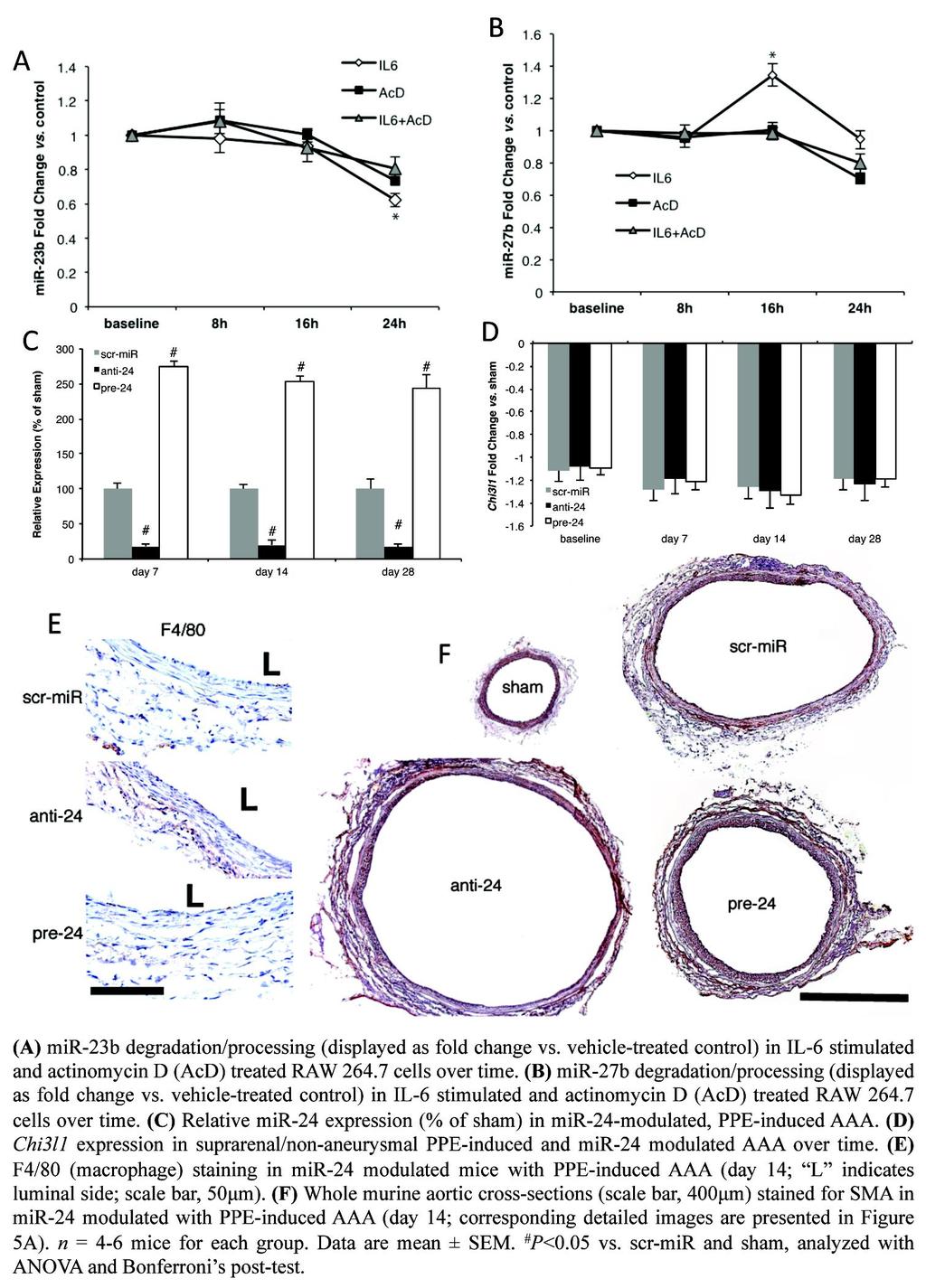
2 Supplementary Figure 2: B A C D E (A) Heatmap showing all significantly up-regulated mir-23b-24-27b target genes (FDR < 1%, fold-change > 1.5) in affected aortic tissue from PPE-induced AAA (Day 7 vs. sham). (B, C) Expression of validated mir-24 target genes (from previous studies) at various time-points during PPE-induced AAA expansion (vs. sham), including the expression of Chi3l1. (D) Double immunofluorescence detection of Chi3l1 (red) and smooth muscle α- actin (SMA) (green, left) or F4-80 (green, right) in aortic sections, 14 days after PPEinduction of AAA ( L indicates luminal side; scale bar, 50μm). (E) Luciferase assay confirming direct suppression of CHI3L1 transcription through 3'UTR binding of mir-24 (E-scr = empty plasmid + scr-mir as negative control, R-scr = random 3'UTR plasmid + scr-mir as positive control, R-pre-24 = random 3'UTR plasmid + pre-24, CHI3L1-pre-24 = CHI3L1 3'UTR plasmid + pre-24). n = 4-8 for each group and time point. Data are mean ± SEM. *P<0.05 vs. sham (or R-scr, R-pre-24); analyzed with ANOVA and Bonferroni s post-test.
 CCL2 expression in IL-6-stimulated and mir-24 modulated hasmc. (C) IL8 expression in IL-6 stimulated and mir-24 modulated hasmc.")
3 Supplementary Figure 3: (A) Chi3l1 expression in interleukin-6 (IL-6)-stimulated (12 or 24 h) and mir-24-modulated peritoneal macrophages. (B) CCL2 expression in IL-6-stimulated and mir-24 modulated hasmc. (C) IL8 expression in IL-6 stimulated and mir-24 modulated hasmc. (D) CCL2 expression in CHI3L1-stimulated hasmc ± pre-stimulation with IL-6 (16 h). (E) FACS analysis of C3-labeled, scr-mir-transfected, peritoneal macrophages. First image displays un-transfected control cells, and second image indicates 82.7% transfection efficiency using scr-mir. All experiments were run in triplicate. Data are mean ± SEM. *P<0.05 vs. untreated or scr-mir, # P<0.05 vs. untreated + scr-mir or vs. anti-/pre-24 (in Suppl. Fig. 2C and 2D) or vs. untreated + CHI3L1 30ng/ml (Suppl. Fig. 3E), ^P<0.05 vs. untreated + CHI3L1 treatment alone, analyzed with ANOVA and Bonferroni s post-test.
 with β- tubulin loading control.")
. (C) Protein expression of total JNK (JNK) and phospho-jnk (p-jnk) in mir-24 modulated hasmc (β-tubulin loading control).")
 Modified Boyden chamber migration assay in CHI3L1- stimulated hasmc at various doses. High power field = HPF. Data are mean ± SEM. *P<0.05 vs.")
4 Supplementary Figure 4: E (A) Protein expression of total JNK (JNK) and phospho-jnk (p-jnk) in low-dose (30ng/ml) and high dose (300ng/ml) CHI3L1 stimulated human aortic smooth muscle cells (hasmc) with β- tubulin loading control. (B) Protein expression of total ERK (ERK) and phospho-erk (p-erk) in low-dose (30ng/ml) and high dose (300ng/ml) CHI3L1 stimulated hasmc (β-tubulin loading control). (C) Protein expression of total JNK (JNK) and phospho-jnk (p-jnk) in mir-24 modulated hasmc (β-tubulin loading control). (D) Protein expression of total ERK (ERK) and phospho-erk (p-erk) in mir-24 modulated hasmc (β-tubulin loading control). (E) Modified Boyden chamber migration assay in CHI3L1- stimulated hasmc at various doses. High power field = HPF. Data are mean ± SEM. *P<0.05 vs. control/scr-mir, # P<0.05 vs. all other treatments in the same experiment, analyzed with ANOVA and Bonferroni s post-test.

5 Supplementary Figure 5:
6 Supplementary Figure 6:
7 Supplementary Figure 7:
8 Supplementary Figure 8:
9 Supplementary Figure 9: Proposed mechanism
 utilized in our manuscript.")
10 Supplementary Figure 10: Un-cropped Western blots for all antibodies (with dilutions) utilized in our manuscript. Technical details are described in the Methods.
11 Supplementary Tables Table 1: Absolute ultrasound diameter measurements in the PPE-model baseline day 3 day 7 day 14 day 21 day 28 sham 0.63 ± ± ± ± ± ± 0.09 PPE 0.62 ± ± ± 0.04 * 1.05 ± 0.03 * 1.12 ± 0.08 * 1.13 ± 0.06 * Absolute measurements (mean ± SEM; in mm) of the abdominal aortic diameter using B-mode ultrasound from the porcine-pancreatic-elastase (PPE) induction model from baseline until day 28. Absolute measurements correspond to Figure 1A (presented as AAD vs. baseline in %) in the manuscript. P < 0.05 is considered significant vs. same- day sham, using Student s t-test for parametric measures (*). Table 2: Absolute ultrasound diameter measurements in the ANGII-model baseline day 7 day 14 day 28 sham 1.10 ± ± ± ± 0.12 ANGII 1.11 ± ± ± 0.17 * 2.01 ± 0.21 * Absolute measurements (mean ± SEM; in mm) of the abdominal aortic diameter using B-mode ultrasound in the angiotensinii-aaa induction model from baseline until day 28. Absolute measurements correspond to Figure 2A (presented as AAD vs. baseline in %). P < 0.05 is considered significant vs. same-day sham, using Student s t-test for parametric measures (*). Table 3: Blood pressure measurements in mice with AAA systolic (mmhg) diastolic (mmhg) p-value PPE (n=6) 112 ± ± 8 sham (for PPE; n=6) 109 ± 8 83 ± ANGII (n=6) 145 ± 9* 92 ± 8 sham (for ANGII; n=6) 116 ± 8 90 ± * Blood pressure measurements (systolic/diastolic) in the porcine-pancreatic-elastase (PPE) and angiotensin II (ANGII)-AAA induction models (day 14). Data are mean ± SEM. P < 0.05 is considered significant, using Student s t-test for parametric measures (to compare PPE vs. sham and ANGII vs. sham; *).
12 Table 4: Absolute ultrasound diameter measurements in the PPE-model after mir-24 modulation baseline day 3 day 7 day 14 day 21 day 28 PPE anti ± ± ± ± * 1.31 ± * 1.32 ± * PPE scr-mir PPE pre ± ± ± ± ± ± ± ± ± ± 1.01 ± 1.01 ± * * * Absolute measurements (mean ± SEM; in mm) of the abdominal aortic diameter using B-mode ultrasound from the porcine-pancreatic-elastase (PPE) induction model from baseline until day 28 in anti-/pre-24 and scr-mir mice with AAA. Absolute measurements correspond to Figure 4B (presented as AAD vs. baseline in %) in the manuscript. P < 0.05 is considered significant, using one-way ANOVA with Bonferroni s post-test (*). Table 5: Absolute ultrasound diameter measurements in the ANGII-model after mir-24 modulation baseline day 7 day 14 day 28 ANGII anti ± ± ± 0.05* 2.25 ± 0.04 * ANGII scr-mir 1.09 ± ± ± ± 0.05 ANGII pre ± ± ± 0.04 * 1.79 ± 0.05 * Absolute measurements (mean ± SEM; in mm) of the abdominal aortic diameter using B-mode ultrasound in the angiotensin II (ANGII)-AAA induction model from baseline until day 28 in anti-/pre-24 and scr-mir mice with AAA. Absolute measurements correspond to Figure 6A (presented as AAD vs. baseline in %) in the manuscript. P < 0.05 is considered significant, using one-way ANOVA with Bonferroni s post test (* at day 14 only for anti-21 vs. pre-24). Table 6: Blood pressure measurements in mir-24 modulated mice systolic (mmhg) diastolic (mmhg) p-value scr-mir (n=4) 106 ± 7 81 ± 1.5 anti-24 (n=4) 105 ± 7 80 ± pre-24 (n=4) 106 ± 9 79 ± Blood pressure measurements (systolic/diastolic) in otherwise-untreated mir-24- modulated mice (anti- and pre-24 compared to scr-mir). Data are mean ± SEM. P < 0.05 is considered significant, using one-way ANOVA with Bonferroni s post-test.
13 Table 7: Pathway analysis of transcriptional profiling of in porcine-pancreaticelastase (PPE)-induced abdominal aortic aneurysm (AAA) post-mir-24 modulation sham>scr-mir/ppe sham>pre-24/ppe overlap GO Pathways Oxidation reduction Cell adhesion Steroid metabolism KEGG pathways Focal adhesion VSMC contraction ECM-receptor interaction scr-mir/ppe>sham pre-24/ppe>sham overlap GO Pathways Positive regulation of immune system Induction of apoptosis KEGG pathways Cytokine-cytokine receptor interaction Leukocyte transendothelial migration Toll-like receptor signaling Scr-miR (scrambled-mir-injected mouse with PPE-induced AAA); pre-24 (pre-mir- 24 transduced mice with PPE-induced AAA); VSMC (vascular smooth muscle cells); ECM (extracellular matrix). Numbers of differentially regulated genes in each pathway at day 7 post-aaa induction shown. See Supplementary Note 1 for details.
14 Table 8: Baseline patient characteristics for plasma measurement of the expression of mir-24 and YKL-40 levels Controls Small AAA Large AAA PVOD p-value n = AAD (in mm) ± 7 * 85 ± 14 * * Age (in years) 65 ± 3 65 ± 2 65 ± 3 66 ± Male Sex (in %) Smokers (in %) egfr 84.3 ± ± ± ± Triglycerides 2.3 ± ± ± ± (mmol/l) Bilirubin 11.9 ± ± ± ± (μmol/l) ASAT (U/L) 26.9 ± ± ± ± HbA1c (rel. %) 5.8 ± ± ± ± NSAID (in %) * * ACEI/ARB (in %) Beta blocker * * (in %) Statins (in %) Data are mean ± standard deviation (SD), number (n) of individuals and percentages (%). P < 0.05 is considered significant (*), using ANOVA with Bonferroni s post test for parametric data with normal distribution. For categorical, non-parametric data (smoking status and medication), Chi-squared test (χ 2 ) was utilized. AAA (abdominal aortic aneurysm); AAD (abdominal aortic diameter measured by ultrasound); ACEI (angiotensin converting enzyme inhibitor); ARB (angiotensin II receptor blocker); ASAT (aspartate aminotransferase); egfr (estimated glomerular filtration rate), HbA1c (glycosylated hemoglobin); PVOD (peripheral vascular occlusive disease); NSAID (non-steroidal anti-inflammatory drug).
15 Supplementary Note 1 mir-24 modulation effects on gene expression in murine AAA (expanded) We collected infra-renal aortic tissue from mirna-modulated and saline-infused control mice 7 days after infusion, and hybridized isolated RNA to Agilent mouse whole genome microarrays. Expression analysis identified numerous up-regulated and down-regulated genes (1,996 and 1,200 respectively, at FDR<1%) in scr-mir-treated PPE-induced AAA vs. scr-mir-treated saline-infused control mice. Ontologic pathway enrichment analysis of significant gene lists at 7-days of treatment focusing on KEGG pathways and GO Biological Processes was performed as previously described 1. Differentially regulated PPE-scr-miR genes described above yielded similar enriched pathways to those seen previously in ANGII-induced aneurysms, featuring pathways related to immune responses, matrix remodeling, and cell signaling. Up-regulated (with PPE-induced aneurysm) pathways included cell cycling, apoptosis, toll-like receptor signaling, and regulation of the NF-κB cascade, as well as MAPK and p53 signaling (Supplemental Table 7). As in our previous work, we identified broad increases in genes associated with immune system activation (e.g. macrophage, T-cell, B-cell and natural killer responses), lymphocyte proliferation and differentiation, and numerous cytokines and cytokine receptors. For example, 104 genes in the GO category positive regulation of immune system process were upregulated in scr-mir aorta when compared with saline control. As expected, Chi3l1 was up-regulated (4.5-fold) with aneurysm formation. Down-regulated gene responses to treatment also mimicked to some extent those seen previously with ANGII-aneurysm formation, with enrichment of SMC contractile genes, focal adhesion, insulin signaling, and numerous metabolism pathways such as oxidative phosphorylation and amino acid/lipid metabolism 1. Notably, ontology of anti-24-modulated PPE-infused gene expression was essentially indistinguishable from that seen in scr-mir-treated PPE-infused mice. Further, when anti-24 PPE-infused expression was compared with saline-infused sham-operated controls, the gene numbers and differentially regulated pathways were very similar to those obtained by comparing scr-mir PPE-mice with saline-infused sham controls. Chi3l1 was further up-regulated with anti-24 treatment. In contrast, gene expression in pre-24-modulated aortic tissue was much harder to distinguish from sham controls, with only 208 unique genes higher in pre-24 than sham, and 630 genes lower at FDR < 1%. While some of the enriched pathways
16 were similar overall to those found in scr-mir-ppe vs. sham, the numbers of genes within each categories were much smaller, including for example only 14 positive regulation of immune system process genes up-regulated with pre-24. Downregulated metabolism pathways were also less well represented in pre-24 modulated aortas. Chi3l1 gene expression was undetectable by microarray in pre-24-treated aortas, confirming our qrt-pcr results. Partial lists of enriched KEGG and GO categories with a direct comparison between significant scr-mir and pre-mir pathways are in Supplemental Table 7. Reference 1. Spin, J.M., et al. Transcriptional profiling and network analysis of the murine angiotensin II-induced abdominal aortic aneurysm. Physiological genomics 43, (2011).
Figure S1. Reduction in glomerular mir-146a levels correlate with progression to higher albuminuria in diabetic patients.
 Supplementary Materials Supplementary Figures Figure S1. Reduction in glomerular mir-146a levels correlate with progression to higher albuminuria in diabetic patients. Figure S2. Expression level of podocyte
Supplementary Figure 1.
 Supplementary Figure 1. Increased expression of cell cycle pathway genes in insulin + Glut2 low cells of STZ-induced diabetic islets. A) random blood glucose measuers of STZ and vehicle treated MIP-GFP
Supplementary Figure 1. Confocal immunofluorescence showing mitochondrial translocation of Drp1. Cardiomyocytes treated with H 2 O 2 were prestained
 Supplementary Figure 1. Confocal immunofluorescence showing mitochondrial translocation of Drp1. Cardiomyocytes treated with H 2 O 2 were prestained with MitoTracker (red), then were immunostained with
SUPPLEMENTARY INFORMATION
 DOI:.38/ncb3399 a b c d FSP DAPI 5mm mm 5mm 5mm e Correspond to melanoma in-situ Figure a DCT FSP- f MITF mm mm MlanaA melanoma in-situ DCT 5mm FSP- mm mm mm mm mm g melanoma in-situ MITF MlanaA mm mm
Supplemental Figure 1. Western blot analysis indicated that MIF was detected in the fractions of
 Supplemental Figure Legends Supplemental Figure 1. Western blot analysis indicated that was detected in the fractions of plasma membrane and cytosol but not in nuclear fraction isolated from Pkd1 null
Supplementary Materials
 Supplementary Materials Figure S1. MTT Cell viability assay. To measure the cytotoxic potential of the oxidative treatment, the MTT [3-(4,5-dimethylthiazol- 2-yl)-2,5-diphenyl tetrazolium bromide] assay
Supplementary Table 1. Baseline Characteristics by Quintiles of Systolic and Diastolic Blood Pressures
 Supplementary Data Supplementary Table 1. Baseline Characteristics by Quintiles of Systolic and Diastolic Blood Pressures Quintiles of Systolic Blood Pressure Quintiles of Diastolic Blood Pressure Q1 Q2
Supplementary Materials for
 www.sciencesignaling.org/cgi/content/full/2/1/ra81/dc1 Supplementary Materials for Delivery of MicroRNA-126 by Apoptotic Bodies Induces CXCL12- Dependent Vascular Protection Alma Zernecke,* Kiril Bidzhekov,
mir-509-5p and mir-1243 increase the sensitivity to gemcitabine by inhibiting
 mir-509-5p and mir-1243 increase the sensitivity to gemcitabine by inhibiting epithelial-mesenchymal transition in pancreatic cancer Hidekazu Hiramoto, M.D. 1,3, Tomoki Muramatsu, Ph.D. 1, Daisuke Ichikawa,
Supplementary fig. 1. Crystals induce necroptosis does not involve caspases, TNF receptor or NLRP3. A. Mouse tubular epithelial cells were pretreated
 Supplementary fig. 1. Crystals induce necroptosis does not involve caspases, TNF receptor or NLRP3. A. Mouse tubular epithelial cells were pretreated with zvad-fmk (10µM) and exposed to calcium oxalate
fasting blood glucose [mg/dl]
![fasting blood glucose [mg/dl]](/thumbs/80/82129652.jpg "fasting blood glucose [mg/dl]") SUPPLEMENTL MTERIL Supplemental Figure I body weight [g] 5 5 5 fasting blood glucose [mg/dl] 5 5 5 C total cholesterol [mg/dl] 8 6 4 WT Has -/- Supplemental Figure I: Has-deficient mice exhibited no apparent
SUPPLEMENTL MTERIL Supplemental Figure I body weight [g] 5 5 5 fasting blood glucose [mg/dl] 5 5 5 C total cholesterol [mg/dl] 8 6 4 WT Has -/- Supplemental Figure I: Has-deficient mice exhibited no apparent
c Ischemia (30 min) Reperfusion (8 w) Supplementary Figure bp 300 bp Ischemia (30 min) Reperfusion (4 h) Dox 20 mg/kg i.p.
 Reperfusion (8 w) Supplementary Figure bp 300 bp Ischemia (30 min) Reperfusion (4 h) Dox 20 mg/kg i.p.") a Marker Ripk3 +/ 5 bp 3 bp b Ischemia (3 min) Reperfusion (4 h) d 2 mg/kg i.p. 1 w 5 w Sacrifice for IF size A subset for echocardiography and morphological analysis c Ischemia (3 min) Reperfusion (8
a Marker Ripk3 +/ 5 bp 3 bp b Ischemia (3 min) Reperfusion (4 h) d 2 mg/kg i.p. 1 w 5 w Sacrifice for IF size A subset for echocardiography and morphological analysis c Ischemia (3 min) Reperfusion (8
Stewart et al. CD36 ligands promote sterile inflammation through assembly of a TLR 4 and 6 heterodimer
 NFκB (fold induction) Stewart et al. ligands promote sterile inflammation through assembly of a TLR 4 and 6 heterodimer a. mrna (fold induction) 5 4 3 2 1 LDL oxldl Gro1a MIP-2 RANTES mrna (fold induction)
fl/+ KRas;Atg5 fl/+ KRas;Atg5 fl/fl KRas;Atg5 fl/fl KRas;Atg5 Supplementary Figure 1. Gene set enrichment analyses. (a) (b)
 (b)") KRas;At KRas;At KRas;At KRas;At a b Supplementary Figure 1. Gene set enrichment analyses. (a) GO gene sets (MSigDB v3. c5) enriched in KRas;Atg5 fl/+ as compared to KRas;Atg5 fl/fl tumors using gene set
KRas;At KRas;At KRas;At KRas;At a b Supplementary Figure 1. Gene set enrichment analyses. (a) GO gene sets (MSigDB v3. c5) enriched in KRas;Atg5 fl/+ as compared to KRas;Atg5 fl/fl tumors using gene set
Title: Smooth muscle cell-specific Tgfbr1 deficiency promotes aortic aneurysm formation by stimulating multiple signaling events
 Title: Smooth muscle cell-specific Tgfbr1 deficiency promotes aortic aneurysm formation by stimulating multiple signaling events Pu Yang 1, 3, radley M. Schmit 1, Chunhua Fu 1, Kenneth DeSart 1, S. Paul
Supplementary Fig. 1. GPRC5A post-transcriptionally down-regulates EGFR expression. (a) Plot of the changes in steady state mrna levels versus
 Plot of the changes in steady state mrna levels versus") Supplementary Fig. 1. GPRC5A post-transcriptionally down-regulates EGFR expression. (a) Plot of the changes in steady state mrna levels versus changes in corresponding proteins between wild type and Gprc5a-/-
Supplementary Fig. 1. GPRC5A post-transcriptionally down-regulates EGFR expression. (a) Plot of the changes in steady state mrna levels versus changes in corresponding proteins between wild type and Gprc5a-/-
Supplemental Figure 1. Signature gene expression in in vitro differentiated Th0, Th1, Th2, Th17 and Treg cells. (A) Naïve CD4 + T cells were cultured
 Naïve CD4 + T cells were cultured") Supplemental Figure 1. Signature gene expression in in vitro differentiated Th0, Th1, Th2, Th17 and Treg cells. (A) Naïve CD4 + T cells were cultured under Th0, Th1, Th2, Th17, and Treg conditions. mrna
Supplemental Figure 1. Signature gene expression in in vitro differentiated Th0, Th1, Th2, Th17 and Treg cells. (A) Naïve CD4 + T cells were cultured under Th0, Th1, Th2, Th17, and Treg conditions. mrna
Intracellular MHC class II molecules promote TLR-triggered innate. immune responses by maintaining Btk activation
 Intracellular MHC class II molecules promote TLR-triggered innate immune responses by maintaining Btk activation Xingguang Liu, Zhenzhen Zhan, Dong Li, Li Xu, Feng Ma, Peng Zhang, Hangping Yao and Xuetao
Supplementary Materials for
 www.sciencesignaling.org/cgi/content/full/9/439/ra78/dc1 Supplementary Materials for Small heterodimer partner mediates liver X receptor (LXR) dependent suppression of inflammatory signaling by promoting
Supplementary Figure 1. Expression of CUGBP1 in non-parenchymal liver cells treated with TGF-β
 Supplementary Figures Supplementary Figure 1. Expression of CUGBP1 in non-parenchymal liver cells treated with TGF-β and LPS. Non-parenchymal liver cells were isolated and treated with or without TGF-β
Supplementary Figure 1 ITGB1 and ITGA11 increase with evidence for heterodimers following HSC activation. (a) Time course of rat HSC activation
 Time course of rat HSC activation") Supplementary Figure 1 ITGB1 and ITGA11 increase with evidence for heterodimers following HSC activation. (a) Time course of rat HSC activation indicated by the detection of -SMA and COL1 (log scale).
Supplementary Figure 1 ITGB1 and ITGA11 increase with evidence for heterodimers following HSC activation. (a) Time course of rat HSC activation indicated by the detection of -SMA and COL1 (log scale).
Introduction. Acute sodium overload produces renal tubulointerstitial inflammation in normal rats
 Acute sodium overload produces renal tubulointerstitial inflammation in normal rats MI Roson, et al. Kidney International (2006) Introduction Present by Kanya Bunnan and Wiraporn paebua Tubular sodium
SUPPLEMENTARY INFORMATION
 DOI: 10.1038/ncb3021 Supplementary figure 1 Characterisation of TIMPless fibroblasts. a) Relative gene expression of TIMPs1-4 by real time quantitative PCR (RT-qPCR) in WT or ΔTimp fibroblasts (mean ±
Serum cytokine levels in control and tumor-bearing male and female mice at day 15.
 Supplementary Table 1. Serum cytokine levels in control and tumor-bearing male and female mice at day 15. Male Female Cytokine Control C-26 Control C-26 IL-1β 2.0 ± 0.8 9.6 ± 1.5* 1.8 ± 0.2 6.8 ± 1.4*
T H E J O U R N A L O F C E L L B I O L O G Y
 T H E J O U R N A L O F C E L L B I O L O G Y Supplemental material Amelio et al., http://www.jcb.org/cgi/content/full/jcb.201203134/dc1 Figure S1. mir-24 regulates proliferation and by itself induces
Programmed necrosis, not apoptosis, is a key mediator of cell loss and DAMP-mediated inflammation in dsrna-induced retinal degeneration
 Programmed necrosis, not apoptosis, is a key mediator of cell loss and DAMP-mediated inflammation in dsrna-induced retinal degeneration The Harvard community has made this article openly available. Please
Supplemental Table I.
 Supplemental Table I Male / Mean ± SEM n Mean ± SEM n Body weight, g 29.2±0.4 17 29.7±0.5 17 Total cholesterol, mg/dl 534.0±30.8 17 561.6±26.1 17 HDL-cholesterol, mg/dl 9.6±0.8 17 10.1±0.7 17 Triglycerides,
Supplementary Figure 1: STAT3 suppresses Kras-induced lung tumorigenesis
 Supplementary Figure 1: STAT3 suppresses Kras-induced lung tumorigenesis (a) Immunohistochemical (IHC) analysis of tyrosine 705 phosphorylation status of STAT3 (P- STAT3) in tumors and stroma (all-time
INTRODUCTION. Induction of Monocyte Chemoattractant Protein-1 (MCP-1) Expression by Angiotensin II (AngII) in the Pancreatic Islets and Beta Cells
 Expression by Angiotensin II (AngII) in the Pancreatic Islets and Beta Cells") Induction of Monocyte Chemoattractant Protein-1 (MCP-1) Expression by Angiotensin II (AngII) in the Pancreatic Islets and Beta Cells Galina Chipitsyna, Qiaoke Gong, Chance F. Gray et al. Endocrinology,
Induction of Monocyte Chemoattractant Protein-1 (MCP-1) Expression by Angiotensin II (AngII) in the Pancreatic Islets and Beta Cells Galina Chipitsyna, Qiaoke Gong, Chance F. Gray et al. Endocrinology,
Supplementary Figure 1 IL-27 IL
 Tim-3 Supplementary Figure 1 Tc0 49.5 0.6 Tc1 63.5 0.84 Un 49.8 0.16 35.5 0.16 10 4 61.2 5.53 10 3 64.5 5.66 10 2 10 1 10 0 31 2.22 10 0 10 1 10 2 10 3 10 4 IL-10 28.2 1.69 IL-27 Supplementary Figure 1.
SUPPLEMENTAL MATERIAL
 SUPPLEMENTAL MATERIAL Clinical perspective It was recently discovered that small RNAs, called micrornas, circulate freely and stably in human plasma. This finding has sparked interest in the potential
Supplementary Figure 1. IHC and proliferation analysis of pten-deficient mammary tumors
 Wang et al LEGENDS TO SUPPLEMENTARY INFORMATION Supplementary Figure 1. IHC and proliferation analysis of pten-deficient mammary tumors A. Induced expression of estrogen receptor α (ERα) in AME vs PDA
Title: Cytosolic DNA-mediated, STING-dependent pro-inflammatory gene. Fig. S1. STING ligands-mediated signaling response in MEFs. (A) Primary MEFs (1
 Primary MEFs (1") 1 Supporting Information 2 3 4 Title: Cytosolic DNA-mediated, STING-dependent pro-inflammatory gene induction necessitates canonical NF-κB activation through TBK1 5 6 Authors: Abe et al. 7 8 9 Supporting
1 Supporting Information 2 3 4 Title: Cytosolic DNA-mediated, STING-dependent pro-inflammatory gene induction necessitates canonical NF-κB activation through TBK1 5 6 Authors: Abe et al. 7 8 9 Supporting
Supplementary Information
 Supplementary Information TABLE S1. SUBJECT CHARACTERISTICS* Normal Control Subjects Subjects with Asthma p Value Number 23 48 Age (years) 35±10 35±10 0.75 Sex, M:F (% F) 9:12 (57) 17:26 (60) 0.76 FEV1
Nature Neuroscience: doi: /nn Supplementary Figure 1
 Supplementary Figure 1 EGFR inhibition activates signaling pathways (a-b) EGFR inhibition activates signaling pathways (a) U251EGFR cells were treated with erlotinib (1µM) for the indicated times followed
EPIGENETIC RE-EXPRESSION OF HIF-2α SUPPRESSES SOFT TISSUE SARCOMA GROWTH
 EPIGENETIC RE-EXPRESSION OF HIF-2α SUPPRESSES SOFT TISSUE SARCOMA GROWTH Supplementary Figure 1. Supplementary Figure 1. Characterization of KP and KPH2 autochthonous UPS tumors. a) Genotyping of KPH2
Supplementary Materials for
 www.sciencesignaling.org/cgi/content/full/7/310/ra11/dc1 Supplementary Materials for STAT3 Induction of mir-146b Forms a Feedback Loop to Inhibit the NF-κB to IL-6 Signaling Axis and STAT3-Driven Cancer
Supplementary Figure 1 IMQ-Induced Mouse Model of Psoriasis. IMQ cream was
 Supplementary Figure 1 IMQ-Induced Mouse Model of Psoriasis. IMQ cream was painted on the shaved back skin of CBL/J and BALB/c mice for consecutive days. (a, b) Phenotypic presentation of mouse back skin
hemodynamic stress. A. Echocardiographic quantification of cardiac dimensions and function in
 SUPPLEMENTAL FIGURE LEGENDS Supplemental Figure 1. Fbn1 C1039G/+ hearts display normal cardiac function in the absence of hemodynamic stress. A. Echocardiographic quantification of cardiac dimensions and
a. b. c. d. e. f. g. h. i. j. k. l. m. n. o. p.
 a. b. c. d. e. f. g. h. i. j. k. l. 2.5 2 1.5 1.5 IL-1β 12 8 6 4 2 IL-1β 9 8 7 6 4 3 3 2.9 IL-1β m. n. o. p. 1.8 1.6 1.4 1.2 1.8.6.4.2 6h LPS 2 15 1 5 6h LPS 2 6h LPS 6 4 3 6h LPS Supplementary Figure
Supplementary Figure 1. A. Bar graph representing the expression levels of the 19 indicated genes in the microarrays analyses comparing human lung
 Supplementary Figure 1. A. Bar graph representing the expression levels of the 19 indicated genes in the microarrays analyses comparing human lung immortalized broncho-epithelial cells (AALE cells) expressing
Supplementary Figure 1. HOPX is hypermethylated in NPC. (a) Methylation levels of HOPX in Normal (n = 24) and NPC (n = 24) tissues from the
 Methylation levels of HOPX in Normal (n = 24) and NPC (n = 24) tissues from the") Supplementary Figure 1. HOPX is hypermethylated in NPC. (a) Methylation levels of HOPX in Normal (n = 24) and NPC (n = 24) tissues from the genome-wide methylation microarray data. Mean ± s.d.; Student
Supplementary Figure 1. HOPX is hypermethylated in NPC. (a) Methylation levels of HOPX in Normal (n = 24) and NPC (n = 24) tissues from the genome-wide methylation microarray data. Mean ± s.d.; Student
Contribution of the intronic mir-338-3p and its Hosting Gene AATK to Compensatory β-cell Mass Expansion
 Contribution of the intronic mir-338-3p and its Hosting Gene AATK to Compensatory β-cell Mass Expansion Cécile Jacovetti 1, Veronica Jimenez 2, Eduard Ayuso 2#, Ross Laybutt 3, Marie-Line Peyot 4, Marc
Medical management of abdominal aortic aneurysms
 Medical management of abdominal aortic aneurysms Definition of AAA - Generally a 50% increase in native vessel diameter - Diameter 3 cm - Relative measures compared with nondiseased aortic segments less
SUPPLEMENTARY INFORMATION
 DOI: 10.1038/ncb2607 Figure S1 Elf5 loss promotes EMT in mammary epithelium while Elf5 overexpression inhibits TGFβ induced EMT. (a, c) Different confocal slices through the Z stack image. (b, d) 3D rendering
Supplementary Materials for
 www.sciencesignaling.org/cgi/content/full/4/199/ra75/dc1 Supplementary Materials for Signaling by the Matrix Proteoglycan Decorin Controls Inflammation and Cancer Through PDCD4 and MicroRNA-21 Rosetta
SUPPLEMENTARY INFORMATION
 doi:10.1038/nature12652 Supplementary Figure 1. PRDM16 interacts with endogenous EHMT1 in brown adipocytes. Immunoprecipitation of PRDM16 complex by flag antibody (M2) followed by Western blot analysis
Supplemental Figure 1
 Supplemental Figure 1 1a 1c PD-1 MFI fold change 6 5 4 3 2 1 IL-1α IL-2 IL-4 IL-6 IL-1 IL-12 IL-13 IL-15 IL-17 IL-18 IL-21 IL-23 IFN-α Mut Human PD-1 promoter SBE-D 5 -GTCTG- -1.2kb SBE-P -CAGAC- -1.kb
Gallic acid prevents isoproterenol-induced cardiac hypertrophy and fibrosis through regulation of JNK2 signaling and Smad3 binding activity
 Gallic acid prevents isoproterenol-induced cardiac hypertrophy and fibrosis through regulation of JNK2 signaling and Smad3 binding activity Yuhee Ryu 1,+, Li Jin 1,2+, Hae Jin Kee 1,, Zhe Hao Piao 3, Jae
MicroRNAs Modulate the Noncanonical NF- B Pathway by Regulating IKK Expression During Macrophage Differentiation
 MicroRNAs Modulate the Noncanonical NF- B Pathway by Regulating IKK Expression During Macrophage Differentiation Tao Li 1 *, Michael J. Morgan 1 *, Swati Choksi 1, Yan Zhang 1, You-Sun Kim 2#, Zheng-gang
GW(g)/BW(g) GW(g)/BW(g) Con Dex Con Dex. GW(g)/BW(g) Relative mrna levels. Atrogin-1 Murf-1. Atrogin-1 Murf-1. Soleus
/BW(g) GW(g)/BW(g) Con Dex Con Dex. GW(g)/BW(g) Relative mrna levels. Atrogin-1 Murf-1. Atrogin-1 Murf-1. Soleus") a b c GW(g) GW(g) GW(g) 0.20 0.15 0.10 0.05 0.00 0.20 0.15 0.10 0.05 Den * ** GW(g)/BW(g) Den Dex Dex GW(g)/BW(g) d 0.00 Fasting GW(g) e 0.15 Cancer Cachexia f GW(g) 0.10 0.05 0.00 Young ** Old g GW(g)/BW(g)
a b c GW(g) GW(g) GW(g) 0.20 0.15 0.10 0.05 0.00 0.20 0.15 0.10 0.05 Den * ** GW(g)/BW(g) Den Dex Dex GW(g)/BW(g) d 0.00 Fasting GW(g) e 0.15 Cancer Cachexia f GW(g) 0.10 0.05 0.00 Young ** Old g GW(g)/BW(g)
Supplementary Figure S I: Effects of D4F on body weight and serum lipids in apoe -/- mice.
 Supplementary Figures: Supplementary Figure S I: Effects of D4F on body weight and serum lipids in apoe -/- mice. Male apoe -/- mice were fed a high-fat diet for 8 weeks, and given PBS (model group) or
SUPPLEMENTAL DATA. Lumen area ( m 2 )
") Elastin Lumen area ( m 2 ) Media to lumen ratio (x1) H.E. Medium thickness ( m) Medium area ( m 2 ) SUPPLEMENTAL DATA A (Bmal1 flox/flox ) (SM-Bmal1 -/- ) B 1 8 8 6 6 4 4 2 2 1µm 5 8 4 6 3 2 4 1 2 Supplemental
Elastin Lumen area ( m 2 ) Media to lumen ratio (x1) H.E. Medium thickness ( m) Medium area ( m 2 ) SUPPLEMENTAL DATA A (Bmal1 flox/flox ) (SM-Bmal1 -/- ) B 1 8 8 6 6 4 4 2 2 1µm 5 8 4 6 3 2 4 1 2 Supplemental
Supplementary Figure 1. Normal T lymphocyte populations in Dapk -/- mice. (a) Normal thymic development in Dapk -/- mice. Thymocytes from WT and Dapk
 Normal thymic development in Dapk -/- mice. Thymocytes from WT and Dapk") Supplementary Figure 1. Normal T lymphocyte populations in Dapk -/- mice. (a) Normal thymic development in Dapk -/- mice. Thymocytes from WT and Dapk -/- mice were stained for expression of CD4 and CD8.
Supplementary Figure 1. Normal T lymphocyte populations in Dapk -/- mice. (a) Normal thymic development in Dapk -/- mice. Thymocytes from WT and Dapk -/- mice were stained for expression of CD4 and CD8.
Crucial role for human Toll-like receptor 4 in the development of contact allergy to nickel
 Supplementary Figures 1-8 Crucial role for human Toll-like receptor 4 in the development of contact allergy to nickel Marc Schmidt 1,2, Badrinarayanan Raghavan 1,2, Verena Müller 1,2, Thomas Vogl 3, György
Supplementary Figures
 Supplementary Figures Supplementary Figure 1. Confirmation of Dnmt1 conditional knockout out mice. a, Representative images of sorted stem (Lin - CD49f high CD24 + ), luminal (Lin - CD49f low CD24 + )
Soft Agar Assay. For each cell pool, 100,000 cells were resuspended in 0.35% (w/v)
") SUPPLEMENTARY MATERIAL AND METHODS Soft Agar Assay. For each cell pool, 100,000 cells were resuspended in 0.35% (w/v) top agar (LONZA, SeaKem LE Agarose cat.5004) and plated onto 0.5% (w/v) basal agar.
SUPPLEMENTARY MATERIAL AND METHODS Soft Agar Assay. For each cell pool, 100,000 cells were resuspended in 0.35% (w/v) top agar (LONZA, SeaKem LE Agarose cat.5004) and plated onto 0.5% (w/v) basal agar.
Supplemental Figure S1. RANK expression on human lung cancer cells.
 Supplemental Figure S1. RANK expression on human lung cancer cells. (A) Incidence and H-Scores of RANK expression determined from IHC in the indicated primary lung cancer subgroups. The overall expression
III. Results and Discussion
 III. Results and Discussion 1. Histological findings in the coronary artery Twenty-four swine had surgical treatments performed in two of the coronary arteries, LAD as well as either the LCX or RCA. A
Supplementary Table 1. Characterization of HNSCC PDX models established at MSKCC
 Supplementary Table 1. Characterization of HNSCC PDX models established at MSKCC Supplementary Table 2. Drug content and loading efficiency estimated with F-NMR and UV- Vis Supplementary Table 3. Complete
Supplementary Material
 Supplementary Material accompanying the manuscript Interleukin 37 is a fundamental inhibitor of innate immunity Marcel F Nold, Claudia A Nold-Petry, Jarod A Zepp, Brent E Palmer, Philip Bufler & Charles
Supplementary Figures
 Supplementary Figures Supplementary Fig. 1. Galectin-3 is present within tumors. (A) mrna expression levels of Lgals3 (galectin-3) and Lgals8 (galectin-8) in the four classes of cell lines as determined
Effects of the angiotensin II type-1 receptor antagonist telmisartan on endothelial activation induced by advanced glycation endproducts
 Effects of the angiotensin II type-1 receptor antagonist telmisartan on endothelial activation induced by advanced glycation endproducts Serena Del Turco, Teresa Navarra, Giuseppina Basta, Raffaele De
Supplementary Figure 1. Western blot of hippocampal lysates from WT and Adcy1 KO mice demonstrates the specificity of the ADCY1 antibody.
 ADCY1 13 kda β-actin 45 kda Supplementary Figure 1. Western blot of hippocampal lysates from and mice demonstrates the specificity of the ADCY1 antibody. a DHPG perk1/2 ERK1/2 Relative level min 1.6 *
Supplementary Figure 1. Deletion of Smad3 prevents B16F10 melanoma invasion and metastasis in a mouse s.c. tumor model.
 A B16F1 s.c. Lung LN Distant lymph nodes Colon B B16F1 s.c. Supplementary Figure 1. Deletion of Smad3 prevents B16F1 melanoma invasion and metastasis in a mouse s.c. tumor model. Highly invasive growth
Supplementary Table 1. List of primers used in this study
 Supplementary Table 1. List of primers used in this study Gene Forward primer Reverse primer Rat Met 5 -aggtcgcttcatgcaggt-3 5 -tccggagacacaggatgg-3 Rat Runx1 5 -cctccttgaaccactccact-3 5 -ctggatctgcctggcatc-3
(A) Cells grown in monolayer were fixed and stained for surfactant protein-c (SPC,
 Cells grown in monolayer were fixed and stained for surfactant protein-c (SPC,") Supplemental Figure Legends Figure S1. Cell line characterization (A) Cells grown in monolayer were fixed and stained for surfactant protein-c (SPC, green) and co-stained with DAPI to visualize the nuclei.
Supplemental Figure Legends Figure S1. Cell line characterization (A) Cells grown in monolayer were fixed and stained for surfactant protein-c (SPC, green) and co-stained with DAPI to visualize the nuclei.
Role of Alpha-lipoic acid(ala) and statin in vascular smooth muscle cell proliferation
 and statin in vascular smooth muscle cell proliferation") Role of Alpha-lipoic acid(ala) and statin in vascular smooth muscle cell proliferation In-kyu Lee, M.D., Ph. D. Dept. of Internal Med, Keimyung Univ., School of Med. Taegu, Korea Oxidative Stress and Atherosclerosis
Role of Alpha-lipoic acid(ala) and statin in vascular smooth muscle cell proliferation In-kyu Lee, M.D., Ph. D. Dept. of Internal Med, Keimyung Univ., School of Med. Taegu, Korea Oxidative Stress and Atherosclerosis
Supplementary Figure 1. SA-β-Gal positive senescent cells in various cancer tissues. Representative frozen sections of breast, thyroid, colon and
 Supplementary Figure 1. SA-β-Gal positive senescent cells in various cancer tissues. Representative frozen sections of breast, thyroid, colon and stomach cancer were stained with SA-β-Gal and nuclear fast
Plasma exposure levels from individual mice 4 hours post IP administration at the
 Supplemental Figure Legends Figure S1. Plasma exposure levels of MKC-3946 in mice. Plasma exposure levels from individual mice 4 hours post IP administration at the indicated dose mg/kg. Data represent
Supplementary Figure 1
 Supplementary Figure 1 A B mir-141, human cell lines mir-2c, human cell lines mir-141, hepatocytes mir-2c, hepatocytes Relative RNA.1.8.6.4.2 Relative RNA.3.2.1 Relative RNA 1.5 1..5 Relative RNA 2. 1.5
Reduction of metastatic and angiogenic potency of malignant cancer by Eupatorium. fortunei via suppression of MMP-9 activity and VEGF production
 Supplementary Information Reduction of metastatic and angiogenic potency of malignant cancer by Eupatorium fortunei via suppression of MMP-9 activity and VEGF production Aeyung Kim, Minju Im, Nam-Hui Yim
Supplementary Materials for
 www.sciencesignaling.org/cgi/content/full/8/375/ra41/dc1 Supplementary Materials for Actin cytoskeletal remodeling with protrusion formation is essential for heart regeneration in Hippo-deficient mice
Supporting Information
 Supporting Information Retinal expression of small non-coding RNAs in a murine model of proliferative retinopathy Chi-Hsiu Liu 1, Zhongxiao Wang 1, Ye Sun 1, John Paul SanGiovanni 2, Jing Chen 1, * 1 Department
Supplementary Figure 1.TRIM33 binds β-catenin in the nucleus. a & b, Co-IP of endogenous TRIM33 with β-catenin in HT-29 cells (a) and HEK 293T cells
 and HEK 293T cells") Supplementary Figure 1.TRIM33 binds β-catenin in the nucleus. a & b, Co-IP of endogenous TRIM33 with β-catenin in HT-29 cells (a) and HEK 293T cells (b). TRIM33 was immunoprecipitated, and the amount of
Supplementary Figure 1.TRIM33 binds β-catenin in the nucleus. a & b, Co-IP of endogenous TRIM33 with β-catenin in HT-29 cells (a) and HEK 293T cells (b). TRIM33 was immunoprecipitated, and the amount of
Protection against doxorubicin-induced myocardial dysfunction in mice by cardiac-specific expression of carboxyl terminus of hsp70-interacting protein
 Protection against doxorubicin-induced myocardial dysfunction in mice by cardiac-specific expression of carboxyl terminus of hsp70-interacting protein Lei Wang 1, Tian-Peng Zhang 1, Yuan Zhang 2, Hai-Lian
Supplementary Table 1. The primers used for quantitative RT-PCR. Gene name Forward (5 > 3 ) Reverse (5 > 3 )
 Reverse (5 > 3 )") 770 771 Supplementary Table 1. The primers used for quantitative RT-PCR. Gene name Forward (5 > 3 ) Reverse (5 > 3 ) Human CXCL1 GCGCCCAAACCGAAGTCATA ATGGGGGATGCAGGATTGAG PF4 CCCCACTGCCCAACTGATAG TTCTTGTACAGCGGGGCTTG
770 771 Supplementary Table 1. The primers used for quantitative RT-PCR. Gene name Forward (5 > 3 ) Reverse (5 > 3 ) Human CXCL1 GCGCCCAAACCGAAGTCATA ATGGGGGATGCAGGATTGAG PF4 CCCCACTGCCCAACTGATAG TTCTTGTACAGCGGGGCTTG
Supplementary Figure 1. The mir-182 binding site of SMAD7 3 UTR and the. mutated sequence.
 Supplementary Figure 1. The mir-182 binding site of SMAD7 3 UTR and the mutated sequence. 1 Supplementary Figure 2. Expression of mir-182 and SMAD7 in various cell lines. (A) Basal levels of mir-182 expression
Supplementary Information
 Supplementary Information An orally available, small-molecule interferon inhibits viral replication Hideyuki Konishi 1, Koichi Okamoto 1, Yusuke Ohmori 1, Hitoshi Yoshino 2, Hiroshi Ohmori 1, Motooki Ashihara
Figure SⅠ: Expression of mir-155, mir-122 and mir-196a in allografts compared with
 Figure SⅠ: Expression of mir-155, mir-122 and mir-196a in allografts compared with isografts (control) at the 2nd week, 4th and 8th week by RT-PCR. At the advanced stage, the expression of these three
Supplementary Figure 1. Dynamic Response of WT and mir-21 -/- mice to caerulein. (a) Representative histological sections of mouse pancreas stained
 Representative histological sections of mouse pancreas stained") Supplementary Figure 1. Dynamic Response of WT and mir-21 -/- mice to caerulein. (a) Representative histological sections of mouse pancreas stained with hematoxylin from caerulein-treated WT and mir-21
Supplementary Figure 1. Dynamic Response of WT and mir-21 -/- mice to caerulein. (a) Representative histological sections of mouse pancreas stained with hematoxylin from caerulein-treated WT and mir-21
2.5. AMPK activity
 Supplement Fig. A 3 B phos-ampk 2.5 * Control AICAR AMPK AMPK activity (Absorbance at 45 nm) 2.5.5 Control AICAR Supplement Fig. Effects of AICAR on AMPK activation in macrophages. J774. macrophages were
Supplementary Figure 1
 Supplementary Figure 1 Constitutive EGFR signaling does not activate canonical EGFR signals (a) U251EGFRInd cells with or without tetracycline exposure (24h, 1µg/ml) were treated with EGF for 15 minutes
Supplementary Figure (OH) 22 nanoparticles did not affect cell viability and apoposis. MDA-MB-231, MCF-7, MCF-10A and BT549 cells were
 22 nanoparticles did not affect cell viability and apoposis. MDA-MB-231, MCF-7, MCF-10A and BT549 cells were") Supplementary Figure 1. Gd@C 82 (OH) 22 nanoparticles did not affect cell viability and apoposis. MDA-MB-231, MCF-7, MCF-10A and BT549 cells were treated with PBS, Gd@C 82 (OH) 22, C 60 (OH) 22 or GdCl
Supplementary Figure 1. Gd@C 82 (OH) 22 nanoparticles did not affect cell viability and apoposis. MDA-MB-231, MCF-7, MCF-10A and BT549 cells were treated with PBS, Gd@C 82 (OH) 22, C 60 (OH) 22 or GdCl
SITA 100 mg (n = 378)
") Supplementary Table 1. Summary of Sulfonylurea Background Therapy at Baseline and During the Treatment Period. Sulfonylurea at baseline, n (%) SITA 100 mg (n = 378) CANA 300 mg (n = 377) Total (N = 755)
Supplementary Table 1. Summary of Sulfonylurea Background Therapy at Baseline and During the Treatment Period. Sulfonylurea at baseline, n (%) SITA 100 mg (n = 378) CANA 300 mg (n = 377) Total (N = 755)
AP VP DLP H&E. p-akt DLP
 A B AP VP DLP H&E AP AP VP DLP p-akt wild-type prostate PTEN-null prostate Supplementary Fig. 1. Targeted deletion of PTEN in prostate epithelium resulted in HG-PIN in all three lobes. (A) The anatomy
Supplementary Figure 1. Repression of hepcidin expression in the liver of mice treated with
 Supplementary Figure 1. Repression of hepcidin expression in the liver of mice treated with DMN Immunohistochemistry for hepcidin and H&E staining (left). qrt-pcr assays for hepcidin in the liver (right).
Nature Immunology: doi: /ni.3866
 Nature Immunology: doi:10.1038/ni.3866 Supplementary Figure 1 The effect of TIPE2 on chemotaxis. a, The expression of TIPE2 in dhl-60c, dhl-60t, TIPE2-expressing and 15/16Q-expressing dhl-60t neutrophils
Supplementary Figure 1. Efficiency of Mll4 deletion and its effect on T cell populations in the periphery. Nature Immunology: doi: /ni.
 Supplementary Figure 1 Efficiency of Mll4 deletion and its effect on T cell populations in the periphery. Expression of Mll4 floxed alleles (16-19) in naive CD4 + T cells isolated from lymph nodes and
SUPPLEMENTARY FIG. S5. ROS regulated the signaling responses of A. gambiae 4a3B cells to human insulin. (A) 4a3B cells were stimulated with 6000
 4a3B cells were stimulated with 6000") Supplementary Data SUPPLEMENTARY FIG. S1. Exogenous H 2 O 2 induced rapid activation of ERK in Anopheles stephensi cells. ASE cells were treated with PBS or with 500 mmh 2 O 2 for 5, 30, 60, and 180 min.
Supplementary Data SUPPLEMENTARY FIG. S1. Exogenous H 2 O 2 induced rapid activation of ERK in Anopheles stephensi cells. ASE cells were treated with PBS or with 500 mmh 2 O 2 for 5, 30, 60, and 180 min.
Peli1 negatively regulates T-cell activation and prevents autoimmunity
 Peli1 negatively regulates T-cell activation and prevents autoimmunity Mikyoung Chang 1,*, Wei Jin 1,5,*, Jae-Hoon Chang 1, Yi-chuan Xiao 1, George Brittain 1, Jiayi Yu 1, Xiaofei Zhou 1, Yi-Hong Wang
T-cell activation T cells migrate to secondary lymphoid tissues where they interact with antigen, antigen-presenting cells, and other lymphocytes:
 Interactions between innate immunity & adaptive immunity What happens to T cells after they leave the thymus? Naïve T cells exit the thymus and enter the bloodstream. If they remain in the bloodstream,
Supplementary Figure 1: Neuregulin 1 increases the growth of mammary organoids compared to EGF. (a) Mammary epithelial cells were freshly isolated,
 Mammary epithelial cells were freshly isolated,") 1 2 3 4 5 6 7 8 9 10 Supplementary Figure 1: Neuregulin 1 increases the growth of mammary organoids compared to EGF. (a) Mammary epithelial cells were freshly isolated, embedded in matrigel and exposed
1 2 3 4 5 6 7 8 9 10 Supplementary Figure 1: Neuregulin 1 increases the growth of mammary organoids compared to EGF. (a) Mammary epithelial cells were freshly isolated, embedded in matrigel and exposed
Supplemental Information
 Supplemental Information Tobacco-specific Carcinogen Induces DNA Methyltransferases 1 Accumulation through AKT/GSK3β/βTrCP/hnRNP-U in Mice and Lung Cancer patients Ruo-Kai Lin, 1 Yi-Shuan Hsieh, 2 Pinpin
Supplementary Figure 1
 Supplementary Figure 1 6 HE-50 HE-116 E-1 HE-108 Supplementary Figure 1. Targeted drug response curves of endometrial cancer cells. Endometrial cancer cell lines were incubated with serial dilutions of
Supplemental Table S1
 Supplemental Table S. Tumorigenicity and metastatic potential of 44SQ cell subpopulations a Tumorigenicity b Average tumor volume (mm ) c Lung metastasis d CD high /4 8. 8/ CD low /4 6./ a Mice were injected
Supplementary Figure 1. Establishment of prostacyclin-secreting hmscs. (a) PCR showed the integration of the COX-1-10aa-PGIS transgene into the
 PCR showed the integration of the COX-1-10aa-PGIS transgene into the") Supplementary Figure 1. Establishment of prostacyclin-secreting hmscs. (a) PCR showed the integration of the COX-1-10aa-PGIS transgene into the genomic DNA of hmscs (PGI2- hmscs). Native hmscs and plasmid
Supplementary Figure 1. Establishment of prostacyclin-secreting hmscs. (a) PCR showed the integration of the COX-1-10aa-PGIS transgene into the genomic DNA of hmscs (PGI2- hmscs). Native hmscs and plasmid
Principles of Genetics and Molecular Biology
 Cell signaling Dr. Diala Abu-Hassan, DDS, PhD School of Medicine Dr.abuhassand@gmail.com Principles of Genetics and Molecular Biology www.cs.montana.edu Modes of cell signaling Direct interaction of a
SUPPLEMENTARY FIGURES AND TABLE
 SUPPLEMENTARY FIGURES AND TABLE Supplementary Figure S1: Characterization of IRE1α mutants. A. U87-LUC cells were transduced with the lentiviral vector containing the GFP sequence (U87-LUC Tet-ON GFP).