MR of Pancreaticobiliary Diseases in Children
|
|
|
- Hortense Randall
- 6 years ago
- Views:
Transcription


")
1 SPR Body Imaging Course Sept 2015 MR of Pancreaticobiliary Diseases in Children Sudha A. Anupindi MD The Children s Hospital of Philadelphia (CHOP) University of Pennsylvania Perelman School of Medicine
2 Disclosures None Dr. Nancy Chauvin Dr. David Biko Dr. Asef Khwaja ACKNOWLEDGEMENTS
3 Objectives Techniques for Magnetic Resonance Cholangiopancreatography (MRCP) Use of different contrast agents to enhance imaging Technique for MRI of the pancreas ->masses Bile duct pathology Choledochal cyst Alagille syndrome Congenital Hepatic Fibrosis(CHF) Primary sclerosing cholangitis (PSC) Pancreas pathology Pancreatitis congenital anomalies masses
4 MRCP After US, useful adjunct tool to evaluate the bile ducts and pancreatic duct Good diagnostic accuracy in adults and children Advantages: Non-invasive and lacks ionizing radiation Advantage over ERCP and percutaneous cholangiography Disadvantages Cannot provide therapy
5 MRCP Technique* Heavily T2W sequences; TE > 500msec (MR hydrography) 2D and 3D imaging SSFSE (HASTE) 2D T2 Cor oblique 2D Thick slab T2 ( can be done radially) 3D thin slice FRFSE seq- enable reformations in any plane and direction *Egbert et al MRI Clinics NA 2013
6 MRCP Technique 2D Disadvantages Poor spatial resolution Low SNR Inability to reconstruct into multiple planes **No difference in visibility of major bile ducts between 3D FSE and 2D SSFSE radial slab Radial imaging showed superior visualization of the PD *3D Advantages Higher SNR and CNR Improved spatial resolution Thin section isotropic images Create multi-planar reformations *Egbert ND et al MRI Clinics of North America 2013 MRI of the Pediatric Pancreaticobiliary system **Chavhan GB et al Ped Rad 2013
7 Optimizing MRCP Use of parallel imaging decrease scan time Improve duct visibility (Lee JH et al JMRI 2014) Breath hold/ respiratory triggering/navigation 3T versus 1.5T High quality diagnostic studies (Chavhan et al Radiographics 2009, Merkle Em et al Eur J Rad 2006) Higher SNR More artifacts (Almehdar A & Chavhan GB et al B J Radiology 2013)
8 MRCP/MRI pancreas protocol Institutional protocol 3D T2 SPACE Coronal T2 TSE Coronal thick slab T2 High resolution BLADE FSE Axial T2 HASTE FS Coronal (oblique) (long TE) +/- 25 T2 HASTE FS AXIAL (long TE) T1 in and out of phase DWI + IV Contrast protocol Axial T1 VIBE fs Axial Dynamic post-contrast T1 VIBE fs (3 phases) Optional: Coronal delayed post T1
9 MRCP Technique Contrast options Oral IV gadolinium Hepatocyte-specific agent Gadoxetate disodium (Eovist/Primovist) Secretin IV Secretin


10 MRCP Technique Oral contrast T2 bright fluid in bowel can limit duct evaluation Acai/ blueberry/pineapple juice nulls T2 bowel signal* Manganese results in T2 shortening on heavily T2W sequences Given min prior Contraindications: Allergy, Diabetes I, sedation/anesthesia *Bittman ME, MJ Callahan; Ped Rad 2014
11 MRCP Technique IV contrast (conventional gadolinium agents) Role of IV contrast* (routine MRCP is non-contrast) Evaluation of masses Vascular anatomy, preoperative cases Post-operative ( Whipple or pancreatic resections) Dynamic T1 W fat sat are helpful (3-4 runs)** Delayed and PV phase best detecting peripancreatic and periportal LAN & peritoneal mets *Ly JN et al Rad Clinics of NA 2002 **O Neill E et al Rad Clinics of NA 2014
12 MR pancreas- Technique Role of DWI Helpful in evaluation of masses * Solid tumors restrict Simple pancreatic cysts will not restrict Detect pancreatic masses earlier Detect liver and lymph node mets Distinguishing between benign and malignant masses controversial** Both types of tumors restrict *O Neill E et al MRI Clinics NA 2014 **Wang Y et al. Radiographics 2011
 Evaluation of bile leaks Biliary obstruction Collections in porta hepatis (communicate with bile ducts i.e. biloma) These agents can negatively affect 3D MRCP seq")
13 MRCP Technique-Contrast Role of hepatocyte-specific agent Eovist/Primovist *Delayed T1W images helpful to see excretion into bile ducts (normally excreted min) Evaluation of bile leaks Biliary obstruction Collections in porta hepatis (communicate with bile ducts i.e. biloma) These agents can negatively affect 3D MRCP seq
14 s-mrcp Role of Secretin- Controversial Beneficial - visualize pancreatic duct (PD) Trout AT et al. found effects of secretin on PD dilatation suspect (small change in duct diameter and lack of improvement in visibility of PD)* Gwal et al. report normative PD sizes in children < 11yrs (all MRCP s w/out secretin) * Select Cases Chronic pancreatitis, trauma, congenital abn Assess exocrine response of pancreas Quantitatively or qualitatively (fluid filling in duodenum and small bowel after secretin) Suboptimal, mild, moderate *Trout AT et al Ped Rad 2012 * Gwal K et al Ped Rad 2015


15 MRCP Technique-Contrast Technique s-mrcp 0.2 mg/kg IV slow infusion up to 16 mg Thick slab SS T2 FSE Q30 secs over 5 mins 10 yr-old with cystic fibrosis
16 Pancreatic Pathology Pancreatitis Congenital anomalies Masses
17 Acute pancreatitis (AP) Abdominal pain Terminology Serum amylase and lipase > 3 times normal Imaging findings of AP complete structural + functional restitution Acute Recurrent Pancreatitis (ARP) More than 2 episodes of AP with intervening return to baseline (asymptomatic and normal labs) Chronic pancreatitis (CP) Chronic atypical abd pain Structural changes in the pancreas and pancreatic duct on imaging Plus exocrine and/or endocrine insufficiency
 J Clin Gastroenterol *Bai HX (2011) JPGN *Joergensen M(2010) Dig Dis Sci")
18 Etiology for Pancreatitis in Children Acute pancreatitis Biliary disorders Systemic conditions Medications Trauma Idiopathic Acute Recurrent Idiopathic Structural anomalies Genetic, hereditary Chronic Cystic fibrosis Fibrosing pancreatitis Hereditary chronic pancreatitis Inborn errors of metabolism *Benifla M (2003) J Clin Gastroenterol *Bai HX (2011) JPGN *Joergensen M(2010) Dig Dis Sci *Minen F (2012) Scand J Gastroenterol
19 Identify an etiology Pancreatitis Role of MRCP Stone disease Structural congenital duct variants Complications, progression of disease Guide IR or surgical treatment
 joins CBD drains major")
20 Pancreas Divisum (PDV) Most common congenital anomaly failure of fusion of the ventral and dorsal anlage 12-26% of patients with recurrent pancreatitis have PDV Main dorsal duct is in continuity with the duct of Santorini and drains minor papilla Ventral duct (not communicating with dorsal duct) joins CBD drains major papilla

21 Accuracy of MRCP in Pancreas Divisum detection is 100% ( c/w ERCP)* Variations in anatomy Clinical relevance remains controversial** Most pts are asymptomatic *Bret P et al Radiology 1996 **Yu J et al AJR 2006


22 Pancreas Divisum Adolescent with normal anatomy of CBD and PD 15 year old with ARP and Pancreas divisum PD CBD
23 Anomalous Pancreaticobiliary Junction (APBJ) Premature union of the bile & pancreatic duct (PD) outside of the duodenal wall (long common channel) Why does it cause pancreatitis? Reflux of secretions into the PD when bile duct pressure > PD pressure High association with choledochal cysts(chc)* Kim M et al 2002 (n=20); 60% of pts with ChC had APBJ seen at MRCP, on ERCP(80%) *Kim M et al AJR 2002
24 APBJ 3 types Early detection helpful for surgical planning not easily seen on MRCP Suspect in Type 1 ChC Treatment: bile duct resection and biliaryenteric anastomosis Kim M et al 2002 AJR


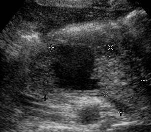
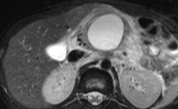
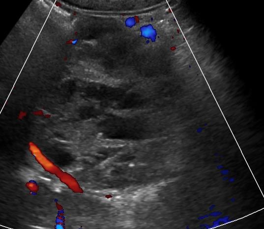
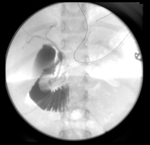
25 1 year old girl with biliary dilatation on US and elevated pancreatic enzymes (Type 1 ChC, long channel & stone)
26 Complications of Pancreatitis Hemorrhage Necrosis Role of MR/MRCP Vascular: splenic vein thrombosis, pseudoaneurysms Pseudocyst formation


27 Hemorrhagic pancreatitis 12 yr old with B-cell ALL T2 with fat sat T2 with fat sat T1 fat sat vibe pre


28 7 yr old Necrotizing pancreatitis T2 fat sat T1 post contrast
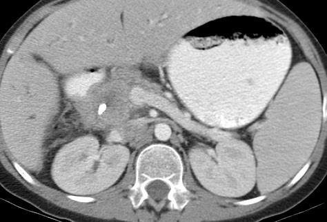
29 Pancreatic pseudocyst 12 yr old boy

30 Chronic Pancreatitis Role of MR/MRCP Imaging features Tissue loss, atrophy PD dilatation and dilation and visualization of side branches Irregular pancreas contour calcifications


31 14 year old girl with chronic hereditary pancreatitis Pancreatic duct stricture (arrows)
32 Pancreatic tumors Overall small percent of all pediatric tumors One study over 10 yrs-92 pancreatic disorders, n=10 panc tumors (0.1%) Epithelial Non-epithelial Exocrine and Endocrine
33 Pancreatic tumors Epithelial Ductal Origin Rare Acinar origin Pancreaticoblastoma Acinar cell carcinoma Solid pseudopapillary tumor (SPEN) Endocrine Congenital hyperinsulinemic hypoglycemia Insulinoma Non-Epithelial Lymphoma (Burkitt s) Sarcomas Lymphangioma PNET


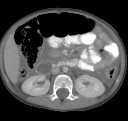
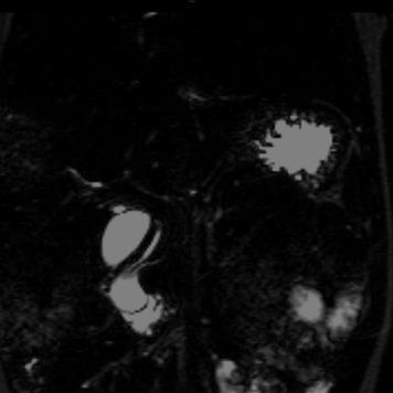
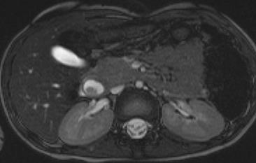
34 11 year old with weight loss & fatigue


35 11 year old with weight loss & fatigue T1 Early Post Gad T2 Delayed Post Gad


36 SPEN Female predominance (Asian & Blacks) 10% males Low malignant potential Comprised of solid and cystic (bloody components) Fibrous capsule Dark T1 and T2 capsule 95% surgical cure
37 Islet Cell Tumors Most common functional islet cell tumor of pancreas insulinomas and gastrinomas All others not reported in children Multiple islet cell tumors seen in MEN type 1 Von Hippel-Lindau phacomatoses Gastrinomas seen in Zollinger-Ellison syndrome
38 Imaging Insulinoma Small hypervascular Majority in the body and tail of pancreas US is usually normal CT and MRI hypervascular MRI- Iso-low SI on T1, bright on T2, hypervascular MRI better for liver mets

39 Insulinoma on MRI and PET
40 Biliary Pathology Choledochal Cyst Alagille Syndrome Congenital Hepatic Fibrosis Primary Sclerosing Cholangitis (PSC)
 II: diverticulum of CBD III: choledochocele IVa &")
41 Choledochal Cysts Not true cysts, ductal ectasia Jaundice, RUQ pain, palpable mass 80% dx in childhood 20% dx in adulthood MRI provides Extent of cyst into the liver Assoc duct dilatation Compression on adj structures Type 4 Result in pancreatitis APBJ /external compression Todani Classification I: involves CBD, saccular, diffuse or focal (80-90%) II: diverticulum of CBD III: choledochocele IVa & IVb: +intra & extrahepatic dilatation; +extrahepatic saccular

42 9 month old Type 1 Choledochal Cyst
, compared to thick slab (right) GB CC GB")

43 Better anatomical detail on MIP from SPACE (left), compared to thick slab (right) GB CC GB CC Coronal MIP from SPACE sequence. Coronal thick slab image. GB: Gallbladder CC: Choledochal cyst
44 Choledochocele Type 3


45 Type 4a Choledochal Cyst causing mass effect on PD pancreatitis CC PD
46 Choledochal Cyst Type V Todani equates this with Caroli disease Purists disagree Caroli disease pure intrahepatic ductal ectasia without any hepatic abn Caroli Syndrome: CHF and Caroli s disease occur together


47 12 year old girl with Caroli s (type V) Dilated CBD Axial HASTE image Arrow: dilated intra-hepatic biliary ducts Axial HASTE image HASTE images reveal dilated intra and extra-hepatic biliary ducts. Liver is normal
48 Alagille Syndrome Role of MRI Portal Hypertension Screen for masses Risk for Hepatocellular ca Recognize large regenerative nodules
 Portal")
49 Congenital Hepatic Fibrosis MR/MRCP ARPKD (nephromegaly with cysts) Portal HTN


50 Congenital Hepatic Fibrosis MR/MRCP Ductal ectasia to varying degrees Choledochal cysts, Caroli syndrome
, consistent with stricture.")
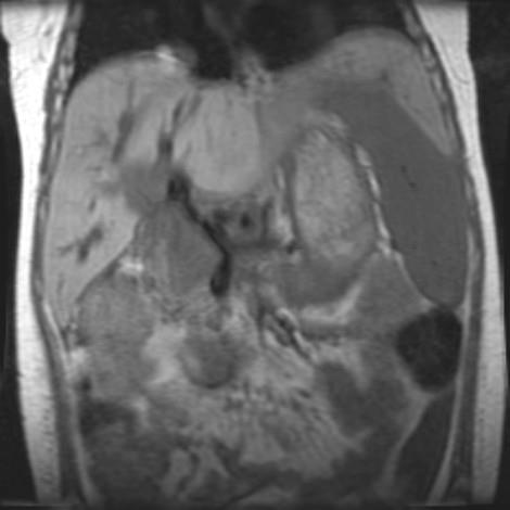
51 5 year old girl with known congenital hepatic fibrosis- stricture of CBD GB S Coronal HASTE image reveals abrupt tapering of dilated CBD (arrow), consistent with stricture. Also seen is splenomegaly (S)

52 Primary Sclerosing Cholangitis Role of MRI Strictures and dilated ducts beaded appearance Risk for malignancy MRCP 100% specificity, PPD; accuracy 85% * MRCP Classification system based on severity** Pitfall of MRCP Overestimate the degree of stricture Coronal 3D MIP with SPACE at 3T *Ferrara C et al Ped Rad 2002 **Majoie CB et al AJR 1991 Vitellas KM AJR 2002

53 12 yr old UC, PSC with Cholangiocarcinoma
54 Conclusion MRCP is an excellent adjunct to US Optimal imaging can be performed at 3T with 2D & 3D seq with BH or respiratory triggered/navigator gated Several contrast options- enhance Focused role MR in the evaluation of common pancreatic and biliary pathology
55 Thank You!
A patient with an unusual congenital anomaly of the pancreaticobiliary tree
 A patient with an unusual congenital anomaly of the pancreaticobiliary tree Thomas Hocker, HMS IV BIDMC Core Radiology Case Presentation September 17, 2007 Review of Normal Pancreaticobiliary Tract Anatomy
IMAGING OF ACUTE AND CHRONIC PANCREATITIS, INCLUDING EXOCRINE FUNCTION
 IMAGING OF ACUTE AND CHRONIC PANCREATITIS, INCLUDING EXOCRINE FUNCTION Andrew T. Trout, MD @AndrewTroutMD Disclosures Grant support National Pancreas Foundation In-kind support - ChiRhoClin modified from:
Biliary MRI w Eovist
 Biliary MRI w Eovist Is there any added value? Elmar M. Merkle, MD Director of MR Imaging Duke University Medical Center elmar.merkle@duke.edu Declaration of Conflict of Interest or Relationship Research
Anatomical and Functional MRI of the Pancreas
 Anatomical and Functional MRI of the Pancreas MA Bali, MD, T Metens, PhD Erasme Hospital Free University of Brussels Belgium mbali@ulb.ac.be Introduction The use of MRI to investigate the pancreas has
MRI Abdomen Protocol Pancreas/MRCP with Contrast
 MRI Abdomen Protocol Pancreas/MRCP with Contrast Reviewed By: Brett Mollard, MD; Anna Ellermeier, MD Last Reviewed: July 2018 Contact: (866) 761-4200 Standard uses: 1. Characterization of cystic and solid
Classification of choledochal cyst with MR cholangiopancreatography in children and infants: special reference to type Ic and type IVa cyst
 Classification of choledochal cyst with MR cholangiopancreatography in children and infants: special reference to type Ic and type IVa cyst Poster No.: C-1333 Congress: ECR 2011 Type: Educational Exhibit
Original Policy Date 12:2013
 MP 6.01.30 Magnetic Resonance Cholangiopancreatography Medical Policy Section Radiology Is12:2013sue 3:2005 Original Policy Date 12:2013 Last Review Status/Date 12:2013 Return to Medical Policy Index Disclaimer
16 April 2010 Resident Teaching Conference. Pancreatitis. W. H. Nealon, M.D., F.A.C.S. J.J. Smith, M.D., D.W.D.
 16 April 2010 Resident Teaching Conference Pancreatitis W. H. Nealon, M.D., F.A.C.S. J.J. Smith, M.D., D.W.D. Santorini Wirsung anatomy.med.umich.edu/.../ duodenum_ans.html Bud and ductology Ventral pancreatic
Anatomy of the biliary tract
 Harvard-MIT Division of Health Sciences and Technology HST.121: Gastroenterology, Fall 2005 Instructors: Dr. Jonathan Glickman Anatomy of the biliary tract Figure removed due to copyright reasons. Biliary
Imaging of liver and pancreas
 Imaging of liver and pancreas.. Disease of the liver Focal liver disease Diffusion liver disease Focal liver disease Benign Cyst Abscess Hemangioma FNH Hepatic adenoma HCC Malignant Fibrolamellar carcinoma
Case 7729 Child with choledochal cyst presenting with episodes of vomitting and jaundice
 Case 7729 Child with choledochal cyst presenting with episodes of vomitting and jaundice dos Santos R 1, Almeida J 1, Mendes PP 2, Pereira S 3, Borges C 3, Soares E 4. 1) Radiology resident, 2) Radiology
Normal anatomy and anatomic variants of the biliary tree and pancreatic ductal system at MRCP - what the clinicians want to know
 Normal anatomy and anatomic variants of the biliary tree and pancreatic ductal system at MRCP - what the clinicians want to know Poster No.: C-1696 Congress: ECR 2014 Type: Educational Exhibit Authors:
Magnetic resonance cholangiopancreatography (MRCP) is an imaging. technique that is able to non-invasively assess bile and pancreatic ducts,
 is an imaging. technique that is able to non-invasively assess bile and pancreatic ducts,") SECRETIN AUGMENTED MRCP Riccardo MANFREDI, MD, MBA, FESGAR Magnetic resonance cholangiopancreatography (MRCP) is an imaging technique that is able to non-invasively assess bile and pancreatic ducts, in
SECRETIN AUGMENTED MRCP Riccardo MANFREDI, MD, MBA, FESGAR Magnetic resonance cholangiopancreatography (MRCP) is an imaging technique that is able to non-invasively assess bile and pancreatic ducts, in
Congenital dilatation of the common bile duct and pancreaticobiliary maljunction clinical implications
 Langenbecks Arch Surg (2009) 394:209 213 DOI 10.1007/s00423-008-0330-6 CURRENT CONCEPT IN CLINICAL SURGERY Congenital dilatation of the common bile duct and pancreaticobiliary maljunction clinical implications
Dr Claire Smith, Consultant Radiologist St James University Hospital Leeds
 Dr Claire Smith, Consultant Radiologist St James University Hospital Leeds Imaging in jaundice and 2ww pathway Image protocol Staging Limitations Pancreatic cancer 1.2.4 Refer people using a suspected
Spectrum of congenital pancretico-biliary ductal anomalies Demonstration by MRCP positively impacts the management
 Spectrum of congenital pancretico-biliary ductal anomalies Demonstration by MRCP positively impacts the management protocol Poster No.: C-1539 Congress: ECR 2012 Type: Educational Exhibit Authors: S. Patwari,
Pediatric Hepatobiliary, Pancreatic & Splenic US
 Pediatric Hepatobiliary, Pancreatic & Splenic US Susan J. Back, MD Department of Radiology, The Children s Hospital of Philadelphia No Disclosures Objectives Normal Abnormal: cases and US advances Objectives
Biliary tree dilation - and now what?
 Biliary tree dilation - and now what? Poster No.: C-1767 Congress: ECR 2012 Type: Educational Exhibit Authors: I. Ferreira, A. B. Ramos, S. Magalhães, M. Certo; Porto/PT Keywords: Pathology, Diagnostic
Navigating the Biliary Tract with CT & MR: An Imaging Approach to Bile Duct Obstruction
 Navigating the Biliary Tract with CT & MR: An Imaging Approach to Bile Duct Obstruction Ann S. Fulcher, MD Medical College of Virginia Virginia Commonwealth University Richmond, Virginia Objectives To
Neuro-endocrine and pancreatic non-adenocarcinomas. Marc Engelbrecht, AMC, Amsterdam
 Neuro-endocrine and pancreatic non-adenocarcinomas Marc Engelbrecht, AMC, Amsterdam Pancreatic Tumors q Epithelial Exocrine q Mesenchymal Ductal Adenocarcinoma (85-95%) Metastasis Lymfoma Acinar Cell Carcinoma
Hilar cholangiocarcinoma. Frank Wessels, Maarten van Leeuwen, UMCU utrecht
 Hilar cholangiocarcinoma Frank Wessels, Maarten van Leeuwen, UMCU utrecht Content Anatomy Biliary strictures (Hilar) Cholangiocarcinoom Staging Biliary tract 1 st order Ductus hepatica dextra Ductus hepaticus
4/9/2018 OBJECTIVES PANCREAOTO BILIARY ULTRASOUND: BEYOND CHOLECYSTITIS
 PANCREAOTO BILIARY ULTRASOUND: BEYOND CHOLECYSTITIS Jean Yves Sewah Kaiser Permanente West Los Angeles 1 OBJECTIVES Discuss the role of ultrasound in the evaluation of the gallbladder, biliary tree and
The Pancreas. Basic Anatomy. Endocrine pancreas. Exocrine pancreas. Pancreas vasculature. Islets of Langerhans. Acinar cells Ductal System
 SGNA: Back to Basics Rogelio G. Silva, MD Assistant Clinical Professor of Medicine University of Illinois at Chicago Department of Medicine Division of Gastroenterology Advocate Christ Medical Center GI
X-ray Corner. Imaging of The Pancreas. Pantongrag-Brown L
 X-ray Corner 125 Imaging of The Pancreas Modern imaging modalities commonly used in pancreas include ultrasound (US), CT, and MRI. Pancreas is a retroperitoneal organ which makes it difficult to visualize
Pancreatic Lesions. Valerie Jefford Pediatric Surgery Rounds June 6, 2003
 Pancreatic Lesions Valerie Jefford Pediatric Surgery Rounds June 6, 2003 Embryology 4 th week 2 buds of endodermal origin from caudal foregut Dorsal and ventral bud Ventral migrates dorsally with CBD (below/behind
Role of Imaging Methods in Diagnosis of Acute Pancreatitis. Válek V. Radiologická klinika, FN Brno a LF MU v Brně
 Role of Imaging Methods in Diagnosis of Acute Pancreatitis Válek V. Radiologická klinika, FN Brno a LF MU v Brně New Classification: Acute Pancreatitis 2007 revision of Atlanta classification and definitions
Autoimmune Pancreatitis: A Great Imitator
 Massachusetts General Hospital Harvard Medical School Autoimmune Pancreatitis: A Great Imitator Dushyant V Sahani MD dsahani@partners.org Autoimmune Pancreatitis: Learning Objectives Clinical manifestations
CTA/MRA of Pediatric Hepatic Masses Radiology-Pathology Correlation
 Acta Radiológica Portuguesa, Vol.XVIII, nº70, pág. 41-50, Abr.-Jun., 2006 CTA/MRA of Pediatric Hepatic Masses Radiology-Pathology Correlation Marilyn J. Siegel Mallinckrodt Institute of Radiology, Washington
Primary Sclerosing Cholangitis and Cholestatic liver diseases. Ahsan M Bhatti MD, FACP Bhatti Gastroenterology Consultants
 Primary Sclerosing Cholangitis and Cholestatic liver diseases Ahsan M Bhatti MD, FACP Bhatti Gastroenterology Consultants I have nothing to disclose Educational Objectives What is PSC? Understand the cholestatic
State of the Art Imaging for Hepatic Malignancy: My Assignment
 State of the Art Imaging for Hepatic Malignancy: My Assignment CT vs MR vs MRCP Which one to choose for HCC vs Cholangiocarcinoma What special protocols to use for liver tumors Role of PET and Duplex US
MR imaging of primary sclerosing cholangitis (PSC) using the hepatobiliary specific contrast agent Gd-EOB-DTPA
 using the hepatobiliary specific contrast agent Gd-EOB-DTPA") MR imaging of primary sclerosing cholangitis (PSC) using the hepatobiliary specific contrast agent Gd-EOB-DTPA Poster No.: C-0019 Congress: ECR 2010 Type: Educational Exhibit Topic: Abdominal Viscera (Solid
MR imaging of primary sclerosing cholangitis (PSC) using the hepatobiliary specific contrast agent Gd-EOB-DTPA Poster No.: C-0019 Congress: ECR 2010 Type: Educational Exhibit Topic: Abdominal Viscera (Solid
CT 101 :Pancreas and Spleen
 CT 101 :Pancreas and Spleen Shikha Khullar,, MD, MPH Division of Radiology University of South Alabama The Pancreas Normal Pancreas 3 Phase Pancreatic CT Non contrast Arterial phase : 30-35 35 second
MRI of the Hepatobiliary System
 BODY APPLICATIONS OF MRI 51 Chapter Four C H A P T E R F O U R MRI of the Hepatobiliary System After completing this chapter, the reader will be able to: Develop a protocol for liver MRI Identify benign
To describe normal anatomy and common variants in biliary tree anatomy including the prevalence of these.
 Up for debate: Should 3.0T magnetic resonance pancreatography (MRCP) become a routine part of preoperative planning for patients undergoing laparoscopic cholecystectomy? Poster No.: C-0196 Congress: ECR
Cholangiocarcinoma (Bile Duct Cancer)
") Cholangiocarcinoma (Bile Duct Cancer) The Bile Duct System (Biliary Tract) A network of bile ducts (tubes) connects the liver and the gallbladder to the small intestine. This network begins in the liver
Cholangiocarcinoma (Bile Duct Cancer) The Bile Duct System (Biliary Tract) A network of bile ducts (tubes) connects the liver and the gallbladder to the small intestine. This network begins in the liver
Common and unusual CT and MRI manifestations of pancreatic adenocarcinoma: a pictorial review
 Review Article Common and unusual CT and MRI manifestations of pancreatic adenocarcinoma: a pictorial review Min-Jie Yang, Su Li, Yong-Guang Liu, Na Jiao, Jing-Shan Gong Department of Radiology, Shenzhen
Malignant Focal Liver Lesions
 Malignant Focal Liver Lesions Other Than HCC Pablo R. Ros, MD, MPH, PhD Departments of Radiology and Pathology University Hospitals Cleveland Medical Center Case Western Reserve University Pablo.Ros@UHhospitals.org
CASE 1 11/1/2016 HEPATOBILIARY IMAGING CASE PRESENTATIONS DECLARATION. Dr. Chirag Patel ORGAN IMAGING yr old lady
 HEPATOBILIARY IMAGING CASE PRESENTATIONS DECLARATION No financial disclosures or affiliations with commercial organisations No discussion of investigational or off-label use of medical devices, products
Newcastle HPB MDM updated radiology imaging protocol recommendations. Author Dr John Scott. Consultant Radiologist Freeman Hospital
 Newcastle HPB MDM updated radiology imaging protocol recommendations Author Dr John Scott. Consultant Radiologist Freeman Hospital This document is intended as a guide to aid radiologists and clinicians
Hepatobiliary Contrast Agents
 Hepatobiliary Contrast Agents SCBT/MR Annual Meeting Salt Lake City September 21, 2016 Scott B. Reeder, MD, PhD Department of Radiology University of Wisconsin Madison, WI Disclosures University of Wisconsin-Madison
Evidence based imaging of the pancreas
 Evidence based imaging of the pancreas D.Vanbeckevoort, D.Bielen, K.Op de beeck, R.Vanslembrouck Department of Radiology Chairman Prof. Dr. R.Oyen Non-invasive imaging tests available for the diagnosis
CT & MRI of Benign Liver Neoplasms Srinivasa R Prasad
 CT & MRI of Benign Liver Neoplasms Srinivasa R Prasad No financial disclosures Acknowledgements Many thanks to Drs. Heiken, Narra & Menias (MIR) Dr. Sahani (MGH) for sharing images Benign Liver Tumors:
Pediatric Retroperitoneal Masses Radiologic-Pathologic Correlation
 Acta Radiológica Portuguesa, Vol.XVIII, nº 70, pág. 61-70, Abr.-Jun., 2006 Pediatric Retroperitoneal Masses Radiologic-Pathologic Correlation Marilyn J. Siegel Mallinckrodt Institute of Radiology, Washington
Lesions of the pancreaticoduodenal groove, a pictorial review
 Lesions of the pancreaticoduodenal groove, a pictorial review Poster No.: C-2131 Congress: ECR 2013 Type: Educational Exhibit Authors: E. Ni Mhurchu, L. Lavelle, I. Murphy, S. Skehan ; IE, Dublin/ IE Keywords:
Variations of the Union between the Terminal Bile Duct and the Pancreatic Duct in Patients with Pancreaticobiliary Maljunction
 Yamanashi Med. J. 18(4), 67~ 75, 2003 Review Variations of the Union between the Terminal Bile Duct and the Pancreatic Duct in Patients with Pancreaticobiliary Maljunction Hideki FUJII 1) 1) Department
Cholangiocarcinoma: Radiologic evaluation and interventions
 November 2014 Cholangiocarcinoma: Radiologic evaluation and interventions Colin Nevins, Harvard Medical School Year III Agenda Initial course and work-up Endoscopic retrograde cholangiopancreatography
Primary sclerosing cholangitis: Evaluation with MR cholangiopancreatography (MRCP)
") The Egyptian Journal of Radiology and Nuclear Medicine (2011) 42, 351 356 Egyptian Society of Radiology and Nuclear Medicine The Egyptian Journal of Radiology and Nuclear Medicine www.elsevier.com/locate/ejrnm
The Egyptian Journal of Radiology and Nuclear Medicine (2011) 42, 351 356 Egyptian Society of Radiology and Nuclear Medicine The Egyptian Journal of Radiology and Nuclear Medicine www.elsevier.com/locate/ejrnm
GASTROINTESTINAL IMAGING STUDY GUIDE
 GASTROINTESTINAL IMAGING STUDY GUIDE Pharynx Diverticula Foreign bodies Trauma o Motility Disorders Esophagus Diverticula Trauma Esophagitis Barrett esophagus Rings, webs, and strictures Varices Benign
Hepatobiliary Contrast Agents for Liver MRI
 Hepatobiliary Contrast Agents for Liver MRI Scott B. Reeder, MD, PhD International Society for Magnetic Resonance in Medicine Sociedad Mexicana de Radiologia e Imagen (SMRI) Mexico City June 4, 2014 Department
What to do and not do before seeking surgical consultation for a patient with suspected pancreatic cancer
 What to do and not do before seeking surgical consultation for a patient with suspected pancreatic cancer 9 Th Annual Symposium on Gastrointestinal Cancers, St. Louis University School of Medicine Carlos
Cystic lesions of the pancreato-duodenal confluence. Who is who?
 Cystic lesions of the pancreato-duodenal confluence. Who is who? Poster No.: C-0183 Congress: ECR 2014 Type: Educational Exhibit Authors: L. Goiburu Gonzalez, M. Paraira Beser, A. Pedrerol Perez, 1 3 1
Liver MRI in 30 minutes
 X Liver MRI in 30 minutes SCBT/MR Annual Meeting Salt Lake City September 18, 2016 Scott B. Reeder, MD, PhD Department of Radiology University of Wisconsin Madison, WI Disclosures University of Wisconsin-Madison
BILIARY TRACT & PANCREAS, PART II
 CME Pretest BILIARY TRACT & PANCREAS, PART II VOLUME 41 1 2015 A pretest is mandatory to earn CME credit on the posttest. The pretest should be completed BEFORE reading the overview. Both tests must be
MR cholangiopancreatography; Predicting imaging findings for differentiation of malignant bile ductal obstruction versus benign lesion
 Acta Med Kindai Univ Vol.43, No.1 1-8, 2018 1 MR cholangiopancreatography; Predicting imaging findings for differentiation of malignant bile ductal obstruction versus benign lesion Shojiro Hidaka 1,2,
Role of Gd-EOB-DTPA enhanced MR Imaging in the evaluation of the transplanted liver: Advantages and Limitations
 Role of Gd-EOB-DTPA enhanced MR Imaging in the evaluation of the transplanted liver: Advantages and Limitations Robinson Yu, MD, Amir A. Borhani, MD, Alessandro Furlan, MD, Matthew T. Heller, MD, Mitchell
IT 의료융합 1 차임상세미나 복부질환초음파 이재영
 IT 의료융합 1 차임상세미나 2013-4-3 복부질환초음파 이재영 나는오늘누구를위하여 종을울리나? 전통적의료 의사 공학설계자 의사 최첨단진단장비들 USG, CT, MRI 환자 환자 현대의료 사용자중심의사고 US in the Abdomen Detection DDx Look Behavior Response by external stimuli Guiding Tool
Abdominal MRI Techniques in Pediatric Oncology
 Abdominal MRI Techniques in Pediatric Oncology Jonathan R. Dillman, M.D. Assistant Professor Departments of Radiology & Urology Section of Pediatric Radiology C.S. Mott Children s Hospital Disclosures
LIVER IMAGING TIPS IN VARIOUS MODALITIES. M.Vlychou, MD, PhD Assoc. Professor of Radiology University of Thessaly
 LIVER IMAGING TIPS IN VARIOUS MODALITIES M.Vlychou, MD, PhD Assoc. Professor of Radiology University of Thessaly Hepatocellular carcinoma is a common malignancy for which prevention, screening, diagnosis,
Radiology of hepatobiliary diseases
 GI cycle - Lecture 14 436 Teams Radiology of hepatobiliary diseases Objectives 1. To Interpret plan x-ray radiograph of abdomen with common pathologies. 2. To know the common pathologies presentation.
Pictorial review of Benign Biliary tract abnormality on MRCP/MRI Liver with Endoscopic (including splyglass) and Endoscopic Ultrasound correlation
 and Endoscopic Ultrasound correlation") Pictorial review of Benign Biliary tract abnormality on MRCP/MRI Liver with Endoscopic (including splyglass) and Endoscopic Ultrasound correlation Poster No.: C-2617 Congress: ECR 2015 Type: Educational
Pictorial review of Benign Biliary tract abnormality on MRCP/MRI Liver with Endoscopic (including splyglass) and Endoscopic Ultrasound correlation Poster No.: C-2617 Congress: ECR 2015 Type: Educational
Liver imaging takes a step forward with Ingenia
 Publication for the Philips MRI Community ISSUE 49 2013 / 2 Liver imaging takes a step forward with Ingenia Lyon South Hospital strives to move from several studies first CT, then MR or PET to using just
Chronic Pancreatitis: When to Scope? Gregory A. Cote, MD, MS Assistant Professor of Medicine Indiana University School of Medicine
 Chronic Pancreatitis: When to Scope? Gregory A. Cote, MD, MS Assistant Professor of Medicine Indiana University School of Medicine Endoscopy & Chronic Pancreatitis Diagnosis EUS ERCP Exocrine Function
Case Report Uncommon Mixed Type I and II Choledochal Cyst: An Indonesian Experience
 Case Reports in Surgery Volume 2013, Article ID 821032, 4 pages http://dx.doi.org/10.1155/2013/821032 Case Report Uncommon Mixed Type I and II Choledochal Cyst: An Indonesian Experience Fransisca J. Siahaya,
Ruptured choledochal cyst: a rare presentation and unique approach to management
 Case Report Ruptured choledochal cyst: a rare presentation and unique approach to management Michael Meschino, Carlos García-Ochoa, Roberto Hernandez-Alejandro London Health Sciences Centre, Western University,
Vesalius SCALpel : Biliary (see also: biliary/pancreatic folios) Physiology
 Physiology") Vesalius SCALpel : Biliary (see also: biliary/pancreatic folios) Physiology 95% of bile acids reabsorbed; colic and chenodeoxycolic primary bile acids cholecystokinin (CCK) major stimulus of gallbladder
Vesalius SCALpel : Biliary (see also: biliary/pancreatic folios) Physiology 95% of bile acids reabsorbed; colic and chenodeoxycolic primary bile acids cholecystokinin (CCK) major stimulus of gallbladder
IMAGING OF LIVER, BILIARY TREE, PANCREAS
 IMAGING OF LIVER, BILIARY TREE, PANCREAS Department of Radiology West China Hospital, Sichuan University Yao Jin Learning Points The methodology for imaging the LBP (liver, biliary tree, and pancreas )
Overview of PSC Making the Diagnosis
 Overview of PSC Making the Diagnosis Tamar Taddei, MD Assistant Professor of Medicine Yale University School of Medicine Overview Definition Epidemiology Diagnosis Modes of presentation Associated diseases
Liver Tumors. Prof. Dr. Ahmed El - Samongy
 Liver Tumors Prof. Dr. Ahmed El - Samongy Objective 1. Identify the most important features of common benign liver tumors 2. Know the risk factors, diagnosis, and management of hepatocellular carcinoma
Essentials of Clinical MR, 2 nd edition. 65. Benign Hepatic Masses
 65. Benign Hepatic Masses Pulse sequences acquired for abdominal MRI typically consist of fast acquisition schemes such as single-shot turbo spin echo (i.e. HASTE) and gradient echo schemes such as FLASH
Magnetic resonance cholangiopancreatography: the ABC of MRCP
 Insights Imaging (2012) 3:11 21 DOI 10.1007/s13244-011-0129-9 PICTORIAL REVIEW Magnetic resonance cholangiopancreatography: the ABC of MRCP Nyree Griffin & Geoff Charles-Edwards & Lee Alexander Grant Received:
Afternoon Session Cases
 Afternoon Session Cases Case 1 19 year old woman Presented with abdominal pain to community hospital Mild incr WBC a14, 000, Hg normal, lipase 100 (normal to 75) US 5.2 x 3.7 x 4 cm mass in porta hepatis
Pediatric Pancreatic Lesions
 Pediatric Pancreatic Lesions Pediatric Surgery Grand Rounds 9 October 2013 Tim Weatherall, PGY2 The University of Tennessee Health Science Center Memphis, TN Disclosures No financial interests to disclose
BODY MRI PROTOCOLS 1.5T
 BODY MRI PROTOCOLS 1.5T Table of Contents Non Specific...3-5 Renals.....6-8 Adrenals.....9-11 Pancreas..12-14 Liver 15-17 Liver No Gad.18-21 Liver Eovist..22-24 Liver Iron Quant 25-29 MRCP.30-34 ENTEROGRAPHY.35-38
Pancreas Quizzes c. Both A and B a. Directly into the blood stream (not using ducts)
") Pancreas Quizzes Quiz 1 1. The pancreas produces hormones. Which type of hormone producing organ is the pancreas? a. Endocrine b. Exocrine c. Both A and B d. Neither A or B 2. Endocrine indicates hormones
Pancreas Quizzes Quiz 1 1. The pancreas produces hormones. Which type of hormone producing organ is the pancreas? a. Endocrine b. Exocrine c. Both A and B d. Neither A or B 2. Endocrine indicates hormones
Personal Profile. Name: 劉 XX Gender: Female Age: 53-y/o Past history. Hepatitis B carrier
 Personal Profile Name: 劉 XX Gender: Female Age: 53-y/o Past history Hepatitis B carrier Chief complaint Fever on and off for 2 days Present illness 94.10.14 Sudden onset of epigastric pain 94.10.15 Fever
Enhancements in Hepatobiliary Imaging:
 Enhancements in Hepatobiliary Imaging: S. Channual 1, MD; A. Pahwa 2, MD; S. Raman 1, MD. 1 UCLA Medical Center, Department of Radiologic Sciences 2 Olive-View UCLA Medical Center, Department of Radiology
Biliary cancers: imaging diagnosis. Study of 30 cases
 Biliary cancers: imaging diagnosis. Study of 30 cases N Hammoune, S Semlali, M Eddarai, T. Amil, M Zentar, S. El Kandri,, M Benameur,, S Chaouir. Radiology Department. Mohamed V Military Hospital. Rabat-
Post-operative complications following hepatobiliary surgery: imaging findings and current radiological treatment options
 Post-operative complications following hepatobiliary surgery: imaging findings and current radiological treatment options Poster No.: C-1501 Congress: ECR 2015 Type: Educational Exhibit Authors: A. Hadjivassiliou,
Pancreatic Benign April 27, 2016
 Department of Surgery Pancreatic Benign April 27, 2016 James Choi Dr. Hernandez Objectives Medical Expert: 1. Anatomy and congenital anomalies of the pancreas and pancreatic duct (divisum, annular pancreas
JOHN M UECKER, MD, FACS COMPLEX PANCREATICODUODENAL INJURIES
 JOHN M UECKER, MD, FACS COMPLEX PANCREATICODUODENAL INJURIES THE PROBLEM DUODENAL / PANCREATIC INJURIES Difficult to diagnose Not very common Anatomic and physiologic challenges 90% rate of associated
Management of Pancreatic Fistulae
 Management of Pancreatic Fistulae Jose Ramos University of the Witwatersrand Donald Gordon Medical Centre Fistula definition A Fistula is a permanent abnormal passageway between two organs (epithelial
Disclosures. Overview. Case 1. Common Bile Duct Sizes 10/14/2016. General GI + Advanced Endoscopy: NAFLD/Stones/Pancreatitis
 Disclosures General GI + Advanced Endoscopy: NAFLD/Stones/Pancreatitis 123 Blank Blank, LLC Aldo Maspons, MD Assistant Professor Director of Endoscopy Department of Pediatrics Texas Tech University Health
PANCREATIC PSEUDOCYSTS. Madhuri Rao MD PGY-5 Kings County Hospital Center
 PANCREATIC PSEUDOCYSTS Madhuri Rao MD PGY-5 Kings County Hospital Center 34 yo M Case Presentation PMH: Chronic pancreatitis (ETOH related) PSH: Nil Meds: Nil NKDA www.downstatesurgery.org Symptoms o Chronic
Imaging of Neuroendocrine Metastases
 Imaging of Neuroendocrine Metastases Aoife Kilcoyne, Shaunagh McDermott, Colin McCarthy,Manuel Patino, Dushyant Sahani, Michael Blake Abdominal Imaging Division Massachusetts General Hospital Disclosure
Imaging of common diseases of hepatobiliary and GI system
 Imaging of common diseases of hepatobiliary and GI system Natthaporn Tanpowpong, M.D. Diagnostic radiology Faculty of Medicine, Chulalongkorn University Normal plain radiograph A = Common bile duct
Case 1. Intro to Gallbladder & Pancreas Pathology. Case 1 DIAGNOSIS??? Acute Cholecystitis. Acute Cholecystitis. Helen Remotti M.D.
 Cholecystitis acute chronic Gallbladder tumors Adenomyoma (benign) Adenocarcinoma Pancreatitis acute chronic Pancreatic tumors Intro to Gallbladder & Pancreas Pathology Helen Remotti M.D. Case 1 70 year
Alice Fung, MD Oregon Health and Science University
 Alice Fung, MD Oregon Health and Science University Disclosure Comments The speaker Alice Fung, MD Has relevant financial relationships to disclose. Received honorarium from (Guerbet). This individual
X-Ray Corner. Imaging Approach to Cystic Liver Lesions. Pantongrag-Brown L. Solitary cystic liver lesions. Hepatic simple cyst (Figure 1)
") THAI J 136 Imaging Approach to Cystic Liver Lesions GASTROENTEROL 2013 X-Ray Corner Imaging Approach to Cystic Liver Lesions Pantongrag-Brown L Cystic liver lesions are common findings in daily practice
THAI J 136 Imaging Approach to Cystic Liver Lesions GASTROENTEROL 2013 X-Ray Corner Imaging Approach to Cystic Liver Lesions Pantongrag-Brown L Cystic liver lesions are common findings in daily practice
Unusual Pancreatic Neoplasms RTC 2/11/2011
 Unusual Pancreatic Neoplasms RTC 2/11/2011 Objectives Intraductal Papillary Mucinous Neoplasm (IPMN) Mucinous Cystic Neoplasm (MCN) Islet Cell Tumors Insulinoma Glucagonoma VIPoma Somatostatinoma Gastrinoma
ABDOMINAL DIFFUSION WEIGHTED MR
 ABDOMINAL DIFFUSION WEIGHTED MR Frank Miller, M.D. FACR Professor of Radiology Chief, Body Imaging Section Medical Director, MR Imaging Northwestern University Feinberg School of Medicine fmiller@northwestern.edu
Endoscopic Management of Biliary Strictures. Sammy Ho, MD Director of Pancreaticobiliary Services and Endoscopic Ultrasound Montefiore Medical Center
 Endoscopic Management of Biliary Strictures Sammy Ho, MD Director of Pancreaticobiliary Services and Endoscopic Ultrasound Montefiore Medical Center Malignant Biliary Strictures Etiologies: Pancreatic
Imaging of the Pancreas: Technique and Clinical Applications
 Clinical Abdominal Imaging MAGNETOM Flash (69) 3/2017 Imaging of the Pancreas: Technique and Clinical Applications Giovanni Morana 1 ; Alex Faccinetto 1 ; Alberto Dorigo 1 ; Pierluigi Ciet 2 ; Silvia Venturini
Noncalculous Biliary Disease Dean Abramson, M.D. Gastroenterologists, P.C. Cedar Rapids. Cholestasis
 Noncalculous Biliary Disease Dean Abramson, M.D. Gastroenterologists, P.C. Cedar Rapids Cholestasis Biochemical hallmark Impaired bile flow from liver to small intestine Alkaline phosphatase is primary
Caroli s disease: magnetic resonance imaging features
 Eur Radiol (2002) 12:2730 2736 DOI 10.1007/s00330-002-1471-6 HEPATOBILIARY PANCREAS France Guy François Cognet Marie Dranssart Jean-Pierre Cercueil Laurent Conciatori Denis Krausé Caroli s disease: magnetic
6 th August 2018 Day 1 - Gallbladder & Bile duct Topic
 Venue: Sterling Hospital Auditorium, Sterling Hospitals, Gurukul Road Ahmedabad, Gujarat 6 th August 2018 Day 1 - Gallbladder & Bile duct Registration(8:00am-8:15am) Inauguration(8:15am-8:30am) Welcome
UTILITY OF THREE DIMENSIONAL MAGNETIC RESONANCE CHOLANGIOPANCREATOGRAPHY IN EVALUATION OF BILIARY OBSTRUCTION IN ADULTS
 - 842 - UTILITY OF THREE DIMENSIONAL MAGNETIC RESONANCE CHOLANGIOPANCREATOGRAPHY IN EVALUATION OF BILIARY OBSTRUCTION IN ADULTS Moanes M. Enaba MD *Tarek H. ELKammash**MD,, Mansour M. Morsy ***MD * Department
Jaundice. Agnieszka Dobrowolska- Zachwieja, MD, PhD
 Jaundice Agnieszka Dobrowolska- Zachwieja, MD, PhD Jaundice definition Jaundice, as in the French jaune, refers to the yellow discoloration of the skin. It arises from the abnormal accumulation of bilirubin
Indications: Abdomen and pelvis for malignancy. If there is history of small bowel or mesentery tumor, see "contrast" section for oral contrast**
 MRB AB01AMR 01aAbdomen (with dynamics) + Pelvis (pre/post) A74183 A72197 Indications: Abdomen and pelvis for malignancy. If there is history of small bowel or mesentery tumor, see "contrast" section for
Case Study: #3: Gallbladder Carcinoma?
 Case Study: #3: Gallbladder Carcinoma? By: Megan Wyatt K. SON Wyatt 225 2B1 RDMS, RVT Patient: Male 85 YOA Caucasian Indication: Elevated Alkaline Phosphatase History Annual physical showed elevated alkaline