FUNCTIONAL ASPECTS OF DRUSEN REGRESSION IN AGE-RELATED MACULAR DEGENERATION
|
|
|
- Adele Casey
- 6 years ago
- Views:
Transcription
1 FUNCTIONAL ASPECTS OF DRUSEN REGRESSION IN AGE-RELATED MACULAR DEGENERATION Ferenc B Sallo 1,2, Ehud Rechtman 1, Tunde Peto 1, Dinu Stanescu-Segall 1, Gabor Vogt 3, Alan C Bird 1,2, Frederick W Fitzke 2 1 Moorfields Eye Hospital, London, UK 2 UCL Institute of Ophthalmology, London, UK 3 National Health Centre, Budapest, Hungary Corresponding author: Ferenc B Sallo The Reading Centre Department of Research and Development Moorfields Eye Hospital 162 City Road London EC1V 2PD Keywords: Macula; Degeneration; Psychophysics; Imaging; AMD; drusen; natural history; regression Word count: 2710 (excluding title page, abstract, references, tables and figures)
2 ABSTRACT Aims: To investigate the functional implications of macular soft drusen regression in AMD eyes. Methods: Patients were selected from a large ongoing collection of clinical data at Moorfields Eye Hospital. Phenotyping based on standard colour fundus images was performed according to the system defined by the International Classification for ARM, by certified graders masked to the main aim of the study. Fundus Autofluorescence (FA) was recorded using a Heidelberg Retina Angiograph 2. Where drusen regression was confirmed by independent grading, the patient was invited for photopic and scotopic Fine Matrix Mapping (FMM). Phenotype and functional data were analyzed for correlations between fundus appearance, autofluorescence and retinal sensitivity. Results: Fundus and FA images of 960 patients were screened, soft drusen regression was detected in 34 cases, 14 patients agreed to participate in the study, ranging in age from 52 to 84 years (median 72). The mean follow-up period was 5.9 years (range years). FMM showed generalised threshold elevation relative to normal controls both under photopic and scotopic conditions. Scotopic sensitivity loss exceeded photopic loss in all cases. Sensitivity loss over areas with drusen or regressed drusen did not differ significantly from that over non-drusen areas. Conclusion: Macular soft drusen may fade or disappear without detectable ophthalmoscopic, FA or psychophysical signs of local dysfunction. This phenomenon is a potential source of misclassification. The prognosis for cases with true regression of drusen compared with those without needs to be considered in future studies on AMD.
3 INTRODUCTION Age-Related Macular Degeneration (AMD) is the leading cause of severe irreversible central visual loss among the elderly in western industrialized countries.[1, 2] The aetiology and pathogenesis of AMD are poorly understood. Risk factors identified include age, certain genetic variants, smoking and the presence of macular soft drusen with high-risk characteristics.[1, 2, 3, 4] Clinically, early AMD is characterized by abnormalities of the retinal pigment epithelium (RPE), soft drusen and mild visual symptoms, predominantly under dark-adapted conditions.[5, 6] Late AMD is dominated by either geographic atrophy (GA) or neovascularisation (NV), typically with severe central function loss.[5] AMD affects primarily the RPE, Bruch's Membrane (BrM) and choriocapillaris (the RPE/BrM complex). In early AMD, a thickening of BrM and an accumulation of focal and diffuse lipid-rich deposits (drusen, basal linear and laminar deposits) under the RPE and within BrM are prominent.[7] The consequential reduction in hydraulic conductivity may impede transport through BrM and thus contribute to the pathogenesis of lesions seen in late disease.[8] The origin and mechanism of debris deposition are little known. Of the sub-rpe deposits, drusen alone are detectable clinically. The natural history of soft drusen may involve an increase in size, area and confluence with subsequent NV, or fading and disappearance which is believed to be associated with atrophy of the overlying RPE and photoreceptors.[3] The functional and prognostic implications of true drusen regression, however, have not been demonstrated. Since the RPE/BrM complex is essential for photoreceptor survival, psychophysical assessment of photoreceptor function may yield information about the significance of clinically observable retinal changes that may not be provided by other methods until later or at all.[9] Imaging the distribution of autofluorescence in the fundus (fundus autofluorescence, FA) represents an additional tool for evaluating RPE health.[10] Visualised by a confocal Scanning Laser Ophthalmoscope (cslo, excitation λ=488nm) FA is mainly derived from lipofuscin (LF),[10, 11] a complex material produced in RPE cells from residues of incomplete lysosomal degradation of photoreceptor outer segment (POS) discs. LF contains components toxic to the RPE (primarily A2-E isomers), its excessive accumulation is believed to precede photoreceptor degeneration in AMD.[11] The aim of this study was to investigate the functional implications of macular soft drusen regression in AMD eyes.
4 PATIENTS AND METHODS Patients were selected from a large ongoing collection of clinical data at the Reading Centre of Moorfields Eye Hospital. Inclusion criteria were: a clinical diagnosis of AMD, both manifest and resolved soft macular drusen, a level of fixation and general fitness sufficient to perform the full test sequence. Patients with exudative late-stage AMD in the eye studied were excluded. Imaging Standard 30º stereo colour images (SCI) of the fundus, centred on the fovea (Field 2) were recorded digitally. FA imaging was performed using a cslo (Heidelberg Retina Angiograph 2, Heidelberg Engineering GmbH, Dossenheim, Germany). The fundus was illuminated using a solid-state laser (488 nm), induced fluorescence was recorded through a long-pass filter with a short-wavelength cut-off at 500nm. The image of a 30 x30 retinal area centred on the fovea was recorded digitally at 1536x1536 pixel (5µm/pixel) and 256-level grey-scale resolution. To reduce random noise, a master image was produced by averaging 16 individually recorded images for each eye in the study. Lens opacities - where present - were in all cases diffuse and permitted adequate imaging of FA. Phenotyping Detailed phenotyping was performed by grading based on Field 2 colour and FA images, according to the system defined in the International Classification for ARM and AMD,[5] independently by two certified graders masked to the identity of the patients and the main aim of the study. These gradings were compared for (both inter- and intra-observer) reliability. A final copy was created by adjudication and changes over time in the final copy were analysed. In cases where the simultaneous presence of AMD, disappearance of drusen and absence of exudative disease were confirmed, the patient was invited for functional testing. Psychophysical testing For psychophysical testing Fine Matrix Mapping (FMM) was selected due to its superior spatial resolution and its ability to measure wide ranges of sensitivity changes. The full test sequence included a visual acuity test, a standard visual field examination using the 30-2 program of the Humphrey Field Analyzer (HFA, Carl Zeiss Ltd., UK), a photopic FMM test, pupil dilatation (with 1.0% tropicamide and 2.5% phenylephrine), dark adaptation for 45 minutes, a scotopic 30-2 visual field test, scotopic FMM and finally the determination of the location and stability of fixation. Fine-Matrix Mapping FMM was performed using a modified HFA. Test flashes were positioned over the retinal area of interest, including both drusen and areas where the resolution of drusen was confirmed by grading. The coordinates of four interlaced 5x5 grids (25 locations with 2 intervals) were entered in the Custom Grid feature of the perimeter. Each grid was offset relative to the other grids by 1 on either the x, y, or both axes. Data thus obtained were merged into a single 9 x 9 matrix of 100 test locations with 1 intervals. A standard (Goldmann) size 3 stimulus was used. Photopic testing was done using a white stimulus with a background illuminance of 31.5 apostilbs, scotopic measurements were done using a blue stimulus with no background illumination. Fixation stability was monitored through an infrared camera. Detection threshold sensitivity was expressed in decibels and thresholds as
5 log units. The maximum stimulus illuminance of 10,000 apostilbs was interpreted as 0dB. Data processing FMM thresholds were processed by (3x3) Gaussian filtering to improve repeatability.[12] The attenuation of the original signal and loss of detail inherent in all smoothing methods was in our case insignificant and outweighed by the benefits with respect to noise reduction (repeatability).[13] For each patient, sensitivity data were analyzed intra-individually as well as compared with age-matched normal data (collected under identical conditions) that have been published,[14, 15, 16] through point-by-point subtraction of averaged normal values at corresponding coordinates within the FMM matrix. Filtered data were used to calculate the mean and the maximum threshold elevation from baseline as well as elevation relative to averaged threshold levels of the normal control group. Interpolated threshold values at 0.25 intervals were used to generate a contour plot showing luminance sensitivity gradients across the matrix. Three-dimensional threshold profiles were also generated. Composite images containing overlays of aligned sequential colour fundus and FA images and sensitivity threshold plots were analyzed for correlations between fundus appearance, FA and retinal sensitivity. Total area covered by drusen at baseline and at FMM testing was measured for each zone within the IC grading grid.[5] Areas with disappearing drusen, persistent drusen and drusen-free areas were identified and associated levels of sensitivity loss were compared. Software used include: Adobe Photoshop v7 with the FoveaPro plug-in, ImageJ v1.38x, Microsoft Excel and SPSS v11. Fixation stability A major factor influencing FMM test reliability is the stability of fixation. For the measurement of fixation stability, a customized cslo was used in a way similar to a technique described previously.[17] Values thus obtained reflect eye movements and are used to calculate the bivariate contour ellipse area (BCEA), which is the area of an ellipse on the retinal surface within which the centre of the target was imaged 68% of the time. BCEA is a standardized measure which provides a means for quantification and comparison of fixation stability. Smaller BCEA values correspond to more precise fixation. The study protocol adhered to the tenets of the Declaration of Helsinki and was approved by the local ethics committee. Before inclusion, written, informed consent was obtained from each participating patient after explanation of the nature of the study. Maximum retinal irradiance of lasers used was well below the limits established by the American National Standards Institute (ANSI Z136.1; 1993) and other international standards.
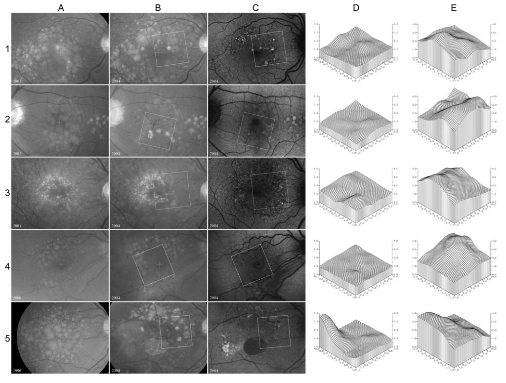
6 RESULTS Sequential colour fundus and FA images of 960 patients were screened for disappearing drusen. Soft drusen regression was detected in 34 cases (25 spontaneous, 9 following prophylactic laser treatment). 19 patients met all inclusion criteria, 14 (10 female, 4 male) agreed to participate in the study, ranging in age from 52 to 84 years (median age was 72 years). The baseline of the study was defined as the earliest date when both clinical data and fundus SCI were available, and the endpoint as the date the patient was last seen clinically. The mean follow-up period was 5.9 years (ranging from 2.8 to 14.4 years). Phenotype The predominant phenotype in the study eye at baseline was soft drusen in all cases. Disappearance of drusen was followed by NV in one, GA in 3 cases, while in 10 cases no indications of end-stage disease were seen in the colour fundus images. In most cases, parallel to fading drusen, new drusen in other locations appeared and grew in size and confluence. New drusen tended to form at increasingly peripheral locations relative to the fovea (Table 1). Repeated appearance of drusen in the same retinal location was not seen. Representative images are shown in Figure 1. Table 1. Change in area covered by drusen. Columns: (A) Reduction in area of soft drusen present at baseline in respective zone of the IC grading grid. (B) Overall change in area covered by drusen, including drusen appearing after baseline. Negative numbers indicate net decrease from baseline. Autofluorescence FA associated with drusen varied from decreased to increased, no good correspondence was detectable. FA corresponding to areas with disappearing drusen in the absence of pigmentary changes was normal in 7 cases. In two patients increased FA was seen, in one case in an area adjacent to the junctional zone of a GA, in the other adjacent to a large crystalline druse. One other patient showed widely varying levels of FA in connection with regressed drusen (Figure 1, row 3). GA was associated with decreased FA centrally and increased FA along the boundaries. Crystalline drusen showed decreased, granular hyperpigmentation increased FA.
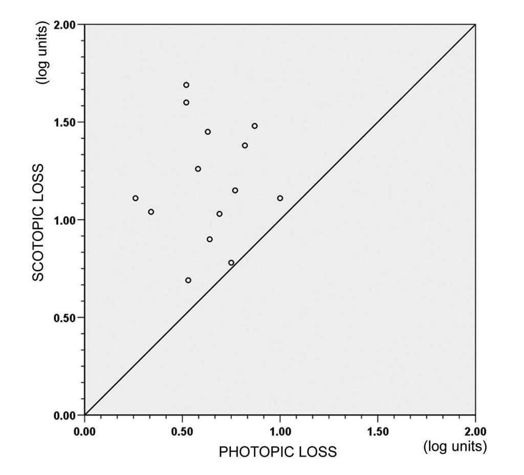
7 Functional characteristics Best corrected visual acuities in the study eye assessed at the time of FMM testing ranged from 6/12 to 6/5 (median=6/6). All patients had less than two lines loss in BCVA compared to the baseline value. Fixation Stability was good in all cases (Table 2). Two patients (2 and 4), both with end-stage disease, showed significant (more than 2 lines) deterioration in BCVA at the endpoint as compared to the baseline value. Fine Matrix Mapping showed generalised threshold elevation relative to normal controls both under photopic and scotopic conditions (Table 2). Scotopic sensitivity loss exceeded photopic loss in all cases (Figure 2). Scotopic loss over areas with drusen or regressed drusen did not differ significantly from that over non-drusen areas (p=0.289 and p=0.989 respectively, ANOVA, Figure 3). Elevated scotopic thresholds were seen associated with GA, crystalline drusen and coarse granular hyperpigmentation, all in connection with abnormal FA. Photopic thresholds showed little topographic variation except in areas with GA. Table 2. Functional characteristics. FMM Global estimates of sensitivity loss are based on Gaussian filtered data and are expressed in log units.
8 DISCUSSION Regression of macular soft drusen has been described in clinical,[2, 4, 18] as well as in histopathological studies.[3, 19] Gass noted that drusen may fade and disappear, leaving only an irregular mottling of the RPE, while visual acuity may not be affected.[3] Gass also observed that most cases GA follow the fading of drusen or the collapse of a serous detachment of the RPE.[3] Drusen disappearance is thus believed to be associated with subsequent degeneration and atrophy of the RPE and photoreceptors.[3] It was however noted in large population-based studies that in some patients, drusen may also regress without residual signs.[2, 4, 18] The prognostic implications of true drusen regression is unknown. From the clinical aspect, it raises the possibility that arrested progression or even regression of the disease process may exist naturally. In this study, ten out of 14 patients showed no ophthalmoscopic indications of manifest or incipient end-stage disease in the fundus following drusen regression. However, in nine, parallel to regression, new drusen in other locations appeared and grew in size and confluence, signifying the continued activity of the disease, with a tendency toward the periphery. Mean retinal sensitivity relative to normal controls was reduced in all patients tested, both in the light and dark adapted states, with significantly higher loss under scotopic conditions. This observation confirms that in AMD, rods are at increased risk for degeneration and function loss occurs before progression to the late clinical stage.[9, 20] Earlier psychophysical studies found that rod-mediated sensitivity declines faster with age than photopic sensitivity and patients with early AMD have significantly lower mean central scotopic sensitivity than age-matched controls. Also, in most AMD patients, mean scotopic sensitivity loss exceeded mean photopic sensitivity loss and the peak deficit in scotopic sensitivity was within 9 of fixation, corresponding to the parafovea.[20] Histopathologic studies show direct correlates to these findings. In early AMD, a preferential loss of macular rod photoreceptors was demonstrated, with the greatest loss occurring in the parafovea. In late AMD this leads to a reversal of the macular (9:1) rod predominance seen in the young.[9] The rod system also shows altered kinetics with aging and in AMD.[6] One possible explanation for this preferential vulnerability of rods is provided by the retinoid deficiency hypothesis.[9] RPE cells are responsible for the transport of nutrients from the choriocapillary circulation to the photoreceptors, as well as for the recycling of the end-products of POS disc degradation. 11-cis retinal (a derivative of Vitamin A) is essential for the regeneration of photoreceptor pigment after bleaching by light as well as for photoreceptor survival. In AMD, diffuse sub-rpe deposits may act as a diffusion barrier between the choriocapillaris and the RPE,[8] thereby disrupting transport across BrM and leading to a local scarcity of 11-cis-retinal. Vitamin A deprivation is a known cause of outer segment degeneration and photoreceptor death. Cones have an additional retinoid delivery pathway involving Müller cells and possibly the neurosensory retina and may thus be less vulnerable to reduced transport across Bruch s membrane.[9, 21] Although generalised sensitivity loss was measured in all our patients, topographic variation in scotopic (and photopic) sensitivity loss over drusen relative to areas with normal appearance was not significant. This confirms earlier observations that the presence of macular soft drusen seems to have little effect on the local sensitivity of affected retinal areas,[22, 23] with the exception of large, soft foveal drusen. These may be regarded as small RPE detachments and show mildly reduced photopic and considerably reduced scotopic sensitivity as well as increased FA.[23] It appears reasonable, that the impact of focal barriers such as drusen on RPE metabolism and photoreceptor function may be more limited than that of diffuse deposits. We also found that scotopic sensitivity over areas with regressed drusen was not substantially different from that over unaffected retinal areas, thus we did not find functional evidence for manifest or incipient photoreceptor atrophy. In diseases, where the RPE is primarily affected,
9 the presence of abnormal FA may be an early sign of progression. Areas with increased FA were found to precede the development of new or the enlargement of existing atrophic patches.[24] Decreased FA over the drusen overall may also reflect incipient atrophy.[25] In eyes with soft drusen but without apparent GA, if focal decreased FA is present, patches with increased FA in areas adjacent to rather than corresponding to drusen may be markers for progression to atrophy.[26] In this study, autofluorescence of retinal areas with normal appearance in the colour image following drusen regression was in most cases normal and none of the above FA patterns associated with impending atrophy was seen. Thus, although the generalised disease persists, retinal sensitivity and FA, two direct and sensitive measures of retinal health do not indicate that disappearance of drusen is necessarily followed by local function loss and atrophy. In summary, although the limitations of this study in terms of subject numbers and follow-up time as well as the lack of psychophysical testing before the drusen disappeared may not permit definitive conclusions, our results do indicate that macular soft drusen may fade or disappear without detectable ophthalmoscopic, FA or psychophysical signs of local dysfunction or incipient atrophy. This phenomenon suggests that some normal-appearing eyes at the present time may have had some features of AMD in the past and is thus a potential source of misclassification and needs to be remembered in epidemiologic studies investigating the natural history of the disease as well as in clinical trials that evaluate the efficacy of possible therapies.[18] It is not known to what extent focal and diffuse sub-rpe deposits coincide and whether the regression of drusen is accompanied by the regression of diffuse deposits. The prognosis for cases with true regression of drusen compared with those without needs to be considered in future studies on AMD.
10 ACKNOWLEDGEMENTS The authors thank Mr Vy Luong for his technical support and Mr Tibor Fermon and Mr Istvan Toth for useful discussions and general support. COMPETING INTERESTS: None declared. (All authors declare that the answer to the questions on your competing interest form are all No and therefore have nothing to declare.) FUNDING: NONE LICENCE FOR PUBLICATION The Corresponding Author has the right to grant on behalf of all authors and does grant on behalf of all authors, an exclusive licence (or non exclusive for government employees) on a worldwide basis to the BMJ Publishing Group Ltd to permit this article (if accepted) to be published in BJO and any other BMJPGL products and sublicences such use and exploit all subsidiary rights, as set out in our licence
11 REFERENCES 1 Klein R, Peto T, Bird A, Vannewkirk MR. The epidemiology of age-related macular degeneration. Am J Ophthalmol Mar;137(3): Review. PMID: Klein R, Klein BE, Tomany SC, Meuer SM, Huang GH. Ten-year incidence and progression of age-related maculopathy: The Beaver Dam eye study. Ophthalmology Oct;109(10): PMID: Gass JD. Drusen and disciform macular detachment and degeneration. Arch Ophthalmol Sep;90(3): PMID: Also in: Retina Dec;23(6 Suppl): PMID: Sparrow JM, Dickinson AJ, Duke AM, Thompson JR, Gibson JM, Rosenthal AR. Seven year follow-up of age-related maculopathy in an elderly British population. Eye. 1997;11 ( Pt 3): PMID: Bird AC, Bressler NM, Bressler SB, Chisholm IH, Coscas G, Davis MD, de Jong PT, Klaver CC, Klein BE, Klein R, et al.an international classification and grading system for agerelated maculopathy and age-related macular degeneration. The International ARM Epidemiological Study Group. Surv Ophthalmol Mar-Apr;39(5): Review. PMID: Steinmetz RL, Haimovici R, Jubb C, Fitzke FW, Bird AC. Symptomatic abnormalities of dark adaptation in patients with age-related Bruch's membrane change. Br J Ophthalmol Sep;77(9): PMID: Sarks SH. Ageing and degeneration in the macular region: a clinico-pathological study. Br J Ophthalmol May;60(5): PMID: Zarbin MA. Current concepts in the pathogenesis of age-related macular degeneration. Arch Ophthalmol Apr;122(4): Review. PMID: Curcio CA, Owsley C, Jackson GR. Spare the rods, save the cones in aging and age-related maculopathy. Invest Ophthalmol Vis Sci Jul;41(8): Review. PMID: von Rückmann A, Fitzke FW, Bird AC. Fundus autofluorescence in age-related macular disease imaged with a laser scanning ophthalmoscope. Invest Ophthalmol Vis Sci Feb;38(2): PMID:
12 11 Dorey CK, Wu G, Ebenstein D, Garsd A, Weiter JJ. Cell loss in the aging retina. Relationship to lipofuscin accumulation and macular degeneration. Invest Ophthalmol Vis Sci Aug;30(8): PMID: Fitzke FW, Crabb DP, McNaught AI, Edgar DF, Hitchings RA. Image processing of computerised visual field data. Br J Ophthalmol Mar;79(3): PMID: Fitzke FW, Kemp CM. Probing visual function with psychophysics and photochemistry. Eye. 1989;3: Westcott MC, McNaught AI, Crabb DP, Fitzke FW, Hitchings RA. High spatial resolution automated perimetry in glaucoma. Br J Ophthalmol Jun;81(6): PMID: Guymer RH, Gross-Jendroska M, Owens SL, Bird AC, Fitzke FW. Laser treatment in subjects with high-risk clinical features of age-related macular degeneration. Posterior pole appearance and retinal function. Arch Ophthalmol May;115(5): PMID: Wu D, Bird AC, Mcnaught A, Buckland MS, Fitzke FW. Fine matrix mapping of the macular region in normal subjects. Zhonghua Yan Ke Za Zhi Jul;31(4): PMID: Culham LE, Fitzke FW, Timberlake GT, Marshall J. Assessment of fixation stability in normal subjects and patients using a scanning laser ophthalmoscope. Clin Vision Sci. 1993;8: Bressler NM, Munoz B, Maguire MG, Vitale SE, Schein OD, Taylor HR, West SK. Five-year incidence and disappearance of drusen and retinal pigment epithelial abnormalities. Waterman study. Arch Ophthalmol Mar;113(3): PMID: Sarks JP, Sarks SH, Killingsworth MC. Evolution of geographic atrophy of the retinal pigment epithelium. Eye. 1988;2 (Pt 5): PMID: Owsley C, Jackson GR, Cideciyan AV, Huang Y, Fine SL, Ho AC, Maguire MG, Lolley V, Jacobson SG. Psychophysical evidence for rod vulnerability in age-related macular degeneration. Invest Ophthalmol Vis Sci Jan;41(1): PMID: Mata NL, Radu RA, Clemmons RC, Travis GH. Isomerization and oxidation of vitamin a in cone-dominant retinas: a novel pathway for visual-pigment regeneration in daylight. Neuron Sep 26;36(1): PMID:
13 22 Sunness JS, Johnson MA, Massof RW, Marcus S. Retinal sensitivity over drusen and nondrusen areas. A study using fundus perimetry. Arch Ophthalmol Aug;106(8): PMID: Scholl HP, Bellmann C, Dandekar SS, Bird AC, Fitzke FW. Photopic and scotopic fine matrix mapping of retinal areas of increased fundus autofluorescence in patients with agerelated maculopathy. Invest Ophthalmol Vis Sci Feb;45(2): PMID: Holz FG, Bellman C, Staudt S, Schütt F, Völcker HE. Fundus autofluorescence and development of geographic atrophy in age-related macular degeneration. Invest Ophthalmol Vis Sci Apr;42(5): PMID: Sunness JS, Ziegler MD, Applegate CA. Issues in quantifying atrophic macular disease using retinal autofluorescence. Retina Jul-Aug;26(6): PMID: Smith RT, Chan JK, Busuoic M, Sivagnanavel V, Bird AC, Chong NV. Autofluorescence characteristics of early, atrophic, and high-risk fellow eyes in age-related macular degeneration. Invest Ophthalmol Vis Sci Dec;47(12): PMID:
14 APPENDICES Figure legends: Figure 1. Representative images from selected cases. Columns: (A) the fundus at baseline, (B-C) colour and FA images taken at FMM testing, white rectangles mark the placement of respective FMM test grid, (D-E) surface plots of photopic and scotopic FMM thresholds. Rows 1-2 and 4 (patients 3, 6 and 11) illustrate the typical picture seen in the majority of patients. Row 3 shows a case (patient 7) where areas with regressed drusen appear normal in the colour fundus image but show a speckled variation in FA. Row 5 shows a case where progression to atrophy was noted (patient 2). Figure 2. Photopic versus scotopic sensitivity loss of the 14 patients in the study. Figure 3. Regional differences in scotopic sensitivity loss at retinal locations with (C) drusen, (B) regressed drusen and (A) areas with normal appearance.
15
16
17
L ipofuscin (LF) accumulates with age within the lysosomal
 accumulates with age within the lysosomal") 1381 EXTENDED REPORT Fundus autofluorescence imaging compared with different confocal scanning laser ophthalmoscopes C Bellmann, G S Rubin, S A Kabanarou, A C Bird, F W Fitzke... See end of article for
1381 EXTENDED REPORT Fundus autofluorescence imaging compared with different confocal scanning laser ophthalmoscopes C Bellmann, G S Rubin, S A Kabanarou, A C Bird, F W Fitzke... See end of article for
Retinal pigment epithelial detachments in the elderly:
 British Journal of Ophthalmology, 1985, 69, 397-403 Retinal pigment epithelial detachments in the elderly: classification and outcome A G CASSWELL, D KOHEN, AND A C BIRD From Moorfields Eye Hospital, City
Diagnosis in AMD. Managing your AMD Patients
 Managing your AMD Patients Robert W. Dunphy, O.D., F.A.A.O. Diagnosis in AMD Have suspicion Identify relative risk Conduct surveillance Biometry Utilize technology to facilitate detection of change / stability
Fundus Autofluorescence
 Brittany Bateman, BS Fundus autofluorescence imaging is used to record fluorescence that may occur naturally in ocular structures or as a byproduct of a disease process. This technique allows the topographic
Fundus autofluorescence in exudative age-related macular degeneration
 Fundus autofluorescence in exudative age-related macular degeneration Q. Peng*, Y. Dong* and P.Q. Zhao Department of Ophthalmology, Xinhua Hospital Affiliated to Shanghai JiaoTong University School of
Fundus Autofluorescence. Jonathan A. Micieli, MD Valérie Biousse, MD
 Fundus Autofluorescence Jonathan A. Micieli, MD Valérie Biousse, MD The retinal pigment epithelium (RPE) has many important functions including phagocytosis of the photoreceptor outer segments Cone Rod
Vitreous! Retinal pigment epithelium! and the visual cycle! Retinal degenerations and pigment epithelium!
 Vitreous Bruch s membrane Retinal pigment epithelium and the visual cycle Retinal degenerations and pigment epithelium Basic Science course 2017 Swiss Eye Week, Neuchâtel Ch. E. Remé, Zürich Ch.E. Remé
Clinical Trial Endpoints for Macular Diseases
 Clinical Trial Endpoints for Macular Diseases Developed in collaboration Learning Objective Upon completion, participants should be able to: Summarize types of biomarkers of progression and treatment response
Original Policy Date
 MP 9.03.08 Photocoagulation of Macular Drusen Medical Policy Section Miscellaneous Policies Issue 12/2013 Original Policy Date 12/2013 Last Review Status/Date Reviewed with literature search/12/2013 Return
Geographic atrophy (GA) is the atrophic late-stage manifestation
 is the atrophic late-stage manifestation") Clinical and Epidemiologic Research Semiautomated Image Processing Method for Identification and Quantification of Geographic Atrophy in Age-Related Macular Degeneration Steffen Schmitz-Valckenberg, 1
Clinical and Epidemiologic Research Semiautomated Image Processing Method for Identification and Quantification of Geographic Atrophy in Age-Related Macular Degeneration Steffen Schmitz-Valckenberg, 1
Age-related macular degeneration (ARMD) is related to. Blue-Light versus Green-Light Autofluorescence: Lesion Size of Areas of Geographic Atrophy
 is related to. Blue-Light versus Green-Light Autofluorescence: Lesion Size of Areas of Geographic Atrophy") Clinical Trials Blue-Light versus Green-Light Autofluorescence: Lesion Size of Areas of Geographic Atrophy Ute E. K. Wolf-Schnurrbusch, 1,2 Valéry V. Wittwer, 1,2 Ramzi Ghanem, 1,3 Martin Niederhaeuser,
Clinical Trials Blue-Light versus Green-Light Autofluorescence: Lesion Size of Areas of Geographic Atrophy Ute E. K. Wolf-Schnurrbusch, 1,2 Valéry V. Wittwer, 1,2 Ramzi Ghanem, 1,3 Martin Niederhaeuser,
4/19/2018 FUNDUS AUTOFLUORESCENCE. Fluorescence Imaging. Fundus Autofluorescence (FAF) Fluorescence. Fluorescence
 Fluorescence. Fluorescence") I have no financial or proprietary interest in the subject matter of this presentation. FUNDUS AUTOFLUORESCENCE Timothy J. Bennett, CRA, OCT-C, FOPS Penn State Eye Center Hershey, PA Fluorescence Imaging
I have no financial or proprietary interest in the subject matter of this presentation. FUNDUS AUTOFLUORESCENCE Timothy J. Bennett, CRA, OCT-C, FOPS Penn State Eye Center Hershey, PA Fluorescence Imaging
Acquired vitelliform detachment in patients with subretinal drusenoid deposits (reticular pseudodrusen)
") Zurich Open Repository and Archive University of Zurich Main Library Strickhofstrasse 39 CH-8057 Zurich www.zora.uzh.ch Year: 2011 Acquired vitelliform detachment in patients with subretinal drusenoid
Zurich Open Repository and Archive University of Zurich Main Library Strickhofstrasse 39 CH-8057 Zurich www.zora.uzh.ch Year: 2011 Acquired vitelliform detachment in patients with subretinal drusenoid
T he cone and cone-rod dystrophies are a clinically and
 332 EXTEED REPORT A detailed phenotypic study of cone dystrophy with supernormal rod ERG M Michaelides, G E Holder, A R Webster, D M Hunt, A C Bird, F W Fitzke, J D Mollon, A T Moore... See end of article
Cirrus TM HD-OCT. Details defi ne your decisions
 Cirrus TM HD-OCT Details defi ne your decisions 2 With high-defi nition OCT Carl Zeiss Meditec takes you beyond standard spectral domain Built on 10 years experience at the vanguard of innovation, Carl
Widefield Retinal Imaging with Auto Fluorescence Technology in the Optometric Practice
 Widefield Retinal Imaging with Auto Fluorescence Technology in the Optometric Practice This course will define ultra-widefield retinal imaging and autofluorescence for the attendee. Will show how it is
Spectral-domain Optical Coherence Tomography Imaging of Age-related Macular Degeneration
 Imaging Spectral-domain Optical Coherence Tomography Imaging of Age-related Macular egeneration Carlos Alexandre de Amorim Garcia Filho, 1 Philip J Rosenfeld, 2 Zohar Yehoshua 3 and Giovanni Gregori 3
NIH Public Access Author Manuscript Br J Ophthalmol. Author manuscript; available in PMC 2010 December 1.
 NIH Public Access Author Manuscript Published in final edited form as: Br J Ophthalmol. 2010 December ; 94(12): 1618 1623. doi:10.1136/bjo.2009.166843. Dynamic soft drusen remodelling in age-related macular
The first sign of impending pathology in age-related macular
 Autofluorescence Distribution Associated with Drusen in Age-Related Macular Degeneration François C. Delori, 1,2 Mark R. Fleckner, 1 Douglas G. Goger, 1 John J. Weiter, 1,2 and C. Kathleen Dorey 1,2 PURPOSE.
Photocoagulation of disciform macular lesions
 British Journal of Ophthalmology, 1979, 63, 669-673 Photocoagulation of disciform macular lesions with krypton laser A. C. BIRD AND R. H. B. GREY From the Institute of Ophthalmology, Moorfields Eye Hospital,
HHS Public Access Author manuscript Ophthalmic Surg Lasers Imaging Retina. Author manuscript; available in PMC 2016 January 14.
 High-Speed Ultrahigh-Resolution OCT of Bruch s Membrane in Membranoproliferative Glomerulonephritis Type 2 Mehreen Adhi, MD, Sarah P. Read, MD, PhD, Jonathan J. Liu, PhD, James G. Fujimoto, PhD, and Jay
Optical Coherence Tomography in Diabetic Retinopathy. Mrs Samantha Mann Consultant Ophthalmologist Clinical Lead of SEL-DESP
 Optical Coherence Tomography in Diabetic Retinopathy Mrs Samantha Mann Consultant Ophthalmologist Clinical Lead of SEL-DESP Content OCT imaging Retinal layers OCT features in Diabetes Some NON DR features
The New Frontier of Microperimetry
 Macular Integrity Assessment The New Frontier of Microperimetry Index 4 Company Profile Microperimetry is attracting our attention more and more as a method that is superior to standard automated perimetry
The New Frontier of Microperimetry
 Macular Integrity Assessment The New Frontier of Microperimetry Microperimetry is attracting our attention more and more as a method that is superior to standard automated perimetry for visual function
Cirrus TM HD-OCT. Details define your decisions
 Cirrus TM HD-OCT Details define your decisions 2 With high-definition OCT Carl Zeiss Meditec takes you beyond standard spectral domain Built on 10 years experience at the vanguard of innovation, Carl Zeiss
OCT Angiography in Primary Eye Care
 OCT Angiography in Primary Eye Care An Image Interpretation Primer Julie Rodman, OD, MS, FAAO and Nadia Waheed, MD, MPH Table of Contents Diabetic Retinopathy 3-6 Choroidal Neovascularization 7-9 Central
Fluorescein Angiography
 Last revision: October 2011 by Luis Arias Fluorescein Angiography Authors: Luis Arias, MD Hospital Universitari de Bellvitge - University of Barcelona. Spain Jordi Monés, MD Institut de la Màcula i de
Geographic atrophy (GA) represents the atrophic late-stage
 represents the atrophic late-stage") Clinical Trials Progression of Age-Related Geographic Atrophy: Role of the Fellow Eye Monika Fleckenstein, 1,2 Steffen Schmitz-Valckenberg, 1,2 Christine Adrion, 3 Sivatharisini Visvalingam, 1 Arno P.
Clinical Trials Progression of Age-Related Geographic Atrophy: Role of the Fellow Eye Monika Fleckenstein, 1,2 Steffen Schmitz-Valckenberg, 1,2 Christine Adrion, 3 Sivatharisini Visvalingam, 1 Arno P.
Use of Scanning Laser Ophthalmoscope Microperimetry in Clinically Significant Macular Edema in Type 2 Diabetes Mellitus
 Use of Scanning Laser Ophthalmoscope Microperimetry in Clinically Significant Macular Edema in Type 2 Diabetes Mellitus Fumihiko Mori, Satoshi Ishiko, Norihiko Kitaya, Taiichi Hikichi, Eiichi Sato, Akira
Agreement between. image grading of conventional (451) and ultra. wide-angle (2001) digital images in the macula in the Reykjavik eye study
 and ultra. wide-angle (2001) digital images in the macula in the Reykjavik eye study") (2010) 24, 1568 1575 & 2010 Macmillan Publishers Limited All rights reserved 0950-222X/10 $32.00 www.nature.com/eye CLINICAL STUDY Agreement between image grading of conventional (451) and ultra wide-angle
(2010) 24, 1568 1575 & 2010 Macmillan Publishers Limited All rights reserved 0950-222X/10 $32.00 www.nature.com/eye CLINICAL STUDY Agreement between image grading of conventional (451) and ultra wide-angle
F luorescence angiography (FL-A) with fluorescein (FL)
 with fluorescein (FL)") 1609 EXTENDED REPORT Lower limits of fluorescein and indocyanine green dye for digital cslo fluorescence angiography A Bindewald, O Stuhrmann, F Roth, S Schmitz-Valckenberg, H-M Helb, A Wegener, N Eter,
1609 EXTENDED REPORT Lower limits of fluorescein and indocyanine green dye for digital cslo fluorescence angiography A Bindewald, O Stuhrmann, F Roth, S Schmitz-Valckenberg, H-M Helb, A Wegener, N Eter,
Long-Term Effect of Acetozolomide in o Patient with Retinitis Pigmentoso
 Investigative Ophthalmology & Visual Science. Vol. 31, No., September Copyright Association for Research in Vision and Ophthalmology Reports LongTerm Effect of Acetozolomide in o Patient with Retinitis
Distribution of fundus autofluorescence with a scanning laser ophthalmoscope
 British Journal of Ophthalmology 1995; 79: 407-412 407 Distribution of fundus autofluorescence with a scanning laser ophthalmoscope A von Ruickmann, F W Fitzke, A C Bird Institute of Ophthalmology, Moorfields
Geographic atrophy (GA) of the retinal pigment epithelium
 of the retinal pigment epithelium") Correlation between the Area of Increased Autofluorescence Surrounding Geographic Atrophy and Disease Progression in Patients with AMD Steffen Schmitz-Valckenberg, 1 Almut Bindewald-Wittich, 1 Joanna Dolar-Szczasny,
Correlation between the Area of Increased Autofluorescence Surrounding Geographic Atrophy and Disease Progression in Patients with AMD Steffen Schmitz-Valckenberg, 1 Almut Bindewald-Wittich, 1 Joanna Dolar-Szczasny,
Senile disciform macular degeneration
 British Journal of Ophthalmology, 1977, 61, 141-147 Senile disciform macular degeneration in the second eye Z. GREGOR, A. C. BIRD, AND I. H. CHISHOLM From Moorfields Eye Hospital and Institute of Ophthalmology,
You can see clearly now. Heidelberg Retina Angiograph 2
 You can see clearly now Heidelberg Retina Angiograph 2 Wishes come true The way ahead is clear Highest image contrast and detail Lowest light exposure Simultaneous FA and ICGA Infra-red and Blue Reflectance
Fundus Autofluorescence Imaging
 March 2009 Meena Chakrabarti - Fundus Autofluorescence Imaging 55 OPHTHALMIC INSTRUMENTATION Fundus Autofluorescence Imaging Dr. Meena Chakrabarti MS DO DNB Fundus auto fluorescence (FAF) imaging is a
Autofluorescence Imaging for Diagnosis and Follow-up of Cystoid Macular Edema
 Autofluorescence Imaging for Diagnosis and Follow-up of Cystoid Macular Edema Nazanin Ebrahimiadib 1, MD; Mohammad Riazi-Esfahani 1,2, MD 1Eye Research Center, Farabi Eye Hospital, Tehran University of
OCT Image Analysis System for Grading and Diagnosis of Retinal Diseases and its Integration in i-hospital
 Progress Report for1 st Quarter, May-July 2017 OCT Image Analysis System for Grading and Diagnosis of Retinal Diseases and its Integration in i-hospital Milestone 1: Designing Annotation tool extraction
Long-term Management of AMD. Motasem Al-latayfeh, MD Assistant Prof. Ophthalmology Hashemite University Jordan
 Long-term Management of AMD Motasem Al-latayfeh, MD Assistant Prof. Ophthalmology Hashemite University Jordan DEFINITION 1 Age-related macular degeneration (AMD) is a disorder of the macula characterized
Fundus Autofluorescence
 E-ISSN 0976-2892 Major Review Fundus Autofluorescence Delhi J Ophthalmol 2013; 24 (2): 80-87 DOI: http://dx.doi.org/10.7869/djo.2013.18 * Vivek Pravin Dave, # Rajeev R. Pappuru * Consultant Retina, Uvea
Parafoveal Scanning Laser Polarimetry for Early Glaucoma Detection
 Yamanashi Med. J. 18(1), 15~ 20, 2003 Original Article Parafoveal Scanning Laser Polarimetry for Early Glaucoma Detection Satoshi KOGURE, Yoshiki TODA, Hiroyuki IIJIMA and Shigeo TSUKAHARA Department of
Spontaneous Large Serous Retinal Pigment Epithelial Tear
 This is an Open Access article licensed under the terms of the Creative Commons Attribution-NonCommercial-NoDerivs 3.0 License (www.karger.com/oa-license), applicable to the online version of the article
Posterior Segment Age-related Macular Degeneration
 Posterior Segment Age-related Macular egeneration Spectral-domain Optical oherence Tomography Imaging of Age-related Macular egeneration arlos Alexandre de Amorim Garcia Filho, M, 1 Philip J Rosenfeld,
The Evolution of Fundus Perimetry
 The Evolution of Fundus Perimetry Company Profile CenterVue designs and manufactures highly automated medical devices for the diagnosis and management of ocular pathologies, including those that represent
Prevalence, Natural Course, and Prognostic Role of Refractile Drusen in Age-Related Macular Degeneration
 Retina Prevalence, Natural Course, and Prognostic Role of Refractile Drusen in Age-Related Macular Degeneration Akio Oishi, 1 Sarah Thiele, 1 Jennifer Nadal, 2 Maho Oishi, 1 Monika Fleckenstein, 1 Matthias
Sorsby's pseudoinflammatory macular dystrophy
 British Journal of Ophthalmology, 1981, 65, 859-865 Sorsby's pseudoinflammatory macular dystrophy A. HOSKIN, K. SEHMI, AND A. C. BIRD From the Department of Clinical Ophthalmology, Institute of Ophthalmology,
eye as a camera Kandel, Schwartz & Jessel (KSJ), Fig 27-3
, Fig 27-3") eye as a camera Kandel, Schwartz & Jessel (KSJ), Fig 27-3 retinal specialization fovea: highest density of photoreceptors, aimed at where you are looking -> highest acuity optic disk: cell-free area, where
eye as a camera Kandel, Schwartz & Jessel (KSJ), Fig 27-3 retinal specialization fovea: highest density of photoreceptors, aimed at where you are looking -> highest acuity optic disk: cell-free area, where
25% of normal 2/20/2018. Practical Guidelines for the Treatment of AMD 78% 22% Overview. AMD Information Overload
 I am a consultant / or have financial interest in: Practical Guidelines for the Treatment of Maculogix J&J Acuvue Vision Source Ocusoft Pogo Tech Jeffrey W. Jones, OD Longview, TX 75605 jjoneseye@gmail.com
optic disc neovascularisation
 British Journal of Ophthalmology, 1979, 63, 412-417 A comparative study of argon laser and krypton laser in the treatment of diabetic optic disc neovascularisation W. E. SCHULENBURG, A. M. HAMILTON, AND
The Role of Phenotype in Selectively Enriching Patients for Clinical Studies
 The Role of Phenotype in Selectively Enriching Patients for Clinical Studies Developing Treatments for Dry Age-Related Macular Degeneration (AMD) Workshop November 15, 2014 National Academy of Sciences
Dry AMD: the regulatory view
 EMA Ophthalmology Workshop 2011 Dry AMD: the regulatory view Marco Coassin, MD PhD University of Rome Campus Bio-Medico - Italy In this presentation Personal views Previous scientific advices No currently
OCT Interpretation in Retinal Disease
 OCT Interpretation in Retinal Disease Jay M. Haynie, OD, FAAO Financial Disclosure I have received honoraria or am on the advisory board for the following companies: Carl Zeiss Meditec Advanced Ocular
In 1990 our group first described reticular pseudodrusen as a. Choroidal Changes Associated with Reticular Pseudodrusen. Retina
 Retina Choroidal Changes Associated with Reticular Pseudodrusen Giuseppe Querques, 1,2 Lea Querques, 1,2 Raimondo Forte, 1 Nathalie Massamba, 1 Florence Coscas, 1 and Eric H. Souied 1 PURPOSE. To analyze
Macular edema consists of an accumulation of fluid in
 Photoreceptor Function in Eyes with Macular Edema Charlotte W. T. A. Lardenoye, Kiki Probst, Peter Jaap DeLint, and Aniki Rothova PURPOSE. The irreversible loss of visual acuity in macular edema is usually
Abstract Aims To analyse the histopathology of classic and occult choroidal neovascular membrane surgical specimens in age
 Br J Ophthalmol 2000;84:239 243 239 ORIGINAL ARTICLES Clinical science Clinicopathological correlation in exudative age related macular degeneration: histological diverentiation between classic and occult
Foveal cone photopigment bleaching in central serous retinopathy
 Foveal cone photopigment bleaching in central serous retinopathy Stephen A. Burns, Ann E. Elsner, and Louis A. Lobes, Jr. Color-matching techniques were used to follow the course of central serous retinopathy
Anthony G. Robson Æ Michel Michaelides Æ Zubin Saihan Æ Alan C. Bird Æ Andrew R. Webster Æ Anthony T. Moore Æ Fred W. Fitzke Æ Graham E.
 Doc Ophthalmol (2008) 116:79 89 DOI 10.1007/s10633-007-9087-4 ORIGINAL RESEARCH ARTICLE Functional characteristics of patients with retinal dystrophy that manifest abnormal parafoveal annuli of high density
Prevalence of age related maculopathy in a representative Japanese population: the Hisayama study
 Br J Ophthalmol 2001;85:1153 1157 1153 WORLD VIEW (Series editor: Emmett T Cunningham Jr) Department of Ophthalmology, Faculty of Medicine, Kyushu University, Fukuoka, Japan Y Oshima T Ishibashi T Murata
Optical Coherence Tomograpic Features in Idiopathic Retinitis, Vasculitis, Aneurysms and Neuroretinitis (IRVAN)
") Columbia International Publishing Journal of Ophthalmic Research (2014) Research Article Optical Coherence Tomograpic Features in Idiopathic Retinitis, Vasculitis, Aneurysms and Neuroretinitis (IRVAN)
Columbia International Publishing Journal of Ophthalmic Research (2014) Research Article Optical Coherence Tomograpic Features in Idiopathic Retinitis, Vasculitis, Aneurysms and Neuroretinitis (IRVAN)
Comparison of Geographic Atrophy Measurements from the OCT Fundus Image and the Sub-RPE Slab Image
 CLINICAL SCIENCE Comparison of Geographic Atrophy Measurements from the OCT Fundus Image and the Sub-RPE Slab Image Zohar Yehoshua, MD, MHA; Carlos Alexandre A. Garcia Filho, MD; Fernando M. Penha, MD,
Pseudoxanthoma elasticum (PXE) is a rare systemic disease
 is a rare systemic disease") Multimodal Imaging Including Spectral Domain OCT and Confocal Near Infrared Reflectance for Characterization of Outer Retinal Pathology in Pseudoxanthoma Elasticum Peter Charbel Issa, Robert P. Finger,
Multimodal Imaging Including Spectral Domain OCT and Confocal Near Infrared Reflectance for Characterization of Outer Retinal Pathology in Pseudoxanthoma Elasticum Peter Charbel Issa, Robert P. Finger,
Macular Function Impairment in Eyes With Early Age- Related Macular Degeneration
 Macular Function Impairment in Eyes With Early Age- Related Macular Degeneration Edoardo Midena, Claudia Degli Angeli, Maria C. Blarzino, Massimo Valenti, and Tatiana Segato urpose. To study different
A Comparative Study of Age Related Macular Degeneration In Relation To SD-OCTand Fundus Photography.
 IOSR Journal of Dental and Medical Sciences (IOSR-JDMS) e-issn: 2279-0853, p-issn: 2279-0861.Volume 14, Issue 11 Ver. III (Nov. 2015), PP 33-37 www.iosrjournals.org A Comparative Study of Age Related Macular
Do You See What I See!!! Shane R. Kannarr, OD
 Do You See What I See!!! Shane R. Kannarr, OD skannarr@kannarreyecare.com Define Specialty Testing Additional Test to: Prove/Disprove Diagnosis To monitor progression of a condition To document a condition
Manual. Manual Welsh Eye Care Initiative. A Welsh Eye Care Initiative. Protocol. The Assessment and Management of Age-related Macular Degeneration
 A Protocol 1.0 Definitions The following terms are important in this text: Wet Macular Degeneration Condition caused by the growth of abnormal blood vessels under the retina. Symptoms appear suddenly and
The ideal tool for early detection and monitoring of AMD.
 The ideal tool for early detection and monitoring of AMD. presenting maia 1 MAIA, the new frontier of Fundus Perimetry (microperimetry) assesses the function of the macula representing an effective clinical
Angio-OCT. Degenerazione Maculare Legata all Eta. Giuseppe Querques
 Angio-OCT Degenerazione Maculare Legata all Eta Giuseppe Querques Department of Ophthalmology, IRCCS Ospedale San Raffaele, University Vita Salute San Raffaele, Milan, Italy Financial Disclosure ADVISORY
Fundus Autofluorescenceclinical applications
 Northwestern University Feinberg School of Medicine Fundus Autofluorescenceclinical applications Amani A. Fawzi, MD Associate Professor of Ophthalmology Northwestern University, Feinberg School of Medicine
The Measure of Confidence
 Heidelberg_936357.qxd:Layout 1 5/9/08 12:01 PM 12:02 Page 1 (Cyan (Magenta (Yellow (Black (UV Five Powerful Solutions to Fit Your Practice PowerCheck Glaucoma FastCheck+ GPS Software and Retina Edema Index
CANALOPLASTY. RESTORATIVE GLAUCOMA SURGERY. Retinal Rejuvenation. Naturally, from Ellex. ACCESSORIES SPECIFICATIONS INDICATIONS FOR USE
 ACCESSORIES SPECIFICATIONS Tablet Interface The intuitive tablet interface allows for easy adjustment of key treatment parameters. Laser Source Q-switched Nd: YAG laser, frequency doubled Wavelength 532
SEGMENTATION OF MACULAR LAYERS IN OCT DATA OF TOPOLOGICALLY DISRUPTED MACULA
 SEGMENTATION OF MACULAR LAYERS IN OCT DATA OF TOPOLOGICALLY DISRUPTED MACULA Athira S C 1, Reena M Roy 2 1 P G Scholar, L.B.S Institute of Technology for women Poojappura Trivandrum, 2 Assistant Professor,
Geographic atrophy (GA), a late-stage manifestation of various
, a late-stage manifestation of various") Clinical Trials Fundus Autofluorescence and Spectral-Domain Optical Coherence Tomography Characteristics in a Rapidly Progressing Form of Geographic Atrophy Monika Fleckenstein, 1 Steffen Schmitz-Valckenberg,
Clinical Trials Fundus Autofluorescence and Spectral-Domain Optical Coherence Tomography Characteristics in a Rapidly Progressing Form of Geographic Atrophy Monika Fleckenstein, 1 Steffen Schmitz-Valckenberg,
A population based study of macular choroidal neovascularization using optical coherence tomography in Eastern China
 EXPERIMENTAL AND THERAPEUTIC MEDICINE 8: 371-376, 2014 A population based study of macular choroidal neovascularization using optical coherence tomography in Eastern China JIE ZHAO, JUN HU, HAO LU and
RETINA 2018 OBJECTIVES OCT VERY USEFUL INFORMATION SAFE AND FRIENDLY 1/11/2018 KELLY MITCHELL
 RETINA 2018 KELLY MITCHELL OBJECTIVES HIGHLIGHT NEW DIAGNOSTIC & TREATMENT OPTIONS REVIEW DIAGNOSTIC KEYS OF SELECT RETINAL DISEASES DISCUSS USE OF IMAGING AND REFERRAL RECOURSES FOR PATIENT BENEFIT OCT
ZEISS AngioPlex OCT Angiography. Clinical Case Reports
 Clinical Case Reports Proliferative Diabetic Retinopathy (PDR) Case Report 969 PROLIFERATIVE DIABETIC RETINOPATHY 1 1-year-old diabetic female presents for follow-up of proliferative diabetic retinopathy
AGE-RELATED MACULAR DEGENERATION (AMD) IS
 IS") Progression of Geographic Atrophy and Impact of Fundus Autofluorescence Patterns in Age-related Macular Degeneration FRANK G. HOLZ, MD, ALMUT BINDEWALD-WITTICH, MD, MONIKA FLECKENSTEIN, MD, JENS DREYHAUPT,
Progression of Geographic Atrophy and Impact of Fundus Autofluorescence Patterns in Age-related Macular Degeneration FRANK G. HOLZ, MD, ALMUT BINDEWALD-WITTICH, MD, MONIKA FLECKENSTEIN, MD, JENS DREYHAUPT,
Age-related macular degeneration (AMD) is a complex disease
 is a complex disease") Clinical Trials Concordance of Disease Progression in Bilateral Geographic Atrophy Due to AMD Monika Fleckenstein, 1,2 Christine Adrion, 2,3 Steffen Schmitz-Valckenberg, 1 Arno P. Göbel, 1 Almut Bindewald-Wittich,
Clinical Trials Concordance of Disease Progression in Bilateral Geographic Atrophy Due to AMD Monika Fleckenstein, 1,2 Christine Adrion, 2,3 Steffen Schmitz-Valckenberg, 1 Arno P. Göbel, 1 Almut Bindewald-Wittich,
What do we perceive?
 THE VISUAL SYSTEM Aditi Majumder What do we perceive? Example: Switch off the light in room What we perceive Not only the property of the scene But also that of the visual system Our perception is filtered
Clinical Features of Bilateral Acute Idiopathic Maculopathy
 Clinical Features of Bilateral Acute Idiopathic Maculopathy Toru Nakazawa,, Katsuhiro Yamaguchi, Masahiko Shimura, Madoka Yoshida, Yuki Yoshioka and Makoto Tamai Department of Ophthalmology, Katta General
Clinical Study Autofluorescence Images with Carl Zeiss versus Topcon Eye Fundus Camera: A Comparative Study
 Ophthalmology Volume 213, Article ID 39192, 4 pages http://dx.doi.org/1.1155/213/39192 Clinical Study Autofluorescence Images with Carl Zeiss versus Topcon Eye Fundus Camera: A Comparative Study Juan M.
Efficient phenotyping of nonneovascular age-related macular
 Clinical Trials Quantitative Comparison of Drusen Segmented on SD-OCT versus Drusen Delineated on Color Fundus Photographs Nieraj Jain, 1 Sina Farsiu, 1,2 Aziz A. Khanifar, 1 Srilaxmi Bearelly, 1,3 R.
CLINICAL SCIENCES. Laser Burn Intensity and the Risk for Choroidal Neovascularization in the CNVPT Fellow Eye Study
 CLINICAL SCIENCES Laser Burn Intensity and the Risk for Choroidal Neovascularization in the CNVPT Fellow Eye Study Richard S. Kaiser, MD; Jeffrey W. Berger, MD, PhD ; Maureen G. Maguire, PhD; Allen C.
af Diagnostic Atlas A Retinal Reference Guide Building The Retina Company
 af Diagnostic Atlas A Retinal Reference Guide Building The Retina Company af Diagnostic Atlas A Retinal Reference Guide Optos core devices produce ultra-widefield (UWF ), high resolution digital images
Ganglion cell complex scan in the early prediction of glaucoma
 Original article in the early prediction of glaucoma Ganekal S Nayana Super Specialty Eye Hospital and Research Center, Davangere, Karnataka, India Abstract Objective: To compare the macular ganglion cell
3/16/2018. Perimetry
 Perimetry The normal visual field extends further away from fixation temporally and inferiorly than superiorly and nasally. From the center of the retina this sensitivity decreases towards the periphery,
DOME SHAPED MACULOPATHY. Ιωάννης Ν. Βαγγελόπουλος Χειρ. Οφθαλμίατρος - Βόλος
 DOME SHAPED MACULOPATHY Ιωάννης Ν. Βαγγελόπουλος Χειρ. Οφθαλμίατρος - Βόλος DOME SHAPED MACULOPATHY-DEFINITIONS The entity Dome Shaped Macula ( DSM ) was first described by Gaucher and associates in 2008
Disease-Specific Fluorescein Angiography
 Ruth E. Picchiottino, CRA Disease-Specific Fluorescein Angiography 15 Disease-Specific Fluorescein Angiography Recommendations for tailoring retinal fluorescein angiography to diabetic retinopathy, macular
QUANTIFICATION OF PROGRESSION OF RETINAL NERVE FIBER LAYER ATROPHY IN FUNDUS PHOTOGRAPH
 QUANTIFICATION OF PROGRESSION OF RETINAL NERVE FIBER LAYER ATROPHY IN FUNDUS PHOTOGRAPH Hyoun-Joong Kong *, Jong-Mo Seo **, Seung-Yeop Lee *, Hum Chung **, Dong Myung Kim **, Jeong Min Hwang **, Kwang
PRIMUS 200 from ZEISS The essential OCT
 PRIMUS 200 from ZEISS The essential OCT Seeing beyond the surface. ZEISS PRIMUS 200 // INNOVATION MADE BY ZEISS Clear Visualization. Advanced Technology. Reliability. Essential elements of your first OCT.
The short-wavelength autofluorescence (SW-AF; 488 nm
 Retina Distinct Characteristics of Inferonasal Fundus Autofluorescence Patterns in Stargardt Disease and Retinitis Pigmentosa Tobias Duncker, 1 Winston Lee, 1 Stephen H. Tsang, 1,2 Jonathan P. Greenberg,
Supplementary Appendix
 This appendix has been provided by the authors to give readers additional information about their work. Supplement to: Edwards TL, Jolly JK, MacLaren RE, et al.. N Engl J Med 206;374:996-8. DOI: 0.056/NEJMc50950
CENTRAL SEROUS CHORIORETINOPATHY (CSR) IS A
 IS A") Imaging Polarimetry in Central Serous Chorioretinopathy MASAHIRO MIURA, MD, ANN E. ELSNER, PHD, ANKE WEBER, MD, MICHAEL C. CHENEY, MS, MASAHIRO OSAKO, MD, MASAHIKO USUI, MD, AND TAKUYA IWASAKI, MD PURPOSE:
Imaging Polarimetry in Central Serous Chorioretinopathy MASAHIRO MIURA, MD, ANN E. ELSNER, PHD, ANKE WEBER, MD, MICHAEL C. CHENEY, MS, MASAHIRO OSAKO, MD, MASAHIKO USUI, MD, AND TAKUYA IWASAKI, MD PURPOSE:
An A to Z guide on Epiretinal Membranes (ERMs) Paris Tranos PhD,ICO,FRCS OPHTHALMICA Vitreoretinal & Uveitis Department
 Paris Tranos PhD,ICO,FRCS OPHTHALMICA Vitreoretinal & Uveitis Department") An A to Z guide on Epiretinal Membranes (ERMs) Paris Tranos PhD,ICO,FRCS OPHTHALMICA Vitreoretinal & Uveitis Department Types of ERM Natural history OCT prognostic factors ERM with co-existing pathology
An A to Z guide on Epiretinal Membranes (ERMs) Paris Tranos PhD,ICO,FRCS OPHTHALMICA Vitreoretinal & Uveitis Department Types of ERM Natural history OCT prognostic factors ERM with co-existing pathology
Doyne honeycomb retinal dystrophy functional improvement following subthreshold nanopulse laser treatment: a case report
 Cusumano et al. Journal of Medical Case Reports (2019) 13:5 https://doi.org/10.1186/s13256-018-1935-1 CASE REPORT Open Access Doyne honeycomb retinal dystrophy functional improvement following subthreshold
2018 OPTIONS FOR INDIVIDUAL MEASURES: REGISTRY ONLY. MEASURE TYPE: Process
 Quality ID #14 (NQF 0087): Age-Related Macular Degeneration (AMD): Dilated Macular Examination National Quality Strategy Domain: Effective Clinical Care 2018 OPTIONS FOR INDIVIDUAL MEASURES: REGISTRY ONLY
On Different Wavelengths: The Spectrum of Retinal Imaging. On Different Wavelengths: The Spectrum of Retinal Imaging. Wavelength Specific Imaging
 On Different Wavelengths: The Spectrum of Retinal Imaging Timothy J. Bennett, CRA, FOPS, OCT-C Penn State Hershey Eye Center Hershey, PA On Different Wavelengths: The Spectrum of Retinal Imaging Wavelengths
Intraocular Radiation Therapy for Age-Related Macular Degeneration
 Medical Policy Manual Medicine, Policy No. 134 Intraocular Radiation Therapy for Age-Related Macular Degeneration Next Review: April 2019 Last Review: June 2018 Effective: August 1, 2018 IMPORTANT REMINDER
Age Related Macular Degeneration - An Overview Ajay Kapoor MS, Nishank Mittal MBBS, MS, Fel.(V.R.), Ankur Gupta MBBS, DNB,Fel.(V.R.
, Ankur Gupta MBBS, DNB,Fel.(V.R.") MEDICAL OPHTHALMOLOGY Age Related Macular Degeneration - An Overview Ajay Kapoor MS, Nishank Mittal MBBS, MS, Fel.(V.R.), Ankur Gupta MBBS, DNB,Fel.(V.R.) Age related macular degeneration (AMD) is one
MEDICAL OPHTHALMOLOGY Age Related Macular Degeneration - An Overview Ajay Kapoor MS, Nishank Mittal MBBS, MS, Fel.(V.R.), Ankur Gupta MBBS, DNB,Fel.(V.R.) Age related macular degeneration (AMD) is one
EasyScan: Smart Retinal Imaging
 easyscan EasyScan: Smart Retinal Imaging Superior Imaging Enjoy the benefits of SLO technology and capture high-quality images easily for accurate diagnosis. Never Dilate Reduce examination time, capture