Drusen of the Iris : in Advanced Malignant Choroidal Melanoma*
|
|
|
- Evelyn Hawkins
- 6 years ago
- Views:
Transcription

1 Albrecht v. Graefes Arch. klin. exp. Ophthal. 197, (1975) 9 by Springer-Verlag 1975 Drusen of the Iris : in Advanced Malignant Choroidal Melanoma* J. l~eimer Wolter and Ronald D. Cox The Departments of Ophthalmology and Pathology of the University of Michigan Medical Center, Ann Arbor, Michigan Received October 20, 1974 Summary. Drusen were found in the iris between the layers of the pigment epithelium and the dilator muscle in an eye of a 52 year old patient that contained a large malignant choroidal melanoma. The significance of this unusual observation in comparison to the common drusen of Brueh's membrane Of the choroid is discussed. Zusammen]assung. In einem Auge einer 52j~hrigen Patierltin, das wegen eines Melanoms der Aderhaut enukleiert werden mu~te, fanden sieh Drusen der Regenbogenhaut, die zwisehen dem Pigmentepithel und dem Dilatator-Muskel lagen. Die Bedeutung dieser Drusen im Vergleich zu den Drusen der Aderhaut wird ersrtert. Drusen in the region of the ehoroid are well known to all ophthalmologists as button-like hyaline deposits on Bruch's membrane which may be seen ophthalmoscopically as round yellowish spots deep to the retina and often surrounded by borders of increased pigmentation. Drusen in the neuroectodermal layers of the iris, in contrast, appear to be a rarety. I have never seen any before and I have been unable to find reports about drusen of the iris in the literature. To report the occurrence of numerous drusen between the layers of the pigment epithelium and the dilatator muscle of the iris in an eye with a large malignant melanoma is the purpose of the present paper. Case Report This 52 year-old white female patient had noticed slowly progressive loss of vision in her right eye for more than two years. For several months the eye had been totally blind. Pain developed in the eye recently and this brought the patient to see an ophthalmologist. Clinical examination revealed secondary glaucoma, early rubeosis iridis and retinal detachment that was solid in some parts and exudative in others. In the eareas of solid detachment a large dark tumor was recognized under the retina. A clinical diagnosis of advanced malignant choroidal melanoma causing secondary glaucoma was made and enucleation was advised. The left eye was normal. It was of special interest that occasional small drusen were recognized ophthalmologically in the retinal periphery of this normal left eye. The right eye was enucleated without difficulties in general anaesthesia on Inspection of the removed right eye did not reveal any evidence of extraocular tumor extension. Pathological study showed the removed eye to contain a round dark ehoroidal tumor measuring mm that was protruding from the ehoroid into the retroretinal space. The retina was detached. Microscopic study of sections stained with Hematoxylin Eosin and PAS Sehiff stain gave the following results. * Supported by the Research to Prevent Blindness, Inc., New York, N.Y.
2
 located between pigment epithelium and dilatator muscle of the iris.")
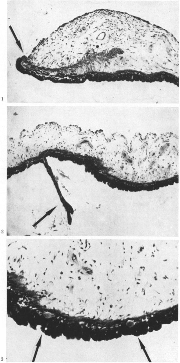
3 Drusen of the Iris 27 Fig. 1. Low power of cross section of the iris showing slight extropion uveae (arrow). -paraffin section, H and E stain, photomicrograph 100 Fig. 2. Displaced ciliary process on the peripheral iris (arrow). -paraffin section, tt and E stain, photomicrograph 100 Fig. 3. Two drusen (arrows) located between pigment epithelium and dilatator muscle of the iris. -paraffin section, H and E stain, Photomicrograph 250 :Fig. 4. Druse of the iris in the present case (arrow) seen at higher power. -paraffin section, H and E stain, photomicrograph X 600 :Fig. 5. Another druse in the iris of the present case (arrow) with distinctly layered structure. -paraffin section, H and E stain, photomicrograph 600

4 28 J.R. Wolter and R. D. Cox Fig. 6. Druse on Bruch's membrane of the choroid (arrow) in the present case. -paraffin section, tt and E stain, photomicrograph 600 The corneal epithelium exhibited an erosion and a few areas with bullous changes. Bowman's membrane was continuous. The corneal stroma appeared to be slightly edematous. Descemct's membrane was continuous and the corneal endothelium was about normal. The iris exhibited some thin-walled blood vessels on its anterior surface and the anterior chamber angle was narrow. Early ectropin uveae was recognized on the pupillary border (Fig. 1). Occasional lymphocytes and plasma cells were found in the iris stroma. A displaced ciliary process was seen on the peripheral iris (Fig. 2). Surprisingly, numerous drusen were observed in the iris in a zone between the pigment epithelium and the myo-epithelial cells of the dilatator muscle (Figs. 3, 4 and 5). These drusen were round bodies of hyaline substance which gave the typical stains with both, the I-I and E and the PAS stains and exhibited the lamellar structure (Fig. 5) that is typically seen in drusen. Both layers, the pigment epithelium and the dilatator muscle were well preserved in the region of the drusen as well as elsewhere in this iris. The ciliary body, showed some mononuelear infiltration as well as some distortion due to the extensive retinal detachment. The lens exhibited early subcapsular cataractous changes. The vitreous was involved with advanced degeneration. The detached retina showed neuronal atrophy, gliosis and distortion everywhere. It was directly involved with the large choroidal tumor that had broken through Bruch's membrane and was composed of spindle-shaped ceils mostly. Some of these cells contained pigment. Most of the cells had a nucleolated nucleus. Serous exudate was found in the retro-retinal space surrounding the tumor. The tumor had at its base in the mid-periphery extended into the inner most layers of the selera, but it had not grown through the sclera to the outside of the eye. The optic nerve was not directly involved, but it exhibited distinct atrophy. It was of special interest that the pigment epithelium in the region of the choroid exhibited many small drusen (Fig. 6) which were in size and staining characteristics very similar to the drusen of the iris. The histopathological diagnosis, thus, was: large malignant choroidal melanoma of a spindle B cell type without direct extraocular extensions. The eye also exhibited bullous keratopathy, early rubeosis iridis, early ectropin uveae, partly exudative and partly solid retinal detachment with secondary atrophy of retina and optic nerve. Small drusen of the

5 Drusen of the Iris 29 senile type were observed on the ehoroid and, surprisingly, very similar drusen were also found in the iris between pigment epithelium and the myo-epithelial cells. Discussion The first extensive histological study of drusen of Bruch's membrane of the choroid was done in 1855 by H. Mueller [1]. These drusen are almost constant in people over 60 years of age, common in people over 45 years and not unusual in younger people. The literature contains more than a dozen different theories to explain the development of drusen. The space of this paper does not allow for a discussion of all of them, but three major theories have survived and deserve attention. Mueller [i] and Aft [2] suggested drusen were a secretion of an intact pigment epithelium. Donders [3] proposed the transformation theory by suggesting they were the result of the direct conversion of degenerating pigment epithelium into drusen material. A vascular theory of drusen formation has been advanced by Friedman, Smith, and Kuwabara [4] who noted pink staining bodies around collecting venules of vortex veins. They believed these bodies to be exudative material which result from vascular decompensation in the aged and which then migrate through porous spaces in the lamina vitrea to be deposited beneath the pigment epithelium. Relatively recent work by Hogan [5, 6] in electron microscopy and further such studies including the histochemical characterization of drusen by Farkas, Sylvester, Archer and Altona [7, 8] have further eluciated the nature of drusen and the probable mechanism of their formation. Hogan [5, 6] has observed that drusen material accumulates in the retinal pigment epithelium cells and in the region of Bruch's membrane. Farkas, Sylvester and Archer [7] have shown that the retinal pigment epithelial cells degenerated into an amorphous material which fills the inner collagenous zone of Brueh's membrane forming drusen. They have demonstrated that a large number of lysosomes are present in the degenerating retinal pigment epithelial cells and drusen material. This, they suggest, supports the theory that drusen formation is due to uncontrolled lysosomal cytolysis of retinal pigment epithelium. It is also noted by Farkas, Sylvester and Archer [7] that the choriocapillaries layer is free from drusen material and that the elastic lamina of Bruch's membrane prevents passage of this material. Thus the site of drusen formation could not be choriocapillaries as suggested by Friedman, Smith and Kuwabara [4]. Histoehemical studies by Farkas, Sylvester, Archer and Altona [8] have shown the composition of drusen of Bruch's membrane to consist of the mucopolysaceharide, sialie acid, and a lipid found to be eerebroside. They suggest that drusen are formed by abnormal lysosomal activity causing the autophagic destruction of retinal pigment epithelium. Mitochondria contain sialic acid. l~efinal pigment epithelium contains lipofuscin granules which could be a source of cerebrosides. Thus the presence of the two major constituents of drusen is explained. The above-mentioned findings are of special interest if one considers their possible application to the present ease of typical drusen between the pigment 3 Albrech~ v. Graefes Arch. klin. exp. Ophthal.
6 30 J.R. Wolter and R. D. Cox epithelium and dilator muscle of the iris. This may represent uncontrolled lysosomal eytolysis of the iris pigment epithelium. This iris drusen in the present case of ocular melanoma may, of course, have to be classified as so-called secondary drusen [9]. The occurrence of druseu in the iris appears to be extremely rare, however, and thus it would not be wise to make more speculations and conclusions concerning this unusual finding in a single eye with rubeosis as well as a large intraoeular tumor. All emphasis should be one the most interesting observation of these drusen under the conditions of the present ease. References 1. Mueller, H.: Anatomische Beitr~ge zur Ophthalmologie. Arch. 0phthal. 2, 1 (pt 2) (1855) 2. Aft, A. : Beitr~ge zur pathologischcn Anatomie des menschlichen Auges. Arch. Ophthal. Otolaryng. 6, 304 (1877) 3. Donders, F.C.: Beitr~ge zur pathologischen Anatomie des Auges. Arch. Ophthal. 1, 106 (et. 2) (1855) 4. Friedman, E., Smith, T., Kuwabara, T. : Senile choroidal vascular patterns and drusen. Arch. Ophthal. 69, 114 (1963) 5. Hogan, M. J.: Bruch's membrane and disease of the macula. Role of elastic tissue and collagen. Trans. ophthal. Soc. U.K. 87, 113 (1967) 6. Hogan, M. J., Alvarado, J. A., Weddell, J. E. : Histology of the human eye, p Philadelphia: W. B. Saunders Farkas, T.G., Sylvester, V., Archer, D.: The ultrastructure of drusen. Amer. J. Ophthal. 71, 1196 (1971) 8. Farkas, T.G., Sylvester, V., Archer, D., Altona, M.: The histochemistry of drusen. Amer. J. Ophthal. 71, 1206 (1971) 9. Welter, J. 1~o, Falls, H. F. : Bilateral confluent drusen. Arch. Ophthal. 68, 219 (1962) J. Reimer Wolter, M. D. Departments of Ophthalmology and Pathology University of Michigan Medical Center Ann Arbor, Michigan 48104, USA
Cystoid macular edema associated with limbal melanoma*
 Graefe's Arch Clin Exp Ophthalmol (1983) 221:101-105 Graefe's Archive for CliniCal and Experimental Ophthalmology Springer-Verlag 1983 Cystoid macular edema associated with limbal melanoma* J. Reimer Wolter
Senile disciform macular degeneration
 British Journal of Ophthalmology, 1977, 61, 141-147 Senile disciform macular degeneration in the second eye Z. GREGOR, A. C. BIRD, AND I. H. CHISHOLM From Moorfields Eye Hospital and Institute of Ophthalmology,
XUE HUI Department of Histology& Embryology, Basic Medicine College of Jilin University
 SENSE ORGAN XUE HUI Department of Histology& Embryology, Basic Medicine College of Jilin University EYE fibrous globe lens photosensitive cells a system of cells and nerves concentric layers the sclera
Around The Globe in 60 Minutes
 Around The Globe in 60 Minutes Around the GLOBE in Sixty Minutes Basic Ocular Anatomy, Examination, and Diagnostic Techniques Introduction Focusing on canine and feline ocular anatomy and basic examination
Uveal Melanoma. Protocol applies to malignant melanoma of the uvea.
 Uveal Melanoma Protocol applies to malignant melanoma of the uvea. Protocol revision date: January 2005 Based on AJCC/UICC TNM, 6 th edition Procedures Cytology (No Accompanying Checklist) Biopsy (No Accompanying
THE EYE: RETINA AND GLOBE
 Neuroanatomy Suzanne Stensaas February 24, 2011, 10:00-12:00 p.m. Reading: Waxman Ch. 15. Your histology and gross anatomy books should be useful. Reading: Histology of the Eye from any histology book
COMMUNICATIONS PHOTOCOAGULATION OF THE RETINA* OPHTHALMOSCOPIC AND HISTOLOGICAL FINDINGS. photocoagulation of the rabbit's retina.
 Brit. J. Ophthal. (1963) 47, 577. COMMUNICATIONS PHOTOCOAGULATION OF THE RETINA* OPHTHALMOSCOPIC AND HISTOLOGICAL FINDINGS BY A. LAVYEL Haifa, Israel SINCE the introduction of the photocoagulator by Meyer-Schwickerath
Glaucoma Glaucoma is a complication which has only recently been confirmed as a feature of
 1.2.4 OPHTHALMOLOGICAL ABNORMALITIES Ocular abnormalities are well documented in patients with NPS 6 62 81 95. 1.2.4.1 Glaucoma Glaucoma is a complication which has only recently been confirmed as a feature
Corneal blood staining after hyphaema
 Brit. J_. Ophthal. (I 972) 56, 589 after hyphaema J. D. BRODRICK Sheffield has been described as a rare complication of contusion injury in which a hyphaema of relatively long duration and a raised intraocular
Vision I. Steven McLoon Department of Neuroscience University of Minnesota
 Vision I Steven McLoon Department of Neuroscience University of Minnesota 1 Eye Cornea Sclera Conjunctiva 2 Eye The conjunctiva lines the inner surface of the eyelids and outer surface of the sclera. 3
Coagulative necrosis in a malignant melanoma of the choroid at the macula with extensive subretinal hemorrhage
 Coagulative necrosis in a malignant melanoma of the choroid at the macula with extensive subretinal hemorrhage Robert D. Yee, Robert Y. Foos, and Bradley R. Straatsma The authors present a case report
Asadi-Amoli et al Adenocarcinoma of RPE Iranian Journal of Ophthalmology - Volume 19, Number 4, 2007
 Adenocarcinoma of Retinal Pigment Epithelium Clinically Diagnosed as Malignant Melanoma; A Case Report with Unsystematic Review of Literature Fahimeh Asadi-Amoli, MD, 1 Hedyeh Moradi, MD 2 Mohammad-Taher
Concentric "Microwaves" of Henle's Fiber Layer: Associated with Horizontal Folding
 Albrecht von Graefes Arch Klin Ophthalmol. (1981) 216:31-39 Graefes Archiv for klinische und experimenlelle Ophthalmologie 9 Springer-Verlag 1981 Concentric "Microwaves" of Henle's Fiber Layer: Associated
Histology of the Eye
 Histology of the Eye Objectives By the end of this lecture, the student should be able to describe: The general structure of the eye. The microscopic structure of:»cornea.»retina. EYE BULB Three coats
Drusen, an Age Related Change in the Retina of the Fisher Rat
 ORIGINAL ARTICLE Drusen, an Age Related Change in the Retina of the Fisher Rat M. Waheed Rana, Ph.D. and Yok Lai, M.D., Ph.D. St. L ouis, Missouri DOI: http://dx.doi.org/10.5915/23-2-14923 Abstract The
Adenocarcinorna of the Ciliary Body A Report of 2 Cases in Dogs
 Path. vet. 5: 122-126 (1968) From the Ophthalmic Pathology Laboratory, Department of Ophthalmology, New York University School of Medicine, New York Adenocarcinorna of the Ciliary Body A Report of 2 Cases
Medical School Histology Basics. VIBS 289 lab. Eye
 Medical School Histology Basics VIBS 289 lab Eye Larry Johnson Texas A&M University Aqueous humor OUTLINE OVERVIEW CELLULAR STRUCTURES THROUGH WHICH LIGHT PASSES A. CORNEA B. LENS C. RETINA STRUCTURES
Year 2 MBChB Clinical Skills Session Ophthalmoscopy. Reviewed & ratified by: Mr M Batterbury Consultant Ophthalmologist
 Year 2 MBChB Clinical Skills Session Ophthalmoscopy Reviewed & ratified by: o Mr M Batterbury Consultant Ophthalmologist Learning objectives o To understand the anatomy and physiology of the external and
Haemorrhagic glaucoma
 Brit. j. Ophthal. (I97I) 55, 444 Haemorrhagic glaucoma Comparative study in diabetic and nondiabetic patients P. H. MADSEN From the Departments of Ophthalmology and Internal Medicine, the University Hospital,
Graefe's Archive. Ophthalmology Springer-Verlag Artificial anterior chamber for the growing of membranes on lens implants*
 Graefe's Arch Clin Exp Ophthalmol (1983) 221:55-60 Graefe's Archive for Clinical and Experimental Ophthalmology Springer-Verlag 1983 Artificial anterior chamber for the growing of membranes on lens implants*
Scrub In. What is the function of vitreous humor? What does the pupil do when exposed to bright light? a. Maintain eye shape and provide color vision
 Scrub In What is the function of vitreous humor? a. Maintain eye shape and provide color vision b. Maintain eye shape and refract light rays c. Provide night vision and color vision d. Provide night vision
A NEW CLINICAL FORM OF HETEROCHROMIA OF
 Brit. J. Ophthal. (1961) 45, 597. A NEW CLINICAL FORM OF HETEROCHROMIA OF THE IRIS WITH PIGMENTATION OF THE SCLERA AND PIGMENTARY GLAUCOMA* BY V. CAVKA Beograd, Yugoslavia THE significance of pigmentation
Benign melanoma of the choroid
 Brit. J. Ophthal. (I969) 53, 621 Benign melanoma of the choroid Recognition of malignant change using clinical photographic techniques E. S. ROSEN* AND A. GARNERt Manchester Roval Eye Hospital* and the
02/03/2014. Average Length: 23mm (Infant ~16mm) Approximately the size of a quarter Volume: ~5mL
 Approximately the size of a quarter Volume: ~5mL") Identify the anatomy of the eye. Explain the basic physiology of the parts of the eye. Briefly discuss various surgeries related to different parts of the anatomy. Average Length: 23mm (Infant ~16mm) Approximately
Identify the anatomy of the eye. Explain the basic physiology of the parts of the eye. Briefly discuss various surgeries related to different parts of the anatomy. Average Length: 23mm (Infant ~16mm) Approximately
The Orbit. The Orbit OCULAR ANATOMY AND DISSECTION 9/25/2014. The eye is a 23 mm organ...how difficult can this be? Openings in the orbit
 The eye is a 23 mm organ...how difficult can this be? OCULAR ANATOMY AND DISSECTION JEFFREY M. GAMBLE, OD COLUMBIA EYE CONSULTANTS OPTOMETRY & UNIVERSITY OF MISSOURI DEPARTMENT OF OPHTHALMOLOGY CLINICAL
Ocular Pathology. I. Congenital and/or developmental. A. Trisomy 21. Hypertelorism (widely spaced eyes) Keratoconus (cone shaped cornea)
 Keratoconus (cone shaped cornea)") I. Congenital and/or developmental Robbins Pathologic Basis of Disease, 6 th Ed. A. Trisomy 21 Hypertelorism (widely spaced eyes) Keratoconus (cone shaped cornea) Focal hypoplasia of iris Cataracts frequently
I. Congenital and/or developmental Robbins Pathologic Basis of Disease, 6 th Ed. A. Trisomy 21 Hypertelorism (widely spaced eyes) Keratoconus (cone shaped cornea) Focal hypoplasia of iris Cataracts frequently
HYPERPLASIA OF THE ANTERIOR LAYER OF THE IRIS STROMA*t
 Brit. J. Ophthal. (1965) 49, 516 HYPERPLASIA OF THE ANTERIOR LAYER OF THE IRIS STROMA*t BY MALCOLM N. LUXENBERG From the Bascom Palmer Eye Institute, Department of Ophthalmology, University of Miami School
Vascular changes in the iris in chronic
 Vascular changes in the iris in chronic anterior uveitis LEILA LAATIKAINEN From the Department of Ophthalmology, University of Helsinki, Finland British Journal of Ophthalmology, 1979, 63, 145-149 SUMMARY
Central venous occlusion
 Central venous occlusion Central venous occlusion (right eye) There are dark haemorrhages at the macula and all over the retina. Choroidal haemangioma A choroidal haemangioma has salmon pink colour. There
OCT Angiography in Primary Eye Care
 OCT Angiography in Primary Eye Care An Image Interpretation Primer Julie Rodman, OD, MS, FAAO and Nadia Waheed, MD, MPH Table of Contents Diabetic Retinopathy 3-6 Choroidal Neovascularization 7-9 Central
Fundus Autofluorescence. Jonathan A. Micieli, MD Valérie Biousse, MD
 Fundus Autofluorescence Jonathan A. Micieli, MD Valérie Biousse, MD The retinal pigment epithelium (RPE) has many important functions including phagocytosis of the photoreceptor outer segments Cone Rod
UNDERSTAND MORE ABOUT UVEITIS UVEITIS
 UNDERSTAND MORE ABOUT UVEITIS UVEITIS Uveitis What is uveitis? Uveitis is inflammation of the uvea, the middle layer of your eye. The eye is shaped much like a tennis ball, with three different layers
Pathology of the lens
 Pathology of the lens Carol Naranjo, LV, DACVP, DECVP, PhD IDEXX Laboratories Embryonal development Gelatt s Veterinary Ophthalmology, 5th Ed. Normal histology Lens capsule Anterior > posterior Lens cortex
Eye Fluids. Dr. Mohamed Saad Daoud
 Eye Fluids 1 Reference Books: Text Book of Medical physiology (Guyton and Hall) Eleventh edition 2 Fluid System of the Eye (Intraocular Fluid) The eye is filled with intraocular fluid, which maintains
OCULAR DISORDERS REPORT BOSTON TERRIER
 OCULAR DISORDERS REPORT BOSTON TERRIER 1991-1999 2000-2009 2010-2012 TOTAL DOGS EXAMINED 2723 6803 2004 Diagnostic Name # % # % # % GLOBE 0.110 microphthalmia 1 0.0% 1 0.0% 0 EYELIDS 20.140 ectopic cilia
Mild NPDR. Moderate NPDR. Severe NPDR
 Diabetic retinopathy Diabetic retinopathy is the most common cause of blindness in adults aged 35-65 years-old. Hyperglycaemia is thought to cause increased retinal blood flow and abnormal metabolism in
DAMAGE TO CORNEA AND LENS*
 Brit. J. Ophthal. (1965) 49, 460 PRIMARY ACUTE ANGLE-CLOSURE GLAUCOMA DAMAGE TO CORNEA AND LENS* BY RONALD F. LOWE From the Glaucoma Unit, the Royal Victorian Eye and Ear Hospital, Melbourne, and the Ophthalmic
Patient AB. Born in 1961 PED
 Clinical Atlas Patient AB Born in 1961 PED Autofluorescence Dilated 45 EasyScan Zero-dilation IR 45 Fundus Dilated 45 In the fundus photos (Canon CX1) the PED is not able to be seen. However, the extent
Outline. Brief history and principles of ophthalmic ultrasound. Types of ocular ultrasound. Examination techniques. Types of Ultrasound
 Ultrasound and Intraocular Tumors 2015 Ophthalmic Photographers' Society Mid-Year Program Cagri G. Besirli MD, PhD Kellogg Eye Center University of Michigan Outline Brief history and principles of ophthalmic
CONGENITAL CORNEAL OPACITIES*t IN A PATIENT WITH RIEGER'S ANOMALY AND DOWN'S SYNDROME
 Brit. J. Ophthal. (1968) 52, 631 CONGENITAL CORNEAL OPACITIES*t IN A PATIENT WITH RIEGER'S ANOMALY AND DOWN'S SYNDROME BY From the Department of Ophthalmology, Royal Hospital, Sheffield THIS report concerns
Diffuse infiltrating retinoblastoma
 Brit. 1. Ophthal. (I 971) 55, 6oo Diffuse infiltrating retinoblastoma GWYN MORGAN Department of Pathology, Institute of Ophthalmology, University of London The term "diffuse infiltrating retinoblastoma"
generalized neurofibromatosis
 Brit. 7. Ophthal. (I972) 56, 487 Glial hamartoma of the retina in generalized neurofibromatosis von Recklinghausen's disease L. J. MNIARTYN AND D. L. KNOX From the li'ilmer Institute, the Johns Hopkins
Electron Microscopic Demonstration of Centrifugal Nerve Fibers in the Human Optic Nerve
 Albrecht v. Graefes Arch. klin. exp. Ophthal. 210, 3141 (1979) Graefes Archiv fijr klinische und experimentelle Ophthalmologie 9 by Springer-Verlag 1979 Electron Microscopic Demonstration of Centrifugal
PART 1: GENERAL RETINAL ANATOMY
 PART 1: GENERAL RETINAL ANATOMY General Anatomy At Ora Serrata At Optic Nerve Head Fundoscopic View Of Normal Retina What Is So Special About Diabetic Retinopathy? The WHO definition of blindness is
Abstract Aims To analyse the histopathology of classic and occult choroidal neovascular membrane surgical specimens in age
 Br J Ophthalmol 2000;84:239 243 239 ORIGINAL ARTICLES Clinical science Clinicopathological correlation in exudative age related macular degeneration: histological diverentiation between classic and occult
 measure of your overall performance. An isolated glucose test is helpful to let you know what your sugar level is at one moment, but it doesn t tell you whether or not your diabetes is under adequate control
OPHTHALMOLOGY AND ULTRASOUND
 Vet Times The website for the veterinary profession https://www.vettimes.co.uk OPHTHALMOLOGY AND ULTRASOUND Author : JAMES OLIVER Categories : Vets Date : April 28, 2008 JAMES OLIVER discusses why ultrasound
Vitreoretinal juncture; epiretinal membranes and vitreous. Robert Y. Foos
 Vitreoretinal juncture; epiretinal membranes and vitreous Robert Y. Foos This report reviews current knowledge of the ultrastructural features of the vitreoretinal juncture in its normal state, in eyes
GENERAL INFORMATION CORNEAL TRANSPLANTATION
 GENERAL INFORMATION CORNEAL TRANSPLANTATION WHAT IS CORNEAL TRANSPLANTATION? A corneal transplant is an operation where a damaged or diseased cornea is replaced with donated, healthy tissue. Also called
Speaker Disclosure Statement. " Dr. Tim Maillet and Dr. Vladimir Kozousek have no conflicts of interest to disclose.
 Speaker Disclosure Statement Dr. Tim Maillet and Dr. Vladimir Kozousek have no conflicts of interest to disclose. Diabetes Morbidity Diabetes doubles the risk of stroke. Diabetes quadruples the risk of
Dr/ Marwa Abdellah EOS /16/2018. Dr/ Marwa Abdellah EOS When do you ask Fluorescein angiography for optic disc diseases???
 When do you ask Fluorescein angiography for optic disc diseases??? 1 NORMAL OPTIC DISC The normal optic disc on fluorescein angiography is fluorescent due to filling of vessels arising from the posterior
CENTRAL AREOLAR CHOROIDAL SCLEROSIS* A HISTO-PATHOLOGICAL STUDY
 Brit. J. Op/ithal. (1953), 37, 140. CENTRAL AREOLAR CHOROIDAL SCLEROSIS* A HISTO-PATHOLOGICAL STUDY BY NORMAN ASHTON Department of Pathology, Institute of Ophthalmology, London THE history and clinical
A study of iris melanoma in Northern Ireland
 British Journal of Ophthalmology, 1989, 73, 591-595 A study of iris melanoma in Northern Ireland J N McGALLIARD AND P B JOHNSTON From the Department of Ophthalmology, Royal Victoria Hospital, Grosvenor
Vitreous! Retinal pigment epithelium! and the visual cycle! Retinal degenerations and pigment epithelium!
 Vitreous Bruch s membrane Retinal pigment epithelium and the visual cycle Retinal degenerations and pigment epithelium Basic Science course 2017 Swiss Eye Week, Neuchâtel Ch. E. Remé, Zürich Ch.E. Remé
4/22/16. Eye. External Anatomy of Eye. Accessory Structures. Bio 40B Dr. Kandula
 Eye Bio 40B Dr. Kandula External Anatomy of Eye Accessory Structures l Eyebrows l Levator Palpebrae Superioris - opens eye l Eyelashes l Ciliary glands modified sweat glands l Small sebaceous glands l
NEPTUNE RED BANK BRICK
 NEPTUNE RED BANK BRICK Diabetes & The Eye Diabetics are more likely to develop Cataracts at a younger age. Diabetics are twice as likely to develop Glaucoma when compared to non-diabetics. The primary
Foreign Body Giant Cells on Intraocular Lens Implants*
 Graefe's Arch Clin Exp Ophthalmol (1982) 219:103-11I Graefe's Archive for Clinical and Experimental Ophthalmology Springer-Verlag 1982 Foreign Body Giant Cells on Intraocular Lens Implants* J. Reimer Wolter
RETINAL DETACHMENT AT THE POSTERIOR POLE*
 Brit. J. Ophthal. (1958) 42, 749. RETINAL DETACHMENT AT THE POSTERIOR POLE* BY CALBERT I. PHILLIPSt Institute of Ophthalmology, University oflondon THE common feature of the cases to be described in this
Dr. D. Y. Patil Medical College, Pimpri, Pune
 Dr. D. Y. Patil Medical College, Pimpri, Pune - 411 018 Period : 04/July/16 to 22/September/16 Semester : 7 th Semester Department : Ophthalmology Lecture Lesson Plan Sr No Date Topic Learning objectives
Retinal Tear and Detachment
 Retinal Tear and Detachment Introduction The retina is the layer of tissue in the back of the eye that is responsible for vision. It is attached to the choroid tissue, which supplies the retina with blood.
The Quick Guide to OCT Mastery 50 Real Cases with Expert Analysis
 OPTICAL COHERENCE TOMOGRAPHY The Quick Guide to OCT Mastery 50 Real Cases with Expert Analysis VOL 1 Sanjay Sharma, MD, FRCS, MSc (Epid), MBA Ophthalmologist, Epidemiologist Queen s University, Canada
FELINE DIFFUSE IRIDAL MELANOMA
 Vet Times The website for the veterinary profession https://www.vettimes.co.uk FELINE DIFFUSE IRIDAL MELANOMA Author : JAMES OLIVER Categories : Vets Date : June 3, 2013 JAMES OLIVER looks at several clinical
2/26/2017. Sameh Galal. M.D, FRCS Glasgow. Lecturer of Ophthalmology Research Institute of Ophthalmology
 Sameh Galal M.D, FRCS Glasgow Lecturer of Ophthalmology Research Institute of Ophthalmology No financial interest in the subject presented 1 Managing cataracts in children remains a challenge. Treatment
Five Things You re Missing with Your Fundus Camera
 ebook Five Things You re Missing with Your Fundus Camera By Donald J. Siegel, OD, Sun City West Eye Care Sponsored by: Before I began incorporating EIDON true-color imaging into my practice, my retinal
Department of Ophthalmology
 Period : 03/July/17 to 07/September/17 Semester : 7 th Semester Department of Ophthalmology Lecture Lesson Plan Sr 1 03.07.17 Uvea-Anatomy, Uvea-Anatomy, Classification of Uveitis Dr R Paranjpe Classification
OUR EYES & HOW WE SEE
 OUR EYES & HOW WE SEE UNDERSTAND MORE ABOUT OUR EYES & HOW WE SEE Our Eyes & How We See The eye is our visual gateway to the world. Within it, an array of delicate components labour away to give us the
PATIENT INFORMATION ON CORNEAL GRAFT
 PATIENT INFORMATION ON CORNEAL GRAFT (TRANSPLANT) SURGERY M ANANDAN What is the cornea? The clear window of the eye approximately 0.5mm thick and 12mm across. It lies in front of the fluid filled anterior
PATHWAY OF CENTRIFUGAL FIBRES IN THE HUMAN
 Brit. J. Ophthal. (1965) 49, 246 PATHWAY OF CENTRIFUGAL FIBRES IN THE HUMAN OPTIC NERVE, CHIASM, AND TRACT*t BY J. REIMER WOL-TER AND ROMAN R. KNOBLICH From the Departments of Ophthalmology and Pathology
Capsular exfoliation syndrome
 Brit. J. Ophthal. (I973) 57, I 20 G. C. SOOD, B. K. SOFAT, S. K. MEHROTRA, AND R. D. CHANDEL Department of Ophthalmology, H.P. Medical College, Simla- i, India The nature ofsenile capsular exfoliation
Department of Ophthalmology
 Department of Ophthalmology Period : 02/July/18 to 30/August/18 Semester : 7 th Semester Lecture Lesson Plan Sr. Date Topic Lesson plan Name of Faculty No. 1 02.07.18 Lens- Lens-Anatomy, Classification
Chorioretinal vascular occlusions with latex microspheres (a long-term study). Part II. Howard Golclor and Andrew J. Gay
. Part II. Howard Golclor and Andrew J. Gay") Chorioretinal vascular occlusions with latex microspheres (a long-term study). Part II Howard Golclor and Andrew J. Gay The results of a long-term study (3 to 9 months) of chorioretinal vascular occlusions
Chorioretinal vascular occlusions with latex microspheres (a long-term study). Part II Howard Golclor and Andrew J. Gay The results of a long-term study (3 to 9 months) of chorioretinal vascular occlusions
Eye and Ocular Adnexa, Auditory Systems
 Eye and Ocular Adnexa, Auditory Systems CPT copyright 2011 American Medical Association. All rights reserved. Fee schedules, relative value units, conversion factors and/or related components are not assigned
Unit VIII Problem 8 Anatomy: Orbit and Eyeball
 Unit VIII Problem 8 Anatomy: Orbit and Eyeball - The bony orbit: it is protecting our eyeball and resembling a pyramid: With a base directed: anterolaterally. And an apex directed: posteromedially. Notes:
It has previously been shown that pine
 The effects of cortisone on pine pollen-induced uveitis in guinea pigs Stan L. Coleman and Samuel Canaan Injection of pine pollen into the vitreous of guinea pigs previously sensitized with Freund's adjuvant
Silicone oil pupillary block after laser retinopexy in aphakic eyes with presumed closed peripheral iridectomy: report of three cases
 Int Ophthalmol (2014) 34:913 917 DOI 10.1007/s10792-013-9862-z CASE REPORT Silicone oil pupillary block after laser retinopexy in aphakic eyes with presumed closed peripheral iridectomy: report of three
(Received 8 March 1965)
") J. Physiol. (1965), 180, pp. 837-845 837 With 1 plate and 4 text-figures Printed in Great Britain THE EFFECT OF OCCLUDING THE RETINAL AND CHOROIDAL CIRCULATIONS ON THE ELECTRO- RETINOGRAM OF MONKEYS BY
J. Physiol. (1965), 180, pp. 837-845 837 With 1 plate and 4 text-figures Printed in Great Britain THE EFFECT OF OCCLUDING THE RETINAL AND CHOROIDAL CIRCULATIONS ON THE ELECTRO- RETINOGRAM OF MONKEYS BY
Bilateral congenital ocular defects in a foal
 Brit. J. Ophthal. (I969) 53, 5 I 3 Bilateral congenital ocular defects in a foal A. GARNER AND P. GRIFFITHS Department of Pathology, Institute of Ophthalmology, University of London; and Burton-on-Trent,
Retinal pigment epithelial detachments in the elderly:
 British Journal of Ophthalmology, 1985, 69, 397-403 Retinal pigment epithelial detachments in the elderly: classification and outcome A G CASSWELL, D KOHEN, AND A C BIRD From Moorfields Eye Hospital, City
uveal melanoma: case report with pathological examination
 British Journal of Ophthalmology, 1986, 70, 33-38 Simultaneous bilateral primary diffuse malignant uveal melanoma: case report with pathological examination SHIGEO TSUKAHARA,' KAICHI WAKUI,2 AND SHINOBU
Complicated Cataract to Intraocular Tumors, Beware of the unexpected
 Complicated Cataract to Intraocular Tumors, Beware of the unexpected Ihab Saad Othman, MD, FRCS Professor of Ophthalmology Cairo University In this part of the world: We Master Phakoemulsification 1 Intraoperative/Second
Clinically Significant Macular Edema (CSME)
") Clinically Significant Macular Edema (CSME) 1 Clinically Significant Macular Edema (CSME) Sadrina T. Shaw OMT I Student July 26, 2014 Advisor: Dr. Uwaydat Clinically Significant Macular Edema (CSME) 2
Clinically Significant Macular Edema (CSME) 1 Clinically Significant Macular Edema (CSME) Sadrina T. Shaw OMT I Student July 26, 2014 Advisor: Dr. Uwaydat Clinically Significant Macular Edema (CSME) 2
_~~~~~~~~~~~~~~... I~~~~~~~~~~~~~~~~~~~~~~~~...x'..'.i ORBITAL BENIGN HAEMANGIO ENDOTHELIOMATA*
 '..,S''''. t ~~~~~~~~~~~~~~~~~~~~~~~. g.# lgl,~~~~~~~~~~~~~~~~~~~~~~~~~~~~~~.%. : Brit. J. Ophthal. (1963) 47, 164. ORBITAL BENIGN HAEMANGIO ENDOTHELIOMATA* BY ALY MORTADA Faculty of Medicine, Cairo University,
Mesectodermal suprauveal iridociliary leiomyoma: Transscleral excision without postoperative iris defect
 4 Chapter Mesectodermal suprauveal iridociliary leiomyoma: Transscleral excision without postoperative iris defect Lubna Razzaq 1, Ekaterina A Semenova 2, Marina Marinkovic 1, Rob JW de Keizer 1, Sjoerd
let's continue talking about the eye,
 Eye is mainly composed of 3 layers: External layer, which called The Sclera which is a hard connective tissue that gives the eye its round shape. Extension of the sclera into the front is the cornea, which
Ocular Anatomy for the Paraoptometric
 Ocular Anatomy for the Paraoptometric Minnesota Optometric Association Paraoptometric CE Friday September 30, 2016 Lindsay A. Sicks, OD, FAAO Assistant Professor, Illinois College of Optometry lsicks@ico.edu
What Is O.C.T. and Why Should I Give A Rip? OCT & Me How Optical Coherence Tomography Changed the Life of a Small Town Optometrist 5/19/2014
 OCT & Me How Optical Coherence Tomography Changed the Life of a Small Town Optometrist Email: myoder@wcoil.com Mark A. Yoder, O.D. 107 N. Main Street PO Box 123 Bluffton, OH 45817 @yoderod 115.02 Histoplasma
Disease-Specific Fluorescein Angiography
 Ruth E. Picchiottino, CRA Disease-Specific Fluorescein Angiography 15 Disease-Specific Fluorescein Angiography Recommendations for tailoring retinal fluorescein angiography to diabetic retinopathy, macular
Funduscopic Interpretation Understanding the Fundus: is that normal?
 Funduscopic Interpretation Understanding the Fundus: is that normal? Gillian McLellan BVMS PhD DVOphthal DECVO DACVO MRCVS With thanks to Christine Heinrich and all who contributed images Fundus Retina
DEGENERATIONS OF THE DOG RETINA*
 Brit. J. Ophthal. (1954) 38, 545. DEGENERATIONS OF THE DOG RETINA* V. GENERALIZED PROGRESSIVE ATROPHY OF UNCERTAIN AETIOLOGY BY Animal Health Trust, Kennett, Newmarket, Suffolk DURING an investigation
(ALBERS-SCHONBERG DISEASE)*
*") Brit. J. Ophthal. (1964) 48, 218. NEURO-HISTOLOGICAL FINDINGS IN OSTEOPETROSIS (ALBERS-SCHONBERG DISEASE)* BY F. VRABEC AND J. SEDLA(KOVA From the Eye Clinic of the Medical Faculty of Hygiene of the Charles
Brit. J. Ophthal. (1964) 48, 218. NEURO-HISTOLOGICAL FINDINGS IN OSTEOPETROSIS (ALBERS-SCHONBERG DISEASE)* BY F. VRABEC AND J. SEDLA(KOVA From the Eye Clinic of the Medical Faculty of Hygiene of the Charles
Pseudohypopyon in Retinoblastoma. Choroidal Nevus. Masquerade Syndromes. Vision pathways. Flat with uniform color
 Primary Intraocular Tumors Thomas F. Freddo, O.D., Ph.D., F.A.A.O. Professor and Former Director School of Optometry University of Waterloo Masquerade Syndromes
LEUKAEMIA*t INFILTRATION OF THE IRIS IN CHRONIC LYMPHATIC. pattemn * Received for pubiication November io, i967.
 Brit. J. Ophthal. (1968) 52, 781 INFILTRATION OF THE IRIS IN CHRONIC LYMPHATIC LEUKAEMIA*t BY BRIAN MARTIN The General Infirmary, Leeds OCULAR involvement is common in the leukaemias though the anterior
Graefe's Archive. Ophthalmology Springer-Verlag t983. Fusion of maerophages on lens implants resulting in the formation of giant cells
 Graefe's Arch Clin Exp Ophthalmol (1983) 221:1-7 Graefe's Archive for Clinical and Experimental Ophthalmology Springer-Verlag t983 Fusion of maerophages on lens implants resulting in the formation of giant
Retina Center of Oklahoma Sam S. Dahr, M.D. Adult Intraocular Tumors
 Adult Intraocular Tumors Sam S. Dahr, M.D. Retina Center of Oklahoma www.retinacenteroklahoma.com www.rcoklahoma.com Table of Contents Posterior uveal malignant melanoma Uveal metastasis Uveal melanoma
THE CHRONIC GLAUCOMAS
 THE CHRONIC GLAUCOMAS WHAT IS GLAUCOMA? People with glaucoma have lost some of their field of all round vision. It is often the edge or periphery that is lost. That is why the condition can be missed until
Secondary anterior crocodile shagreen of Vogt
 Brit. J. Ophthal. (1975) 59, 59 Secondary anterior crocodile shagreen of Vogt R. C. TRIPATHI AND A. J. BRON From the Department of Pathology, Institute of Ophthalmologyv Nuffield Laboratory of Ophthalmology,
Primary Angle Closure Glaucoma
 www.eyesurgeonlondon.co.uk Primary Angle Closure Glaucoma What is Glaucoma? Glaucoma is a condition in which there is damage to the optic nerve. This nerve carries visual signals from the eye to the brain.
Vision loss in elderly. Erica Weir, April 2015
 Vision loss in elderly Erica Weir, April 2015 1 Burden Enter nursing homes 3 years earlier Twice the risk of falling 4x the risk of hip fracture Independent risk factor for delirium What are the leading