A spectral-domain OCT study of formerly premature children. Prat Itharat MD May 30, 2008 Vanderbilt Eye Institute Preceptor: Dr.
|
|
|
- Valentine McCormick
- 6 years ago
- Views:
Transcription
1 A spectral-domain OCT study of formerly premature children. Prat Itharat MD May 30, 2008 Vanderbilt Eye Institute Preceptor: Dr. Recchia
2 Background: Optical coherence tomography (OCT) OCT analogous to ultrasound imaging Non-invasive Uses light waves Many applications in ophthalmology
3 Background: OCT Zeiss Stratus OCT Time Zeiss Domain Cirrus HD-OCT (Meditec, Dublin, CA) Acquires 6 linear 200 B-scans linear B- scans Fewer data points sampled More data points sampled Slower Faster scanning scanning time time Better resolution: up to 5 Resolution: up to 10 microns microns Less concern for eccentric More fixation concern and for eccentric technical fixation and skills technical skills Zeiss Cirrus HD-OCT Spectral Domain (Meditec, Dublin, CA) Zeiss Stratus OCT Acquires 200 linear B-scans Acquires 6 linear B- scans Fewer data points sampled Slower scanning time Resolution: up to 10 microns More concern for eccentric fixation and technical skills More data points sampled Faster scanning time Better resolution: up to 5 microns Less concern for eccentric fixation and technical skills

4 Background: SD vs TD OCT Spectral Domain Time Domain Courtesy of Zeiss Whitepaper
5 Background: Prematurity
6 Background: Prematurity Escedy et al (2007) noted increase central retinal thickness in formerly premature children Stratus OCT Mainly due to presence to ROP

7 Recchia et al (2007) showed OCT abnormalities in ROP patients - first to suggest that changes may be vestiges of prematurity

8 Recchia et al (2007) showed OCT abnormalities in ROP patients - first to suggest that changes may be vestiges of prematurity

9 Recchia et al (2007) showed OCT abnormalities in ROP patients - first to suggest that changes may be vestiges of prematurity
10 Limitations Technologically Methodologically
11 Hypothesis: OCT abnormalities are more common in formerly premature children than in full term children.
12 OCT and prematurity Study questions: 1. Is there a difference in macular thickness between premature and full-term children? 2. Is there a difference in foveal contour between these groups? 3. Are the OCT findings correlated to the presence of ROP or another variable?
13 Methods IRB approval for prospective study Records of formerly premature infants born prior to 1/1/2001 (treated in the Vanderbilt NICU) Control group matched for age and refractive error
14 Methods Group I: premature with ROP (<32 weeks gestational age) Group II: premature without ROP (<32 weeks gestational age) Group III: full term (>36 weeks gestational age)
15 Methods Gestational age, birth weight, ROP status obtained from records Cycloplegic refraction Best corrected visual acuity
16 Methods Quantitative primary outcome measures (OCT) -central subfield thickness -inner (ring) macular thickness -outer (ring) macular thickness -total macular volume
17 Methods Qualitative primary outcome measures (OCT) -presence of foveal depression -preservation of retinal layers
18 Methods OCT data were reviewed by a trained physician (FMR) masked to the birth history of the patient Quality of all scans was assessed and any scans with a signal strength less than 8/10 were discarded For each eye, a 6 x 6 mm macular cube and 5-line macular raster line scan were interpreted individually

19 Results: Eyes
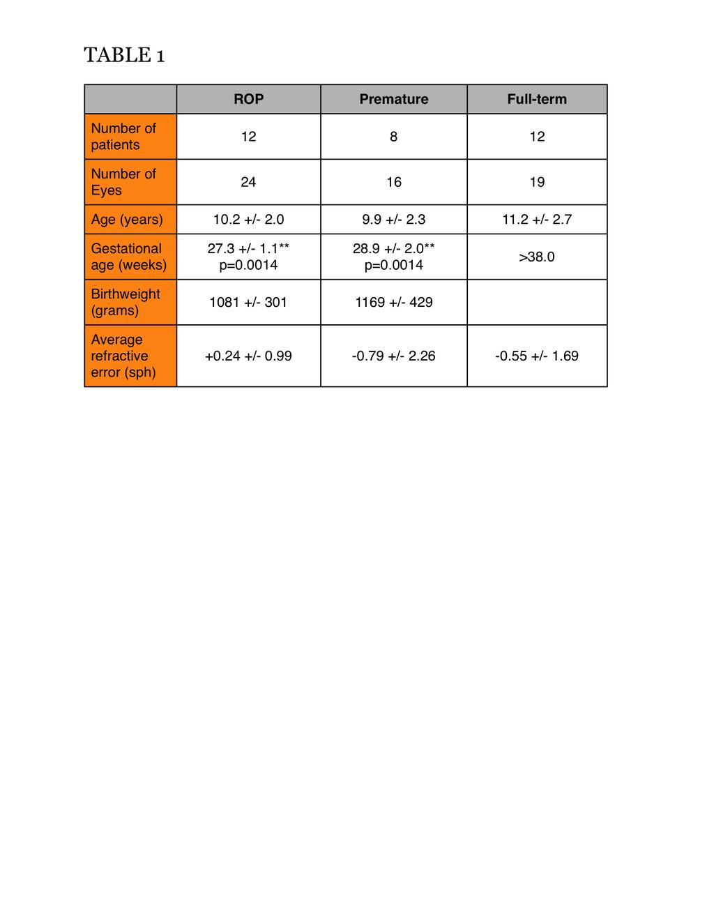
20 Results:
21 Results: Central subfield thickness

22 Results: Inner ring retinal thickness
23 Results: Outer ring retinal thickness
24 Results: Total macular volume

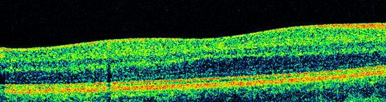
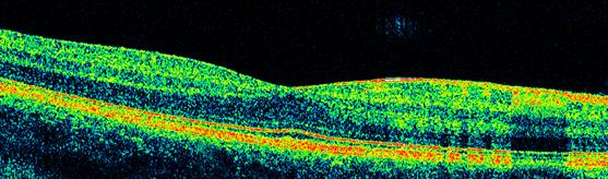
25 Results: Foveal contour

26 Results: Central subfield thickness : inner ring thickness
27 Results: Gestational age effect?

28 Discussion: Significant effect of gestational age Non-significant effect of ROP In contrast to prior studies which suggest that ROP is the major determinant of OCT abnormalities

29 Discussion: Foveal development Foveal depression occurs by a reduction of inner (ganglion cell and inner nuclear) layers of the retina Evident by weeks of gestation Continues until four months postnatally Diagram from Provis et al.
30 Discussion: Foveal development Mintz-Hittner et al described smaller foveal avascular zone (FAZ) in formerly premature children Provis et al showed that the formation of the FAZ (vascular border) precedes foveal depression These two processes may be interrelated
31 Discussion: Foveal development Prematurity may change retinal oxygenation Disruption of FAZ and foveal formation -Blunted/absent foveal depression -Thickened central macula -Preservation of retinal layers Interestingly, most of our patients had excellent visual acuity
32 Limitations Selection bias (less severe ROP, less severe neurological disease) Non-matched gestational age Most ROP staging obtained indirectly through NICU discharge summaries Inter-rater variability of ROP
33 Conclusions OCT findings such as increased central macular thickness and foveal depression blunting may represent hallmarks of prematurity These findings can be associated with normal visual acuity
34 Clinical relevance Greater use of OCT in evaluation of patients with decreased visual acuity OCT abnormalities may not represent ocular disease Research studies in which numerical cutoffs are made for inclusion or evaluation of therapeutic response
35 Acknowledgements
36 References Ecsedy M, Szamosi A, Karko C, Zubovics L, Varsanyl B, Nemeth J, Recsan Z. A comparison of Macular Structure Imaged by Optical Coherence Tomography in Preterm and Full-Term Children. Investigative Ophthalmology & Visual Science. November 2007; 48(11), Fleck BW, McIntosh N. Pathogenesis of retinopathy of prematurity and possible preventive strategies. Early Human Development 2008; 84: Fulton AB, Hansen RM, Moskowitz A, Barnaby Am. Multifocal ERG in subjects with a history of retinopathy of prematurity. Doc. Ophthal. 2005; 111:7-13. Hammer DX, Iftimia N, Ferguson RD, Bigelow CE, Ustun TE, Barnaby AM, Fulton AB. Foveal Fine Structure in Retinopathy of Prematurity: An Adaptive Optics Fourier Domain Optical Coherence Tomography Study. IOVS. May 2008; 49(5), Hendrickson AE and Yuodelis C. The morphological development of the huma fovea. Ophthalmology 1984; 91, Leung CKS, Cheung CYL, Weinreb RN, Lee G, Lin D, Pang CP, Lam DSC. Comparison of macular thickness measurements between time domain and spectral domain optical coherence tomography. IOVS 2008 O Conno AR, Wilson CM, Fielder AR. Ophthalmological problems associated with preterm birth. Eye 2007; 21, Provis JM, Diaz CM, Dreher B. Ontogeny of the primate fovea: a central issue in retinal development. Progress in Neurobiology 1998; 54, Provis JM, Sandercoe T, Hendrickson A. Astrocytes and blood vessels define the foveal rim during primate retinal development. IOVS 2000; 41(10), Provis, JM. Development of the Primate Retinal Vasculature. Progress in Retinal and Eye Research 20(6), Recchia FM, Recchia CC. Foveal Dysplasia Evident by Optical Coherence Tomography in Patients with a History of Retinopathy of Prematurity. Retina 2007; Stout AU, South JM. Retinopathy of Prematuirty. Ped Clin N Am 2003; 50,
Advances in OCT Murray Fingeret, OD
 Disclosures Advances in OCT Murray Fingeret, OD Consultant Alcon, Allergan, Bausch & Lomb, Carl Zeiss Meditec, Diopsys, Heidelberg Engineering, Reichert, Topcon Currently Approved OCT Devices OCT Devices
Deeper visualizations for intervening with confidence.
 CIRRUS OCT with AngioPlex from ZEISS Making the revolutionary routine New vascular quantification Deeper visualizations for intervening with confidence. CIRRUS OCT with AngioPlex from ZEISS can be a much
Optical coherence tomography angiography of foveal hypoplasia
 Optical coherence tomography angiography of foveal hypoplasia Kaivon Pakzad-Vaezi, MD 1, Pearse Keane, MD 1,2, João Nobre Cardoso, MD 1, Catherine Egan, MD 1, Adnan Tufail, MD 1. Moorfields Eye Hospital
Cirrus TM HD-OCT. Details define your decisions
 Cirrus TM HD-OCT Details define your decisions 2 With high-definition OCT Carl Zeiss Meditec takes you beyond standard spectral domain Built on 10 years experience at the vanguard of innovation, Carl Zeiss
Diabetic Retinopathy Clinical Research Network
 Diabetic Retinopathy Clinical Research Network Comparison of Time Domain OCT and Spectral Domain OCT Retinal Thickness Measurement in Diabetic Macular Edema Version 1.0 June 16, 2009 comparison of td vs
Optical Coherence Tomography in Diabetic Retinopathy. Mrs Samantha Mann Consultant Ophthalmologist Clinical Lead of SEL-DESP
 Optical Coherence Tomography in Diabetic Retinopathy Mrs Samantha Mann Consultant Ophthalmologist Clinical Lead of SEL-DESP Content OCT imaging Retinal layers OCT features in Diabetes Some NON DR features
R&M Solutions
 Mohamed Hosny El-Bradey, MD., Assistant Professor of Ophthalmology, Tanta University. Wael El Haig, MD., Professor of Ophthalmology. Zagazeeg University. 1 Myopic CNV is considered the most common vision
Cirrus TM HD-OCT. Details defi ne your decisions
 Cirrus TM HD-OCT Details defi ne your decisions 2 With high-defi nition OCT Carl Zeiss Meditec takes you beyond standard spectral domain Built on 10 years experience at the vanguard of innovation, Carl
Comparison of the Thickness and Volume of the Macula and Fovea in Patients with Anisometropic Amblyopia Prior to and after Occlusion Therapy
 pissn: 1011-8942 eissn: 2092-9382 Korean J Ophthalmol 2018;32(1):52-58 https://doi.org/10.3341/kjo.2016.0127 Original Article Comparison of the Thickness and Volume of the Macula and Fovea in Patients
OCT Interpretation in Retinal Disease
 OCT Interpretation in Retinal Disease Jay M. Haynie, OD, FAAO Financial Disclosure I have received honoraria or am on the advisory board for the following companies: Carl Zeiss Meditec Advanced Ocular
Is OCT-A Needed As An Investigative Tool During The Management Of Diabetic Macular Edema
 Is OCT-A Needed As An Investigative Tool During The Management Of Diabetic Macular Edema Ayman M Khattab MD, FRCS Professor of Ophthalmology Cairo University Diabetic Macular Edema (DME) Diabetic macular
Measuring of the fovea and foveola using line scans and 3D Macular scans obtained with spectral domain optical coherent tomography.
 Measuring of the fovea and foveola using line scans and 3D Macular scans obtained with spectral domain optical coherent tomography. Vakhrameeva O.A. 1, Moiseenko G.A. 1, Maltsev D.S. 2, Sukhinin M.V. 2,
Comparative evaluation of time domain and spectral domain optical coherence tomography in retinal nerve fiber layer thickness measurements
 Original article Comparative evaluation of time domain and spectral domain optical coherence tomography in retinal nerve fiber layer thickness measurements Dewang Angmo, 1 Shibal Bhartiya, 1 Sanjay K Mishra,
Optical Coherence Tomography (OCT) in Uveitis Piergiorgio Neri, BMedSc, MD, PhD Head Ocular Immunology Unit
 in Uveitis Piergiorgio Neri, BMedSc, MD, PhD Head Ocular Immunology Unit") The Eye Clinic Polytechnic University of Marche Head: Prof Alfonso Giovannini November, 1991 Optical Coherence Tomography (OCT) in Uveitis Piergiorgio Neri, BMedSc, MD, PhD Head Ocular Immunology Unit
The Eye Clinic Polytechnic University of Marche Head: Prof Alfonso Giovannini November, 1991 Optical Coherence Tomography (OCT) in Uveitis Piergiorgio Neri, BMedSc, MD, PhD Head Ocular Immunology Unit
Method for comparing visual field defects to local RNFL and RGC damage seen on frequency domain OCT in patients with glaucoma.
 Method for comparing visual field defects to local RNFL and RGC damage seen on frequency domain OCT in patients with glaucoma. Donald C. Hood 1,2,* and Ali S. Raza 1 1 Department of Psychology, Columbia
Retinal, visual, and refractive development in retinopathy of prematurity
 Retinal, visual, and refractive development in retinopathy of prematurity The Harvard community has made this article openly available. Please share how this access benefits you. Your story matters. Citation
History/principles of the OCT What does the normal retinal OCT look like Vitreal disorders Retinal/RPE disorders Choroidal disorders
 Nathan Lighthizer, O.D., F.A.A.O. Assistant Professor Assistant Dean for Clinical Care Director of Continuing Education Chief of Specialty Care Clinics Chief of Electrodiagnostics Clinic Oklahoma College
ROP and Imaging. Deborah M Costakos MD, MS August 12, 2016
 ROP and Imaging Deborah M Costakos MD, MS August 12, 2016 Acknowledgements Adam Dubis, Ph.D. Joseph Carroll, Ph.D. Ryan Vogel, MD Clinton Warren, MD C Devika Subramaniam, M.D. Fouad Zakla, M.D. Alana Trotter,
Swept-Source OCT Angiography: SS OCT Angio TM
 Swept-Source OCT Angiography: SS OCT Angio TM Not available in all countries, please check with your distributor. 2015.09 Swept-Source OCT Angiography: SS OCT Angio TM Introduction Optical coherence tomography
Optical Coherence Tomograpic Features in Idiopathic Retinitis, Vasculitis, Aneurysms and Neuroretinitis (IRVAN)
") Columbia International Publishing Journal of Ophthalmic Research (2014) Research Article Optical Coherence Tomograpic Features in Idiopathic Retinitis, Vasculitis, Aneurysms and Neuroretinitis (IRVAN)
Columbia International Publishing Journal of Ophthalmic Research (2014) Research Article Optical Coherence Tomograpic Features in Idiopathic Retinitis, Vasculitis, Aneurysms and Neuroretinitis (IRVAN)
NIH Public Access Author Manuscript JAMA Ophthalmol. Author manuscript; available in PMC 2013 September 10.
 NIH Public Access Author Manuscript Published in final edited form as: JAMA Ophthalmol. 2013 May ; 131(5): 693 694. doi:10.1001/jamaophthalmol.2013.692. Effect of Intravitreous Anti Vascular Endothelial
Optical Coherence Tomography: Pearls for the Anterior Segment Surgeon Basic Science Michael Stewart, M.D.
 Optical Coherence Tomography: Pearls for the Anterior Segment Surgeon Basic Science Michael Stewart, M.D. Disclosure OCT Optical Coherence Tomography No relevant financial relationships I will refer to
Introducing ANGIOVUE ESSENTIAL. Built on the Avanti Widefield OCT Platform. OCT Angiography for Primary Eye Care
 Introducing ANGIOVUE ESSENTIAL Built on the Avanti Widefield OCT Platform OCT Angiography for Primary Eye Care Transform Your View of the Retina OCT Angiography (OCTA) is a quick non-invasive test that
The fovea is the source of highest resolution vision. Its
 Multidisciplinary Ophthalmic Imaging Noninvasive Visualization and Analysis of the Human Parafoveal Capillary Network Using Swept Source OCT Optical Microangiography Laura Kuehlewein, 1,2 Tudor C. Tepelus,
Research Article Repeatability of Perimacular Ganglion Cell Complex Analysis with Spectral-Domain Optical Coherence Tomography
 Ophthalmology Volume 2015, Article ID 605940, 5 pages http://dx.doi.org/10.1155/2015/605940 Research Article Repeatability of Perimacular Ganglion Cell Complex Analysis with Spectral-Domain Optical Coherence
Il contributo dell'angio-oct: valutazione integrata della componente nervosa e vascolare della malattia glaucomatosa
 SIMPOSIO G.O.A.L. - LE NUOVE FRONTIERE DIAGNOSTICHE E LE LINEE DI INDIRIZZO AMBULATORIALI DEL GLAUCOMA Coordinatore e moderatore: D. Mazzacane Presidente: L. Rossetti Il contributo dell'angio-oct: valutazione
Citation. As Published Publisher. Version
 Effect of Intravitreous Anti Vascular Endothelial Growth Factor Therapy on Choroidal Thickness in Neovascular Age-Related Macular Degeneration Using Spectral-Domain The MIT Faculty has made this article
8/6/17. Disclosures Aerie Pharmaceuticals Alcon BioTissue Diopsys Optovue Shire
 Nathan Lighthizer, O.D., F.A.A.O. Associate Professor Assistant Dean for Clinical Care Director of Continuing Education Chief of Specialty Care Clinics Oklahoma College of Optometry Tahlequah, OK lighthiz@nsuok.edu
3/6/2014. Hoda MH Mostafa MD Associate Professor of Ophthalmology Cairo University. The author has no proprietary interest. Today s Objectives
 Hoda MH Mostafa MD Associate Professor of Ophthalmology Cairo University The author has no proprietary interest Today s Objectives Identify the CLINICAL SCENARIOS IN MACULAR EDEMA where OCT plays a MAJOR
Course # Getting to Know Your OCT
 Course # 140 Getting to Know Your OCT Course Title: Lecturer: Getting to Know Your OCT Brad Sutton, OD, FAAO IU School of Optometry Financial Disclosures No financial disclosures Optical Coherence Tomography-OCT
Flore De Bats, 1 Benjamin Wolff, 2,3 Martine Mauget-Faÿsse, 2 Claire Scemama, 2 and Laurent Kodjikian Introduction
 Case Reports in Medicine Volume 2013, Article ID 260237, 7 pages http://dx.doi.org/10.1155/2013/260237 Case Report B-Scan and En-Face Spectral-Domain Optical Coherence Tomography Imaging for the Diagnosis
OtticaFisiopatologica
 Anno quindicesimo dicembre 2010 How to assess the retinal nerve fiber layer thickness Antonio Ferreras Miguel Servet University Hospital, Zaragoza. Aragón Health Sciences Institute University of Zaragoza
OCT in Diabetic Macular Edema and its Correlation with Flourescein Angiography
 Uvea OCT in Diabetic Macular Edema and its Correlation with Flourescein Angiography Kirti Jaisingh MS Kirti Jaisingh MS, Yashpal Goel* MS, Kshitij Aditya** DO * Guru Nanak Eye Centre, New Delhi ** Baba
IN NICU OCT UTILIZES A CONCEPT KNOWN AS INTERFEROMETRY APPLICATIONS FOR OCT THE PRIMARY USE IN THE EYE - RETINA
 2016 25 YEARS OF OPTICAL COHERENCE TOMOGRAPHY OPTICAL COHERENCE TOMOGRAPHY IN NICU Marcin Stopa, MD, PhD, FEBO Department of Ophthalmology, Chair of Ophthalmology and Optometry. Poznan University of Medical
Retinal Nerve Fiber Layer Measurements in Myopia Using Optical Coherence Tomography
 Original Article Philippine Journal of OPHTHALMOLOGY Retinal Nerve Fiber Layer Measurements in Myopia Using Optical Coherence Tomography Dennis L. del Rosario, MD and Mario M. Yatco, MD University of Santo
A Formula to Predict Spectral Domain Optical Coherence Tomography (OCT) Retinal Nerve Fiber Layer Measurements Based on Time Domain OCT Measurements
 Retinal Nerve Fiber Layer Measurements Based on Time Domain OCT Measurements") pissn: 1011-8942 eissn: 2092-9382 Korean J Ophthalmol 2012;26(5):369-377 http://dx.doi.org/10.3341/kjo.2012.26.5.369 Original Article A Formula to Predict Spectral Domain Optical Coherence Tomography (OCT)
pissn: 1011-8942 eissn: 2092-9382 Korean J Ophthalmol 2012;26(5):369-377 http://dx.doi.org/10.3341/kjo.2012.26.5.369 Original Article A Formula to Predict Spectral Domain Optical Coherence Tomography (OCT)
In Vivo Foveal Development Using Optical Coherence Tomography
 Retina In Vivo Foveal Development Using Optical Coherence Tomography Helena Lee, Ravi Purohit, Aarti Patel, Eleni Papageorgiou, Viral Sheth, Gail Maconachie, Anastasia Pilat, Rebecca J. McLean, Frank A.
Mark Dunbar: Disclosure
 Important Things to Understand About OCT Mark T. Dunbar, O.D., F.A.A.O. Bascom Palmer Eye Institute University of Miami, School of Medicine Mark Dunbar: Disclosure Optometry Advisory Board for: Allergan
Optical Coherence Tomography (OCT)
") Understanding and Interpreting OCT Mark Dunbar: Disclosure The Swiss Army Pocket Knife of Eye Care Mark T. Dunbar, O.D., F.A.A.O. Bascom Palmer Eye Institute University of Miami, School of Medicine Consultant
Understanding and Interpreting OCT Mark Dunbar: Disclosure The Swiss Army Pocket Knife of Eye Care Mark T. Dunbar, O.D., F.A.A.O. Bascom Palmer Eye Institute University of Miami, School of Medicine Consultant
Comparison of Spectral/Fourier Domain Optical Coherence Tomography Instruments for Assessment of Normal Macular Thickness
 Comparison of Spectral/Fourier Domain Optical Coherence Tomography Instruments for Assessment of Normal Macular Thickness The MIT Faculty has made this article openly available. Please share how this access
Optical Coherence Tomography Grid Decentration and Its Effect on Macular Thickness Measurements
 Optical Coherence Tomography Grid Decentration and Its Effect on Macular Thickness Measurements Khalil Ghasemi Falavarjani, MD 1 Joobin Khadamy, MD 2 Nasser Karimi, MD, MPH 2 Anis Alsadat Jazayeri, MD
Translating data and measurements from stratus to cirrus OCT in glaucoma patients and healthy subjects
 Romanian Journal of Ophthalmology, Volume 60, Issue 3, July-September 2016. pp:158-164 GENERAL ARTICLE Translating data and measurements from stratus to cirrus OCT in glaucoma patients and healthy subjects
Macular Thickness Measurement via Heidelberg Spectralis SD-OCT in Pediatric Patients
 Ophthalmology Research: An International Journal 2(6): 384-390, 2014, Article no. OR.2014.6.013 SCIENCEDOMAIN international www.sciencedomain.org Macular Thickness Measurement via Heidelberg Spectralis
CLINICAL SCIENCES. Repeatability and Reproducibility of Fast Macular Thickness Mapping With Stratus Optical Coherence Tomography
 CLINICAL SCIENCES Repeatability and Reproducibility of Fast Macular Thickness Mapping With Stratus Optical Coherence Tomography Antonio Polito, MD; Michele Del Borrello, MD; Miriam Isola, MHS; Nicola Zemella,
Influence of cataract on image quality and macular thickness measured using spectral domain optical coherence tomography: a prospective cohort study
 International Journal of Advances in Medicine Lathika VK et al. Int J Adv Med. 2017Apr;4(2):546-550 http://www.ijmedicine.com pissn2349-3925 eissn 2349-3933 Original Research Article DOI: http://dx.doi.org/10.18203/2349-3933.ijam20171058
Research Article Comparison of Central Macular Thickness Measured by Three OCT Models and Study of Interoperator Variability
 The Scientific World Journal Volume 2012, Article ID 842795, 6 pages doi:10.1100/2012/842795 The cientificworldjournal Research Article Comparison of Central Macular Thickness Measured by Three OCT Models
Incorporating OCT Angiography Into Patient Care
 Incorporating OCT Angiography Into Patient Care Beth A. Steele, OD, FAAO OCT A: Introduction Isolates microvascular circulation from OCT image data Axial resolution = 5 microns (i.e. fine capillaries visible)
Title: Detection of Airbag Impact-induced Cone Photoreceptor Damage by Adaptive Optics Scanning Laser Ophthalmoscopy: A Case Report
 Author s response to reviews Title: Detection of Airbag Impact-induced Cone Photoreceptor Damage by Adaptive Optics Scanning Laser Ophthalmoscopy: A Case Report Authors: shintaro nakao (snakao@med.kyushu-u.ac.jp)
Journal Articles: Ophthalmology
 University of Nebraska Medical Center DigitalCommons@UNMC Journal Articles: Ophthalmology Ophthalmology 1-1-212 Comparison of time domain and spectral domain optical coherence tomography in measurement
Structural examina.on: Imaging
 ManaMa: Glaucoma Structural examina.on: Imaging Luís Abegão Pinto, MD, PhD Department of Ophthalmology CHLC Lisbon Faculty of Medicine, Lisbon University 1 11-10- 2013 Structural changes Qualitative changes
Macular Thickness and Amblyopia
 Original Article Macular Thickness and Amblyopia Zhale Rajavi 1, MD; Hossein Moghadasifar 1, MD; Mohadese Feizi 1, MD; Narges Haftabadi 1, MS Reza Hadavand 1, BS; Mehdi Yaseri 2,3, PhD; Kourosh Sheibani
Clinical Study Macular Development in Aggressive Posterior Retinopathy of Prematurity
 BioMed Research International Volume 2015, Article ID 808639, 5 pages http://dx.doi.org/10.1155/2015/808639 Clinical Study Macular Development in Aggressive Posterior Retinopathy of Prematurity Hemang
PRIMUS 200 from ZEISS The essential OCT
 PRIMUS 200 from ZEISS The essential OCT Seeing beyond the surface. ZEISS PRIMUS 200 // INNOVATION MADE BY ZEISS Clear Visualization. Advanced Technology. Reliability. Essential elements of your first OCT.
Recurrence of ROP after Anti-VEGF Therapy: How Many, Which Ones, When, What, Where
 Recurrence of ROP after Anti-VEGF Therapy: How Many, Which Ones, When, What, Where Helen Mintz-Hittner, M.D. Department of Ophthalmology and Visual Science University of Texas-Health Science Center-Houston
PRIMUS 200 from ZEISS The essential OCT
 EN 00_00I The contents of the brochure may differ from the current status of approval of the product in your country. Please contact your regional representative for more information. Subject to change
How to Be Efficient and Effective. Disclosure. Topics CASE CM. Case JF 2007 OHTN / POAG? How to Be Efficient and Effective with. with New Technology
 How to Be Efficient and Effective with Disclosure COPE Course ID: 40750 GL Michael Chaglasian has the following disclosures: 1. Advisory Board: Allergan, Inc., Alcon Labs, B+L Carl Zeiss Meditec 2. Research:
Case report 12/10/2014. Delphine Lam ; Dr Mayer Srour Service d ophtalmologie Professeur E.Souied Université Paris Est
 Case report 12/10/2014 Delphine Lam ; Dr Mayer Srour Service d ophtalmologie Professeur E.Souied Medical history Man, 75 years old Complaint: Vision loss in left eye in June 2014 Past ophthalmologic history:
New Technologies in Glaucoma Management: From ERG to OCT
 What s New and What s Next in Glaucoma New Technologies in Glaucoma Management: From ERG to OCT Ben Gaddie, OD FAAO Murray Fingeret, OD FAAO IOP 24- Hour IOP Role of hysteresis in glaucoma risk Cerebrospinal
Analysis of Peripapillary Atrophy Using Spectral Domain Optical Coherence Tomography
 Analysis of Peripapillary Atrophy Using Spectral Domain Optical Coherence Tomography The MIT Faculty has made this article openly available. Please share how this access benefits you. Your story matters.
Reproducibility of Choroidal Thickness Measurements Across Three Spectral Domain Optical Coherence Tomography Systems
 Reproducibility of Choroidal Thickness Measurements Across Three Spectral Domain Optical Coherence Tomography Systems The MIT Faculty has made this article openly available. Please share how this access
Longitudinal Changes of Retinal Thicknesses in Branch Retinal Artery Occlusion: Spectral-Domain Optical Coherence Tomography Study METHODS.
 Retina Longitudinal Changes of Retinal Thicknesses in Branch Retinal Artery Occlusion: Spectral-Domain Optical Coherence Tomography Study Min-Su Kim, 1 Kyeung-Min Kim, 1 Hyung-Bin Lim, 1,2 Young-Joon Jo,
Expanded spectral domain OCT findings in the early detection of hydroxychloroquine retinopathy and changes following drug cessation
 DOI 10.1186/s40942-016-0042-y International Journal of Retina and Vitreous ORIGINAL ARTICLE Open Access Expanded spectral domain OCT findings in the early detection of hydroxychloroquine retinopathy and
Ultrahigh Speed Imaging of the Rat Retina Using Ultrahigh Resolution Spectral/Fourier Domain OCT
 Ultrahigh Speed Imaging of the Rat Retina Using Ultrahigh Resolution Spectral/Fourier Domain OCT The MIT Faculty has made this article openly available. Please share how this access benefits you. Your
Ganglion cell analysis by optical coherence tomography (OCT) Jonathan A. Micieli, MD Valérie Biousse, MD
 Jonathan A. Micieli, MD Valérie Biousse, MD") Ganglion cell analysis by optical coherence tomography (OCT) Jonathan A. Micieli, MD Valérie Biousse, MD Figure 1. Normal OCT of the macula (cross section through the line indicated on the fundus photo)
Ganglion cell analysis by optical coherence tomography (OCT) Jonathan A. Micieli, MD Valérie Biousse, MD Figure 1. Normal OCT of the macula (cross section through the line indicated on the fundus photo)
What You Should Know About Acute Macular Neuroretinopathy
 What You Should Know About Acute Macular Neuroretinopathy David J. Browning MD, PhD Chong Lee BS Acute macular neuroretinopathy is a condition characterized by the sudden, painless onset of paracentral
Dehiscence of detached internal limiting membrane in eyes with myopic traction maculopathy with spontaneous resolution
 Hirota et al. BMC Ophthalmology 2014, 14:39 RESEARCH ARTICLE Open Access Dehiscence of detached internal limiting membrane in eyes with myopic traction maculopathy with spontaneous resolution Kazunari
An A to Z guide on Epiretinal Membranes (ERMs) Paris Tranos PhD,ICO,FRCS OPHTHALMICA Vitreoretinal & Uveitis Department
 Paris Tranos PhD,ICO,FRCS OPHTHALMICA Vitreoretinal & Uveitis Department") An A to Z guide on Epiretinal Membranes (ERMs) Paris Tranos PhD,ICO,FRCS OPHTHALMICA Vitreoretinal & Uveitis Department Types of ERM Natural history OCT prognostic factors ERM with co-existing pathology
An A to Z guide on Epiretinal Membranes (ERMs) Paris Tranos PhD,ICO,FRCS OPHTHALMICA Vitreoretinal & Uveitis Department Types of ERM Natural history OCT prognostic factors ERM with co-existing pathology
Clinical Study X-Linked Retinoschisis in Juveniles: Follow-Up by Optical Coherence Tomography
 Hindawi BioMed Research International Volume 2017, Article ID 1704623, 5 pages https://doi.org/10.1155/2017/1704623 Clinical Study X-Linked Retinoschisis in Juveniles: Follow-Up by Optical Coherence Tomography
Optical Coherence Tomography-Measured Nerve Fiber Layer and Macular Thickness in Emmetropic, High-Myopic and High-Hyperopic Eyes
 Optical Coherence Tomography-Measured Nerve Fiber Layer and Macular Thickness in Emmetropic, High-Myopic and High-Hyperopic Eyes Mohammad-Mehdi Parvaresh, MD 1 Marjan Imani, MD 2 Mohsen Bahmani-Kashkouli,
OPTOMETRY RESEARCH PAPER
 C L I N I C A L A N D E X P E R I M E N T A L OPTOMETRY RESEARCH PAPER Interocular symmetry of retinal nerve fibre layer thickness in healthy eyes: a spectral-domain optical coherence tomographic study
Study of clinical significance of optical coherence tomography in diagnosis & management of diabetic macular edema
 Original Research Article Study of clinical significance of optical coherence tomography in diagnosis & management of diabetic macular edema Neha Kantilal Desai 1,*, Somesh Vedprakash Aggarwal 2, Sonali
Comparison of Retinal Nerve Fiber Layer Thickness between Stratus and Spectralis OCT
 pissn: 1011-8942 eissn: 2092-9382 Korean J Ophthalmol 2011;25(3):166-173 DOI: 10.3341/kjo.2011.25.3.166 Original Article Comparison of Retinal Nerve Fiber Layer Thickness between Stratus and Spectralis
CLINICAL SCIENCES. Rajeev H. Muni, MD, FRCSC; Radha P. Kohly, MD, FRCSC, PhD; Alexander C. Charonis, MD; Thomas C. Lee, MD
 CLINICL SCIENCES Retinoschisis Detected With Handheld Spectral-Domain Optical Coherence Tomography in Neonates With dvanced Retinopathy of Prematurity Rajeev H. Muni, MD, FRCSC; Radha P. Kohly, MD, FRCSC,
Seiji T. Takagi, Yoshiyuki Kita, Asuka Takeyama, and Goji Tomita. 1. Introduction. 2. Subjects and Methods
 Ophthalmology Volume 2011, Article ID 914250, 5 pages doi:10.1155/2011/914250 Clinical Study Macular Retinal Ganglion Cell Complex Thickness and Its Relationship to the Optic Nerve Head Topography in Glaucomatous
Ganglion cell complex scan in the early prediction of glaucoma
 Original article in the early prediction of glaucoma Ganekal S Nayana Super Specialty Eye Hospital and Research Center, Davangere, Karnataka, India Abstract Objective: To compare the macular ganglion cell
One of the most exciting developments in ophthalmic
 Macular Thickness Measurements in Healthy Eyes Using Six Different Optical Coherence Tomography Instruments Ute E. K. Wolf-Schnurrbusch, 1,2 Lala Ceklic, 1,3 Christian K. Brinkmann, 1,2 Milko E. Iliev,
NIH Public Access Author Manuscript Ophthalmology. Author manuscript; available in PMC 2012 December 01.
 NIH Public Access Author Manuscript Published in final edited form as: Ophthalmology. 2011 December ; 118(12): 2315 2325. doi:10.1016/j.ophtha.2011.05.028. Dynamics of Human Foveal Development after Premature
Andrew J. Barkmeier, MD; Benjamin P. Nicholson, MA; Levent Akduman, MD
 c l i n i c a l s c i e n c e Effectiveness of Laser Photocoagulation in Clinically Significant Macular Edema With Focal Versus Diffuse Parafoveal Thickening on Optical Coherence Tomography Andrew J. Barkmeier,
Individual A-Scan Signal Normalization Between Two Spectral Domain Optical Coherence Tomography Devices
 Multidisciplinary Ophthalmic Imaging Individual A-Scan Signal Normalization Between Two Spectral Domain Optical Coherence Tomography Devices Chieh-Li Chen, 1,2 Hiroshi Ishikawa, 1,2 Gadi Wollstein, 1 Yun
Efficacy of Anti-VEGF Agents in the Treatment of Age-Related Macular Degeneration
 Efficacy of Anti-VEGF Agents in the Treatment of Age-Related Macular Degeneration Marilita M. Moschos Abstract- Purpose: To evaluate by OCT and mf-erg the macular function in eyes with CNV due to ARMD
OCT Angiography: The Next Step in Retinal Imaging Jonathan Zelenak D.O.
 OCT Angiography: The Next Step in Retinal Imaging Jonathan Zelenak D.O. Hillsdale Hospital Michigan State University Overview Evolution of OCT How does OCT angiography work? Clinical examples Potential
Rates of Abnormal Retinal Nerve Fiber Layer and Ganglion Cell Layer OCT Scans in Healthy Myopic Eyes: Cirrus Versus RTVue
 CLINICAL SCIENCE Rates of Abnormal Retinal Nerve Fiber Layer and Ganglion Cell Layer OCT Scans in Healthy Myopic Eyes: Cirrus Versus RTVue Jean-Claude Mwanza, MD, MPH, PhD; Fouad E. Sayyad, MD; Ahmad A.
New Normative Database of Inner Macular Layer Thickness Measured by Spectralis OCT Used as Reference Standard for Glaucoma Detection
 Article https://doi.org/10.1167/tvst.7.1.20 New Normative Database of Inner Macular Layer Thickness Measured by Spectralis OCT Used as Reference Standard for Glaucoma Detection María Nieves-Moreno 1,2,
LEE EYE CENTRE. YOUR VISION, OUR PASSION LEC EyeNews
 LEE EYE CENTRE YOUR VISION, OUR PASSION LEC EyeNews FOR INTERNAL CIRCULATION ONLY www.lec.com.my ISSUE 51/003 SEPT OCT 2017 The American Society of Cataract and Refractive Surgery is one of the leading
Diabetic macular edema (DME) is the primary cause of
 is the primary cause of") Retina Quantification of External Limiting Membrane Disruption Caused by Diabetic Macular Edema from SD-OCT Xinjian Chen,*,1 Li Zhang, 1 Elliott H. Sohn, 2,3 Kyungmoo Lee, 1 Meindert Niemeijer, 1,3 John
Retina Quantification of External Limiting Membrane Disruption Caused by Diabetic Macular Edema from SD-OCT Xinjian Chen,*,1 Li Zhang, 1 Elliott H. Sohn, 2,3 Kyungmoo Lee, 1 Meindert Niemeijer, 1,3 John
Assessment of Retinal Nerve Fiber Layer Changes by Cirrus High-definition Optical Coherence Tomography in Myopia
 Divya Singh et al Original REASEARCH 10.5005/jp-journals-10028-1223 Assessment of Retinal Nerve Fiber Layer Changes by Cirrus High-definition Optical Coherence Tomography in Myopia 1 Divya Singh, 2 Sanjay
Repeatability and Reproducibility of Macular Thickness Measurements Using Fourier Domain Optical Coherence Tomography
 10 The Open Ophthalmology Journal, 009, 3, 10-14 Open Access Repeatability and Reproducibility of Macular Thickness Measurements Using Fourier Domain Optical Coherence Tomography Alison Bruce 1, Ian E.
Clinical Study Spectral Domain OCT: An Aid to Diagnosis and Surgical Planning of Retinal Detachments
 Ophthalmology Volume 2011, Article ID 725362, 4 pages doi:10.1155/2011/725362 Clinical Study Spectral Domain OCT: An Aid to Diagnosis and Surgical Planning of Retinal Detachments Graham Auger and Stephen
NIH Public Access Author Manuscript Br J Ophthalmol. Author manuscript; available in PMC 2010 April 29.
 NIH Public Access Author Manuscript Published in final edited form as: Br J Ophthalmol. 2009 August ; 93(8): 1057 1063. doi:10.1136/bjo.2009.157875. Retinal nerve fibre layer thickness measurement reproducibility
Retinal Nerve Fiber Layer Measurement Variability with Spectral Domain Optical Coherence Tomography
 pissn: 1011-8942 eissn: 2092-9382 Korean J Ophthalmol 2012;26(1):32-38 http://dx.doi.org/10.3341/kjo.2012.26.1.32 Retinal Nerve Fiber Layer Measurement Variability with Spectral Domain Optical Coherence
OCT Assessment of the Vitreoretinal Relationship in CSME
 December 2007 Sonia Rani John et al. - IFIS 375 ORIGINAL ARTICLE OCT Assessment of the Vitreoretinal Relationship in CSME Dr. Manoj S. DNB FRCS, Dr. Unnikrishnan Nair MS DO FRCS, Dr. Gargi Sathish MS Introduction
The impact of multifocal intraocular lens in retinal imaging with optical coherence tomography
 Int Ophthalmol (2015) 35:43 47 DOI 10.1007/s10792-014-0016-8 ORIGINAL PAPER The impact of multifocal intraocular lens in retinal imaging with optical coherence tomography Arnaldo Dias-Santos Lívio Costa
Macular Ganglion Cell Complex Measurement Using Spectral Domain Optical Coherence Tomography in Glaucoma
 Med. J. Cairo Univ., Vol. 83, No. 2, September: 67-72, 2015 www.medicaljournalofcairouniversity.net Macular Ganglion Cell Complex Measurement Using Spectral Domain Optical Coherence Tomography in Glaucoma
Comparison of retinal thickness measurements of normal eyes between topcon algorithm and a graph based algorithm
 University of Iowa Iowa Research Online Proceedings of the Ophthalmic Medical Image Analysis International Workshop 2014 Proceedings Sep 14th, 2014 Comparison of retinal thickness measurements of normal
Does blood pressure affect macular thickness in healthy individuals? And is this altered by type two diabetes mellitus?
 Does blood pressure affect macular thickness in healthy individuals? And is this altered by type two diabetes mellitus? Type 2 diabetes mellitus (T2DM) is commonly associated with a raised blood pressure.
OCT Fundal Angiography Initial Experience The new era in Medical Retina Imaging Based on Cirrus 5000 AngioPlex 2016 Model Sheena George & Nicholas
 OCT Fundal Angiography Initial Experience The new era in Medical Retina Imaging Based on Cirrus 5000 AngioPlex 2016 Model Sheena George & Nicholas Lee Consultants Ophthalmologist at The Hillingdon Hospital
What is the Value of Swept Source oct Technology in Biometry? Experts discussed the IOLMaster 700 at the ESCRS ebook. Content provided by:
 ebook Content provided by: What is the Value of Swept Source oct Technology in Biometry? Experts discussed the IOLMaster 700 at the ESCRS 2016 Participating experts: G. Barrett, MD, Australia; D. Chang,
Macular Thickness by Age and Gender in Healthy Eyes Using Spectral Domain Optical Coherence Tomography
 Macular Thickness by Age and Gender in Healthy Eyes Using Spectral Domain Optical Coherence Tomography Mehreen Adhi 1,2, Sumbul Aziz 1, Kashif Muhammad 1, Mohammad I. Adhi 1 * 1 Department of Ophthalmology,
Advances in the Structural Evaluation of Glaucoma with Optical Coherence Tomography
 Curr Ophthalmol Rep (2013) 1:98 105 DOI 10.1007/s40135-013-0014-4 DIAGNOSIS AND MONITORING OF GLAUCOMA (S SMITH, SECTION EDITOR) Advances in the Structural Evaluation of Glaucoma with Optical Coherence
Retinopathy of Prematurity. Objectives. Normal Retina Development. ROP Pathogenesis 6/8/2018. Thomas W. Hejkal, MD, PhD Eye Consultants, PC
 Retinopathy of Prematurity Thomas W. Hejkal, MD, PhD Eye Consultants, PC Chair Emeritus Department of Ophthalmology UNMC drhejkal@eyeconsultantspc.com (No commercial interests) Objectives Identify risk