Dr Pui Lin Chong. In collaboration with: Academic Unit of Diabetes and Endocrinology, Queen Alexandra Hospital, Portsmouth
|
|
|
- Spencer Osborne Hines
- 6 years ago
- Views:
Transcription
1 VSL#3 Probiotic Supplementation in Subjects with Non-alcoholic Fatty Liver Disease: A Randomised, Double-blinded, Placebo-controlled, Proof-of-Concept Trial Assessing Biophysical Markers of Endothelial Function, Oxidative Stress, Vascular Inflammation, Insulin Sensitivity and Liver Injury. Dr Pui Lin Chong This thesis is submitted in partial fulfillment of the requirements for the award of the degree of Doctor of Medicine of the University of Portsmouth In collaboration with: Academic Unit of Diabetes and Endocrinology, Queen Alexandra Hospital, Portsmouth Submitted July
2 Abstract Background and Aims Nonalcoholic fatty liver disease (NAFLD) is considered the hepatic manifestation of the metabolic syndrome and is strongly linked with obesity and type 2 diabetes. The role of gut-liver interaction is increasingly recognised in the development of NAFLD. Modification of gut microbiota may lower cardiovascular risk and reduce liver injury beyond existing treatment in those with NAFLD. This study tests the hypothesis that probiotic supplementation may improve endothelial function and insulin sensitivity; and reduce oxidative stress, inflammation and liver injury in subjects with NAFLD. Methods This is a randomised, double-blinded, placebo-controlled, proof-of-concept trial in which subjects with NAFLD are allocated to take either two sachets VSL#3 probiotic twice daily or the placebo equivalent for 10 weeks. Biophysical markers for endothelial function, oxidative stress, vascular inflammation, insulin resistance and liver injury were undertaken before and after the intervention period. Results Forty-two patients participated and 35 of them completed the study. There were 28 males and 7 females; and 74% had type 2 diabetes or impaired fasting glycaemia. Mean age was 57 ± 8 years, body mass index 32.6 ± 5.0 kg/m 2, blood pressure 134/82 ± 13/7 mmhg, HbA1c 53 ± 14 mmol/mol (7.0 ± 3.4%), total cholesterol 4.42 ± 1.15mmol/l, HDL 1.06 ± 0.29mmol/l, LDL 2.43 ± 1.06 mmol/l, triglycerides 2.00 ± 0.88 mmol/l, ALT 53 ± 26 iu/l and AST 40 ± 15 iu/l. Median duration of NAFLD was 0.3 ± IQR 2.0 years. No significant difference was seen in markers of cardiovascular risk and liver injury following VSL#3 probiotic supplementation. 2
3 Conclusion There was no significant improvement in the markers of endothelial function, oxidative stress, inflammation, insulin resistance, liver fibrosis scores, liver transaminases or liver imaging in this group of patients with NAFLD treated with 10 weeks of VSL#3 probiotic supplementation. The results may be due to a number of factors such as a small sample size, subjects with relatively good metabolic control and possibly less severe liver disease, and the lack of consensus on an effective dose and duration of probiotic supplementation. 3
4 Table of contents Abstract... 2 Background and Aims... 2 Methods... 2 Results... 2 Conclusion... 3 Declaration... 6 List of tables:... 7 List of figures:... 8 Abbreviations... 9 Acknowledgements Dissemination Introduction Non-alcoholic fatty liver disease Definition Epidemiology and natural history Clinical features and diagnosis Pathophysiology NAFLD and Cardiovascular risk VSL#3 Probiotic VSL#3 and insulin resistance VSl#3 and vascular inflammation VSl#3 and oxidative stress VSl#3 and endothelial dysfunction VSl#3 and liver injury Summary Hypothesis and Aims Methods Trial design Participants Clinical protocol
5 Insulin Resistance Endothelial Function Oxidative Stress Vascular Inflammation Liver Injury Outcomes Primary outcome Secondary outcomes Sample size Randomisation Blinding Statistical analysis Results Participant recruitment Baseline characteristics Outcomes Primary outcome measure Secondary outcomes Medication changes and adherence Harms Discussion Comparison with relevant findings from published studies Limitations of this study Patient Cohort Methodology Chosen parameters Implications of this work and future studies References Ethics Appendix
6 Declaration Whilst registered as a candidate for the above degree, I have not been registered for any other research award. The results and conclusions embodied in this thesis are the work of the named candidate and have not been submitted for any other academic award. Word count: 25, 679 6
7 List of tables Table 1.1: IDF definition of the metabolic syndrome Table 1.2: Predictive models of advanced fibrosis (F3-F4) in NAFLD Table 1.3: Studies on cardiovascular events in patients with NAFLD Table 1.4: Studies on cardiovascular mortality in patients with NAFLD Table 3.1: Baseline demographic characteristics Table 3.2: Baseline metabolic parameters Table 3.3: Comparison of baseline characteristics between treatment groups 62 Table 3.4: Unadjusted and Adjusted Intervention Means and Variability for Post Intervention Mode ASQ score with Pre Intervention Mode ASQ score as a Covariate Table 3.5: Effects of VSL#3 and placebo on markers of insulin resistance, endothelial function, oxidative stress, vascular inflammation and liver injury 65 Table 3.6: Effects of VSL#3 and placebo on metabolic parameters Table 3.7: Correlation analyses of baseline markers of insulin resistance, endothelial function, oxidative stress, vascular inflammation and liver transaminases Table 3.8: Reported adverse events
8 List of figures Figure 1.1: Biological mechanisms potentially linking NAFLD and cardiovascular disease Figure 2.1: Schematic diagram of study design Figure 2.2: HOMA2 calculator Figure 2.3: Classification of the digital volume pulse Figure 2.4: Reflection index Figure 3.1: Participant recruitment Figure 3.2: Subjects on cardiovascular prevention medications Figure 3.3: Subjects taking glucose lowering therapies Figure 3.4: Types of glucose lowering agents taken by subjects Figure 3.5: NAFLD fibrosis risk score before and after VSL# Figure 3.6: FIB-4 index before and after VSL# Figure 3.7: NAFLD fibrosis risk score before and after placebo Figure 3.8: FIB-4 index before and after placebo Figure 3.9: Association between baseline hscrp and svcam Figure 3.10: Association between baseline hscrp and GSH:GSSG ratio Figure 3.11: Association between baseline HOMA-IR and AST
9 Abbreviations ACEI ACE inhibitor AEAC Ascorbic Acid Equivalent Antioxidant Capacity ANCOVA Analysis of covariance ANP Atrial natriuretic peptides ARB Angiotensin receptor blocker ALT Alanine aminotransferase ASQ Acoustic Structural Quantification AST Aspartate aminotransferase BARD BMI, AST/ALT ratio and Diabetes BMI Body mass index BCS Bathocuproinedisulphonic Acid CAD Coronary artery disease CASC Coronary artery calcium score CIMT Carotid intima media thickness ChREBP Carbohydrate response element-binding protein CUPRAC-BCS Cupric ion reducing antioxidant capacitybathocuproinedisulphonic acid disodium salt CV Coefficient of variance CVA Cerebrovascular disease CVD Cardiovascular disease DPP IV Dipeptidyl peptidase IV DVP Digital volume pulse ECG Electrocardiogram EDH Endothelium-derived hyperpolarisation EDTA Ethylenediamine tetraacetic acid ELF Enhanced Liver fibrosis ER Endoplasmic reticulum FFA Free fatty acid FMV Flow-mediated vasodilatation GIR Glucose infusion rate 9
10 GLP-1 GSH GSSG GSH:GSSG GTN HbA1c HCV HDL HOMA-IR HSC hscrp HSL ICAM-1 IDF IFG IKKβ IL-1β IL-6 JNK LDL LHBT LHP LPS M2VP NADPH NAFLD NHANES III NASH NF- κb NO NOS NRES Glucagon-like peptide-1 Glutathione Oxidised glutathione Glutathione ratio Glyceryl trinitrate Glycosylated haemoglobin Hepatitis C infection High density lipoprotein Homeostasis model assessment of insulin resistance Hepatic stellate cells Highly-sensitive C-reactive protein Hormone-sensitive lipase Intercellular adhesion molecule-1 International Diabetes Federation Impaired fasting glycaemia I-κB kinase β Interleukin-1β Interleukin-6 c-jun N-terminal kinase Low density lipoprotein Lactulose hydrogen breath test Lipid hydroperoxide Lipopolysaccharide 1-methyl-2-vinyl-pyridinium trifluoromethanesulfonate Nicotinamide adenine dinucleotide phosphate-oxidase Non-alcoholic fatty liver disease Third National Health and Nutrition Examination Survey Non-alcoholic steatohepatitis Nuclear factor kappa B Nitric oxide Nitric oxide synthase National Research Ethics Service 10
11 PKCε Protein kinase C PPARα Peroxisome proliferator activated receptor alpha PVD Peripheral vascular disease QAH Queen Alexandra Hospital RANTES Regulated on activation, normal T cell, expressed and secreted RI Reflective index RISC Relationship between Insulin Sensitivity and Cardiovascular disease ROS Reactive oxygen species SIBO Small intestinal bacterial overgrowth SOCS Suppressors of cytokine signalling SREBP1c Sterol-regulatory-element binding protein 1c SSA Sulphosalicylic acid SST Serum separation tube svcam-1 Soluble vascular cell adhesion molecule-1 T2DM Type 2 diabetes mellitus TAG Triglycerides TAOS Total antioxidant status TLR4 Toll-like receptor 4 TNFα Tumour necrosis factor α VLDL Very low density lipoprotein 11
12 Acknowledgements My sincere thanks to the following individuals: Prof Michael Cummings Dr David Laight Dr Richard Aspinall Ms Sharon Allard Ms Elaine Hallett Ms Victoria Hunter Dr Tony Higginson Dr Richard Beable Dr Partha Kar Dr Georgina Page Dr Eveleigh Nicholson Dr Jana Bujanova Ms Zoe Hickman Ms Mary Wands Ms Heather Cuell Mr Bernard Higgins Dr Darren Van Laar The subjects who volunteered to take part in the study My family 12
13 Dissemination Abstract entitled Vascular inflammation is associated with endothelial dysfunction and oxidative stress in patients with Non-alcoholic fatty liver disease was submitted and accepted for poster presentation at the Diabetes UK Professional Conference, March 2015 and the Digestives Disorders Federation, June
14 Introduction Non-alcoholic fatty liver disease Definition NAFLD refers to the accumulation of fat in the liver exceeding 5% of liver weight in the absence of excessive alcohol consumption and other underlying secondary causes of chronic liver disease. It is one of the most common causes of abnormal liver enzymes and chronic liver disease in the Western world. Clinically it covers a spectrum of liver pathology including steatosis, nonalcoholic steatohepatitis (NASH), fibrosis and cirrhosis. Epidemiology and Natural history The prevalence of NAFLD is estimated to be 20-30% in the Western countries (1) and 6-35% worldwide (2). The true incidence of this condition is unknown due to lack of large prospective studies. NAFLD is strongly associated with obesity (3) and type 2 diabetes mellitus (T2DM) (4). NAFLD is regarded as the hepatic manifestation of the metabolic syndrome, a constellation of clinical features underpinned by insulin resistance (5). The definition of the metabolic syndrome is illustrated in Table 1.1 (6). Table 1.1: IDF definition of the metabolic syndrome Central obesity (defined as waist circumference which is ethnic-specific) European waist circumference: 94cm for males, 80cm for females BMI > 30kg/m 2 assumed to have central obesity Plus any 2 of the following 4 factors: Raised triglycerides Reduced HDL cholesterol Raised blood pressure (BP) Raised fasting plasma glucose (FPG) 1.7mmol/l or specific treatment for this lipid abnormality < 1.03mmol/l in males < 1.29mmol/l in females or specific treatment for this lipid abnormality Systolic BP 130mmHg Diastolic BP 85mmHg or previously diagnosed hypertension FPG 5.6mmol/l or previously diagnosed type 2 diabetes 14
15 The natural history of NAFLD has been well described in the literature. De Alwis and Day summarised that 12-40% patients with simple steatosis progress to NASH with early fibrosis after 8-13 years; 5-10% patients with NASH and early fibrosis will progress to more advanced liver disease; and up to 50% of those with advanced fibrosis will develop cirrhosis. Approximately 7% of patients with cirrhosis will develop hepatocellular carcinoma within 10 years, and 50% will need a liver transplant or die from a liver-related cause (7). Data from the NASH Clinical Research Network suggested that patients with NASH are more likely to be female, have diabetes and insulin resistance, and higher liver transaminases (8). A Swedish study of patients with biopsy proven NAFLD over a mean of 13.7 years found that the risk of fibrosis progression is associated with insulin resistance, weight gain, higher transaminases, lower platelet count and more pronounced hepatic fatty infiltration (9). Survival was lower in the patients with NAFLD compared to sex and agematched population, and the leading causes of death were ischaemic heart disease, malignancy and liver disease (10,11). Hepatic steatosis per se appears to have a more clinically benign course whereas NASH is associated with more progressive liver disease and increased mortality (9,11,12). Clinical features and diagnosis Many individuals with NAFLD are asymptomatic although reported symptoms include fatigue and right upper quadrant discomfort (13). Often individuals are referred to secondary care with incidental finding of raised liver transaminase(s) or hepatomegaly, or may present acutely with sequelae of liver cirrhosis. Features of the metabolic syndrome are commonly associated with underlying NAFLD and a high index of suspicion is necessary to diagnose NAFLD early. The diagnosis of NAFLD is based on histological or radiological evidence of hepatic fat accumulation in the absence of excessive alcohol intake and secondary causes of chronic liver disease (viral hepatitis, haemochromatosis, Wilson s disease, alpha-1 antitrypsin deficiency and hepatotoxic drugs) (14). 15
16 Whilst raised serum transaminases suggest the presence of liver disease, they are not a sensitive tool for diagnosing NAFLD as a proportion of patients, irrespective of histological severity, have normal liver enzymes (13). Ultrasound, CT and MRI can detect steatosis when fatty infiltration exceeds a third of the liver. However, none of these imaging modalities can accurately distinguish NASH or fibrosis from pure steatosis (15). As previously mentioned, it is important to make such distinction clinically as NASH and more severe forms of NAFLD confer greater morbidity and mortality. A liver biopsy is the gold standard test to diagnose and stage the severity of NAFLD. This procedure is invasive and associated with potentially serious complications (16). Other limitations of liver biopsy include the risk of sampling error (17), and inter- and intra-observer variation in histological interpretation. Furthermore, using liver biopsy to screen a condition that affects approximately a third of the population is neither practical nor financially plausible. Consequently, considerable attention has been directed towards the development of non-invasive markers/tools as a means of predicting advanced fibrosis. Several scoring systems (e.g. NAFLD fibrosis risk score, FIB-4, ELF and BARD) using a number of variables including serum biochemical markers of liver injury may be used in clinical practice to predict the presence of advanced fibrosis in patients with NAFLD (18 21). Details of several predictive models of advanced fibrosis are shown in Table 1.2. Transient elastography (FibroScan) measures liver stiffness and distinguishes NASH and fibrosis with good accuracy, significantly better than biomarkers such as AST/ALT ratio and BARD fibrosis score (22). 16
17 Table 1.2: Predictive models of advanced fibrosis (F3-F4) in NAFLD Predictive model Variables used Threshold for presence of F3-4 NAFLD fibrosis risk score (18) Age, BMI, presence of hyperglycaemia, platelet count, serum albumin and AST/ALT ratio > FIB4 index (19) Age, platelet count, AST and ALT > 2.67 ELF panel (20) BARD score (21) Tissue inhibitor of matrix metalloproteinase -1, hyaluronic acid and aminoterminal peptide of pro-collagen III BMI, AST/ALT ratio and presence of diabetes ELF Enhanced Liver Fibrosis; BARD BMI, AST/ALT ratio and Diabetes 2 However, none of these non-invasive tools can accurately stage the degree of liver injury and as yet, cannot replace liver biopsy. In practice, many clinicians may use such tools to identify patients with possible advanced fibrosis and offer them a liver biopsy instead of performing a routine biopsy in all patients with NAFLD. As simple steatosis appears to have a more favourable outcome, one could argue that a liver biopsy is unlikely to change clinical management which is to offer lifestyle advice and screening for features of the metabolic syndrome. At present there is no proven cure for NAFLD and treatment is aimed at improving cardiometabolic risk profile. In a recent systematic review of treatments in NAFLD, weight loss ( 7%) and pioglitazone improved liver histology (steatosis and inflammation) and cardiometabolic risk profile (23). Pathophysiology The pathogenesis of NAFLD and its progression to more advanced liver disease is complex and not fully understood. A two hit theory was previously described whereby the first hit involves hepatic accumulation of triglycerides increasing hepatocyte susceptibility to injury. The second hit is caused by the generation of reactive oxygen species (ROS) with subsequent lipid peroxidation and release 17
18 of proinflammatory cytokines resulting in liver injury, inflammation and fibrosis (24). However, it is increasingly recognised that other mechanisms such as free fatty acid (FFA) mediated inflammation, endoplasmic reticulum (ER) stress and gut-derived endotoxinaemia contribute to the development and progression of NAFLD (25,26). Hepatic steatosis Hepatic accumulation of triglycerides (TAG) is a consequence of increased FFA in the liver from three main sources (27): i. Lipolysis in adipose tissue ii. iii. De novo lipogenesis A fat-rich diet Expanded adipose tissue is a metabolically active organ that promotes lowgrade inflammation (28). In mouse models, macrophage infiltration into expanding adipocytes was associated inflammation and release of proinflammatory cytokines which in turn impair insulin signalling pathways resulting in insulin resistance (29). In an insulin resistant state, there is inadequate suppression of hormone-sensitive lipase (HSL) causing increased free fatty acid (FFA) formation and delivery to the liver. In the liver, FFAs undergo β-oxidation or esterification with glycerol to form TAG. TAG are either stored in hepatocytes causing fat accumulation in the liver or exported as very low density lipoprotein (VLDL). Hyperinsulinaemia and hyperglycaemia upregulate the transcriptional activity of sterol-regulatory-element binding protein 1c (SREBP1c) and carbohydrate response element-binding protein (ChREBP) stimulating lipogenic genes involved in de novo lipogenesis (30). These mechanisms coupled with increased influx of intrahepatic FFAs from a fat rich diet lead to the development of hepatic steatosis. Inflammation and insulin resistance Hepatic steatosis is associated with a chronic inflammatory state within the liver as a result of the activation of I-κB kinase β (IKKß)/nuclear factor kappa β (NF- 18
19 κb) pathway and subsequent production of proinflammatory cytokines such as TNFα, IL-6 and IL-1β shown in animal studies (31). FFAs can also directly activate the IKKß/NF-κB pathway via lysosomal destabilisation and release of cathepsin- B which leads to production of TNFα (32). Hepatic inflammation causes hepatic and systemic insulin resistance via the activation of IKKß/NF-κB, c-jun N- terminal kinase (JNK) and protein kinase C (PKCε) pathways, and overexpression of suppressors of cytokine signalling (SOCS) (31,33,34). Oxidative stress FFAs are ligands for peroxisome proliferator activated receptor alpha (PPARα), a transcription factor involved in regulating genes responsible for peroxisomal, mitochondria and microsomal fat oxidation in the liver. With increased hepatic FFAs, upregulation of these genes leads to increased fat oxidation (26). Reactive oxygen species (ROS) are generated as a consequence of augmented β- oxidation causing oxidative stress with subsequent activation of inflammatory pathways, lipid peroxidation and mitochondrial dysfunction (26,35). Oxidative stress associated with CYP2EI induction (cytochrome P450 isoform) was demonstrated in patients with steatosis and exacerbated in NASH (36). Endoplasmic reticulum stress Endoplasmic reticulum (ER) stress occurs when metabolic demands, from increased protein load in the ER, exceed the ER capacity to process these proteins. This imbalance can be caused by a variety of biological stresses such as hyperinsulinaemia and lipotoxicity, which result in activation of transcription factors and kinases. Hepatic ER stress has been implicated in the development of NAFLD (37). In animal studies, hepatic ER stress is associated with insulin resistance and hepatic steatosis via JNK (38), and SREBP-1c activation respectively (39). Gut-liver interaction Gut microbiota and gut-derived endotoxinaemia have received much interest in the development of NAFLD. This was comprehensively reviewed by Abu-Shanab 19
20 and Quigley (40). Normally the upper small intestine is populated by only small numbers of gram positive bacteria whereas coliforms and anaerobes inhabit the distal jejunum and colon in larger concentrations. SIBO is defined as the presence of excessive bacteria in the small intestine (41). Notably, there is a higher prevalence of small intestinal bacteria overgrowth (SIBO) in patients with NAFLD (42 44). In the context of SIBO, gram negative bacteria in the small intestine produce endotoxins and their active component, lipopolysaccharide (LPS), exerts metabolic and inflammatory effects on the liver. Alterations in gut microbiota can be influenced by diet with a high fat diet enhancing the proportion of LPSproducing bacteria in the gut and a 2-3 fold increase in LPS concentration (45). LPS disrupt tight junctions between intestinal epithelial cells, compromising intestinal barrier integrity and increasing gut permeability therefore allowing translocation of endotoxins into the portal circulation where they are transported to the liver (40,42). Within the liver, LPS activates the TLR4 (Toll-like receptor 4)-dependent pathway in Kupffer cells resulting in activation of IKKB/NF- κb and JNK pathways which in turn triggers the release of proinflammatory cytokines (such as TNFα and IL-8), and impairs insulin signalling pathways therefore contributing to insulin resistance. This is supported by increased expression of TLR4 on CD14- positive cells and higher IL-8 levels in patients with NASH (44), and increased expression of cytokines and phosphorylated forms of IKKB/NF- κb in obese mice infused with LPS (45). In addition, LPS promotes the production of ROS and may be involved with hepatic fibrogenesis (40,46). Separately, intestinal bacteria also produce potentially hepatotoxic by-products such as ethanol and ammonia. Ethanol contributes to intestinal barrier impairment, and promotes inflammation and production of ROS similar to LPS (40,46). 20
21 Adipokines Adipocyte-derived cytokines may be involved in the pathogenesis of NAFLD. Hypoadiponectinaemia is associated with NAFLD, particularly a more severe form of the disease (47,48). Adiponectin increased insulin sensitivity, reduced hepatic fat accumulation and reduced TNFα in animal studies of NAFLD and AFLD, suggesting protective properties against fatty liver disease (49). Conversely, leptin levels are raised in NAFLD (50,51). Leptin enhances inflammation in an already injured liver with evidence of augmented TNFα expression and worsening necroinflammatory changes. However, its exact role in modulating proinflammatory responses is not fully understood (52). Hepatic fibrosis Hepatic fibrosis is characterised by activation of hepatic stellate cells (HSC) resulting in the production and deposition of extracellular matrix proteins. This process is secondary to chronic inflammation and hepatocyte injury, and is considered a healing response (26). Leptin, angiotensin II and norepinephrine were shown to activate HSCs whilst reduced adiponectin contributes to liver fibrosis (52 56). LPS may activate HSCs through the TLR4-dependent pathway as TLR4 are also expressed in HSCs (57). Hyperglycaemia and hyperinsulinaemia have a direct fibrogenic role through expression of connective tissue growth factor in HSCs (58). Protracted fibrosis results in the development of cirrhosis. Taken together, the above mechanisms act in concert resulting in hepatocyte injury. Insulin resistance plays a central role in perpetuating these processes resulting in a vicious cycle of hepatocyte inflammation, injury and cell death. Over time simple steatosis may develop into NASH, fibrosis and ultimately cirrhosis. NAFLD and cardiovascular risk Existing evidence that suggest patients with NAFLD are at a higher risk of developing cardiovascular disease (CVD), independent of insulin resistance, 21
22 metabolic syndrome, and conventional cardiovascular risk factors such as hypertension and hyperlipidaemia are discussed below. Subclinical cardiovascular disease Carotid intima media thickness (CIMT) is a subclinical marker of atherosclerosis which predicts future cardiovascular events (59). A number of studies including a systematic review have demonstrated increased CIMT and higher prevalence of carotid plaques in patients with NAFLD (60 64). It is noteworthy that the severity of liver disease correlated positively with CIMT (62). Although it is not possible to determine the severity of liver disease based on serum transaminases, raised alanine aminotransferase (ALT) was associated with higher risk of developing carotid atherosclerosis in patients with NAFLD (65). The RISC study examined the relation between insulin resistance and cardiovascular risk in a clinically healthy European Caucasian population. The presence of NAFLD was predicted using the fatty liver index (FLI). Those with FLI >60 (i.e. 78% likelihood of NAFLD) had more insulin resistance, higher 10-year coronary heart disease score and increased carotid IMT (64). Other studies have also reported a higher 10 year cardiovascular risk in those with NAFLD (66). Endothelial dysfunction is marker of early atherosclerosis and plays an important role in the pathogenesis of atherosclerosis (67). Villanova et al. used ultrasound guided assessment of flow-mediated brachial artery vasodilatation (FMV) in response to ischaemia, a measure of endothelial function, in patients with NAFLD. Percentage FMV, following ischaemia, was significantly reduced in those with NAFLD and inversely correlated with the severity of liver disease (66). Using serum transaminases as a surrogate marker of NAFLD, elevated ALT was negatively associated with FMV (68). Endothelial dysfunction in patients with NAFLD was also reported using strain-gauge plethysmography (69), and the PulsePen device measuring pulse wave velocity (70). 22
23 Plasma biomarkers of inflammation and endothelial dysfunction (highlysensitive CRP, fibrinogen, von Willebrand factor and plasminogen activator inhibitor-1 activity) were significantly raised in apparently healthy men with NAFLD (71). Highly-sensitive CRP (hscrp) is a novel biomarker of cardiovascular risk (72) which has been shown to be elevated in patients with NAFLD independent of other features of the metabolic syndrome (73,74). Coronary artery calcification is an indicator of subclinical coronary artery disease which can be measured using the coronary artery calcium score (CACS). This score is strongly associated with risk of coronary events (75,76). Studies have demonstrated a significant association between NAFLD and CACS (77,78). As previously discussed, hypoadiponectinaemia has been reported in NAFLD (47,48). In patients with type 2 diabetes, hypoadiponectinaemia was associated with increased CIMT (79). Adiponectin inhibits monocyte adhesion on the endothelium, expression of various adhesion molecules (e.g. VCAM-1) and growth factors (e.g. platelet-derived growth factor), and the proliferation and migration of smooth muscle cells (80). This supports an atherogenic role of hypoadiponectinaemia in NAFLD. Clinical cardiovascular disease A number of studies have reported increased risk of cardiovascular events and mortality in individuals with NAFLD independent of conventional risk factors and the metabolic syndrome (4,9,11,73,81 84). Details of these studies are summarised in Table 1.3 and Table 1.4 for cardiovascular events and mortality respectively. It should be noted that these data are mainly derived from population-based studies and therefore, have associated limitations such as heterogeneity in the diagnosis of NAFLD and outcome measures, presence of confounding factors, and difficulty establishing causality of relationships between NAFLD and cardiovascular outcomes. 23
24 The exact mechanisms in which NAFLD leads to a phenotype with higher cardiovascular risk are not completely understood. Figure 1.1 illustrates biological mechanisms involved in the development of NAFLD and how these potentially contribute to accelerated atherosclerosis. Importantly, NAFLD and atherosclerosis are chronic inflammatory conditions that seem to share common pathways (endothelial dysfunction, oxidative stress and inflammation). It is not known whether the liver is the primary site that drives atherosclerotic processes or whether the liver is one of the target organs of obesity-related systemic insulin resistance and inflammation which drives atherosclerosis. The close relationship between NAFLD and insulin resistance makes it challenging to distinguish the cause-effect relationship leading to increased cardiovascular risk. Nonetheless, given the current evidence, it is important to identify patients with NAFLD to assess their overall cardiometabolic status and address risk factors appropriately. 24
25 FIGURE 1.1: BIOLOGICAL MECHANISMS POTENTIALLY LINKING NAFLD AND CARDIOVASCULAR DISEASE HYPERINSULINAEMIA DYSGLCYAEMIA DIABETES Upregulation SREBP1c by insulin and ChREBP by hyperglycaemia INSULIN RESISTANCE SIBO Translocation of endotoxins Activation of IKKß/NF-Κb, JNK and PKCε pathways, and overexpression of SOCS OBESITY ADIPOSE TISSUE LIPOLYSIS TAG FFA (HSL) HIGH FAT DIET FFA SREBP 1c ChREBP LIPOGENESIS FFA PPARα KUPFFER CELLS: LPS ACTIVATION OF TLR4 DEPENDENT PATHWAY FFA ß-OXIDATION ++ ER stress REACTIVE OXYGEN SPECIES IKKß/NF-κB activation PROINFLAMMATORY CYTOKINES (TNFα, IL6, ADIPOKINES) PROMOTE CHRONIC LOW GRADE INFLAMM n CRP fibrinogen adiponectin GENETIC AND ENVIROMENTAL FACTORS Keys: FFA free fatty acid TAG triacylglycerol VLDL very low density lipoprotein HSL hormone sensitive lipase Apo B apolipoprotein B NASH non-acoholic steatohepatitis CRP C reactive protein PPARα peroxisome proliferator activated receptor α SREBP 1c - sterol-regulatory-element binding-protein-1c ChREBP - carbohydrate response element-binding protein IKKß- inhibitor of NFκB kinase ß NF-κB nuclear factor κb JNK c-jun N terminal kinase SOCS Suppressors of cytokine signalling PKCε Protein kinase C LPS - lipopolysaccharide TAG NET FAT DEPOSITION STEATOSIS VLDL DYSLIPIDAEMIA HDL TAG/LDL OXIDATIVE STRESS NO ENDOTHELIAL PRODUCTION 25 ENDOTHELIAL DYSFUNCTION CHRONIC LIVER INFLAMMATION ATHEROSCLEROSIS CARDIOVASCULAR DISEASE Inflammatory cytokines promotes atherosclerosis NASH FIBROSIS CIRRHOSIS
26 Table 1.3: Studies on cardiovascular events in patients with NAFLD (4,73,81 83) Authors Study details Results Targher et al. Diab Care 2007; 30: Diabetes outpatient population reviewed from Jan 2005 to Jan 2006 n=2392 (1974 with NAFLD; 418 without) Targher et al. Diab Care 2007; 30: US screening for NAFLD in the absence of excessive alcohol consumption and other causes of chronic liver disease CAD = MI, angina or revascularisation CVA = ischaemic stroke, recurrent TIA, carotid endarterectomy or carotid stenosis 70% on carotid Doppler PVD = rest pain or claudication by echo Doppler, or lower extremity amputation or revascularisation Valpolicella Heart Diabetes Study subjects; 6.5 year follow-up T2DM patients attending diabetes clinics n=2103 (157 with NAFLD; 1946 no NAFLD) 70% T2DM have NAFLD. Higher prevalence of CVD in NAFLD (p<0.001) despite adjusting for age and sex, BMI, smoking, diabetes duration, A1c, LDL-C, medications (OR approx 1.8 [95% CI ]) Adjusting for metabolic syndrome and above variables, results remain significant (OR 1.6 [ ], p=0.03) CAD 26.6% v 18.3% (= 1.45x increase) CVA 20.0% v 13.3% (= 1.5x increase) PVD 15.4% v 10% (= 1.54x increase) 384 had CV events; 96 of 384 had NAFLD NAFLD association with incident CVD = HR 2.01 (95% CI ; p<0.01) Hamaguchi et al. WJG 2007; 13: US screening for NAFLD in the absence of other causes of chronic liver disease but 10% drank >20g ethanol/day. Cardiovascular event = non-fatal MI or revascularisation, non-fatal CVA or cardiovascular death Follow-up of healthy workers on routine company medical checkups after approx. 5.8 years n=1221 (231 with NAFLD; 990 no NAFLD); 426 were lost to follow-up US screening for NAFLD in the absence of excessive alcohol consumption and other causes of liver disease Endpoint: First cardiovascular event (unstable angina, acute MI, silent MI, ischaemic stroke, cerebral bleed) After adjusting for age, sex, smoking, diab duration, A1c, LDL and medications HR 1.96 ( , p<0.001) Adjusting for above and metabolic syndrome HR 1.87 ( , p<0.001) 12 cardiovascular events in those with NAFLD and 10 events in those without NAFLD NAFLD significantly assoc with CVD independent of conventional RFs (age, smoking, SBP and LDL) OR 3.57 (95% CI , p=0.005) Independent of risk factors and metabolic syndrome OR 4.12 (95% CI , p=0.004) 26
27 Authors Study details Results Lizardi-Cervera et al. Dig Dis Sci 2007; 52: Healthy company workers, routine medical checkups in Mexico City n=936 (301 with NAFLD and 635 controls) US screening for NAFLD in the absence of excessive alcohol consumption and viral hepatitis CVD risk calculated based on bivariate model described by Ridker using hscrp and LDL-C Mean concentration of ucrp was higher in subjects with HS (4.50 vs mg/l; P<0.001) The relative risk for cardiovascular disease was significantly higher for subjects with NAFLD (OR 4.7 v 2.8; P<0.05) [i.e. 1.68x higher] Wong et al. Gut 2011; 60: Patients undergoing elective coronary angiograms Mean follow-up 87 ± 22 wks n=612 (356 with NAFLD; 256 without NAFLD) US screening for NAFLD in the absence of excessive alcohol intake and other causes of fatty liver Significant coronary artery disease (CAD) = 50% stenosis in at least 1 coronary artery Endpoint: CV deaths, non-fatal MI and need for further revascularisation at follow-up NAFLD more prevalent in those with CAD than without (64.7% v 37.4%, p<0.001) 301 patients with NAFLD had significant CAD versus 164 patients without NAFLD (85% v 64%) (p<0.001) Univariate analysis: NAFLD increased risk of CAD 3.07 (2.09 to 4.51) <0.001 Mutivariate analysis: NAFLD independent factor associated with CAD OR 2.31 (95% CI , p<0.001) after adjusting for demographic and metabolic factors. No increased risk of developing endpoint in NAFLD (adjusted HR 0.89; 95% CI 0.49 to 1.60; p=0.70) 27
28 Table 1.4: Studies on cardiovascular mortality in patients with NAFLD (9,11,84) Paper Study detail Result Ekstedt et al. Hepatology 2006; 44: Biopsy proven NAFLD n=129 at baseline with 88 participating in follow-up Mean follow-up of 13.7 ± 1.3 years 71 patients with NASH, 12 with steatosis and nonspecific inflammation and 46 with simple steatosis Increased cardiovascular-related death in NASH patients compared with reference population 15.5% v 7.5%, p=0.04 Soderberg Hepatology 2010; 51: Dunn Am J Gastroenterol 2008; 103: Biopsy proven NAFLD n=256 patients with raised transaminases; of these 118 had NAFLD Mean follow-up of 21 ± 7.7 years Participants from Third National Health and Nutrition Examination Survey (NHANES III) Suspected NAFLD based upon unexplained raised ALT (ALT > 30 for men, ALT > 19 for woman) Absence of excessive alcohol intake and other causes of chronic liver disease 980 with suspected NAFLD, 6594 without Mean follow-up of 8.7 years No survival difference in patients with steatosis 51 NASH and 67 simple steatosis Compared with population adjusted for age, sex and calendar period: NAFLD had increased all-cause mortality SMR 1.69, 95% CI Simple steatosis: SMR 1.6, 95% CI (p=0.062) NASH: SMR % CI (p=0.007) CVD leading cause of death (30%) Increased all cause mortality [HR 4.10, 95% CI ] and cardiovascular mortality [HR 8.15, 95% CI ] after adjusting for age, gender, SBP, DBP, waist circumference, total cholesterol, HDL, triglycerides, smoking, CRP, daily alcohol, physical activity, diabetes and use of HMG-CoA reductase inhibitor 28
29 VSL#3 Probiotic Probiotics are non-pathogenic live micro-organisms which are beneficial to gut health. A number of human studies using various types of probiotics have shown improved lipid profile (85 88), reduced systolic blood pressure (85,87), improved insulin sensitivity (89), increased antioxidant activity (90) and decreased inflammation (91). VSL#3 contains 8 different strains of live freeze-dried lactic acid bacteria: streptococcus thermophilus, bifidobacterium breve, bifidobacterium longum, bifidobacterium infantis, lactobacillus acidophilus, lactobacillus plantarum, lactobacillus paracasei and lactobacillus bulgaricus. It is a highly concentrated probiotic product with 450million bacteria per sachet. It is classed as a food supplement within the definition of Directive 2002/46/EC of the European Parliament and of the Council of 10 June 2002 on the approximation of the laws of the Member States relating to food supplements. Studies exploring the effects of VSL#3 on biophysical markers of insulin resistance, vascular inflammation, oxidative stress, endothelial dysfunction and liver injury are, thus far, limited and mainly based on animal models. In the following sections, the effects of VSL#3 on each of these markers will be discussed. VSL#3 and insulin resistance Li et al. [2003] examined the effects of VSL#3 and anti-tnf antibodies in genetically obese mice (ob/ob). VSL#3 inhibited hepatic JNK and NF- κb activity suggesting treatment improves hepatic insulin sensitivity (92). Although insulin sensitivity was not physically measured in the study, it is known that activation of JNK and NF- κb pathways promotes insulin resistance so it is reasonable to deduce VSL#3 improves insulin sensitivity via inhibition of these pathways. 29
30 Ma et al. [2008] demonstrated depletion of hepatic natural killer T (NKT) cells in mice fed with a high fat diet (HFD). NKT cells balance the production of proinflammatory and anti-inflammatory cytokines. Treatment with VSL#3 improved insulin sensitivity by increasing hepatic NKT cells which was associated with reduced TNFα expression and inhibition of IKK-β activity (93). Mencarelli et al. [2012] treated mice models of atherosclerosis, hyperlipidaemia and steatosis (ApoE -/- ) with dextran sulphate sodium (DSS; induces gut inflammation) and/or VSL#3. DSS-treated mice had low grade intestinal inflammation with increased gut permeability, mesenteric adiposity, insulin resistance, progressed from steatosis to steatohepatitis, and had more severe atherosclerotic lesions in the aorta. VSL#3 reduced insulin concentration and improved insulin signalling in the liver and adipose tissue (94). Other effects of VSL#3 in this study are described in relevant sections below. In healthy overweight adults treated with VSL#3, there was significant improvement in insulin sensitivity. Insulin resistance was associated with significantly lower lactobacilli and bifidobacteria count, and higher concentrations of E. coli and bacteroides. Modification of gut flora with probiotics improved insulin sensitivity supporting the role of gut microbiota in driving insulin resistance (95). VSL#3 and vascular inflammation Rajkumar et al. [2014] evaluated serum hscrp and proinflammatory markers in overweight individuals and found a significant decrease in hscrp but only a modest reduction in TNFα, IL-6 and IL-1β following VSL#3 treatment (95). Sanaie et al. [2013] demonstrated a significant reduction in hscrp in critically ill, enterally-fed patients treated with VSL#3 compared with placebo (96). 30
31 VSL#3 and oxidative stress In mice fed a HFD and treated with VSL#3, there was a significant decrease in hepatic markers of lipid peroxidation (malondialdehyde) and oxidative stress (inducible nitric oxide synthase and 3-nitrotyrosine) compared to mice on HFD only (97). Loguercio et al. [2005] assessed the effects of VSL#3 in patients with various chronic liver diseases including NAFLD and revealed a significant reduction in plasma markers of lipid peroxidation (malondialdehyde and 4-hydroxynonenal) and oxidative stress (S-nitrosothiols) (98). VSL#3 and endothelial dysfunction Mencarelli [2012] reported severe aortic plaque disease in DSS-treated ApoE -/- mice. Treatment with VSL#3 reduced plaque development and decreased aortic levels of inflammatory mediators such as ICAM-1 (intercellular adhesion molecule-1), VCAM-1 (vascular cell adhesion molecule-1) and RANTES (regulated on activation, normal T cell expressed and secreted) (94). Rashid et al [2014] assessed the effects of VSL#3 on endothelial dysfunction in an animal model of biliary cirrhosis and portal hypertension. VSL#3 improved endothelium-derived hyperpolarisation (EDH)-mediated relaxation to acetylcholine in mesentery artery rings and this was associated with reduced arterial wall oxidative stress, reduced local vascular angiotensin activation and decreased circulating proinflammatory cytokines (99). No human studies in this area have been reported so far. VSL#3 and liver injury Mencarelli et al. [2012] reported that VSL#3 reversed histological progression of liver inflammation and fibrosis (but not steatosis) with associated decrease in inflammatory mediators (TNFα, ICAM-1, RANTES and macrophage 31
32 inflammatory protein-1α) within the liver (94). Anti-inflammatory effects of VSL#3 in mice models may be partly due to modulation of NF-κB pathway (97). Similar histological findings were seen in ob/ob mice treated with VSL#3 (92). In contrast, another study showed significant improvement in hepatic steatosis following VSL#3 treatment in mice fed a high fat diet (93). Several animal studies have shown significant decrease in ALT and/or AST levels after VSL#3 treatment (92,94,97,98). Experiments in the methionine-cholinedeficient diet induced mouse model of NASH demonstrated significant antifibrotic effects in animals fed VSL#3 (100). In obese children with biopsy proven NAFLD, VSL#3 improved ultrasounddefined liver steatosis and reduced BMI. Glucagon like peptide-1 (GLP-1) was raised so it was speculated that VSL#3-dependent GLP-1 rise may contribute to positive effects of VSL#3 (101). A small study on 8 patients with liver cirrhosis treated with VSL#3 illustrated a non-significant reduction in plasma endotoxaemia (102). Two other studies supported reductions in endotoxin levels with improvement in liver prognosis score when patients with cirrhosis were treated with different probiotic strains (103,104). Summary Patients with NAFLD have increased risk of developing CVD beyond established cardiovascular risk factors and a proportion of them progress to more severe forms of liver disease. Unsurprisingly, NAFLD is associated with significant cardiovascular and liver-related morbidity and mortality. It is debatable whether fatty liver is the source that drives atherosclerosis or merely a target organ of obesity-related inflammation and insulin resistance. Being closely linked with obesity and type 2 diabetes, NAFLD is rapidly becoming a major public health issue worldwide. At present, there is no proven cure for NAFLD and treatment is based on weight loss and addressing 32
33 cardiovascular risk factors. Gut microbiota is considered a potential therapeutic target to treat this condition. Existing data on the use of probiotic supplementation in patients with NAFLD is sparse, particularly its effects on cardiovascular risk markers. VSL#3 probiotic seems a promising therapy for NAFLD with beneficial effects attained through modification of gut microbiota, displacement of pathogenic strains of SIBO, reduction in gut-derived endotoxinaemia, reduction in inflammation and improvement in insulin sensitivity. This may alleviate liver damage and stop the progression of liver disease, and improve overall cardiovascular risk profile. Hypotheses and Aims The primary aim of this study is to examine the hypothesis that: (i) VSL#3 improves markers of oxidative stress, inflammation, endothelial function and insulin resistance in patients with NAFLD. (ii) VSL#3 improves markers of liver injury in the same patients. The secondary aims are: (i) To test the hypothesis that insulin resistance, oxidative stress, endothelial function, vascular inflammation and liver injury are interdependent. (ii) To explore the possibility of identifying a primary outcome measure which can be used in future definitive studies as this is an exploratory study without a primary outcome measure. This study will add further information to currently limited literature on the effects of VSL#3 probiotic supplementation in patients with NAFLD. Should the hypotheses be proven, larger clinical trials ought to be conducted to confirm that VSL#3 confers an overall improvement in cardiovascular risk and reduce the progression of liver injury in patients with NAFLD. 33
34 Methods Trial Design This is a randomised, double-blinded, placebo-controlled, proof-of-concept study to assess the effects of VSL#3 probiotics supplementation on endothelial function, oxidative stress, inflammation and insulin sensitivity in individuals with NAFLD. Participants Potentially suitable participants were identified from Hepatology and Diabetes clinics, and the Radiology department at Queen Alexandra Hospital (QAH), community-based obesity clinic and Primary Care in Portsmouth. Correspondence, including a patient information sheet, was sent to these individuals with the option to consider participating in the study by means of a reply slip. Prospective participants are screened for eligibility via a face-to-face interview. The diagnosis of NAFLD was defined by evidence of fatty infiltration of the liver on abdominal ultrasound, the absence of secondary cause of liver disease by means of a biochemical liver screen (viral hepatitis screen, autoimmune profile, ferritin, caeruloplasmin [only individuals under the age of 50], and alpha-1 antitrypsin) and the absence of excessive alcohol consumption ( 21 units per week in men and 14 units per week in women) (14). Participants eligible for this study were patients with confirmed NAFLD (either biopsy proven or based on imaging), age between 18 and 70 years, HbA1c less than 86mmol/mol (10%), and at least 20% risk of a cardiovascular event over the next 10 years. Cardiovascular risk was calculated using the Qrisk2 score which factors in age, sex, ethnicity, UK postcode, smoking status, diabetes status, family history of angina or myocardial infarction in first degree relatives under the age of 60, blood pressure treatment, presence of atrial fibrillation, chronic kidney disease or rheumatoid arthritis, cholesterol/hdl ratio, systolic 34
35 blood pressure, height and weight (105). Existing cardiovascular risk algorithms do not include NAFLD and this may underestimate the risk of cardiovascular disease. Qrisk2 score was multiplied by a factor of 1.87 to reflect the effect of NAFLD on cardiovascular risk (81). Exclusion criteria were established cardiovascular disease (defined as ischaemic heart disease, cerebrovascular disease or peripheral vascular disease), decompensated liver cirrhosis determined by the presence of encephalopathy, ascites, variceal bleed and jaundice (106), allergy or intolerance to VSL#3 probiotic, chronic excess alcohol intake (>21units per week for men and >14units per week for women in the last 2 years) (107), antibiotic treatment 4 weeks prior to the study and/or more than 3 courses of antibiotic treatment over the preceding 6 months (104), solid organ or bone marrow transplantation and oral steroid therapy. An electrocardiogram was performed at the screening visit to rule out incidental findings suggestive of ischaemic heart disease. Written informed consent was obtained from all eligible participants. Ethical approval for the study was sought and granted by NRES Committee South Central Southampton B (REC ref: 11/SC/0532). Clinical protocol This was a 10-week, randomised, double blinded, placebo-controlled study conducted at the Diabetes Centre, QAH. Figure 2.1 summaries the study design. Each subject attended 2 study visits having fasted for at least 12 hours prior to the visit. Subjects on insulin therapy were asked to omit insulin dose(s) the day before their visit (i.e. at least 12 hours before the visit and no Lantus administration 24 hours before the visit) and not to smoke or exercise 30 minutes on the day of their visit (Appendix 1). They were also asked not to consume other probiotic products during the study period. 35
36 Figure 2.1: Schematic Diagram of Study Design Assessment of suitability, Recruitment, Patient information leaflet supplied, ECG, and Consent obtained Visit 1 Physical examination Urine sampling, blood sampling, lactulose hydrogen breath test, photoplethysmography and ASQ liver imaging Randomisation 2 sachets VSL#3 twice daily for 10 weeks Placebo equivalent twice daily for 10 weeks Visit 2 Urine sampling, blood sampling, lactulose hydrogen breath test, photoplethysmography, and ASQ liver imaging At the first visit, a clinical history and routine physical examination was undertaken. Body weight was measured using a digital column scale (Seca; model 778) without shoes and wearing light clothing only. Waist circumference was measured midway between the bottom of the ribs and the top of the pelvic bone. Metabolic parameters measured were systolic and diastolic blood pressure, HbA1c, fructosamine, total cholesterol, HDL cholesterol, LDL cholesterol and triglycerides. 36
37 At each visit, subjects had their blood pressure measured with an automated sphygmomanometer (Welch Allyn; series) after sitting quietly in a stressfree environment for at least 5 minutes (British Hypertension Society). Fasting venous blood samples were taken to measure insulin resistance, lipid profile (total cholesterol, HDL cholesterol, LDL cholesterol and triglycerides), glycaemic control (HbA1c and fructosamine), liver enzymes (ALT and AST), markers of synthetic liver function (albumin and INR), and platelet count (for calculation of fibrosis risk score). Analyses of these measurements were undertaken at the Department of Blood Sciences, QAH, with the exception of fructosamine which was analysed at the Department of Biochemistry, Royal United Hospital, Bath. Additional venous blood samples for markers of endothelial function and oxidative stress were obtained for analysis at the School of Pharmacy and Biomedical Sciences, University of Portsmouth. The coefficient of variation for these tests was <10%. Venous blood samples for hscrp were analysed at the Department of Chemical Pathology/Metabolic Medicine, Guys and St Thomas Hospital, London. A subset of subjects had liver ultrasound using Acoustic Structural Quantification (ASQ) at the Radiology Department, QAH. Methodologies for analyses of these blood samples, digital photoplethysmography and ASQ are described below. Having obtained all fasting venous blood samples, a lactulose hydrogen breath test (LHBT) was performed to detect the presence of small intestinal bacterial overgrowth (108,109). Hydrogen breath test protocols are highly heterogeneous with multiple definitions of a positive test (110). In this study, a baseline breath sample was collected (Micro meter H2, Micro Medical Rochester, Kent, UK) prior to the administration of lactulose solution (10g in 200ml of water). Further breath samples were taken every 20 minutes for 3 hours (111). An increase in hydrogen concentration of more than 20 parts per million from baseline within 90 minutes of the test and a second peak at least 37
38 15 minutes following the initial peak constitute a positive LHBT (108,109). All subjects were asked to avoid foods high in fibre and starch (apart from rice) the day before their visit for the purpose of the LHBT. Study measurements were undertaken at the beginning and the end of the intervention period. At the end of the first visit, subjects were randomly assigned to receive either VSL#3 probiotic supplementation or its placebo equivalent. The dose of VSL#3 (or placebo) was 2 sachets twice a day for 10 weeks. The rationale for using this particular dose and duration of VSL#3 is discussed in the Discussion section under Dose and Duration of VSL#3. Both participants and the study team were blinded to the intervention assigned. In terms of the study methodology, I carried out the recruitment of participants, the venous sampling, the digital photoplethysmography, the lactulose hydrogen breath test, and the bench analyses of biomarkers of oxidative stress and endothelial function. The latter analyses were performed under the supervision of Dr David Laight. Insulin Resistance Homeostasis Model Assessment Background Several direct and indirect methods of measuring insulin resistance have been developed with hyperinsulinaemic euglycaemic clamp study considered the gold standard test (112,113). However, this method is time consuming, labourintensive and technically challenging therefore limiting its use. Homeostasis model assessment (HOMA) is a surrogate marker of insulin resistance and has been extensively used as a predictor of insulin resistance in epidemiological and clinical studies (114). 38
39 HOMA was first described in 1985 based on a mathematical model to predict insulin resistance and beta-cell function from steady-state plasma glucose and insulin concentrations (115). The updated HOMA2 (1996) is a computer-based model which takes into account variations in hepatic and peripheral glucose resistance as well as renal glucose losses; and allows the use of total and specific insulin assays. There is good correlation between estimates of insulin resistance derived from HOMA and euglycaemic clamp studies in patients with dysglycaemia and normal glucose tolerance (114). HOMA has also been used to predict insulin resistance in patients with NAFLD (116). The HOMA2 calculator (Figure 2.2), introduced in 2004, provides easy access to generating estimates of insulin sensitivity and beta-cell function based on the HOMA2 model (downloadable at Technique Three paired fasting venous glucose and insulin were taken at 5-minute intervals from study subjects. Obtaining 3 samples provide more accuracy in assessing real-time glucose and insulin levels compared with a single sample alone (114). Glucose samples were analysed at the Department of Blood Sciences, QAH, and insulin samples were frozen immediately and analysed at the Department of Chemical Pathology, University Hospital Southampton. The mean glucose and insulin values were entered into the HOMA2 calculator to obtain HOMA-IR estimates. In this study, subjects on exogenous insulin therapy were included as the use of HOMA to estimate insulin resistance and beta cell function has been reported previously ( ). 39
 Endothelial Function The endothelium is a major regulator of vascular homeostasis, maintaining a balance between endothelium-derived vasoconstricting and relaxing factors.")
40 Figure 2.2: HOMA2 calculator ( Endothelial Function The endothelium is a major regulator of vascular homeostasis, maintaining a balance between endothelium-derived vasoconstricting and relaxing factors. Damage to the endothelium (e.g. from cardiovascular risk factors such as diabetes, smoking, hypertension and hyperlipidaemia) results in an imbalance of vasoconstricting and relaxing factors which lead to processes that promote atherosclerosis. The earliest changes that precede the formation of atherosclerotic lesions occurring in the endothelium include increased endothelial permeability, upregulation of endothelial adhesion molecules (such as VCAM-1, ICAM-1 and E-selectin), upregulation of leucocyte adhesion molecules (such as L-selectin, integrins and platelet-endothelial-cell adhesion molecule 1) and the migration of leucocytes into arterial wall (120). Nitric oxide (NO) is an endothelium-derived relaxing factor which plays a pivotal role in controlling vascular tone and vasomotor function. NO is formed from the enzymatic action of endothelial nitric oxide synthase (enos) on L-arginine, in the presence of co-factors such as tetrahydrobiopterin and nicotinamide adenine dinucleotide phosphate (NADPH). NO stimulates guanylyl cyclase in vascular smooth muscle to produce cyclic guanosine monophosphate (cgmp) which causes vasodilatation. Apart from vasodilatation, NO inhibits leucocyte 40
41 adhesion, platelet aggregation and proliferation of vascular smooth muscle cells. Impaired NO-mediated endothelium-dependent vasodilatation is considered the hallmark of endothelial dysfunction, an early sign of atherosclerosis (121,122). In this study, several techniques were used to measure endothelial function. Digital photoplethysmography Background The most widely used clinical endpoint to assess endothelial function is endothelial dependent vasodilatation following stimulation of endothelial NO release by pharmacological agents (123). One of the earliest techniques involved cardiac catheterisation and direct assessment of the coronary circulation by measuring the change in coronary artery diameter following local infusion of vasoactive agents such as acetylcholine (124,125). This method is invasive limiting its use in general. Endothelial dysfunction is a systemic process with impaired endothelial responses seen in peripheral circulation and this has led to the development of less invasive methods of assessing endothelial function. These include flow-mediated dilatation (FMD; measures changes in brachial artery diameter using ultrasound), forearm perfusion technique (measures changes in forearm blood flow using strain gauge plethysmography), pulse wave analysis (discussed below) and laser Doppler flowmetry of the skin (measures microvascular endothelial function of the skin) (123,126). Of these methods, forearm perfusion studies would still be considered invasive as it requires brachial arterial cannulation for local infusion of vasoactive drugs. In patients with NAFLD, assessment of endothelial function using FMD (66,68), strain gauge plethysmography (69) and pulse wave analysis (70) have been described. Pulse wave analysis is an assessment of arterial stiffness based on arterial pulse waveform. Endothelial function can be determined by changes in the peripheral 41
42 pressure waveform in response to β-adrenergic stimulation as measured by radial artery tonometry and quantified using the augmentation index(127). Another means of assessing the pulse wave is based on measuring the digital volume pulse (DVP) using finger photoplethysmography (pulse contour analysis). Whilst the amplitude of the pulsatile component of the DVP is by factors that influence local perfusion such as respiration and the sympathetic nervous system, the contour of the pulse remains fairly unchanged. Instead, the contour of the DVP is primarily influenced by characteristics of the systemic circulation (128). Dawber et al. measured the DVP of 1778 individuals from the Framingham cohort and suggested that DVP is categorised into 4 classes (Figure 2.3). Class I is seen in young healthy individuals whereas Class IV in older people and those with established cardiovascular disease (129). Figure 2.3: Classification of the digital volume pulse (129) Class I Class II Class III Class IV Class I: A distinct notch is seen on the downward slope of the pulse wave Class II: No notch develops but the line of descent becomes horizontal Class III: No notch is present but a well-defined change in the angle of descent is observed Class IV: No evidence of a notch is seen or no change in angle of descent occurs. There are two parts to the waveform - the systolic component of the DVP arises from a forward-going pressure wave transmitted from the heart (left ventricle) to the finger, and the diastolic component arises from a pressure wave reflected backward from peripheral arteries mainly in the lower body which then propagates to the finger. The reflection index (RI) is calculated as a ratio of the reflected wave to the first peak and changes in RI in response to vasoactive 42
43 drugs can be used to determine endothelial function. For example, Chowienczyk et al. (1999) evaluated changes in RI following the administration of Salbutamol (endothelium-dependent vasodilator) and glyceryl trinitrate (endothelium-independent vasodilator) in patients with uncomplicated T2DM. Changes in RI was blunted following Salbutamol but preserved after GTN administration. As T2DM is associated with impaired endothelial-dependent vasodilation, the authors suggest using changes in RI in response to Salbutamol as a means of assessing endothelial function (130). Figure 2.4: Reflection index a b Reflection index (%) = b/a x 100 b is the reflected wave from the lower body to the finger and a is the transmitted wave from the heart to the finger. Technique Subjects were asked to lay on their back in a quiet room for at least 15 minutes prior to the test. A pulse trace probe was placed onto the index finger and measurements were obtained whilst subjects laid still and relaxed (Micro Medical Pulse Trace, Rochester, Kent, UK). Three baseline reflective index (RI) readings were taken 5 minutes apart followed by the administration of 400mcg sublingual glycerol trinitrate (GTN; an endothelium-independent vasodilator). Subsequent RI readings were taken at 3 and 5 minutes after GTN. There was a washout period of 30 minutes, after which 3 baseline RI readings were taken 5 minutes apart to ensure RI values have returned to baseline. Inhaled Salbutamol 400mcg (an endothelium-dependent vasodilator) was then administered via a spacer device and RI readings were taken at 10, 12 and 15 minutes after Salbutamol inhalation. The mean of the RI readings at baseline, post-gtn and post-salbutamol was calculated and based on these values; the change in RI was calculated for GTN ( RI-GTN) and Salbutamol ( RI-Salb) to 43
44 determine endothelium-independent and dependent vasodilator changes respectively (130). svcam-1 Background Vascular cell adhesion molecule-1 (VCAM-1) is a member of the immunoglobulin superfamily of endothelial adhesion molecules. VCAM-1 is expressed on endothelial cells and mediates leucocyte adhesion to the endothelium with subsequent transendothelial migration, a key step in the formation of atherosclerotic lesions (120). Animal experiments demonstrated upregulation of VCAM-1 expression in atherosclerotic lesions supporting the role of VCAM-1 in early atherosclerosis (131,132). VCAM-1 has been found in human atherosclerotic plaque but was more prevalent in intimal neovasculature and nonendothelial cells, and this was associated with increased leucocyte recruitment (133). Soluble VCAM-1 (svcam-1) has been used as predictor of cardiovascular risk in a number of clinical studies ( ). Technique 4ml of venous blood were collected in an EDTA (Ethylenediamine tetraacetic acid) bottle and centrifuged at 1000g for 15 minutes. The plasma supernatant was extracted and stored at -80 o C. svcam-1 was measured using the Quantikine Human svcam-1 immunoassay kit purchased from R&D Systems. The assay employs the quantitative sandwich enzyme immunoassay technique. All reagents were prepared according to manufacturer s kit instructions. A 96- well plate coated with mouse monoclonal antiboby against human svcam-1 was used. Thawed plasma supernatant was diluted 20-fold with Calibrator Diluent RD5P (buffered protein solution). 100 l of svcam-1 Conjugate (monoclonal antibody against svcam-1 conjugated to horseradish peroxidase) and 100 l of diluted sample were added to each well. This was incubated for 1.5 hours at room temperature. Wells were then aspirated and washed four times with Wash Buffer (solution of buffered surfactant). 100 l of Substrate Solution (50:50 mix stabilised hydrogen peroxide and chromogen) was added to 44
45 each well and incubated, protected from light, for 20 minutes at room temperature. 50 l Stop Solution (2 N sulfuric acid) was added to each well to stop the reaction. The optical density was measured using a microplate reader set at 450nm with wavelength correction set at 540nm. All samples were analysed in duplicate and the mean value was calculated. The mean and range of svcam-1 in healthy volunteers were 531ng/ml and ng/ml respectively. cgmp Background Nitric-oxide induced endothelial smooth muscle relaxation is mediated through the activation of guanylyl cyclase and formation of cyclic guanosine monophosphate (cgmp) (138). Thus, measuring cgmp indirectly quantify endothelial nitric oxide bioactivity. Several studies have demonstrated a reduction in plasma cgmp levels associated with improved endothelial function after anthocyanin supplementation (anthocyanin-rich food can activate enos) in patients with hypercholesterolaemia (139), after Benipine (calcium channel blocker) in individuals with coronary vasospasm (140), and after vitamin B12 and folate supplementation in patients with metabolic syndrome (141). Technique 4ml of venous blood were collected in an EDTA bottle and centrifuged at 1000g for 15 minutes. The plasma supernatant was extracted and stored at -80 o C. cgmp is measured using the cgmp Assay kit purchased from R&D Systems. The assay is based on a competitive binding technique in which human cgmp competes with a fixed amount of horseradish peroxidase (HRP)-labeled cgmp for sites on a rabbit polyclonal antibody. All reagents were prepared according to manufacturer s kit instructions. A 96-well microplate coated with a goat antirabbit polyclonal antibody was used. Thawed plasma supernatant was diluted 20-fold with Calibrator Diluent RD5-5 (buffered protein solution). 100 l of diluted sample, 50 l of cgmp Conjugate (cgmp conjugated to horseradish Peroxidise) and 50 l of Primary Antibody Solution (rabbit polyclonal 45
46 antibody to cgmp) were added to the wells. The plate was then incubated and gently shaken at room temperature on a horizontal microplate shaker for 3 hours. Wells were aspirated and washed four times with Wash Buffer (solution of buffered surfactant). 200 l Substrate Solution (50:50 mixed stabilised hydrogen peroxide and stabilised chromogen) was added to each well and incubated for 30 minutes at room temperature protected from light. 50 l Stop Solution (2 N sulfuric acid) was added to each well to stop the reaction. The optical density was measured using a microplate reader set at 450nm with wavelength correction set at 540nm. All samples were analysed in duplicate and the mean value was calculated. The mean and range of cgmp in healthy volunteers were 152pmol/ml and pmpl/ml respectively. Albumin Creatinine ratio Background Microalbuminuria is defined as urinary albumin excretion of mg in 24hours. It has been established as an independent predictor of cardiovascular disease ( ). Instead of quantifying urine albumin excretion in a 24hour urine collection, it can be calculated as the urine albumin:creatinine (ACR) ratio from a spot urine collection. The criteria for microalbuminuria as measured by ACR is 3-30mg/mmol and sex-specific range at the lower threshold of ACR can be applied with males >2.5mg/mmol and females >3.5mg/mmol (145). Technique Subjects collected an early morning urine sample on the day of their visit and provided the sample on attendance. This was analysed at the Department of Blood Sciences, QAH, Portsmouth. Urine albumin was measured by radioimmunoassay and urine creatinine concentration was measured by an end-point Jaffe reaction. 46
47 Oxidative Stress In the endothelium, excessive production of reactive oxygen species (ROS) in the presence of impaired and/or insufficient antioxidant defence mechanism leads to oxidative stress. Cardiovascular risk factors such as smoking, hypertension, hypercholesterolaemia and diabetes increase the expression and/or activity of NADPH oxidases (NOX) in the vascular wall leading to the formation of ROS such as hydrogen peroxide. These risk factors are also associated with uncoupling of enos causing production of ROS (hydrogen peroxide and superoxide). Xanthine oxidase is another potential source of ROS production, generating hydrogen peroxide and superoxide. Over time, chronic ROS production causes functional damage of enos such that it becomes an enzyme that predominantly generates superoxide at the expense of NO formation (122). In this study, markers of oxidative stress measured are blood glutathione (GSH:GSSG) ratio, plasma antioxidant capacity and plasma lipid hydroperoxides. Glutathione ratio Background Glutathione (GSH) is a tripeptide found in the cytosol of cells and is the most abundant non-protein thiol that defends against oxidative stress. GSH removes hydrogen peroxide, under the action of gluthathione peroxidase, and is converted to oxidised glutathione (GSSG). GSSG is subsequently reduced back to GSH by glutathione reductase, at the expense of NADPH, forming a redox cycle. Severe oxidative stress overwhelms the ability to reduce GSSG to GSH resulting in the accumulation of GSSG. This leads to a depletion of GSH and a decrease in the ratio of GSH to GSSG (146). The glutathione ratio (GSH:GSSG) is often used as an indicator of cellular redox state. Studies using GSH:GSSG as a marker of oxidative stress in patients with NAFLD have shown a reduction in GSH:GSSG (147,148). Ashfaq et al. (2006) suggested that glutathione redox state (expressed as E h GSH/GSSG) is an independent predictor of early atherosclerosis in healthy adults (149). 47
48 Technique 2ml of venous blood was collected into an EDTA bottle and mixed with 1ml 0.5mM EDTA/10% (w/v) SSA (Sulphosalicylic acid). The sample was centrifuged at g for 15 minutes. The supernatant was extracted and stored at - 80 o C. Glutathione ratio was assessed using the GSSG reductase/5,5 -dithiobis(2-nitrobenzoic acid) re-circulating method as originally described by Tietze [1969] (150) and more recently by Shaik and Mehvar [2006] (151). GSH was assessed photometrically in a microplate reader at 37 C. Thawed samples were diluted 1:20 with phosphate-buffered saline (PBS) (120mM, ph 7.4). 70 l of 5,5 -dithio-bis(2-nitrobenzoic acid) (DTNB) (0.857mM), 10 l of diluted sample, 10 l β-nadph (5mM) and 10 l GSH reductase (25units/ml) were added to each in a microplate. Reagents were dissolved in PBS + 6.3mM EDTA. The recirculating assay was initiated after incubation of 10 minutes at 37 C by the addition of GSH reductase. The initial rate determined from the absorbance increase measured at 405nm every 10 seconds over 1 minute. For the selective measurement of GSSG (i.e. equivalent to 2 GSH), thiols were first derivatised with 1-methyl 2-vinylpyridine (M2VP) added to blood upon collection as described by Somparn et al. (2007) and thawed plasma were analysed following the steps above (152). All samples were analysed in duplicate, and the mean value was calculated and results were expressed as GSH:GSSG ratio. CUPRAC-BCS Assay Background Several methods have been developed to assess total antioxidant capacity (TAC), one being the cupric ion reducing antioxidant capacity (CUPRAC). This assay is based on the reduction of copper (II) complex by antioxidants. There is inconsistency in results using this assay which is related to the selected reaction time. Campos et al. (2009) have chosen to stop the reaction after 3 minutes by adding a strong chelating agent, bathocuproinedisulfonic acid disodium salt 48
49 (BCS) and suggested that the CUPRAC-BCS assay is a suitable method to assess TAC in heparinised plasma samples (153). Technique 4ml venous blood was collected into a heparinised tube and spun in a centrifuged at 1000g for 15 minutes. Separated supernatant was extracted and stored in a plan tube at -80 o C. 585 l of 0.25mM BCS dissolved in 10mM PBS (ph 7.4) was added to 15 l of thawed sample. 200 l of mixed sample was added to a 96-well plate in duplicate and the spectrophotometric absorbance (at 490nm) was measured (pre read). 50 l Copper (II) Sulphate solution (0.5mM) was added to each well and incubated at room temperature for 3 minutes. 50 l EDTA (10mM) was then added, which stopped further chemical reaction in vitro, and spectrophotometric absorbance (at 490nm) was re-measured (post read). The absorbance change due to reducing activity was calculated = (post read-pre read for sample) (post read-pre read for blank ). The steps above were repeated for graded dilutions of ascorbate to obtain a standard curve. Absorbance change was then converted into ascorbate equivalent antioxidant concentration (AEAC) based on linear regression. All samples were analysed in duplicate and the mean value calculated. Lipid Hydroperoxides Background Lipid peroxidation is a process in which ROS attack lipids resulting in the formation of lipid hydroperoxides (LHP). Quantification of LHP serves as a direct index of oxidative stress. In the PREVENT study, elevated LHP levels were predictive of future cardiovascular events in a cohort with angiographic evidence of CAD (154). Ruiz et al (1997) described a method that measures LHP involving a coupled glutathione peroxidase glutathione reductase reaction (155). 49
50 Technique 4ml of venous blood were collected in an EDTA bottle and centrifuged at 1000g for 15 minutes. The plasma supernatant was extracted and stored at -80 o C. Samples were thawed and butylated hydroxytoluene (BHT) solution (20mg/ml) was added at 1% of the sample volume, which stopped lipid peroxidation. 175 l of TRIS base buffer (ph 7.6) was added to a 96-plate well. 50 l of sample (mixed with BHT) was added to the wells and incubated at room temperature for 5 minutes. 50 l of bovine serum albumin (mimics plasma) was added to separate wells to act as the blank. Then, 50 l of NADPH (2mmol/l), 10 l of glutathione peroxidase (16kU/l) and 100 l of reduced glutathione (4.25mmol/l) were added. The samples were incubated at 37 o C for 15 minutes and spectrophotometric absorbance at 340nm was measured. 50 l of glutathione reductase (100kU/l) was added to samples, incubated at 33 o C for another 15 minutes and spectrophotometric measurement at 340nm was repeated. The difference between the two absorbances is proportional to the sample LHP content and the values were obtained by comparing with standards of t-butylhydroperoxide. Vascular Inflammation Highly-sensitive CRP Atherosclerosis is well recognised as an inflammatory condition (156). Highlysensitive C-Reactive Protein (hscrp) is considered a cardiovascular risk predictor with cut-offs applied to the degree of cardiovascular risk i.e. <1mg/L as low risk, 1-3mg/L as moderate risk and >3mg/L as high risk (72). Technique 2ml of venous blood was collected into a serum separation tube and allowed to clot for at least 30 minutes. The sample is then spun in a centrifuge (Heraeus Labofuge 200, Thermo Scientific, DJB Labcare, Bucks, UK) at 1000g for 15 minutes. Serum was extracted and stored at -20 o C for analysis at the 50
51 Department of Chemical Pathology/Metabolic Medicine, Guys and St Thomas Hospital, London. Liver Injury Acoustic Structural Quantification Background Acoustic Structure Quantification (ASQ, Toshiba Imaging Systems) is a novel high definition ultrasonographic modality that processes spatial echopatterns from scanned tissues. Unlike conventional ultrasonography, ASQ incorporates analysis of pure acoustical radiofrequency data in addition to B mode imaging, enabling measurement of fibrous structures that reflect the ultrasound beam. These parameters have been validated against liver biopsy of patients with Hepatitis C (157). ASQ scanning promises to be a useful non-invasive method of quantifying liver fibrosis, enabling larger areas of the liver to be accurately assessed. Technique A subset of subjects underwent ASQ liver scan (Toshiba Aplio XG) by two Radiologists at the Radiology Department, QAH, Portsmouth. Radiologists obtained images of regions of interest in the liver as per manufacturer s manual. The mode, average and standard deviation of ASQ data were generated based on a statistical test (modified chi square) which is built into the software. The mode ASQ score (expressed as C 2 m) was used to compare the degree of liver fat/fibrosis instead of the average and standard deviation values as these can be affected by small vessels within the liver. Fibrosis risk score Background NAFLD fibrosis risk score and FIB4 index were discussed earlier in the Background section (Table 1.2). The former uses 6 variables (age, body mass index, AST/ALT ratio, platelet count and hyperglycaemia) to distinguish between patients with and without advanced fibrosis. The diagnostic accuracy 51
52 of this test was assessed in patients with NAFLD and achieved an AUROC of 0.82 ± 0.03 (95% CI = ). By applying a low cutoff point (score < ), only 12% was incorrectly staged as absence of advanced fibrosis (a negative predictive value of 88%). By applying a high cutoff point (score > 0.676), 18% was incorrectly identified with advanced fibrosis (a positive predictive value was 82%) (18). FIB4 index uses 4 variables (age, aspartate transferase and aminotransferase levels, and platelet count) to identify patients with advanced fibrosis. In patients with NAFLD, the AUROC for diagnostic accuracy of this test was (95% CI ). A FIB had a 90% negative predictive value and a FIB had a 80% positive predictive value (19). Technique NAFLD fibrosis risk score can be calculated using the equation below: age (years) BMI (kg/m 2 ) IFG/diabetes (yes = 1, no = 0) AST/ALT platelet count ( 10 9 /L) albumin (g/dl) Alternatively, an online calculator (nafldscore.com) can be used to generate a score based on the equation above. A score of > predicts the presence of significant fibrosis (stage F3-F4 fibrosis); < predicts the absence of significant fibrosis (F0-F2 fibrosis); and between these two cut-offs, it cannot be determined whether significant fibrosis exists. FIB4 index can be calculated using the equation below: Age (years) x AST (iu/l) / platelet count (10 9 /L) x ALT (iu/l) A score of > 2.67 predicts the presence of advanced fibrosis (stage F3-F4 fibrosis); < 1.30 predicts the absence of advanced fibrosis (F0-F2 fibrosis); and between these cut-offs, the score is indeterminate. 52
53 Outcomes Primary outcome The primary outcome of the study is to detect changes in markers of: insulin resistance oxidative stress endothelial dysfunction vascular inflammation liver injury Secondary outcomes The secondary outcomes are to explore whether a primary outcome measure can be identified for use in future definitive studies; and define whether there is any relationship between insulin resistance, oxidative stress, endothelial function, vascular inflammation and liver transaminases at baseline. Sample size During the design of the study, input was sought from a Statistician (Mr Reuben O Gollah) at the Research Office, QAH, to ascertain a sample size. As there were no clinical human studies assessing the effects of VSL#3 on biomarkers of endothelial dysfunction, oxidative stress, insulin sensitivity and inflammation in patients with NAFLD as described in this study, it was not possible to identify a primary outcome measure from the existing literature which would allow for a sample size calculation. Consequently, through discussions with Mr O Gollah, this study was regarded as an exploratory proof-of-concept study. Randomisation Subjects were randomised in a 1:1 ratio between the treatment and placebo arms by a computer-generated code using random permuted blocks of randomly varying size. Two stratifying variables were used for randomisation biopsy (with or without) and gender (male or female). As the study recruits both biopsy and non-biopsy proven NALFD individuals, stratification should ensure 53
54 equal allocation of biopsy-proven NAFLD participants in each treatment group. Randomisation was undertaken by the Clinical Trials Pharmacy Service, QAH at the time of dispensing the study product. Blinding Investigators and study subjects were blinded to the intervention throughout the study period. VSL#3 and placebo were supplied by VSL#3 Pharmaceuticals, Italy directly to the Clinical Trials Pharmacy Service, QAH who were responsible for packaging boxes containing identical, non-identifiable sachets of VSL#3 and placebo. Statistical analysis All analyses were treated as preliminary and exploratory, and were mainly descriptive. Response to treatment was analysed on the basis of intention to treat for all subjects who completed the study. The Shapiro-Wilk test was used to assess normality of data. Data were presented as mean and standard deviation (SD) or median and inter-quartile range (IQR) for non-parametric data. Difference in baseline characteristics between the treatment groups was analysed using independent t-test or Mann Whitney U test for non-parametric data. Analysis of co-variance (ANCOVA) was used to compare the outcomes at the end of the study between the two treatment groups with the baseline measures entered as covariates. Descriptive statistics for ANCOVA were presented as mean and SD. Wilcoxon Signed Rank test was used to determine whether there was difference in fibrosis risk scores before and after treatment. Spearman s correlation was used to determine any association between markers of insulin resistance, endothelial function, oxidative stress, vascular inflammation and liver transaminases at baseline. Multivariate analysis was used to determine if associations remain significant after adjustment for confounding factors. 54
55 Baseline characteristics and ANCOVA were analysed for participants who completed the study only, whereas Spearman s correlation was analysed for all participants including those who did not complete the study. All analyses will be performed using Statistical software IBM SPSS Statistics 22.0 for windows (IBM Corporation 2013). All tests will be performed at a 5% level of significance. 55
56 Results Participant recruitment Medical notes and letters of 80 patients with NAFLD were reviewed and 11 were not suitable for eligibility assessment. Three patients had acute medical issues (1 investigated for cardiac disease and 1 for malignancy, the other had acute gout flare-up), and were not screened. Out of the 66 patients assessed for eligibility to participate in this study, 17 patients were excluded as they did not satisfy the study criteria 12 had low cardiovascular risk (QRISK2 <20%), 1 had probable haemochromatosis, 1 had antibiotics within 4 weeks, 1 had multiple courses of antibiotics, 1 exceeded alcohol consumption criteria and 1 had poor glycaemic control (HbA1c > 86 mmolmol). Five patients had other ongoing personal or medical issues so they did not wish to participate in the study. One patient was withdrawn as investigators were concerned that gut absorption may be affected by previous bowel surgery (right hemicolectomy and terminal ileum resection) and another withdrawn as it was not possible to draw venous blood samples despite multiple attempts. Of the 42 patients who participated, 35 completed the study. One patient was withdrawn as he was due to undergo bariatric surgery whilst 6 patients withdrew from the study for various reasons (taste of product, personal reasons, recurrent infections, nausea or diarrhoea). Figure 3.1 summaries participant recruitment. Recruitment began in May 2012 and ended in March 2014 when the study closed. Follow-up of the last participant was completed in January
57 Figure 3.1: Participant recruitment Case notes/letters reviewed n = 80 Assessed eligibility n = 66 Recruited n = 43 Randomised n = 42 Completed n = 35 Pre-screening failure (n=11) Ongoing acute medical issues, not screened (n=3) Excluded (n=17), not meeting criteria Declined to participate (n=5) Withdrawn due to previous history of small bowel resection (n=1) Withdrawn, unable to obtain blood sample (n=1) Discontinued intervention (n=7) - Due bariatric surgery (n=1) - Other reasons (taste of products, personal reasons, recurrent infections, nausea or diarrhoea) (n=6) Baseline characteristics Thirty-five subjects were predominantly men with 28 males and 7 females. Their mean age was 57 ± 8 years (mean ± standard deviation) with a relatively short duration of NAFLD (median duration of NAFLD 0.3 ± IQR 2.0 years). 74% had T2DM or impaired fasting glucose. Majority were obese with a mean of BMI 32.6 ± 5.0 kg/m 2 and a mean waist circumference of ± 12.6cm (mean for men was ± 11.4cm and women was ± 17.3cm). Baseline demographic characteristics are shown in Table 3.1 including those who did not complete the study as their baseline measurements of insulin resistance, 57
58 endothelial function, oxidative stress, vascular inflammation and liver transaminases were included in Spearman s correlation analysis (in Outcomes ). Baseline metabolic parameters included a mean baseline blood pressure of 134/82 ± 13/7 mmhg, total cholesterol 4.42 ± 1.15mmol/l, HDL 1.06 ± 0.29mmol/l, LDL 2.43 ± 1.06 mmol/l, triglycerides 2.00 ± 0.88 mmol/l and HbA1c 53 ± 14 mmol/mol. Table 3.2 shows baseline metabolic parameters of subjects who completed the study. Baseline characteristics of majority of the subjects fit the definition of the metabolic syndrome presented in Table 1.1. About half the subjects were taking cardiovascular prevention medications (Figure 3.2) 17/35 (49%) on an ACE inhibitor or angiotensin receptor blocker; 16/35 (46%) on other antihypertensive agents and 17/35 (49%) on a statin. Only a small proportion was taking aspirin (6/35; 17%). 60% (21/35) of subjects were taking glucose lowering medications (Figure 3.3) and all of them took metformin either alone or in combination with other glucose lowering agents (Figure 3.4). Comparison of baseline characteristics of the VSL#3 - treated group and the placebo-treated group showed no significant difference in these parameters (Table 3.3). Exclusion of an outlier in the diastolic BP (DBP) and total cholesterol data did not change the analysis results. Baseline HOMA-IR was lower in the VSL#3 -treated group with this difference just reaching statistical significance (1.6 ± IQR 1.7 v placebo 3.0 ± IQR 1.8; p=0.04). This difference remained even with the exclusion of subjects on insulin therapy. 58
59 Table 3.1: Baseline demographic characteristics Trial No Age (yrs) Sex Duration of NAFLD (yrs) Weight (kg) Height (m) BMI (kg/m2) Waist circ (cm) CV risk (%) F * F M M M M M M F M M M M M M M M M F M * F M M F M M M M M M M M M M F ^ 67 F ^ 56 M ^ 70 M * 007^ 59 F ^ 49 M ^ 52 F ^ 64 F ^ did not complete the study * original Qrisk2 score was used as multiplying by 1.87 would exceed 100% 59
60 Table 3.2: Baseline metabolic parameters Trial No SBP (mmhg) DBP (mmhg) T-chol (mmol/l) HDL (mmol/l) LDL (mmol/l) Trig (mmol/l) HbA1c (mmol/mol) na SBP systolic blood pressure; DBP diastolic blood pressure; t-chol total cholesterol; HDL high density lipoprotein; LDL low density lipoprotein; trig triglycerides; HbA1c glycosylated haemoglobin 60
61 % patients % patients Figure 3.2: Subjects on cardiovascular prevention medications ACEI/ARB other anti-htn statin aspirin ACEI ACE inhibitor; ARB Angiotensin receptor blocker Anti-HTN antihypertensive medications Figure 3.3: Subjects taking glucose lowering therapies No. of glucose therapies taken 0 not on any glucose lowering agents; 1-4 one or a combination of glucose lowering therapies 61
62 % patients Figure 3.4: Types of glucose lowering agents taken by subjects MET SU TZD DPPIV inhibitor GLP-1 analogue Insulin Table 3.3: Comparison of baseline characteristics between treatment groups Measurements VSL#3 group Placebo group p value (n=19) (n=16) Age (y) 57 ± 8 58 ± Gender (male/female) 15/4 13/ Duration NAFLD (y) 0.25 ± IQR ± IQR T2DM (or IFG) 15 (79%) 11 (69%) 0.61 Smoker 2 (11%) 2 (13%) 0.94 Alcohol (units/wk) 1 ± IQR 8 6 ± IQR BMI (kg/m 2 ) 31.2 ± IQR ± IQR Waist circ (cm) ± ± SBP (mmhg) 133 ± ± DBP (mmhg) 82 ± 8 82 ± Total chol (mmol/l) 4.51 ± ± HDL (mmol/l) 0.98 ± IQR ± IQR LDL (mmo/l) 2.58 ± ± Trig (mmol/l) 1.80 ± IQR ± IQR HbA1c (mmol/mol) 54 ± IQR ± IQR ALT (iu/l) 43 ± IQR ± IQR AST (iu/l) 40 ± ± Mann Whitney U test for non-parametric data; ^one missing value in LDL data as triglyceride (>4.5 mmol/l) was too high for LDL calculation. 62
63 Outcomes Six of 35 subjects (17%) had a positive LBHT suggesting the presence of small intestinal bacterial overgrowth. In the VSL#3 -treated group, the presence of SIBO increased from 11% (2/19) to 16% (3/19). SIBO in the placebo-treated group was unchanged (25%; 4/16). Primary outcome measure There was no statistically significant difference between the effects of VSL#3 and placebo treatment on markers of insulin resistance, endothelial function, oxidative stress, vascular inflammation and liver transaminases (ALT and AST). Exclusion of outliers and log transformation of these parameters did not change the analysis results significantly. In addition, there was no significant difference in insulin resistance between the two treatment groups after exclusion of subjects on insulin therapy. Lipid hydroperoxide levels were undetectable in all but one subject so it was excluded from data analysis. Comparison of measurements before and after both interventions is shown in Table 3.5. The mean difference in post intervention mode ASQ score between VSL#3 and placebo treatment just reached statistical significance suggesting that the placebo-treated group had lower post intervention mode ASQ score than the VSL#3 -treated group (p=0.048) [Table 3.4]. VSL#3 and placebo treatment did not change NAFLD fibrosis risk score and FIB4-index score significantly (Figure 3.5 and 3.6 illustrate NAFLD fibrosis risk score and FIB-4 index for VSL#3 group, Figure 3.7 and 3.8 for placebo group). Four cgmp values were not measurable (patient ID 021, 032 and 037 from placebo group, and 044 from VSL#3 group). HOMA-IR data had two sets of missing values as insulin levels were outside the HOMA calculator range (subject ID 029 from placebo group and 036 from VSL#3 group). HbA1c in the placebo group had one missing value (subject ID 017). LDL cholesterol had 63
64 missing data for subject ID 021 from placebo group and 035 from VSL#3 group as triglycerides were > 4.5mmol/l which preclude LDL cholesterol calculation. SBP and DBP values were missing after intervention in the placebo group (patient ID 026). These data were excluded from respective analyses. There was no statistically significant difference in outcomes of metabolic parameters following treatment with either VSL#3 or placebo. Table 3.6 illustrates these parameters before and after both interventions. Table 3.4: Unadjusted and Adjusted Intervention Means and Variability for Post Intervention Mode ASQ score with Pre Intervention Mode ASQ score as a Covariate Unadjusted Adjusted N Mean SD Mean SE Placebo VSL# N = number of participants, SD = standard deviation, SE = standard error Covariate: pre-intervention mode ASQ score = 95 Mode ASQ score measured in C 2 m 64
65 Table 3.5: Effects of VSL#3 and placebo on markers of insulin resistance, endothelial function, oxidative stress, vascular inflammation and liver injury Before After Before After VSL#3 VSL#3 placebo placebo HOMA-IR 2.2 ± ± ± ± 1.4 PPG-GTN 28 ± 6 25 ± 8 24 ± 7 23 ± 6 PPG-Salb 9 ± 8 8 ± 4 9 ± 6 9 ± 6 VCAM-1 (ng/ml) 536 ± ± ± ± 423 cgmp (pmol/l) 178 ± ± ± ± 57 ACR (mg/mol) 2.6 ± ± ± ± 5.5 GSH:GSSG 22 ± ± ± ± 9 CuPRAC (mm Asc 0.46 ± ± ± ± 0.07 [AEAC]) hscrp (mg/l) 3.0 ± ± ± ± 2.7 ALT (iu/l) 56 ± ± ± ± 26 AST (iu/l) 40 ± ± ± ± 17 Mode ASQ (C 2 m) 91 ± ± ± ± 13 Missing data: HOMA-IR patient ID 029 [A] and 036 [B]; cgmp patient ID 021, 032, 037 [A] and 044 [B]. Subset analysis of ASQ: no data for patient ID 021, 025, 032 and 045 [A]; 016, 020, 027, 033, 036, 039 and 044 [B]. Missing ASQ data: patient ID 002 [A], 010 and 040 [B]. [A] = placebo and [B] = VSL#3 Table 3.6: Effects of VSL#3 and placebo on metabolic parameters Before After Before After VSL#3 VSL#3 placebo placebo SBP (mmhg) 133 ± ± ± ± 17 DBP (mmhg) 82 ± 8 80 ± 7 82 ± ± 11 Total chol (mmol/l) 4.51 ± ± ± ± 1.06 HDL (mmol/l) 1.07 ± ± ± ± 0.35 LDL (mmol/l) 2.58 ± ± ± ± 0.96 Trig (mmol/l) 1.89 ± ± ± ± 1.42 HbA1c (mmol/mol) 54 ± ± ± ± 15 Fructosamine ( mol/l) 257 ± ± ± ± 64 Missing data: SBP and DBP patient ID 026 [A]; LDL patient ID 021 [A] and 035 [B]; HbA1c patient ID 017 [A]. [A] = placebo and [B] = VSL#3 65
66 No. of patients No. of patients Figure 3.5: NAFLD fibrosis risk score before and after VSL#3 (n=19) prerx postrx 2 0 F3-F4 indeterminate F0-F1 prerx before treatment; postrx after treatment Figure 3.6: FIB4-index before and after VSL#3 (n=19) prerx postrx F3-F4 indeterminate F0-F1 66
67 No. of patients No. of patients Figure 3.7: NAFLD fibrosis risk scores before and after placebo (n=16) prerx postrx F3-F4 indeterminate F0-F1 prerx before treatment; postrx after treatment Figure 3.8: FIB4-index before and after placebo (n=16) prerx postrx F3-F4 indeterminate F0-F1 67
; and moderate correlation between HOMA-IR and AST (rho=0.489, p<0.01) at baseline. These relationships are illustrated in Figure 3.9, 3.10 and 3.11 respectively.")
68 Secondary outcome measure There was moderate correlation between VCAM-1 and hscrp (rho=0.392, p=0.01); moderate correlation between hscrp and GSH:GSSG (rho=0.325, p=0.04); and moderate correlation between HOMA-IR and AST (rho=0.489, p<0.01) at baseline. These relationships are illustrated in Figure 3.9, 3.10 and 3.11 respectively. A summary of correlation analysis results is presented in Table 3.7. Urinary ACR was not analysed as a number of ACR values were undetectable and despite categorising them into clinically meaningful groups, the sample size in these groups were too uneven to run correlation analysis on the urinary ACR data. There were 3 missing values for cgmp (subject ID 015, 037 and 044) and HOMA-IR (subject ID 004, 029 and 036) respectively, and these were excluded from Spearman s correlation analysis. Figure 3.9: Association between baseline hscrp and svcam % confidence interval 68


69 Figure 3.10: Association between baseline hscrp and GSH:GSSG ratio % confidence interval Figure 3.11: Association between baseline HOMA-IR and AST 69
70 Table 3.7: Correlation analyses of baseline markers of insulin resistance, endothelial function, oxidative stress, vascular inflammation and liver transaminases Measurements rho p value VCAM + cgmp VCAM + PPG-Salb VCAM + GSH:GSSG VCAM + TAOS VCAM + hscrp * VCAM + HOMA-IR VCAM + ALT VCAM + AST HOMA-IR + cgmp HOMA-IR + PPG-Salb HOMA-IR + GSH:GSSG HOMA-IR + TAOS HOMA-IR + hscrp HOMA-IR + ALT HOMA-IR + AST * cgmp + hscrp cgmp + PPG-Salb cgmp + GSH:GSSG cgmp + TAOS cgmp + ALT cgmp + AST PPG-Salb + hscrp PPG-Salb + GSH:GSSG PPG-Salb + TAOS PPG-Salb + ALT PPG-Salb +AST hscrp + GSH:GSSG * hscrp + TAOS hscrp +ALT hscrp + AST GSH:GSSG + TAOS GSH:GSSG + ALT GSH:GSSG + AST TAOS + ALT TAOS + AST rho Spearman s correlation analysis 70
71 Medication changes and adherence Four subjects had medication alterations, as advised by their medical physicians, during the intervention period. In the VSL#3 -treated group, one had statin dose reduction (simvastatin from 80mg to 40mg) and one had an antihypertensive medication changed (amlodipine to bendroflumethiazide). In the placebo-treated group, one stopped statin therapy and one was started on statin 8 days before the end of their intervention period. Glucose lowering therapies did not change throughout the study period for all subjects. One subject from the VSL#3 -treated group was deemed non-compliant, taking 63% of study products only. These subjects were included in the results analyses on the basis of intention to treat. Harms Of those subjects who reported an adverse event, 6 subjects (ID 005, 007, 015, 019, 020 and 023) were treated with VSL#3 and 4 subjects (ID 003, 028, 031 and 032) with placebo. No serious adverse event was reported, however, there were 15 adverse events (Table 3.8) which were assessed in a trials steering committee. None of these was deemed to be directly related to the study product. Nonetheless, three subjects (ID 007, 015 and 023) who were treated with VSL#3 probably had bloating related to VSL#3. In the VSL#3 -treated group, a total of 10 adverse events were reported, of which 4 were urinary tract infections (UTIs). The frequency of UTIs observed may be related to subjects having a higher risk of developing UTI in view of a background of T2DM and previous history of recurrent UTIs. 71
72 Table 3.8: Reported adverse events AE no. Patient trial no. Symptom(s) Serious (Y/N) Relatedness Expectedness Excessive bloating* Sore lips^ and genital thrush^ N *Possibly ^Unlikely *Expected ^Unexpected Toe infection (traumatic) N Unrelated Unexpected Urinary tract infection N Unrelated Unexpected Urinary tract infection N Unrelated Unexpected Urinary tract infection N Unrelated Unexpected Persistent nausea N Unrelated Unexpected Bloating N Probably Expected Diarrhoea N Unlikely Unexpected Intermittent bloating N Probably Expected Urinary tract infection N Unrelated Unexpected Nausea N Unlikely Unexpected Back pain radiating to leg N Unrelated Unexpected Toe infection (after removal of toe nail) N Unrelated Unexpected Perianal rash N Unrelated Unexpected Abdominal cramps N Possibly Unexpected 72
73 Discussion Probiotics have anti-inflammatory and antioxidant properties with beneficial effects on metabolic parameters. In this study, VSL#3 probiotic supplementation was used to assess the impact of probiotics on markers of insulin resistance, endothelial function, oxidative stress, vascular inflammation and liver injury in patients with NAFLD. The main findings are summarised below: 1. VSL#3 did not improve insulin resistance, endothelial dysfunction, oxidative stress, inflammation or liver injury. 2. There was a significant association between inflammation and endothelial dysfunction, and between inflammation and oxidative stress at baseline. 3. Insulin resistance was significantly associated with AST, a surrogate marker of liver injury, at baseline. 4. Mode ASQ score was significantly reduced after placebo treatment compared with VSL#3 treatment. For point 2, when Bonferroni correction was applied, associations would be considered statistically significant when p value is < In other words, only the correlation between HOMA-IR and AST (p=0.0010) remain statistically significant. Bearing in mind that Bonferroni correction may increase the possibility of Type II errors such that genuinely significant results are dismissed (158), I have written up the correlation results as they are without Bonferroni adjustment. Nevertheless, it is prudent to interpret the association between inflammation and oxidative stress with caution, given a p value of 0.036, even though it seems reasonable to assume that these processes occur in tandem. In the following sections, I will compare and discuss the findings of the study with other relevant published studies, and explore the limitations of the study which may have contributed to the results observed. 73
74 Comparison with relevant findings from published studies There are only a handful of published studies evaluating the effects of VSL#3 on patients with liver disease to date. Most VSL#3 -related studies on liver disease are based on experimental animal models previously discussed in Background. Of human studies published, liver diseases from various causes (such as viral hepatitis) have been included and the majority of them assessed the benefits of VSL#3 on hepatic encephalopathy and portal hypertension. Of note, there are currently no published studies exploring the effects of VSL#3 on endothelial function in patients with NAFLD. Alisi et al. [2014] conducted a double-blind, randomised controlled trial on 40 obese children with biopsy-proven NAFLD using 1 sachet VSL#3 (concentration of bacteria not stated) per day for children aged < 10 years and 2 sachets per day for children > 10 years over 4 months. Compared with the placebo group, those on VSL#3 were less likely to develop moderate-severe fatty changes defined by liver ultrasound and had significant reduction in BMI. There was no difference in insulin resistance, triglyceride and ALT levels between VSL#3 and placebo treatment (101). Compared with my study, subjects in the Alisi [2014] study were treated for a longer duration and prescribed a low calorie diet with a recommended exercise programme. As lifestyle interventions were the same between treatment groups, it would seem that VSL#3 supplementation leads to weight loss beyond lifestyle adjustments. There was no significant difference in BMI in my study following either VSL#3 or placebo intervention (median BMI pre v post intervention: 31.2kg/m 2 v 31.8kg/m 2 ; 31.9kg/m 2 v 32.1kg/m 2 respectively). Perhaps a longer duration of treatment in my study may have shown possible beneficial effects of VSL#3 on the parameters measured. Similarly, another study reported positive findings of VSL#3 when given for a slightly longer duration. Loguercio et al. [2005] assessed the effects of VSL#3 on oxidative stress in patients with various chronic liver diseases (22 patients 74
75 with NAFLD, 20 with alcoholic fatty liver disease and 36 with hepatitis C). All patients received 2 sachets of VSL#3 twice daily (450 billion bacteria per sachet) for 3 months. There was a significant reduction in plasma markers of lipid peroxidation (malondialdehyde and 4-hydroxynonenal) and oxidative stress (S-nitrosothiols) in patients with NAFLD (98). Rajkumar et al. [2014] assessed the effects of VSL#3 and omega-3 fatty acid on 60 overweight, otherwise healthy, Indian adults in a randomised controlled trial (95). VSL#3 was administered at a dose of billion bacteria (one capsule) per day over 6 weeks. There was improvement in insulin sensitivity and lipid profile, and reduction in hscrp and proinflammatory cytokines (TNFα, IL-1β and IL-1) following VSL#3 treatment. Given the ethnicity of patients in that study, majority would be considered obese with a mean BMI of 28.8kg/m 2 (range 27-30). As there is a strong association between obesity and NAFLD, these results may be applicable to the NAFLD population even though they could not be replicated in the present study. Conversely, a recently published study reported no significant change in hscrp and inflammatory markers (TNFα, IL-6 and IL-10) in healthy adults aged years after an 8 week intervention with a personalised diet and VSL#3 (224 billion bacteria per day) compared with diet alone (159). However, there was an improvement in erythrocyte sedimentation rate in both treatment groups although it was only significant in the diet alone group. There are no reported studies investigating ASQ data in response to VSL#3 in patients with NAFLD and this is the first study to do so. However, it should be noted that ASQ has not been fully validated in patients with NAFLD and the threshold that defines the severity of fatty infiltration has not been determined. Toyoda et al. [2009] reported good correlation between ASQ data and grades of liver fibrosis in 148 patients with histologically proven chronic hepatitis C. The severity of liver fibrosis according to the median values of mode ASQ (expressed 75
76 as C 2 m) was classified as (range ) for patients without fibrosis or mild fibrosis, ( ) for patients with moderate fibrosis, and ( ) for patients with severe fibrosis (157). The median values of mode ASQ (C 2 m) of subjects in the present study suggest that the majority of them did not have significant fibrosis [placebo group: median value of mode C 2 m was (range ) before treatment and 93.0 ( ) after treatment; VSL#3 group: 89.0 ( ) before treatment and 96.0 ( ) after treatment]. However, no further interpretation can be made of the data due to lack of validated studies. Limitations of the study Several factors may have contributed to the results of this study. Here, the limitations are explored broadly in 3 categories: patient cohort, methodology and chosen parameters. Patient Cohort The 10-year CV risk may have been overestimated by multiplying the QRISK2 score with a factor of 1.87 based on epidemiological data of CV events in patients with NAFLD (81). Conversely, all these patients may be at high risk of CVD but the majority of them have reasonably well-controlled cardiometabolic parameters such as blood pressure, lipid profile and glycaemic control. Approximately 50% of patients were taking statin therapy and an ACE inhibitor (or ARB) and 60% taking metformin. These medications have been shown to reduce endothelial dysfunction, vascular inflammation and oxidative stress ( ). It is possible that processes underlying atherosclerosis have been attenuated by these medications, thus reducing the potential beneficial effects that would, otherwise, be seen with VSL#3 supplementation. The average duration of NAFLD was relatively short with 54% of subjects diagnosed less than a year before participating in the study. One may speculate 76
77 that these subjects have less severe liver disease and epidemiological studies have shown that steatosis alone is relatively benign (9,11,12). Methodology Sample size The number of subjects in the study was small so significant changes with VSL#3 may be less easily detected. It is worth noting that a sample size calculation could not be made at the outset of the study because it was not possible to identify a primary outcome measure from the existing literature to make such a calculation As a result, this study is treated as an exploratory, proof-of-concept study. Matched controls It was assumed that patients with NAFLD, in this study, are at high cardiovascular risk with raised markers of oxidative stress, inflammation, endothelial dysfunction and insulin resistance. The absence of age and sexmatched controls to confirm this assumption is seen as a limitation of the study design. However, other studies comparing patients with and without NAFLD (i.e. controls) have shown that those with NAFLD were more insulin resistant (62) with higher circulating pro-inflammatory markers (71) and had evidence of endothelial dysfunction (66). Dose and duration of VSL#3 There is limited published literature on VSL#3 in patients with NAFLD particularly in evaluating its effects on cardiovascular risk. The majority of these studies assessed liver-related endpoints in cirrhotic patients. The dose and duration of VSL#3 used in human studies vary considerably (112.5 billion bacteria to 3,600 billion bacteria per day; 6 weeks to 6 months) (95,98,102,163). So far, no ideal dose and duration have been recommended. Notably, favourable alteration of gut flora associated with improvement in insulin sensitivity and metabolic parameters were documented with 1 capsule of VSL#3 (112.5 billion bacteria) per day over 6 weeks (95). There was reduction 77
78 in markers of oxidative stress with 3 months of 1,800 billion bacteria daily (98). In the absence of a consensus on VSL#3 dose and duration, 1,800 billion bacteria per day administered over 10 weeks seemed reasonable. Compliance Subjects were asked to return all study products, used and unused sachets, to the study team at their last visit. These were counted and verified by the research team before disposal by the Pharmacy Department, QAH. All subjects were telephoned about a week after starting the study products to ensure they are taking the correct dose, keeping used products and storing unused products appropriately. An instruction sheet was also given to each participant. There was no biochemical test to assess compliance to study products and it relied upon subjects returning study products, self-reporting of missed doses and sachet counts. Liver biopsy The majority of the subjects did not have pre-existing liver biopsy so the severity of their liver disease was largely unknown. One may speculate that subjects in the study had less severe liver disease so no discernible benefits were seen with VSL#3. A liver biopsy before and after intervention would identify histopathological changes within the liver following VSL#3 which may not be detected on fibrosis risk scores, liver enzymes and liver imaging. On the other hand, having a liver biopsy as part of the study protocol may be a deterrent to participation for some patients. In clinical practice, not all patients diagnosed with NAFLD require a liver biopsy at the outset as management of simple steatosis differs from that of NASH, fibrosis and cirrhosis. Fibrosis risk scores are used as a tool to predict the likelihood of advanced fibrosis and rationalise the need for a liver biopsy. Patients with more severe liver disease require long term liver surveillance in Secondary Care, whereas those with simple steatosis require lifestyle adjustment and addressing cardiovascular risk factors which are usually 78
79 undertaken by their GPs. Liver biopsy is invasive with potentially serious, lifethreatening complications; is expensive; and not without sampling error. The risks of a liver biopsy outweigh the benefits in most patients with simple steatosis. Hence the inclusion of patients with and without liver biopsy allows the results of the study to better reflect real-world practice. Composition of gut flora and endotoxins levels The study did not include a test to establish whether VSL#3 changed gut microbiota or endotoxin levels, which are both implicated in the pathogenesis of NAFLD. These measurements may have provided supporting mechanistic evidence that VSL#3 modifies gut microbiota and reduces gut-derived endotoxaemia in patients with NAFLD; and to a certain extent, changes in composition of gut microbiota in response to VSL#3 would indirectly indicate that subjects were compliant with treatment. Chosen parameters Insulin resistance The hyperinsulinaemic euglycaemic glucose clamp technique, developed by De Fronzo et al. [1979], is generally considered the reference standard for quantitative assessment of insulin sensitivity/resistance in humans (112). It involves infusion of insulin at a constant rate to achieve a higher basal insulin state (hyperinsulinaemic) and simultaneous infusion of 20% dextrose at a variable rate to maintain plasma glucose levels in the normal range (euglycaemic). It is assumed a steady-state of plasma insulin, blood glucose and glucose infusion rate (GIR) is achieved following constant insulin infusion and that there is complete suppression of hepatic glucose production by insulin. Under steady-state clamp conditions, the GIR is equivalent to the glucose disposal rate (or metabolised glucose, M) which is an index of tissue sensitivity to exogenous insulin. M is inversely proportional to the degree of insulin resistance. This method is laborious, time consuming and expensive, therefore would not be feasible for this study. 79
80 HOMA described by Matthews et al. [1985] is a surrogate marker of insulin resistance calculated from fasting plasma insulin and glucose samples. It correlates well with hyperinsulinaemic euglycaemic clamp studies in normal subjects and those with diabetes (115). Bonora et al. (2000) demonstrated a strong correlation between these two methods irrespective of age, gender, obesity, diabetes and hypertension (164). In this study, three samples of paired fasting venous insulin and glucose were obtained 5 minutes apart to reduce intra-subject variability seen in single samples (114). This tool is not reliable in patients with severely impaired β-cell function and not validated for use in patients on insulin therapy. Even though the use of HOMA-IR in patients on insulin therapy has been previously reported ( ), it has to be considered as a weakness of this study that HOMA-IR was used as a surrogate measure of insulin resistance in about 15% of subjects who were on insulin therapy. In a Brazilian study, HOMA-IR cut-off was determined from 116 patients with NAFLD without diabetes. Those with NAFLD were significantly more insulin resistant than age and sex matched controls (HOMA-IR: 3.9 ± 2.8 v 1.2 ± 0.6 respectively) (116). Other studies have reported similar HOMA-IR values in NAFLD (66,71). HOMA-IR was even higher in patients with NAFLD and diabetes (4.1 ± 2.1) and with metabolic syndrome (5.7 ± 4.3) (62,74). Subjects in this study had less insulin resistance than reported (HOMA-IR: 2.6 ± 1.8; or 2.2 ± 1.3 if those on insulin treatment were excluded). This may be related to co-existing metformin use which has been shown to reduce HOMA-IR by a third in patients with T2DM (165). A systematic review and meta-analysis of treatments in NAFLD have shown significant reductions in HOMA-IR with metformin (23). There was a positive correlation between HOMA-IR and AST at baseline suggesting a relationship between insulin resistance and liver injury. This supports the pathogenesis of NAFLD in which insulin resistance and liver inflammation are closely linked. The IRAS study has shown a similar relationship between insulin resistance and liver transaminases (AST and ALT), however in that study insulin sensitivity index instead of HOMA was used (166). 80
81 Endothelial function Assessment of endothelial function can be undertaken with several methods and there is currently no consensus as to which method is best. In this study, peripheral vasomotor changes were measured by digital photoplethysmography (a pulse contour analysis) in response to an endothelium-independent vasodilator (GTN) and an endothelium-dependent vasodilator (Salbutamol). Donald et al. (2006) made a direct comparison between flow-mediated dilation (FMD) and pulse wave analysis (PWA)/pulse contour analysis (PCA) in adults and children. Reproducibility of FMD was much better than PWA and PCA, and the investigators suggested that FMD is the noninvasive technique of choice for assessing endothelial function (167). Whilst both PCA and FMD are non-invasive, FMD is more technically demanding and requires an experienced investigator to perform the test. Whilst endothelial function measured by pulse wave velocity (70) has been reported, digital photoplethysmography has not been undertaken in patients with NAFLD. The baseline reflection index after Salbutamol (a well-known endothelium-dependent vasodilator) in our subjects was higher than patients with T2DM (9.0 ± 6.9% vs. 5.9 ± 1.8%) but reduced compared with the age and sex-matched control group in that study (11.8 ± 1.8%) (130). About 75% of patients in the study have T2DM and this may have contributed to endothelial dysfunction detected. Insulin plays a role in maintaining vascular endothelial function via its effects on nitric oxide (NO) production. When insulin signalling is impaired, for example in T2DM, there is a reduction in endothelial NO synthesis contributing to endothelial dysfunction (168). The degree of endothelial dysfunction may have been masked by subjects taking medications such as statins and ACE inhibitors (160,161). Of note, the reproducibility of digital volume pulse as measured by digital photoplethysmograpy is dependent on a number of factors including the ambient temperature, the perfusion of subjects index finger, subjects position, and sudden movements (e.g. a sneeze or cough) when measurement were 81
82 undertaken. These factors would be considered limitations of the test even though every effort was made to maintain similar conditions in which all subjects undergo digital photoplethysmography. Restriction on caffeine intake was not literally specified although subjects were instructed to fast for 12 hours and not to exercise 30 minutes before their study visit (Appendix 1). Endothelial function can be affected by caffeine intake and strenuous exercise; and in retrospect, subjects should have abstained from caffeine intake and strenuous exercise for 24 hours before each study visit. This omission is, therefore, regarded as a weakness of the study protocol. The mean baseline cgmp in this cohort of patients was slightly higher than healthy volunteers (172pmol/ml and 152pmol/ml respectively), however the overall range between the two were similar ( pmol/ml and pmol/ml). The cgmp values for apparently healthy volunteers were taken from the manufacturer s guide. One would expect reduced cgmp levels in NAFLD given the mechanisms involved in its development and its association with increased cardiovascular risk. Paradoxically, raised cgmp has also been reported recently. Felipo et al. [2013] examined alterations in cgmp homeostasis in morbidly obese patients with NAFLD and whether these changes reversed with bariatric surgery. Plasma cgmp concentration was significantly elevated in patients with NASH, but not steatosis, when compared with control subjects. This was reversed 18 months after bariatric surgery and weight loss. Interestingly plasma cgmp did not correlate with NO metabolites but with atrial natriuretic peptides (ANP). Under physiological conditions, the production of cgmp is modulated by two mechanisms: 1) soluble guanylyl cyclase activated by NO and 2) particulate guanylyl cyclase activated by ANP. In NASH and morbid obesity, it is possible that cgmp synthesis is upregulated by increased ANP and the release of cgmp into plasma is enhanced by cgmp transporter activity resulting in elevated plasma cgmp concentrations (169). To date, no studies have examined the relationship between cgmp and endothelial function in patients with NAFLD. 82
83 The mean baseline svcam-1 in the study (714ng/ml, range ng/ml) was higher than healthy volunteers (531ng/ml, range ng/ml) suggesting that these patients with NAFLD have underlying endothelial dysfunction. The values for apparently healthy volunteers were taken from the manufacturer s guide. Other studies have reported lower levels of svcam-1. Lucero et al. [2011] compared svcam-1 levels between patients with metabolic syndrome and those with metabolic syndrome and hepatic steatosis (74). There was no significant difference between the two groups (510 ± 129ng/ml without NAFLD v 511 ± 144ng/ml with NAFLD). Another study evaluated adhesion molecules in patients with idiopathic portal hypertension from various causes of liver disease including NAFLD. In the NAFLD group, svcam-1 was similar to the control group (453 ± 148ng/ml and 415 ± 216ng/ml respectively) (170). Patients with NASH have a higher frequency of microalbuminuria independent of age, gender, body mass index, smoking status, insulin resistance (HOMA-IR), and components of the metabolic syndrome suggesting a population at greater risk of CVD (171). Majority of subjects have low-undetectable urine ACR indicative of a cohort with good metabolic control which may be related to medications taken and a relatively short duration of NAFLD presumably with steatosis alone. Oxidative stress Narashimhan et al. [2010] demonstrated a significantly lower glutathione ratio in patients with NAFLD and T2DM compared with those with T2DM alone, those with NAFLD alone and controls (GSH:GSSG ratio 4.1 ± 2.0 vs. 6.1 ± 2.0 vs ± 7.2 vs ± 5.0 respectively) (148). In a study evaluating plasma markers of oxidative stress in patients with NASH, reduced GSH and glutathione ratio were significantly lower compared with healthy volunteers (21.1 ± 18.3 M vs ± 22.2 M; and 0.9 ± 0.7 vs. 1.5 ± 0.8 respectively) (147). These levels were much lower than those found in my study (reduced GSH ± M; GSH:GSSG ratio 20 ± 10). The reason for this is unclear but a previous study conducted in 83
84 patients with T2DM, using the same technique for glutathione measurements, showed that subjects had higher reduced GSH (464 ± 195 M), and glutathione ratio (63 ± IQR 73). The author speculated that low oxidative stress levels may be related to a group of patients with pre-existing good metabolic control, the majority of whom were taking cardiovascular preventative medications (172). The subjects in my study probably have fairly benign liver disease given its duration (i.e. steatosis alone) and were taking cardiovascular disease prevention medications so they may also have relatively good metabolic control. There are a number of methods to quantify total antioxidant capacity (TAC) and the advantages of using the CUPRAC-BCS method are that it is simple and inexpensive, measures hydrophilic and lipophilic antioxidants, and works at physiological ph therefore there is no risk of underestimating (under acidic conditions) or overestimating (under alkaline conditions) TAC (173). However, CUPRAC-BCS has not been reported in patients with NAFLD and no direct comparison with the literature can be made. Inflammation Highly-sensitive CRP is a well established independent predictor of cardiovascular risk in addition to conventional risk factors therefore it was chosen as a marker to assess inflammation (72). The JUPITER study supports this as evidence by a significant reduction in cardiovascular risk associated with decrease in hscrp on rosuvastatin (174). In this study, baseline hscrp levels were suggestive of a cohort at high cardiovascular risk (hscrp > 3mg/l) according to guidelines from the American Heart Association and Centers of Disease Control and Prevention (mean hscrp 4.0mg/l) (175). Similar levels were reported in another study (mean hscrp 4.5mg/l) in which hscrp was associated with a significantly higher relative risk of CVD in patients with NAFLD compared with controls (73). 84
85 A moderate positive correlation was detected between hscrp and svcam-1 at baseline. A similar relationship has been noted in a study evaluating proinflammatory and atherogenic markers in NAFLD (74). This supports an association between endothelial dysfunction and inflammation in patients with NAFLD. There was also a moderate positive correlation between hscrp and GSH:GSSG ratio which was unexpected. Elevated hscrp, an indicator of inflammation, ought to be associated with reduced GSH:GSSG ratio, a marker of oxidative stress. Perhaps, this is due to upregulation of antioxidant mechanisms to compensate for pathological states such as chronic inflammation and once these mechanisms are overwhelmed, GSH:GSSG ratio falls. This is supported by increased total glutathione levels in endothelial and smooth muscle cells following exposure to peroxynitrite-mediated oxidative stress which suggests upregulation of glutathione synthesis as an adaptive response in the presence of oxidative stress (176). Liver transaminases Both ALT and AST are surrogate markers of liver injury. At baseline the mean ALT (52 ± 26 iu/l) and AST (42 ± 18 iu/l) levels were above the normal reference range suggesting the presence of liver injury. The definition of normal for liver transaminases has been debated. Prati et al. [2002] evaluated serum ALT in blood donors and suggested new thresholds for normal ALT defined as <30 iul/l for males and <19 iu/l for females which improved sensitivity (39.7% to 61.1%) and a slight reduction in specificity (97.6% to 95.5%) in screening patients for HCV (177). Whilst these ALT cut-offs are more for diagnostic purposes, they support the presence of liver injury in this cohort (males 52 ± 23 iu/l; females 54 ± 35 iu/l). Lactulose hydrogen breath test The prevalence of SIBO was lower in this study (17%) compared with quoted figures of 50-60% in other studies (42,43). As mentioned previously, LHBT has its own limitations due to heterogeneity of test protocols and definitions of a positive test. The sensitivity (68%) and specificity (44%) of LHBT at detecting 85
86 SIBO is relatively poor which may explain the discrepancy of SIBO rates seen in this study (110). Conversely, the lack of SIBO in our cohort may indicate that these subjects with relatively short duration of NAFLD were metabolically healthier and thus, the potential beneficial effects of probiotics were not elicited in this study. Although jejunal aspirates obtained from endoscopy is considered the gold standard test to identify SIBO, this method is invasive, not cost-effective and culture results are not always representative. Hence, it is a simpler test to perform hydrogen breath test as it does not require other technical expertise and is non-invasive. ASQ Whilst ASQ provided information on fibrosis in the subset of subjects scanned, it was not possible to quantify the severity of hepatic steatosis due to lack of studies validating its use in patients with NAFLD. Most subjects did not have pre-existing liver biopsy to correlate ASQ data. Analysing baseline ASQ data of all subjects scanned, the median value of mode C 2 m for the placebo group was 99.5 (range ) compared with 98.0 ( ) for the VSL#3 group. Each subject, from the two groups, with the highest mode C 2 m had a liver biopsy pre-dating the study. The one from the VSL#3 group (C 2 m 124) had steatohepatitis and severe fibrosis, and the one from the placebo group (C 2 m 121) had steatohepatitis and a degree of fibrosis which was not graded on the histological report. Although only two patients had a liver biopsy, one speculates that ASQ thresholds for fibrosis described in patients with chronic hepatitis C can be applied in patients with NAFLD. A recently published abstract suggests using a statistical model on ASQ (focal disturbance ratio) to quantify liver fat in patients with NAFLD (178). Unfortunately it was not possible to obtain this measurement retrospectively as a different software version was required. Other imaging modalities to evaluate fatty infiltration in the liver include CT, MRI and MR spectroscopy. Of these, MRI 86
87 and MR spectroscopy had the best diagnostic accuracy in detecting hepatic steatosis and quantifying the degree of fatty infiltration (179). None of these modalities was used as CT poses additional radiation exposure, and there were logistical issues in organising MRI and MR spectroscopy locally. Implication of this work and future studies This study does not support the hypothesis that probiotics improve markers of cardiovascular risk and liver injury in patients with NAFLD. Despite this a metaanalysis of probiotics in NAFLD illustrated significant reductions in HOMA-IR, TNFα, total cholesterol, ALT and AST (180). A larger sample size would be needed to adequately assess the effects of probiotics in patients with NAFLD. The present study demonstrated a positive association between endothelial dysfunction and inflammation, and between oxidative stress and inflammation. Studies to explore these relationships further may help identify a primary outcome measure allowing sample size calculation for future clinical trials. As human studies examining markers of cardiovascular risk in NAFLD are lacking, the inclusion of matching controls will provide important baseline comparison of cardiovascular parameters. It would be interesting to explore whether probiotic supplementation prevents the progression of liver disease in patients with liver steatosis alone as NASH and more advanced forms of liver disease are associated with increased morbidity and mortality. A proposed new cut-off for ALT (males <30 iu/l and females <19 iu/l) that is more sensitive at detecting liver disease can be used to screen patients for future studies (177). Another area of research is the validation of ASQ liver imaging in patients with NAFLD. Currently ASQ is only validated to quantify the severity of fibrosis in patients with HCV. This imaging modality has the potential to quantify liver fat and if proven to do so, it offers clinicians a more accessible means of 87
88 determining the severity of fatty infiltration in the liver instead of a liver biopsy or MR spectroscopy. A number of limitations recognised in this study could have affected the outcome. Modificaton of gut microbiota with probiotics remains a potential therapeutic option in NAFLD, perhaps as an adjunct to lifestyle adjustments and weight loss. As NAFLD is closely linked with obesity and T2DM, it is rapidly becoming a global problem with significant health and socio-economic implications. To that end, further research on preventative strategies to delay the development of NAFLD and on therapeutic options to treat the condition is urgently needed. Going forward with future research, I suggest a randomised crossover trial with a longer intervention period of 24 weeks using the same dose as in this study, and recruiting subjects who are insulin-naive with biopsy proven NAFLD. Having biopsy proven subjects allows for staging of the severity of liver disease which may have some impact on the outcome as NASH (or more severe liver disease) seems to confer a worse prognosis. Sample size calculation should be undertaken, with the help of a statistician, to provide adequate statistical power for data analysis. Measurement of lipopolysaccharide, an indicator of gut endotoxinaemia, and analysis of changes in gut microflora from faecal samples should be included in the study. With extension on the intervention period, changes in carotid intima medial thickness (CIMT) as a measure of subclinical atherosclerosis may offer a clinically more meaningful way of assessing cardiovascular risk. Flow-mediated dilatation may be considered as an alternative non-invasive technique for assessing endothelial function provided there is appropriate training of investigators to perform it. For measurements of endothelial function, the study protocol ought to have clear instructions on strenuous physical activity, caffeine intake and alcohol intake i.e. abstinence for at least 24 hours before study visit. I would include hscrp, biomarkers of endothelial function and oxidative stress, and liver imaging in future trials. Ideally a histological analysis of the liver before and after treatment should be 88
89 undertaken but liver biopsies are invasive and not realistic in a clinical trial setting. Therefore, the use of liver imaging and fibrosis risk scores remain useful tools to assess liver fat content and the presence of fibrosis. At present, Acoustic Structural Quantification is not a validated tool to measure either of these in subjects with NAFLD. Until data emerge on its validity in NAFLD, future trials should use well-described methods to measure liver fat content such as MR spectroscopy and a FibroScan to assess the presence of liver fibrosis. 89
90 References 1. Neuschwander-Tetri BA, Caldwell SH. Nonalcoholic steatohepatitis: summary of an AASLD Single Topic Conference. Hepatology May;37(5): Vernon G, Baranova A, Younossi ZM. Systematic review: the epidemiology and natural history of non-alcoholic fatty liver disease and non-alcoholic steatohepatitis in adults. Aliment Pharmacol Ther. 2011;34(3): Machado M, Marques-Vidal P, Cortez-Pinto H. Hepatic histology in obese patients undergoing bariatric surgery. J Hepatol Oct;45(4): Targher G, Bertolini L, Padovani R, Rodella S, Tessari R, Zenari L, et al. Prevalence of Nonalcoholic Fatty Liver Disease and Its Association With Cardiovascular Disease Among Type 2. Diabetes Care. 2007;30(5): Marchesini G, Brizi M, Bianchi G, Tomassetti S, Bugianesi E, Lenzi M, et al. Nonalcoholic fatty liver disease: a feature of the metabolic syndrome. Diabetes. 2001;50(8): International Diabetes Federation. The IDF consensus worldwide definition of the metabolic syndrome [Internet]. The IDF consensus worldwide definition of the metabolic syndrome p Available from: 7. De Alwis NMW, Day CP. Non-alcoholic fatty liver disease: the mist gradually clears. J Hepatol Jan;48 Suppl 1:S Neuschwander-Tetri BA, Clark JM, Bass NM, Van Natta ML, Unalp-Arida A, Tonascia J, et al. Clinical, laboratory and histological associations in adults with nonalcoholic fatty liver disease. Hepatology. 2010;52(3): Ekstedt M, Franzén LE, Mathiesen UL, Thorelius L, Holmqvist M, Bodemar G, et al. Long-term follow-up of patients with NAFLD and elevated liver enzymes. Hepatology Oct;44(4): Adams L a., Lymp JF, St. Sauver J, Sanderson SO, Lindor KD, Feldstein A, et al. The Natural History of Nonalcoholic Fatty Liver Disease: A Population-Based Cohort Study. Gastroenterology Jul;129(1): Söderberg C, Stål P, Askling J, Glaumann H, Lindberg G, Marmur J, et al. Decreased survival of subjects with elevated liver function tests during a 28-year follow-up. Hepatology Feb;51(2): Matteoni C a, Younossi ZM, Gramlich T, Boparai N, Liu YC, McCullough a J. Nonalcoholic fatty liver disease: a spectrum of clinical and pathological severity. Gastroenterology Jun;116(6):
91 13. Mofrad P, Contos MJ, Haque M, Sargeant C, Fisher RA, Luketic VA, et al. Clinical and histologic spectrum of nonalcoholic fatty liver disease associated with normal ALT values. Hepatology Jun;37(6): Chalasani N, Younossi Z, Lavine JE, Diehl AM, Brunt EM, Cusi K, et al. The diagnosis and management of non-alcoholic fatty liver disease: practice Guideline by the American Association for the Study of Liver Diseases, American College of Gastroenterology, and the American Gastroenterological Association. Hepatology Jun;55(6): Saadeh S, Younossi ZM, Remer EM, Gramlich T, Ong JP, Hurley M, et al. The utility of radiological imaging in nonalcoholic fatty liver disease. Gastroenterology. 2002;123(3): Bravo A, Sheth S, Chopra S, Bravo. Liver biopsy. N Engl J Med. 2001;344(7): Ratziu V, Charlotte F, Heurtier A, Gombert S, Giral P, Bruckert E, et al. Sampling Variability of Liver Biopsy in Nonalcoholic Fatty Liver Disease. Gastroenterology Jun;128(7): Angulo P, Hui JM, Marchesini G, Bugianesi E, George J, Farrell GC, et al. The NAFLD fibrosis score: a noninvasive system that identifies liver fibrosis in patients with NAFLD. Hepatology Apr;45(4): Shah A, Lydecker A, Murray K, Tetri BN, Contos MJ, Sanyal AJ, et al. Use of the FIB4 Index for Non-Invasive Evaluation of Fibrosis in Nonalcoholic Fatty Liver Disease. Clin Gastroenterol Hepatol. 2009;7(10): Guha IN, Parkes J, Roderick P, Chattopadhyay D, Cross R, Harris S, et al. Noninvasive markers of fibrosis in nonalcoholic fatty liver disease: Validating the European Liver Fibrosis Panel and exploring simple markers. Hepatology Feb;47(2): Harrison SA, Oliver D, Arnold HL, Gogia S, Neuschwander-Tetri BA. Development and validation of a simple NAFLD clinical scoring system for identifying patients without advanced disease. Gut Oct;57(10): Wong VW-S, Vergniol J, Wong GL-H, Foucher J, Chan HL-Y, Le Bail B, et al. Diagnosis of fibrosis and cirrhosis using liver stiffness measurement in nonalcoholic fatty liver disease. Hepatology Feb;51(2): Musso G, Cassader M, Rosina F, Gambino R. Impact of current treatments on liver disease, glucose metabolism and cardiovascular risk in non-alcoholic fatty liver disease (NAFLD): A systematic review and meta-analysis of randomised trials. Diabetologia p Day CP, James OFW. Steatohepatitis: A tale of two hits? Gastroenterology. Elsevier; 1998 Aug 19;114(4): Day CP. From fat to inflammation. Gastroenterology Jan;130(1):
92 26. Cortez-Pinto H, de Moura MC, Day CP. Non-alcoholic steatohepatitis: from cell biology to clinical practice. J Hepatol Jan;44(1): Donnelly KL, Smith CI, Schwarzenberg SJ, Jessurun J, Boldt MD, Parks EJ. Sources of fatty acids stored in liver and secreted via lipoproteins in patients with nonalcoholic fatty liver disease. J Clin Invest. 2005;115(5): Rocha VZ, Folco EJ. Inflammatory concepts of obesity. Int J Inflam Jan;2011: Xu H, Barnes GT, Yang Q, Tan G, Yang D, Chou CJ, et al. Chronic inflammation in fat plays a crucial role in the development of obesity-related insulin resistance. J Clin Invest. 2003;112(12): Postic C, Girard J. Contribution of de novo fatty acid synthesis to hepatic steatosis and insulin resistance: lessons from genetically engineered mice. J Clin Invest. 2008;118(3): Cai D, Yuan M, Frantz DF, Melendez PA, Hansen L, Lee J, et al. Local and systemic insulin resistance resulting from hepatic activation of IKK-beta and NFkappaB. Nat Med. 2005;11(2): Feldstein AE, Werneburg NW, Canbay A, Guicciardi ME, Bronk SF, Rydzewski R, et al. Free fatty acids promote hepatic lipotoxicity by stimulating TNF-alpha expression via a lysosomal pathway. Hepatology Jul;40(1): Samuel VT, Liu Z-X, Qu X, Elder BD, Bilz S, Befroy D, et al. Mechanism of hepatic insulin resistance in non-alcoholic fatty liver disease. J Biol Chem Jul 30;279(31): Ueki K, Kondo T, Tseng Y-H, Kahn CR. Central role of suppressors of cytokine signaling proteins in hepatic steatosis, insulin resistance, and the metabolic syndrome in the mouse. Proc Natl Acad Sci U S A Jul 13;101(28): Pessayre D, Fromenty B. NASH: a mitochondrial disease. J Hepatol Jun;42(6): Videla LA, Rodrigo R, Orellana M, Fernandez V, Tapia G, Quiñones L, et al. Oxidative stress-related parameters in the liver of non-alcoholic fatty liver disease patients. Clin Sci (Lond). 2004;106(3): Malhi H, Kaufman RJ. Endoplasmic reticulum stress in liver disease. J Hepatol. European Association for the Study of the Liver; 2011 Apr;54(4): Ozcan U, Cao Q, Yilmaz E, Lee A-H, Iwakoshi NN, Ozdelen E, et al. Endoplasmic reticulum stress links obesity, insulin action, and type 2 diabetes. Science Oct 15;306(5695): Kammoun HL, Chabanon H, Hainault I, Luquet S, Magnan C, Koike T, et al. GRP78 expression inhibits insulin and ER stress-induced SREBP-1c activation and reduces hepatic steatosis in mice. J Clin Invest. 2009;119(5):
93 40. Abu-Shanab A, Quigley EMM. The role of the gut microbiota in nonalcoholic fatty liver disease. Nat Rev Gastroenterol Hepatol. Nature Publishing Group; 2010 Dec;7(12): Dukowicz AC, Lacy BE, Levine GM. Small intestinal bacterial overgrowth: a comprehensive review. Gastroenterol Hepatol (N Y). 2007;3(2): Miele L, Valenza V, La Torre G, Montalto M, Cammarota G, Ricci R, et al. Increased intestinal permeability and tight junction alterations in nonalcoholic fatty liver disease. Hepatology Jun;49(6): Wigg AJ, Roberts-Thomson IC, Dymock RB, McCarthy PJ, Grose RH, Cummins AG, et al. The role of small intestinal bacterial overgrowth, intestinal permeability, endotoxaemia, and tumour necrosis factor alpha in the pathogenesis of non-alcoholic steatohepatitis. Gut. 2001;48(2): Shanab AA, Scully P, Crosbie O, Buckley M, O Mahony L, Shanahan F, et al. Small intestinal bacterial overgrowth in nonalcoholic steatohepatitis: association with toll-like receptor 4 expression and plasma levels of interleukin 8. Dig Dis Sci. 2011;56(5): Cani PD, Amar J, Iglesias MA, Poggi M, Knauf C, Bastelica D, et al. Metabolic Endotoxemia Initiates Obesity and Insulin Resistance. Diabetes. 2007;56(July): Solga SF, Diehl AM. Non-alcoholic fatty liver disease: Lumen-liver interactions and possible role for probiotics. J Hepatol. 2003;38(5): Hui JM, Hodge A, Farrell GC, Kench JG, Kriketos A, George J. Beyond insulin resistance in NASH: TNF-alpha or adiponectin? Hepatology Jul;40(1): Kaser S, Moschen a, Cayon a, Kaser a, Crespo J, Pons-Romero F, et al. Adiponectin and its receptors in non-alcoholic steatohepatitis. Gut Jan;54(1): Xu A, Wang Y, Keshaw H, Xu LY, Lam KSL, Cooper GJS. The fat-derived hormone adiponectin alleviates alcoholic and nonalcoholic fatty liver diseases in mice. J Clin Invest. 2003;112(1): Chitturi S, Farrell G, Frost L, Kriketos A, Lin R, Fung C, et al. Serum leptin in NASH correlates with hepatic steatosis but not fibrosis: a manifestation of lipotoxicity? Hepatology Aug;36(2): Huang X-D, Fan Y, Zhang H, Wang P, Yuan J-P, Li M-J, et al. Serum leptin and soluble leptin receptor in non-alcoholic fatty liver disease. World J Gastroenterol May 14;14(18): Ikejima K, Honda H, Yoshikawa M, Hirose M, Kitamura T, Takei Y, et al. Leptin augments inflammatory and profibrogenic responses in the murine liver induced by hepatotoxic chemicals. Hepatology Aug;34(2):
94 53. Saxena NK, Ikeda K, Rockey DC, Friedman SL, Frank A. Leptin in Hepatic Fibrosis: Evidence for Increased Collagen Production in Stellate Cells and Lean Littermates of ob/ob Mice. Hepatology. 2002;35(4): Bataller RR, Schwabe RF, Choi YH, Yang L, Paik YH, Lindquist J, et al. NADPH oxidase signal transduces angiotensin II in hepatic stellate cells and is critical in hepatic fibrosis. J Clin Invest. 2003;112(9): Oben J, Roskams T, Yang S, Lin H, Sinelli N, Torbenson M, et al. Hepatic fibrogenesis requires sympathetic neurotransmitters. Gut Mar 1;53(3): Handy JA, Fu PP, Kumar P, Mells JE, Sharma S, Saxena NKK, et al. Adiponectin inhibits leptin signalling via multiple mechanisms to exert protective effects against hepatic fibrosis. Biochem J. 2011;440(3): Paik Y-H, Schwabe RF, Bataller R, Russo MP, Jobin C, Brenner DA. Toll-like receptor 4 mediates inflammatory signaling by bacterial lipopolysaccharide in human hepatic stellate cells. Hepatology May;37(5): Paradis V, Perlemuter G, Bonvoust F, Dargere D, Parfait B, Vidaud M, et al. High glucose and hyperinsulinemia stimulate connective tissue growth factor expression: a potential mechanism involved in progression to fibrosis in nonalcoholic steatohepatitis. Hepatology Oct;34(4): Lorenz MW, Markus HS, Bots ML, Rosvall M, Sitzer M. Prediction of clinical cardiovascular events with carotid intima-media thickness: a systematic review and meta-analysis. Circulation Jan 30;115(4): Targher G, Bertolini L, Padovani R, Zenari L, Zoppini G, Falezza G. Relation of Nonalcoholic Hepatic Steatosis to Early Carotid Atherosclerosis in Healthy Men. Diabetes Care. 2004;27(10): Brea A, Mosquera D, Martín E, Arizti A, Cordero JL, Ros E. Nonalcoholic fatty liver disease is associated with carotid atherosclerosis: a case-control study. Arterioscler Thromb Vasc Biol May;25(5): Targher G, Bertolini L, Padovani R, Rodella S, Zoppini G, Zenari L, et al. Relations between carotid artery wall thickness and liver histology in subjects with nonalcoholic fatty liver disease. Diabetes Care Jun;29(6): Sookoian S, Pirola CJ. Non-alcoholic fatty liver disease is strongly associated with carotid atherosclerosis: A systematic review. J Hepatol. 2008;49(4): Gastaldelli A, Kozakova M, Højlund K, Flyvbjerg A, Favuzzi A, Mitrakou A, et al. Fatty liver is associated with insulin resistance, risk of coronary heart disease, and early atherosclerosis in a large European population. Hepatology May;49(5): Wang C-C, Lin S-K, Tseng Y-F, Hsu C-S, Tseng T-C, Lin HH, et al. Elevation of serum aminotransferase activity increases risk of carotid atherosclerosis in 94
95 patients with non-alcoholic fatty liver disease. J Gastroenterol Hepatol Aug;24(8): Villanova N, Moscatiello S, Ramilli S, Bugianesi E, Magalotti D, Vanni E, et al. Endothelial dysfunction and cardiovascular risk profile in nonalcoholic fatty liver disease. Hepatology Aug;42(2): Ross R. Atherosclerosis--an inflammatory disease. N Engl J Med. 1999;340(2): Schindhelm RK, Diamant M, Bakker SJL, van Dijk RAJM, Scheffer PG, Teerlink T, et al. Liver alanine aminotransferase, insulin resistance and endothelial dysfunction in normotriglyceridaemic subjects with type 2 diabetes mellitus. Eur J Clin Invest. 2005;35(6): Sciacqua A, Perticone M, Miceli S, Laino I, Tassone EJ, Grembiale RD, et al. Endothelial dysfunction and non-alcoholic liver steatosis in hypertensive patients. Nutr Metab Cardiovasc Dis. 2011;21(7): Salvi P, Ruffini R, Agnoletti D, Magnani E, Pagliarani G, Comandini G, et al. Increased arterial stiffness in nonalcoholic fatty liver disease: the Cardio-GOOSE study. J Hypertens. 2010;28(8): Targher G, Bertolini L, Scala L, Zoppini G, Zenari L, Falezza G. Non-alcoholic hepatic steatosis and its relation to increased plasma biomarkers of inflammation and endothelial dysfunction in non-diabetic men. Role of visceral adipose tissue. Diabet Med Oct;22(10): Ridker PM. C-reactive protein: eighty years from discovery to emergence as a major risk marker for cardiovascular disease. Clin Chem Feb;55(2): Lizardi-Cervera J, Chavez-Tapia NC, Pérez-Bautista O, Ramos MH, Uribe M. Association among C-reactive protein, Fatty liver disease, and cardiovascular risk. Dig Dis Sci Sep;52(9): Lucero D, Zago V, López GI, Graffigna M, Fainboim H, Miksztowicz V, et al. Proinflammatory and atherogenic circulating factors in non-alcoholic fatty liver disease associated to metabolic syndrome. Clin Chim Acta. 2011;412(1-2): Pletcher MJ, Tice JA, Pignone M, Browner WS, Review AS. Using the coronary artery calcium score to predict coronary heart disease events: a systematic review and meta-analysis. Arch Intern Med. 2004;164(12): LaMonte MJ, FitzGerald SJ, Church TS, Barlow CE, Radford NB, Levine BD, et al. Coronary artery calcium score and coronary heart disease events in a large cohort of asymptomatic men and women. Am J Epidemiol Sep 1;162(5):
96 77. Kim D, Choi SY, Park EH, Lee W, Kang JH, Kim WR, et al. Nonalcoholic fatty liver disease is associated with coronary artery calcification. Hepatology. 2012;56(2): Sung K-C, Wild SH, Kwag HJ, Byrne CD. Fatty liver, insulin resistance, and features of metabolic syndrome: relationships with coronary artery calcium in 10,153 people. Diabetes Care Nov;35(11): Dullaart RPF, de Vries R, van Tol A, Sluiter WJ. Lower plasma adiponectin is a marker of increased intima-media thickness associated with type 2 diabetes mellitus and with male gender. Eur J Endocrinol Mar;156(3): Díez JJ, Iglesias P. The role of the novel adipocyte-derived hormone adiponectin in human disease. Eur J Endocrinol Mar;148(3): Targher G, Bertolini L, Rodella S, Tessari R, Zenari L, Lippi G, et al. Nonalcoholic Fatty Liver Disease Is Independently Associated With an Increased Incidence of Cardiovascular Events in Type 2 Diabetic Patients. Diabetes Care. 2007;30(8): Hamaguchi M, Kojima T, Takeda N, Nagata C, Takeda J, Sarui H, et al. Nonalcoholic fatty liver disease is a novel predictor of cardiovascular disease. World J Gastroenterol Mar 14;13(10): Wong VW-S, Wong GL-H, Yip GW-K, Lo AO-S, Limquiaco J, Chu WC-W, et al. Coronary artery disease and cardiovascular outcomes in patients with nonalcoholic fatty liver disease. Gut Dec;60(12): Dunn W, Xu R, Wingard DDL, Rogers C, Angulo P, Younossi ZMZ, et al. Suspected nonalcoholic fatty liver disease and mortality risk in a population-based cohort study. Am J Gastroenterol. 2008;103(9): Naruszewicz M, Johansson M, Zapolska-downar D, Bukowska H. Effect of Lactobacillus plantarum 299v on cardiovascular disease. Am J Clin Nutr. 2002;76(6): Ataie-Jafari A, Larijani B, Alavi Majd H, Tahbaz F. Cholesterol-lowering effect of probiotic yogurt in comparison with ordinary yogurt in mildly to moderately hypercholesterolemic subjects. Ann Nutr Metab Jan;54(1): Agerholm-Larsen L, Raben A, Haulrik N, Hansen A, Manders M, Astrup A. Effect of 8 week intake of probiotic milk products on risk factors for cardiovascular diseases. Eur J Clin Nutr Apr;54(4): Sadrzadeh-Yeganeh H, Elmadfa I, Djazayery A, Jalali M, Heshmat R, Chamary M. The effects of probiotic and conventional yoghurt on lipid profile in women. Br J Nutr Jun;103(12): Andreasen AS, Larsen N, Pedersen-Skovsgaard T, Berg RMG, Møller K, Svendsen KD, et al. Effects of Lactobacillus acidophilus NCFM on insulin sensitivity and the 96
97 systemic inflammatory response in human subjects. Br J Nutr Dec;104(12): Kullisaar T, Songisepp E, Mikelsaar M, Zilmer K, Vihalemm T, Zilmer M. Antioxidative probiotic fermented goats milk decreases oxidative stressmediated atherogenicity in human subjects. Br J Nutr Mar 9;90(02): Kekkonen R-A, Lummela N, Karjalainen H, Latvala S, Tynkkynen S, Jarvenpaa S, et al. Probiotic intervention has strain-specific anti-inflammatory effects in healthy adults. World J Gastroenterol Apr 7;14(13): Li Z, Yang S, Lin H, Huang J, Watkins PA, Moser AB, et al. Probiotics and antibodies to TNF inhibit inflammatory activity and improve nonalcoholic fatty liver disease. Hepatology Feb;37(2): Ma X, Hua J, Li. Probiotics Improve High Fat Diet-induced Hepatic Steatosis and Insulin Resistance by Increasing Hepatic NKT cells. J Hepatol. 2008;49(5): Mencarelli A, Cipriani S, Renga B, Bruno A, D Amore C, Distrutti E, et al. VSL#3 resets insulin signaling and protects against NASH and atherosclerosis in a model of genetic dyslipidemia and intestinal inflammation. PLoS One Jan;7(9):e Rajkumar H, Mahmood N, Kumar M, Varikuti SR, Challa HR, Myakala SP. Effect of probiotic (VSL#3) and omega-3 on lipid profile, insulin sensitivity, inflammatory markers, and gut colonization in overweight adults: a randomized, controlled trial. Mediators Inflamm Jan;2014: Sanaie S, Ebrahimi-Mameghani M, Mahmoodpoor A, Shadvar K, Golzari SE. Effect of a Probiotic Preparation (VSL#3) on CardiovascularRisk Parameters in Critically-Ill Patients. J Cardiovasc Thorac Res Jan;5(2): Esposito E, Iacono A, Bianco G, Autore G, Cuzzocrea S, Vajro P, et al. Probiotics reduce the inflammatory response induced by a high-fat diet in the liver of young rats. J Nutr. 2009;139(5): Loguercio C, Federico A, Tuccillo C, Terracciano F, D Auria MV, De Simone C, et al. Beneficial effects of a probiotic VSL#3 on parameters of liver dysfunction in chronic liver diseases. J Clin Gastroenterol. 2005;39(6): Rashid SK, Khodja NI, Auger C, Alhosin M, Boehm N, Oswald-Mammosser M, et al. Probiotics (VSL#3) prevent endothelial dysfunction in rats with portal hypertension: role of the angiotensin system. PLoS One Jan;9(5):e Velayudham A, Dolganiuc A, Ellis M, Petrasek J, Kodys K, Mandrekar P, et al. VSL#3 probiotic treatment attenuates fibrosis without changes in steatohepatitis in a diet-induced nonalcoholic steatohepatitis model in mice. Hepatology Mar;49(3):
98 101. Alisi A, Bedogni G, Baviera G, Giorgio V, Porro E, Paris C, et al. Randomised clinical trial: the beneficial effects of VSL#3 in obese children with non-alcoholic steatohepatitis. Aliment Pharmacol Ther. 2014;39(11): Tandon P, Moncrief K, Madsen K, Arrieta MC, Owen RJ, Bain VG, et al. Effects of probiotic therapy on portal pressure in patients with cirrhosis: a pilot study. Liver Int Aug;29(7): Liu Q, Duan ZP, Ha DK, Bengmark S, Kurtovic J, Riordan SM. Synbiotic modulation of gut flora: effect on minimal hepatic encephalopathy in patients with cirrhosis. Hepatology May;39(5): Lata J, Novotný I, Príbramská V, Juránková J, Fric P, Kroupa R, et al. The effect of probiotics on gut flora, level of endotoxin and Child-Pugh score in cirrhotic patients: results of a double-blind randomized study. Eur J Gastroenterol Hepatol. 2007;19(12): Hippisley-Cox J, Coupland C, Vinogradova Y, Robson J, Minhas R, Sheikh A, et al. Predicting cardiovascular risk in England and Wales: prospective derivation and validation of QRISK2. BMJ Jun 28;336(7659): Yoneda M, Mawatari H, Fujita K, Iida H, Yonemitsu K, Kato S, et al. Highsensitivity C-reactive protein is an independent clinical feature of nonalcoholic steatohepatitis (NASH) and also of the severity of fibrosis in NASH. J Gastroenterol. 2007;42(7): Sanyal AJ, Brunt EM, Kleiner DE, Kowdley K V, Chalasani N, Lavine JE, et al. Endpoints and clinical trial design for nonalcoholic steatohepatitis. Hepatology Jul;54(1): Walters B, Vanner SJ. Detection of bacterial overgrowth in IBS using the lactulose H2 breath test: comparison with 14C-D-xylose and healthy controls. Am J Gastroenterol Jul;100(7): Peralta S, Cottone C, Doveri T, Almasio PL, Craxi A. Small intestine bacterial overgrowth and irritable bowel syndrome-related symptoms: Experience with Rifaximin. World J Gastroenterol. 2009;15(21): Simrén M, Stotzer P-O. Use and abuse of hydrogen breath tests. Gut Mar;55(3): Bratten JR, Spanier J, Jones MP. Lactulose breath testing does not discriminate patients with irritable bowel syndrome from healthy controls. Am J Gastroenterol. 2008;103(4): DeFronzo RA, Tobin JD, Andres R. Glucose clamp technique: a method for quantifying insulin secretion and resistance. Am J Physiol. 1979;237(3):E Muniyappa R, Lee S, Chen H, Quon MJ. Current approaches for assessing insulin sensitivity and resistance in vivo: advantages, limitations, and appropriate usage. Am J Physiol Endocrinol Metab Jan;294(1):E
99 114. Wallace TM, Levy JC, Matthews DR. Use and Abuse of HOMA Modeling. Diabetes Care Jun 1;27(6): Matthews DR, Hosker JP, Rudenski AS, Naylor BA, Treacher DF, Turner RC. Homeostasis model assessment: insulin resistance and beta-cell function from fasting plasma glucose and insulin concentrations in man. Diabetologia. 1985;28(7): Salgado ALFDA, De Carvalho L, Oliveira AC, Dos Santos VN, Vieira JG, Parise ER. Insulin resistance index (HOMA-IR) in the differentiation of patients with nonalcoholic fatty liver disease and healthy individuals. Arq Gastroenterol. 2010;47(2): Li Y, Xu W, Liao Z, Yao B, Chen X, Huang Z, et al. Induction of Long-term Glycemic Control in Newly Diagnosed Type 2 Diabetic Patients Is Associated With Improvement of B-Cell Function. Diabetes Care. 2004;27(11): Elkeles RS, Godsland IF, Feher MD, Rubens MB, Roughton M, Nugara F, et al. Coronary calcium measurement improves prediction of cardiovascular events in asymptomatic patients with type 2 diabetes: the PREDICT study. Eur Heart J Sep;29(18): Chen H-S, Wu T-E, Jap T-S, Hsiao L-C, Lee S-H, Lin H-D. Beneficial Effects of Insulin on Glycemic Control and β-cell Function in Newly Diagnosed Type 2 Diabetes With Severe Hyperglycemia After Short-Term Intensive Insulin Therapy. Diabetes Care. American Diabetes Association; 2008;31(10): Ross R. Atherosclerosis - An inflammatory disease. N Engl J Med. 1999;340(2): Davignon J, Ganz P. Role of endothelial dysfunction in atherosclerosis. Circulation Jun 15;109(23 Suppl 1):III Förstermann U, Münzel T. Endothelial nitric oxide synthase in vascular disease: from marvel to menace. Circulation Apr 4;113(13): Deanfield JE, Halcox JP, Rabelink TJ. Endothelial function and dysfunction: testing and clinical relevance. Circulation Mar 13;115(10): Ludmer PL, Selwyn AP, Shook TL, Wayne RR, Mudge GH, Alexander RW, et al. Paradoxical vasoconstriction induced by acetylcholine in atherosclerotic coronary arteries. N Engl J Med. 1986;315(17): Cox DA, Vita JA, Treasure CB, Fish RD, Alexander RW, Ganz P, et al. Atherosclerosis impairs flow-mediated dilation of coronary arteries in humans. Circulation Sep 1;80(3): Barac A, Campia U, Panza JA. Methods for evaluating endothelial function in humans. Hypertension Apr;49(4):
100 127. Wilkinson IB, Hall IR, Maccallum H, Mackenzie IS, McEniery CM, van der Arend BJ, et al. Pulse-Wave Analysis: Clinical Evaluation of a Noninvasive, Widely Applicable Method for Assessing Endothelial Function. Arterioscler Thromb Vasc Biol Jan 1;22(1): Millasseau SC, Ritter JM, Takazawa K, Chowienczyk PJ. Contour analysis of the photoplethysmographic pulse measured at the finger. J Hypertens Aug;24(8): Dawber T, Thomas HJ, McNamara P. Characteristics of the dicrotic notch of the arterial pulse wave in coronary heart disease. Angiology. 1973;24(4): Chowienczyk PJ, Kelly RP, MacCallum H, Millasseau SC, Andersson TL., Gosling RG, et al. Photoplethysmographic assessment of pulse wave reflection. J Am Coll Cardiol. 1999;34(7): Nakashima Y, Raines EW, Plump AS, Breslow JL, Ross R. Upregulation of VCAM-1 and ICAM-1 at Atherosclerosis-Prone Sites on the Endothelium in the ApoE- Deficient Mouse. Arterioscler Thromb Vasc Biol May 1;18(5): Cybulsky MI, Iiyama K, Li H, Zhu S, Chen M, Iiyama M, et al. A major role for VCAM-1, but not ICAM-1, in early atherosclerosis. J Clin Invest May;107(10): O Brien KD, McDonald TO, Chait A, Allen MD, Alpers CE. Neovascular expression of E-selectin, intercellular adhesion molecule-1, and vascular cell adhesion molecule-1 in human atherosclerosis and their relation to intimal leukocyte content. Circulation. 1996;93(4): Peter K, Nawroth P, Conradt C, Nordt T, Weiss T, Boehme M, et al. Circulating vascular cell adhesion molecule-1 correlates with the extent of human atherosclerosis in contrast to circulating intercellular adhesion molecule-1, E- selectin, P-selectin, and thrombomodulin. Arterioscler Thromb Vasc Biol. 1997;17(3): Jager A, van Hinsbergh VW, Kostense PJ, Emeis JJ, Nijpels G, Dekker JM, et al. Increased levels of soluble vascular cell adhesion molecule 1 are associated with risk of cardiovascular mortality in type 2 diabetes: the Hoorn study. Diabetes Mar 1;49(3): Blankenberg S, Rupprecht HJ, Bickel C, Peetz D, Hafner G, Tiret L, et al. Circulating Cell Adhesion Molecules and Death in Patients With Coronary Artery Disease. Circulation Sep 18;104(12): Mulvihill N, Foley J, Crean P, Walsh M. Prediction of cardiovascular risk using soluble cell adhesion molecules. Eur Heart J Oct 15;23(20): Rapoport RM, Murad F. Agonist-induced endothelium-dependent relaxation in rat thoracic aorta may be mediated through cgmp. Circ Res Mar 1;52(3):
101 139. Zhu Y, Xia M, Yang Y, Liu F, Li Z, Hao Y, et al. Purified anthocyanin supplementation improves endothelial function via NO-cGMP activation in hypercholesterolemic individuals. Clin Chem Dec;57(11): Miwa Y, Masai H, Shimizu M. Differential Effects of Calcium-Channel Blockers on Vascular Endothelial Function in Patients With Coronary Spastic Angina. Circ J. 2009;73(4): Setola E, Monti L, Galluccio E, Palloshi a, Fragasso G, Paroni R, et al. Insulin resistance and endothelial function are improved after folate and vitamin B12 therapy in patients with metabolic syndrome: relationship between homocysteine levels and hyperinsulinemia. Eur J Endocrinol Oct 1;151(4): Borch-Johnsen K, Feldt-Rasmussen B, Strandgaard S, Schroll M, Jensen JS. Urinary Albumin Excretion : An Independent Predictor of Ischemic Heart Disease. Arterioscler Thromb Vasc Biol Aug 1;19(8): Arnlöv J, Evans JC, Meigs JB, Wang TJ, Fox CS, Levy D, et al. Low-grade albuminuria and incidence of cardiovascular disease events in nonhypertensive and nondiabetic individuals: the Framingham Heart Study. Circulation Aug 16;112(7): Matsushita K, van der Velde M, Astor BC, Woodward M, Levey AS, de Jong PE, et al. Association of estimated glomerular filtration rate and albuminuria with all-cause and cardiovascular mortality in general population cohorts: a collaborative meta-analysis. Lancet. 2010;375(9731): MacGregor M, Taal M. Clinical Practice Guidelines: Detection, Monitoring and Care of Patients with CKD ;5th Editio: Lu SC. Regulation of glutathione synthesis. Mol Aspects Med. 2009;30(1-2): Machado MV, Ravasco P, Jesus L, Marques-Vidal P, Oliveira CR, Proença T, et al. Blood oxidative stress markers in non-alcoholic steatohepatitis and how it correlates with diet. Scand J Gastroenterol. 2008;43(1): Sandhya N, Gokulakrishnan K, Ravikumar R, Mohan V, Balasubramanyam M. Association of hypoadiponectinemia with hypoglutathionemia in NAFLD subjects with and without type 2 diabetes. Dis Markers Jan;29(5): Ashfaq S, Abramson JL, Jones DP, Rhodes SD, Weintraub WS, Hooper WC, et al. The relationship between plasma levels of oxidized and reduced thiols and early atherosclerosis in healthy adults. J Am Coll Cardiol Mar 7;47(5): Tietze F. Enzymic method for quantitative determination of nanogram amounts of total and oxidized glutathione: applications to mammalian blood and other tissues. Anal Biochem. 1969;27(3):
102 151. Shaik IH, Mehvar R. Rapid determination of reduced and oxidized glutathione levels using a new thiol-masking reagent and the enzymatic recycling method: Application to the rat liver and bile samples. Anal Bioanal Chem. 2006;385(1): Somparn N, Kukongviriyapan U, Tassaneeyakul W, Jetsrisuparb A, Kukongviriyapan V. Modification of CYP2E1 and CYP3A4 activities in haemoglobin E-beta thalassemia patients. Eur J Clin Pharmacol. 2007;63(1): Campos C, Guzmán R, López-Fernández E, Casado Á. Evaluation of the copper(ii) reduction assay using bathocuproinedisulfonic acid disodium salt for the total antioxidant capacity assessment: The CUPRAC-BCS assay. Anal Biochem. 2009;392(1): Walter MF, Jacob RF, Bjork RE, Jeffers B, Buch J, Mizuno Y, et al. Circulating lipid hydroperoxides predict cardiovascular events in patients with stable coronary artery disease: the PREVENT study. J Am Coll Cardiol Mar 25;51(12): Ruiz C, Alegria A, Barbera R, Farre R, Lagarda MJ. Determination of Plasma Lipid Hydroperoxides by an NADPH / NADP + Coupled Enzyme Reaction System. Evaluation of a Method. 1997;35(12): Hansson GK. Inflammation, atherosclerosis, and coronary artery disease. N Engl J Med May 21;352(16): Toyoda H, Kumada T, Kamiyama N, Shiraki K, Takase K, Yamaguchi T, et al. B- mode ultrasound with algorithm based on statistical analysis of signals: evaluation of liver fibrosis in patients with chronic hepatitis C. AJR Am J Roentgenol Oct;193(4): Perneger T V. What s wrong with Bonferroni adjustments. BMJ. 1998;316(7139): Valentini L, Pinto A, Bourdel-Marchasson I, Ostan R, Brigidi P, Turroni S, et al. Impact of personalized diet and probiotic supplementation on inflammation, nutritional parameters and intestinal microbiota The RISTOMED project : Randomized controlled trial in healthy older people. Clin Nutr. 2015;34(4): Liao JK, Laufs U. Pleiotropic effects of statins. Annu Rev Pharmacol Toxicol. 2005;45(8): Montecucco F, Pende A, Mach F. The renin-angiotensin system modulates inflammatory processes in atherosclerosis: evidence from basic research and clinical studies. Mediators Inflamm Jan;2009(Figure 3): Scarpello JHB, Howlett HCS. Metformin therapy and clinical uses. Diab Vasc Dis Res Oct;5(3):
103 163. Dhiman RK, Rana B, Agrawal S, Garg A, Chopra M, Thumburu KK, et al. Probiotic VSL#3 Reduces Liver Disease Severity and Hospitalization in Patients With Cirrhosis: A Randomized, Controlled Trial. Gastroenterology. Elsevier Ltd; 2014 Aug;DOI: Bonora E, Targher G, Alberiche M, Bonadonna RC, Saggiani F, Zenere MB, et al. Homeostasis model assessment closely mirrors the glucose clamp technique in the assessment of insulin sensitivity: Studies in subjects with various degrees of glucose tolerance and insulin sensitivity. Diabetes Care. 2000;23(1): Mather KJ, Verma S, Anderson TJ. Improved endothelial function with metformin in type 2 diabetes mellitus. J Am Coll Cardiol Apr;37(5): Hanley AJG, Williams K, Festa A, Wagenknecht LE, D Agostino RB, Kempf J, et al. Elevations in markers of liver injury and risk of type 2 diabetes: The insulin resistance atherosclerosis study. Diabetes. 2004;53(10): Donald AE, Charakida M, Cole TJ, Friberg P, Chowienczyk PJ, Millasseau SC, et al. Non-invasive assessment of endothelial function: which technique? J Am Coll Cardiol Nov 7;48(9): Cleland SJ, Petrie JR, Small M, Elliott HL, Connell JM. Insulin action is associated with endothelial function in hypertension and type 2 diabetes. Hypertension. 2000;35: Felipo V, Urios A, García-Torres ML, El Mlili N, del Olmo JA, Civera M, et al. Alterations in adipocytokines and cgmp homeostasis in morbid obesity patients reverse after bariatric surgery. Obesity (Silver Spring) Feb;21(2): Yamaguchi N, Tokushige K, Haruta I, Yamauchi K, Hayashi N. Analysis of adhesion molecules in patients with idiopathic portal hypertension. J Gastroenterol Hepatol. 1999;14(4): Targher G, Bertolini L, Rodella S, Lippi G, Zoppini G, Chonchol M. Relationship between kidney function and liver histology in subjects with nonalcoholic steatohepatitis. Clin J Am Soc Nephrol Dec;5(12): Page GLJ. Oral thiamine (Vitamin B1) supplementation in subjects with type 2 diabetes mellitus: a randomised, double-blind, placebo-controlled crossover trial assessing biophysical markers of endothelial function, oxidant stress, insulin sensitivity and vascular in. Portsmouth Univ Portsmouth. 2011; 173. Apak R, Güçlü K, Demirata B, Özyürek M, Çelik SE, Bektaşoǧlu B, et al. Comparative evaluation of various total antioxidant capacity assays applied to phenolic compounds with the CUPRAC assay. Molecules p Ridker PM, Danielson E, Fonseca FA, Genest J, Gotto AM, Kastelein JJ, et al. Reduction in C-reactive protein and LDL cholesterol and cardiovascular event rates after initiation of rosuvastatin: a prospective study of the JUPITER trial. Lancet. Elsevier Ltd; 2009 Apr 4;373(9670):
104 175. Pearson TA, Mensah GA, Alexander RW, Anderson JA, Cannon III RO, Criqui M, et al. Markers of Inflammation and Cardiovascular Disease: Application to Clinical and Public Health Practice: A Statement for Healthcare Professionals From the Centers for Disease Control and Prevention and the American Heart Association. Circulation Jan 28;107(3): Buckley BJ, Whorton AR. Adaptive responses to peroxynitrite: increased glutathione levels and cystine uptake in vascular cells. Am J Physiol Cell Physiol. 2000;279(4):C Prati D, Taioli E, Zanella A, Della Torre E, Butelli S, Del Vecchio E, et al. Updated definitions of healthy ranges for serum alanine aminotransferase levels. Ann Intern Med. 2002;137(1): Karlas T, Berger J, Garnov N, Lindner F, Busse H, Chakaroun R, et al. Acoustic structure quantification (ASQ): a new ultra- sound-based approach for noninvasive quantification of liver fat content (AASLD Abstracts). Hepatology. 2014;60(Suppl 4):633A Van Werven JR, Marsman HA, Nederveen AJ, Smits NJ, ten Kate FJ, van Gulik TM, et al. Assessment of hepatic steatosis in patients undergoing liver resection: comparison of US, CT, T1-weighted dual-echo MR imaging, and point-resolved 1H MR spectroscopy. Radiology. 2010;256(1): Ma Y-Y, Li L, Yu C-H, Shen Z, Chen L-H, Li Y-M. Effects of probiotics on nonalcoholic fatty liver disease: a meta-analysis. World J Gastroenterol Oct 28;19(40):
105 Ethics 105
106 106
107 107
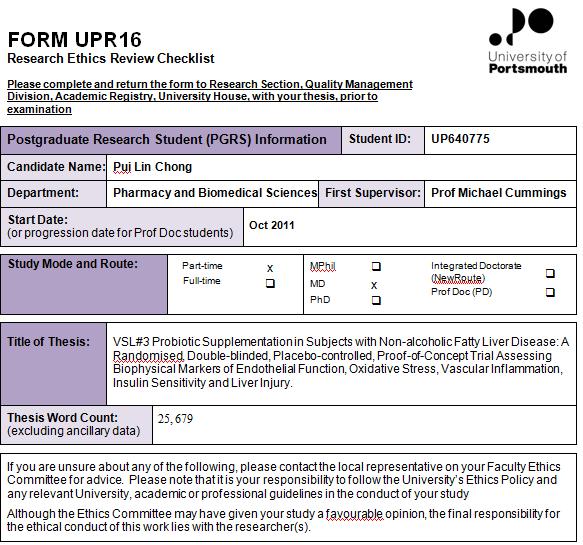

108 108

109 109
Dietary supplementation in treating non-alcoholic fatty liver disease Dr. Ahmad Saedi Associate Professor School of Nutritional Sciences and
 Dietary supplementation in treating non-alcoholic fatty liver disease Dr. Ahmad Saedi Associate Professor School of Nutritional Sciences and Dietetics Tehran University of Medical Sciences Honorary Academic
Early life determinants of Non-Alcoholic Fatty Liver Disease and NASH DR JULIANA MUIVA-GITOBU KENYA PAEDIATRIC ASSOCIATION CONFERENCE APRIL 2016.
 Early life determinants of Non-Alcoholic Fatty Liver Disease and NASH DR JULIANA MUIVA-GITOBU KENYA PAEDIATRIC ASSOCIATION CONFERENCE APRIL 2016. Outline Definition NAFLD and NASH Magnitude of the problem
PATIENTS AND METHODS:
 BACKGROUND: Rheumatoid arthritis (RA) is a chronic systemic inflammatory disease characterized by erosive synovitis that involves peripheral joints and implicates an important influence in the quality
ABNORMAL LIVER FUNCTION TESTS. Dr Uthayanan Chelvaratnam Hepatology Consultant North Bristol NHS Trust
 ABNORMAL LIVER FUNCTION TESTS Dr Uthayanan Chelvaratnam Hepatology Consultant North Bristol NHS Trust INTRODUCTION Liver function tests Cases Non invasive fibrosis measurement Questions UK MORTALITY RATE
ROLE OF INFLAMMATION IN HYPERTENSION. Dr Barasa FA Physician Cardiologist Eldoret
 ROLE OF INFLAMMATION IN HYPERTENSION Dr Barasa FA Physician Cardiologist Eldoret Outline Inflammation in CVDs the evidence Basic Science in Cardiovascular inflammation: The Main players Inflammation as
Index. Note: Page numbers of article titles are in boldface type.
 Note: Page numbers of article titles are in boldface type. A Abetalipoproteinemia NASH and, 537 Acquired errors of metabolism NASH and, 536 537 Amiodarone steatohepatitis due to, 527 Anticonvulsant mood
Complications of Diabetes mellitus. Dr Bill Young 16 March 2015
 Complications of Diabetes mellitus Dr Bill Young 16 March 2015 Complications of diabetes Multi-organ involvement 2 The extent of diabetes complications At diagnosis as many as 50% of patients may have
What to do about the high ALT picked up at the annual review. Dr Michael Yee Consultant in Diabetes and Endocrinology
 What to do about the high ALT picked up at the annual review Dr Michael Yee Consultant in Diabetes and Endocrinology Mrs DC HPC PMH Type 2 Diabetes (decades) Regular retinal screening No foot complications/neuropathy
Vascular disease. Structural evaluation of vascular disease. Goo-Yeong Cho, MD, PhD Seoul National University Bundang Hospital
 Vascular disease. Structural evaluation of vascular disease Goo-Yeong Cho, MD, PhD Seoul National University Bundang Hospital resistance vessels : arteries
Non alcoholic fatty liver and Non alcoholic Steatohepatitis. By Dr. Seham Seif
 Non alcoholic fatty liver and Non alcoholic Steatohepatitis By Dr. Seham Seif Definition NAFL describe a common clinicopathological conditions characterized by significant lipid deposition in the hepatocytes
Clinical Trial Synopsis TL-OPI-518, NCT#
 Clinical Trial Synopsis, NCT# 00225264 Title of Study: A Double-Blind, Randomized, Comparator-Controlled Study in Subjects With Type 2 Diabetes Mellitus Comparing the Effects of Pioglitazone HCl vs Glimepiride
Cho et al., 2009 Journal of Cardiology (2009), 54:
, 54:") Endothelial Dysfunction, Increased Carotid Artery Intima-media Thickness and Pulse Wave Velocity, and Increased Level of Inflammatory Markers are Associated with Variant Angina Cho et al., 2009 Journal
Endothelial Dysfunction, Increased Carotid Artery Intima-media Thickness and Pulse Wave Velocity, and Increased Level of Inflammatory Markers are Associated with Variant Angina Cho et al., 2009 Journal
Improving Access to Quality Medical Care Webinar Series
 Improving Access to Quality Medical Care Webinar Series Presented by The Arizona Telemedicine Program and the Southwest Telehealth Resource Center 2015 UA Board of Regents Welcome AZ, UT, CO, NM & NV FLEX
The Study of Endothelial Function in CKD and ESRD
 The Study of Endothelial Function in CKD and ESRD Endothelial Diversity in the Human Body Aird WC. Circ Res 2007 Endothelial Diversity in the Human Body The endothelium should be viewed for what it is:
Supplementary Online Content
 Supplementary Online Content Larsen JR, Vedtofte L, Jakobsen MSL, et al. Effect of liraglutide treatment on prediabetes and overweight or obesity in clozapine- or olanzapine-treated patients with schizophrenia
1Why lipids cannot be transported in blood alone? 2How we transport Fatty acids and steroid hormones?
 1Why lipids cannot be transported in blood alone? 2How we transport Fatty acids and steroid hormones? 3How are dietary lipids transported? 4How lipids synthesized in the liver are transported? 5 Lipoprotien
Non alcoholic fatty liver disease and atherosclerosis Raul Santos, MD
 Non alcoholic fatty liver disease and atherosclerosis Raul Santos, MD Sao Paulo Medical School Hospital Sao Paulo, Brazil Disclosure Honoraria received for consult and/or speaker : Astra Zeneca, Amgen,
Pathophysiology of Lipid Disorders
 Pathophysiology of Lipid Disorders Henry Ginsberg, M.D. Division of Preventive Medicine and Nutrition CHD in the United States CHD is the single largest killer of men and women 12 million have history
HSN301 REVISION NOTES TOPIC 1 METABOLIC SYNDROME
 HSN301 REVISION NOTES TOPIC 1 METABOLIC SYNDROME What does the term Metabolic Syndrome describe? Metabolic syndrome describes a cluster of cardio-metabolic conditions that increase one's risk of developing
Dyslipidemia Endothelial dysfunction Free radicals Immunologic
 ATHEROSCLEROSIS Hossein Mehrani Professor of Clinical Biochemistry Definition Atherosclerosis: Is a chronic inflammatory process characterized by plaque formation within the vessel wall of arteries and
Metabolic Syndrome. Bill Roberts, M.D., Ph.D. Professor of Pathology University of Utah
 Metabolic Syndrome Bill Roberts, M.D., Ph.D. Professor of Pathology University of Utah Objectives Be able to outline the pathophysiology of the metabolic syndrome Be able to list diagnostic criteria for
3/20/2011. Body Mass Index (kg/[m 2 ]) Age at Issue (*BMI > 30, or ~ 30 lbs overweight for 5 4 woman) Mokdad A.H.
![3/20/2011. Body Mass Index (kg/[m 2 ]) Age at Issue (*BMI > 30, or ~ 30 lbs overweight for 5 4 woman) Mokdad A.H.](/thumbs/82/84935173.jpg "3/20/2011. Body Mass Index (kg/[m 2 ]) Age at Issue (*BMI > 30, or ~ 30 lbs overweight for 5 4 woman) Mokdad A.H.") U.S. Adults: 1988 Nineteen states with 10-14% 14% Prevalence of Obesity (*BMI > 30, or ~ 30 lbs overweight for 5 4 woman) Metabolic John P. Cello, MD Professor of Medicine and Surgery, University of California,
U.S. Adults: 1988 Nineteen states with 10-14% 14% Prevalence of Obesity (*BMI > 30, or ~ 30 lbs overweight for 5 4 woman) Metabolic John P. Cello, MD Professor of Medicine and Surgery, University of California,
Disclosure. Objectives. Smash the Nash: A practical approach to fatty liver disease
 Smash the Nash: A practical approach to fatty liver disease Bruce D. Askey, MS, ANP-BC Associate Lecturer North Andover, MA Adult Nurse Practitioner Dept. of Hepatology/Gastroenterology Guthrie Clinic
Adipose Tissue as an Endocrine Organ. Abdel Moniem Ibrahim, MD Professor of Physiology Cairo University
 Adipose Tissue as an Endocrine Organ Abdel Moniem Ibrahim, MD Professor of Physiology Cairo University Functions of Adipose Tissue Adipose tissue expresses and secretes a variety of bioactive peptides,
METABOLISM of ADIPOSE TISSUE
 METABOLISM of ADIPOSE TISSUE 2. LF UK Prof. Rudolf Poledne, PhD. TYPES OF ADIPOSE TISSUE brown adipose tissue subcutaneous adipose tissue visceral adipose tissue ADIPOSE TISSUE FUNCTIONS: thermal isolation
Dyslipidaemia. Is there any new information? Dr. A.R.M. Saifuddin Ekram
 Dyslipidaemia Is there any new information? Dr. A.R.M. Saifuddin Ekram PhD,FACP,FCPS(Medicine) Professor(c.c.) & Head Department of Medicine Rajshahi Medical College Rajshahi-6000 New features of ATP III
JUPITER NEJM Poll. Panel Discussion: Literature that Should Have an Impact on our Practice: The JUPITER Study
 Panel Discussion: Literature that Should Have an Impact on our Practice: The Study Kaiser COAST 11 th Annual Conference Maui, August 2009 Robert Blumberg, MD, FACC Ralph Brindis, MD, MPH, FACC Primary
Association between arterial stiffness and cardiovascular risk factors in a pediatric population
 + Association between arterial stiffness and cardiovascular risk factors in a pediatric population Maria Perticone Department of Experimental and Clinical Medicine University Magna Graecia of Catanzaro
Ischemic Heart and Cerebrovascular Disease. Harold E. Lebovitz, MD, FACE Kathmandu November 2010
 Ischemic Heart and Cerebrovascular Disease Harold E. Lebovitz, MD, FACE Kathmandu November 2010 Relationships Between Diabetes and Ischemic Heart Disease Risk of Cardiovascular Disease in Different Categories
KEY COMPONENTS. Metabolic Risk Cardiovascular Risk Vascular Inflammation Markers
 CardioMetabolic Risk Poor blood sugar regulation and unhealthy triglyceride and lipoprotein levels often present long before the diagnosis of type 2 Diabetes. SpectraCell s CardioMetabolic and Pre-Diabetes
NASH Bench to Bedside
 NASH Bench to Bedside October 2006 Anna Mae Diehl, M.D. Gastroenterology Division Duke University NonAlcoholic Fatty Liver Disease Common ~1/4-1/3 1/3 US adults Outcome highly variable Course indolent
CRP and fibrinogen imply clinical outcome of patients with type-2 diabetes. and coronary artery disease
 CRP and fibrinogen imply clinical outcome of patients with type-2 diabetes and coronary artery disease Marijan Bosevski 1, *, Golubinka Bosevska 1, Lily Stojanovska 2, Vasso Apostolopoulos 2, * 1 University
UMHS-PUHSC JOINT INSTITUTE
 Role of Visceral Adiposity in the Pathogenesis of Non-Alcoholic Fatty Liver Disease in Lean versus Obese Patients: A Comparative Study between Patients at UMHS versus PUHSC Lai WEI and Anna LOK W Zhang,
METABOLIC SYNDROME AND HCV: FROM HCV
 METABOLIC SYNDROME AND HCV: FROM THEORY TO PRACTICE HCV Steatosis Insulin resistance Arun J Sanyal M.D. Chairman, Div. of Gastroenterology, Hepatology and Nutrition Virginia Commonwealth University Richmond,
2003 World Health Organization (WHO) / International Society of Hypertension (ISH) Statement on Management of Hypertension.
 / International Society of Hypertension (ISH) Statement on Management of Hypertension.") 2003 World Health Organization (WHO) / International Society of Hypertension (ISH) Statement on Management of Hypertension Writing Group: Background Hypertension worldwide causes 7.1 million premature
2003 World Health Organization (WHO) / International Society of Hypertension (ISH) Statement on Management of Hypertension Writing Group: Background Hypertension worldwide causes 7.1 million premature
 A real life example In human studies, we quantify ~120 plasma related proteins Monocytes MMP9 Macrophages TNFα IL1ß platelet derived growth factor IFNγ MMP1 MMP8 MMP13 Myeloid Related Protein14 CD40
Screening cardiac patients for advanced liver disease
 HKASLD 30 th ASM and International Symposium on Hepatology 2017 Screening cardiac patients for advanced liver disease 5 Nov 2017 Dr. Lau Yue Leung Joulen Pamela Youde Nethersole Eastern Hospital NAFLD
Novel Markers of Arterial Dysfunction
 혈관연구회창립심포지움, 3 월 3 일, 2005 Novel Markers of Arterial Dysfunction Kwang Kon Koh, MD, FACC, FAHA Cardiology Gachon Medical School Incheon, Korea Atherosclerosis: A progressive process PHASE I: Initiation
surtout qui n est PAS à risque?
 3*25 min et surtout qui n est PAS à risque? 2018 ESC/ESH Hypertension Guidelines 2018 ESC-ESH Guidelines for the Management of Arterial Hypertension 28 th ESH Meeting on Hypertension and Cardiovascular
Prevalence of non-alcoholic fatty liver disease in type 2 diabetes mellitus patients in a tertiary care hospital of Bihar
 Original Research Article Prevalence of non-alcoholic fatty liver disease in type 2 diabetes mellitus patients in a tertiary care hospital of Bihar Naresh Kumar 1, Jyoti Kumar Dinkar 2*, Chandrakishore
Nonalcoholic Fatty Liver Disease in Children: Typical and Atypical
 Nonalcoholic Fatty Liver Disease in Children: Typical and Atypical Disclosure Naim Alkhouri, MD discloses the following relationships with commercial companies: Membership in the Speakers Bureau for Alexion
Part 1 Risk Factors and Atherosclerosis. LO1. Define the Different Forms of CVD
 Week 3: Cardiovascular Disease Learning Outcomes: 1. Define the difference forms of CVD 2. Describe the various risk factors of CVD 3. Describe atherosclerosis and its stages 4. Describe the role of oxidation,
At Least 1 in 5 Patients in Your Practice Have Fatty Liver
 At Least 1 in 5 Patients in Your Practice Have Fatty Liver What Can You Tell Your Patients Magnus McLeod MD FRCPC Assistant Professor Dalhousie University 30-NOV-2017 NAFLD Non-Alcoholic Fatty Liver Disease
902 Biomed Environ Sci, 2014; 27(11):
:") 902 Biomed Environ Sci, 2014; 27(11): 902-906 Letter to the Editor Curcuminoids Target Decreasing Serum Adipocyte-fatty Acid Binding Protein Levels in Their Glucose-lowering Effect in Patients with Type
902 Biomed Environ Sci, 2014; 27(11): 902-906 Letter to the Editor Curcuminoids Target Decreasing Serum Adipocyte-fatty Acid Binding Protein Levels in Their Glucose-lowering Effect in Patients with Type
SYNOPSIS OF RESEARCH REPORT (PROTOCOL BC20779)
") TITLE OF THE STUDY / REPORT No. / DATE OF REPORT INVESTIGATORS / CENTERS AND COUNTRIES Clinical Study Report Protocol BC20779: Multicenter, double-blind, randomized, placebo-controlled, dose ranging phase
TITLE OF THE STUDY / REPORT No. / DATE OF REPORT INVESTIGATORS / CENTERS AND COUNTRIES Clinical Study Report Protocol BC20779: Multicenter, double-blind, randomized, placebo-controlled, dose ranging phase
NON-ALCOHOLIC STEATOHEPATITIS AND NON-ALCOHOLIC FATTY LIVER DISEASES
 NON-ALCOHOLIC STEATOHEPATITIS AND NON-ALCOHOLIC FATTY LIVER DISEASES Preface Zobair M. Younossi xiii Epidemiology and Natural History of NAFLD and NASH 1 Janus P. Ong and Zobair M. Younossi Understanding
NAFLD: US GUIDELINES. US Guidelines for NAFLD
 NAFLD: US GUIDELINES Arun J Sanyal M.D. Charles Caravati Professor of Medicine Virginia Commonwealth University School of Medicine US Guidelines for NAFLD Represents consensus amongst AGA, AASLD and ACG
WHAT THE EXPERIMENTAL MODELS CAN TEACH US IN NAFLD/NASH? Claudio Tiribelli, MD PhD Scientific Director FIF
 WHAT THE EXPERIMENTAL MODELS CAN TEACH US IN NAFLD/NASH? Claudio Tiribelli, MD PhD Scientific Director FIF ctliver@fegato.it Worldwide estimated prevalence of NAFLD distribution of PNPLA3 genotypes 2017-Younussi
Laboratory analysis of the obese child recommendations and discussion. MacKenzi Hillard May 4, 2011
 Laboratory analysis of the obese child recommendations and discussion MacKenzi Hillard May 4, 2011 aka: What to do with Fasting Labs The Obesity Epidemic The prevalence of obesity in adolescents has tripled
Cardiovascular disease, studies at the cellular and molecular level. Linda Lowe Krentz Bioscience in the 21 st Century September 23, 2009
 Cardiovascular disease, studies at the cellular and molecular level Linda Lowe Krentz Bioscience in the 21 st Century September 23, 2009 Content Introduction The number 1 killer in America Some statistics
300 Biomed Environ Sci, 2018; 31(4):
:") 300 Biomed Environ Sci, 2018; 31(4): 300-305 Letter to the Editor Combined Influence of Insulin Resistance and Inflammatory Biomarkers on Type 2 Diabetes: A Population-based Prospective Cohort Study of
300 Biomed Environ Sci, 2018; 31(4): 300-305 Letter to the Editor Combined Influence of Insulin Resistance and Inflammatory Biomarkers on Type 2 Diabetes: A Population-based Prospective Cohort Study of
Metabolic Syndrome in Asians
 Metabolic Syndrome in Asians Alka Kanaya, MD Asst. Professor of Medicine, UCSF Asian CV Symposium, November 17, 2007 The Metabolic Syndrome Also known as: Syndrome X Insulin Resistance Syndrome The Deadly
Conflicts of Interest in the last 12 months
 STEATOHEPATITIS Richard K. Sterling, MD, MSc, FACP, FACG VCU Hepatology Professor of Medicine Chief, Section of Hepatology Virginia Commonwealth University Richmond, VA Conflicts of Interest in the last
Arteriosclerosis & Atherosclerosis
 Arteriosclerosis & Atherosclerosis Arteriosclerosis = hardening of arteries = arterial wall thickening + loss of elasticity 3 types: -Arteriolosclerosis -Monckeberg medial sclerosis -Atherosclerosis Arteriosclerosis,
Relationship between serum glutathione peroxidase-1activity with endothelial dysfunction level in patients with coronary artery diseases
 Relationship between serum glutathione peroxidase-1activity with endothelial dysfunction level in patients with coronary artery diseases Introduction Reactive oxygen species (ROS),such as superoxide and
Effects of probiotics in the treatment of alcoholic hepatitis: randomized controlled multicenter study
 Effects of probiotics in the treatment of alcoholic hepatitis: randomized controlled multicenter study Lactobacillus subtilis/streptococcus faecium Lactobacillus rhamnosus R0011/acidophilus R0052 Ki Tae
Fatty liver disease: What do we know?
 Fatty liver disease: What do we know? Prof. Dr. Claus Niederau Katholische Kliniken Oberhausen ggmbh St. Josef-Hospital Academic Teaching Hospital University of Duisburg-Essen NAFLD Non-Alcoholic Fatty
Clinical Recommendations: Patients with Periodontitis
 The American Journal of Cardiology and Journal of Periodontology Editors' Consensus: Periodontitis and Atherosclerotic Cardiovascular Disease. Friedewald VE, Kornman KS, Beck JD, et al. J Periodontol 2009;
Pathology of Coronary Artery Disease
 Pathology of Coronary Artery Disease Seth J. Kligerman, MD Pathology of Coronary Artery Disease Seth Kligerman, MD Assistant Professor Medical Director of MRI University of Maryland Department of Radiology
Fatty Liver- how important is it? Jeremy F.L. Cobbold MA PhD MRCP Clinical Lecturer in Hepatology Imperial College London
 Fatty Liver- how important is it? Jeremy F.L. Cobbold MA PhD MRCP Clinical Lecturer in Hepatology Imperial College London Fatty liver- how important is it? Importance in terms of: Prevalence Pathogenesis
Term-End Examination December, 2009 MCC-006 : CARDIOVASCULAR EPIDEMIOLOGY
 MCC-006 POST GRADUATE DIPLOMA IN CLINICAL CARDIOLOGY (PGDCC) 00269 Term-End Examination December, 2009 MCC-006 : CARDIOVASCULAR EPIDEMIOLOGY Time : 2 hours Maximum Marks : 60 Note : There will be multiple
Non-Invasive Assessment of Liver Fibrosis. Patricia Slev, PhD University of Utah Department of Pathology
 Non-Invasive Assessment of Liver Fibrosis Patricia Slev, PhD University of Utah Department of Pathology Disclosure Patricia Slev has no relevant financial relationships to disclose. 2 Chronic Liver Disease
Cardiovascular disease physiology. Linda Lowe-Krentz Bioscience in the 21 st Century November 2, 2016
 Cardiovascular disease physiology Linda Lowe-Krentz Bioscience in the 21 st Century November 2, 2016 Content Introduction The number 1 killer in America Some statistics Recommendations The disease process
The Metabolic Syndrome Update The Metabolic Syndrome: Overview. Global Cardiometabolic Risk
 Update 2013 Marc Cornier, M.D. Associate Professor of Medicine Division of Endocrinology, Metabolism & Diabetes Anschutz Health and Wellness Center University of Colorado School of Medicine Denver Health
Internal and Emergency Medicine Official Journal of the Italian Society of Internal Medicine. ISSN Volume 8 Number 3
 Hepatic Steatosis Index and Lipid Accumulation Product as middle-term predictors of incident metabolic syndrome in a large population sample: data from the Brisighella Heart Study Arrigo F. G. Cicero,
Obesity, Metabolic Syndrome, and Diabetes: Making the Connections
 Obesity, Metabolic Syndrome, and Diabetes: Making the Connections Alka M. Kanaya, M.D. Associate Professor of Medicine & Epi/Biostats University of California, San Francisco February 26, 21 Roadmap 1.
HOW THE MICROBIOME AFFECTS OUR HEALTH
 HOW THE MICROBIOME AFFECTS OUR HEALTH THE INTESTINAL BARRIER AND INTESTINAL PERMEABILITY Intestinal Barrier: a functional body Defense from translocation of dietary antigens, bacteria or bacterial endotoxins
Marshall Tulloch-Reid, MD, MPhil, DSc, FACE Epidemiology Research Unit Tropical Medicine Research Institute The University of the West Indies, Mona,
 Marshall Tulloch-Reid, MD, MPhil, DSc, FACE Epidemiology Research Unit Tropical Medicine Research Institute The University of the West Indies, Mona, Jamaica At the end of this presentation the participant
Metabolic Syndrome. DOPE amines COGS 163
 Metabolic Syndrome DOPE amines COGS 163 Overview - M etabolic Syndrome - General definition and criteria - Importance of diagnosis - Glucose Homeostasis - Type 2 Diabetes Mellitus - Insulin Resistance
IL METABOLISMO EPATICO DEI CARBOIDRATI IN FISIOLOGIA E PATOLOGIA
 UNIGASTRO Il fegato come centrale metabolica e i fattori di danno oltre ai virus epatitici IL METABOLISMO EPATICO DEI CARBOIDRATI IN FISIOLOGIA E PATOLOGIA Dr Elisabetta Bugianesi Divisione di Gastro-Epatologia
National Horizon Scanning Centre. Enhanced Liver Fibrosis Test (ELF) for evaluating liver fibrosis. June 2008
 for evaluating liver fibrosis. June 2008") Enhanced Liver Fibrosis Test (ELF) for evaluating liver fibrosis June 2008 This technology summary is based on information available at the time of research and a limited literature search. It is not intended
Enhanced Liver Fibrosis Test (ELF) for evaluating liver fibrosis June 2008 This technology summary is based on information available at the time of research and a limited literature search. It is not intended
NONALCOHOLIC FATTY LIVER DISEASE
 NONALCOHOLIC FATTY LIVER DISEASE Kiran Bambha, MD, MSc Hepatology and Liver Transplantation University of Colorado Denver April 13, 2012 Non-Alcoholic Fatty Liver Disease (NAFLD) Terminology Pathogenesis
HOW TO DEFINE GOOD BIOMARKERS OF HEALTH? Suzan Wopereis
 HOW TO DEFINE GOOD BIOMARKERS OF HEALTH? Suzan Wopereis My personal objective: identification of biomarkers of (optimal) health Biomarker Definition: a characteristic that is objectively measured and evaluated
Methods. Background and Objectives STRADIVARIUS
 STRADIVARIUS Effect of on Progression of Atherosclerosis in Patients with Abdominal Obesity and Coronary Artery Disease Steven E. Nissen MD Stephen J. Nicholls MBBS PhD, Kathy Wolski MPH, Josep Rodés-Cabau
Assessment of Liver Stiffness by Transient Elastography in Diabetics with Fatty Liver A Single Center Cross Sectional observational Study
 IOSR Journal of Dental and Medical Sciences (IOSR-JDMS) e-issn: 2279-0853, p-issn: 2279-0861.Volume 16, Issue 6 Ver. IV (June. 2017), PP 49-53 www.iosrjournals.org Assessment of Liver Stiffness by Transient
Investigating and Referring Incidental Findings of Abnormal Liver Tests
 Investigating and Referring Incidental Findings of Abnormal Liver Tests Note on Referral Guidelines: these revised guidelines are presented as a tool to aid appropriate referral and management of common
NICE guideline Published: 6 July 2016 nice.org.uk/guidance/ng49
 Non-alcoholic fatty liver disease (NAFLD): assessment and management NICE guideline Published: 6 July 20 nice.org.uk/guidance/ng49 NICE 2017. All rights reserved. Subject to Notice of rights (https://www.nice.org.uk/terms-and-conditions#notice-ofrights).
Lecture 8 Cardiovascular Health Lecture 8 1. Introduction 2. Cardiovascular Health 3. Stroke 4. Contributing Factors
 Lecture 8 Cardiovascular Health 1 Lecture 8 1. Introduction 2. Cardiovascular Health 3. Stroke 4. Contributing Factors 1 Human Health: What s Killing Us? Health in America Health is the U.S Average life
Roadmap. Diabetes and the Metabolic Syndrome in the Asian Population. Asian. subgroups 8.9. in U.S. (% of total
 Diabetes and the Metabolic Syndrome in the Asian Population Alka Kanaya, MD Associate Professor of Medicine, UCSF Feb 26, 2010 Roadmap 1. Diabetes in Asian Americans Prevalence in the U.S. Risk factors
The Metabolic Syndrome: Is It A Valid Concept? YES
 The Metabolic Syndrome: Is It A Valid Concept? YES Congress on Diabetes and Cardiometabolic Health Boston, MA April 23, 2013 Edward S Horton, MD Joslin Diabetes Center Harvard Medical School Boston, MA
John J.P. Kastelein MD PhD Professor of Medicine Dept. of Vascular Medicine Academic Medial Center / University of Amsterdam
 Latest Insights from the JUPITER Study John J.P. Kastelein MD PhD Professor of Medicine Dept. of Vascular Medicine Academic Medial Center / University of Amsterdam Inflammation, hscrp, and Vascular Prevention
Supplementary table 1 Demographic and clinical characteristics of participants by paraoxonase-1 (PON-1) gene polymorphisms
 gene polymorphisms") Supplementary table 1 Demographic and clinical characteristics of participants by paraoxonase-1 (PON-1) gene polymorphisms QQ QR/RR n = 36 n = 80 Men (%) 20 (55) 54 (67) 0.216 Age (years) 57 ± 10 56 ±
Supplementary table 1 Demographic and clinical characteristics of participants by paraoxonase-1 (PON-1) gene polymorphisms QQ QR/RR n = 36 n = 80 Men (%) 20 (55) 54 (67) 0.216 Age (years) 57 ± 10 56 ±
JOSHUA K. KAYIMA INTERLINKING CARDIOVASCULAR DISEASE, CHRONIC KIDNEY DISEASE, AND OBESITY
 INTERLINKING CARDIOVASCULAR DISEASE, CHRONIC KIDNEY DISEASE, AND OBESITY JOSHUA K. KAYIMA ASSOCIATE PROFESSOR DEPT. OF CLINICAL MEDICINE AND THERAPEUTICS UNIVERSITY OF NAIROBI Introduction According to
HSN301 Diet and Disease Entire Note Summary
 Topic 1: Metabolic Syndrome Learning Objectives: 1. Define obesity 2. Factors causing obesity 3. Obesity as a risk factor for metabolic syndrome 4. The pathogenesis of metabolic syndrome 5. Treatment of
In Search of New Biomarkers for Nonalcoholic Fatty Liver Disease
 REVIEW In Search of New Biomarkers for Nonalcoholic Fatty Liver Disease Ting-Ting Chan, M.R.C.P., and Vincent Wai-Sun Wong, M.D. Nonalcoholic fatty liver disease (NAFLD) affects 15% to 40% of the general
The 10 th International & 15 th National Congress on Quality Improvement in Clinical Laboratories
 The 10 th International & 15 th National Congress on Quality Improvement in Clinical Laboratories Cardiac biomarkers in atherosclerosis Najma Asadi MD-APCP Ross and Colleagues in 1973: Response to Injury
Review of guidelines for management of dyslipidemia in diabetic patients
 2012 international Conference on Diabetes and metabolism (ICDM) Review of guidelines for management of dyslipidemia in diabetic patients Nan Hee Kim, MD, PhD Department of Internal Medicine, Korea University
Intima-Media Thickness
 European Society of Cardiology Stockholm, 30th August 2010 Intima-Media Thickness Integration of arterial assessment into clinical practice Prof Arno Schmidt-Trucksäss, MD Institute of Exercise and Health
Correlation of novel cardiac marker
 Correlation of novel cardiac marker and mortality in EGAT population. Soluble ST2 hscrp Poh Chanyavanich, MD SukitYamwong, MD Piyamitr Sritara, MD Ramathibodi hospital Background hscrp - the most widely
Targeting Glucose Metabolism to Stop Strokes IRIS: Insulin Resistance In Stroke study
 Targeting Glucose Metabolism to Stop Strokes IRIS: Insulin Resistance In Stroke study Professor Gary Ford Chief Executive Officer, Oxford Academic Health Science Network Consultant Stroke Physician, Oxford
Metabolic Syndrome: An overview. Kevin Niswender MD, PhD Vanderbilt University School of Medicine
 Metabolic Syndrome: An overview. Kevin Niswender MD, PhD Vanderbilt University School of Medicine Setting the scene GB, 43 yo AA man followed for hypothyroidism returns on LT4 125 mcg/d and has a TSH=1.1
Cardiovascular Complications of Diabetes
 VBWG Cardiovascular Complications of Diabetes Nicola Abate, M.D., F.N.L.A. Professor and Chief Division of Endocrinology and Metabolism The University of Texas Medical Branch Galveston, Texas Coronary
Nonalcoholic Steatohepatitis National Digestive Diseases Information Clearinghouse
 Nonalcoholic Steatohepatitis National Digestive Diseases Information Clearinghouse National Institute of Diabetes and Digestive and Kidney Diseases NATIONAL INSTITUTES OF HEALTH Nonalcoholic steatohepatitis
EASL EASD EASO Clinical practice guidelines for the management of nonalcoholic fatty liver disease.
 Commentary. EASL EASD EASO Clinical practice guidelines for the management of nonalcoholic fatty liver disease. Christopher D. Byrne 1,2, Giovanni Targher 3 1 Nutrition and Metabolism, Faculty of Medicine,
EVALUATION OF ABNORMAL LIVER TESTS
 EVALUATION OF ABNORMAL LIVER TESTS MIA MANABAT DO PGY6 MOA 119 TH ANNUAL SPRING SCIENTIFIC CONVENTION MAY 19, 2018 EVALUATION OF ABNORMAL LIVER TESTS Review of liver enzymes vs liver function tests Clinical
The JUPITER trial: What does it tell us? Alice Y.Y. Cheng, MD, FRCPC January 24, 2009
 The JUPITER trial: What does it tell us? Alice Y.Y. Cheng, MD, FRCPC January 24, 2009 Learning Objectives 1. Understand the role of statin therapy in the primary and secondary prevention of stroke 2. Explain
Role of apolipoprotein B-containing lipoproteins in the development of atherosclerosis Jan Borén MD, PhD
 Role of apolipoprotein B-containing lipoproteins in the development of atherosclerosis Jan Borén MD, PhD Our laboratory focuses on the role of apolipoprotein (apo) B- containing lipoproteins in normal
Cardiovascular disease, studies at the cellular and molecular level. Linda Lowe Krentz Bioscience in the 21 st Century October 4, 2010
 Cardiovascular disease, studies at the cellular and molecular level Linda Lowe Krentz Bioscience in the 21 st Century October 4, 2010 Content Introduction The number 1 killer in America Some statistics
Cardiac Pathophysiology
 Cardiac Pathophysiology Evaluation Components Medical history Physical examination Routine laboratory tests Optional tests Medical History Duration and classification of hypertension. Patient history of
[Sem Liver Disease 21(1):81-88, Thieme Medical Publishers, Inc.]
![[Sem Liver Disease 21(1):81-88, Thieme Medical Publishers, Inc.]](/thumbs/72/66425194.jpg "[Sem Liver Disease 21(1):81-88, Thieme Medical Publishers, Inc.]") Treatment of Nonalcoholic Fatty Liver: Present and Emerging Therapies [Sem Liver Disease 21(1):81-88, 2001. 2001 Thieme Medical Publishers, Inc.] Paul Angulo, M.D., and Keith D. Lindor, M.D., Division
Treatment of Nonalcoholic Fatty Liver: Present and Emerging Therapies [Sem Liver Disease 21(1):81-88, 2001. 2001 Thieme Medical Publishers, Inc.] Paul Angulo, M.D., and Keith D. Lindor, M.D., Division