SOCT Copernicus REVO. * - Currently import and overlay are avaibale in manual mode only
|
|
|
- John Dickerson
- 6 years ago
- Views:
Transcription

1
 Voice guide (support patient through")
 Widefield scans 12 x 12mm area Peripherial scanning Normative")

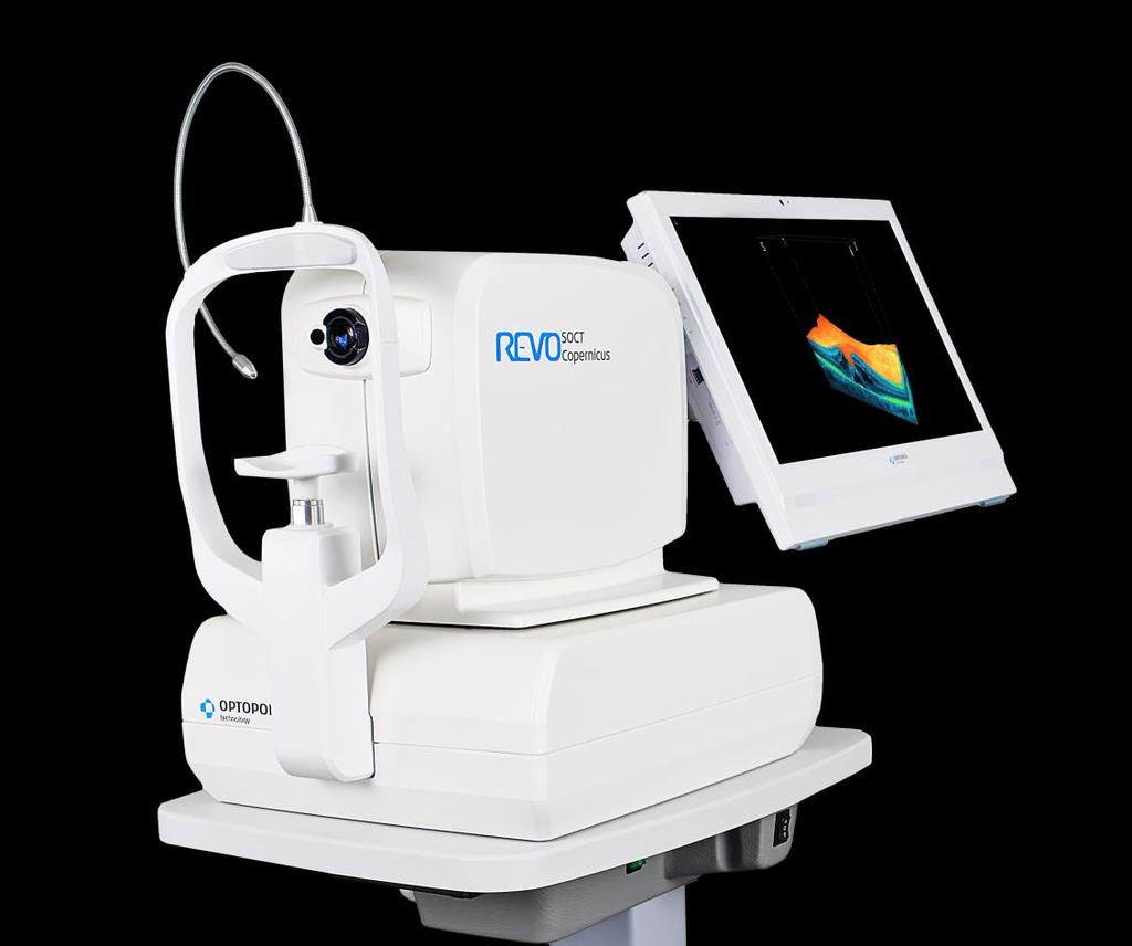
2 SOCT Copernicus REVO Easy Operation (Full auto & Auto mode) Auto alignment (Z-position, C-gate, Focus, Tomogram) Voice guide (support patient through examination) Powerful analysis tools Enhanced tomograms - 2 µm (digital resolution) Widefield scans 12 x 12mm area Peripherial scanning Normative database Analysis program Retina Glaucoma Anterior Anterior - Angle to angle Import of fundus camera image* Multilingual interface * - Currently import and overlay are avaibale in manual mode only


")
3 SOCT Copernicus REVO 8 retina layer recognition Examination review: o Single exam analysis o Asymmetry analysis o Follow up Retina analysis: Retina thickness, Inner retinal thickness, Outer retinal thickness, IS/OS thickness, RPE deformation Glaucoma analysis: Optic Nerve Head scan: RNFL, ONH morphology, DDLS Retina scan: Ganglion analysis as RNFL+GCL+IPL and GCL+IPL, OU and Hemisphere asymmetry Anterior analysis Pachymetry measurement, Epithelium map, LASIK flap, Angle measurement (AOD, TIA, TISA) 3D reconstruction


4 A Complete OCT Device Retina 2048lines/0.08sec Disc Cornea Anterior Wide Anterior


5 Retinal Analysis Single Exam Symmetry


6 Follow-Up Retinal Analysis Retina follow up analysis modes: Morphology view Quantification analysis Trend analysis
")

7 SOCT Copernicus REVO Central scan mode - 12 mm width (fovea and disc on one scan) Peripherial scans (Below are combined two examinations of peripheral area 12mm + 12 mm)
8 Averaging 2 µm Ultra High (Digital) Resolution


9 Enhance Depth modes Vitreous mode Choroid mode
10 Choroid visualisation


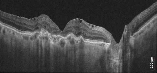
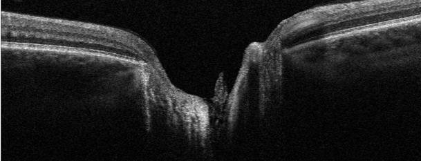
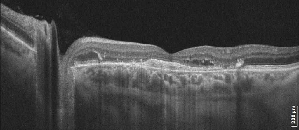
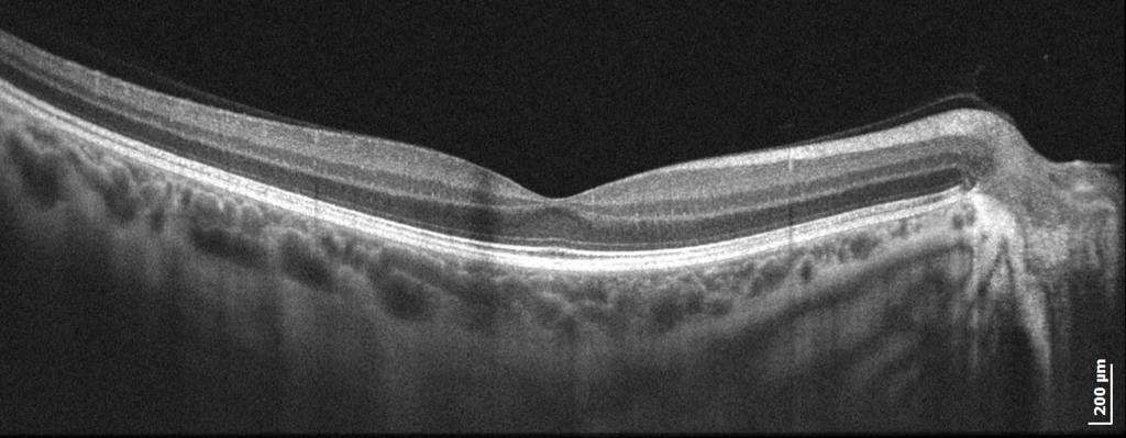
11 Clinical images Case 1 CSR Case 2

12 Clinical images Case 3 Case 4

13 Clinical images Case 5

14 Clinical images Case 6 Case 7


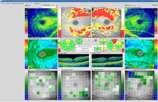
15 Glaucoma analysis Optic Nerve Head analysis Ganglion cells layer analysis Pachymetry and Gonioscopy

16 Corneal analysis Pachymetry map Epithelium thickness Zonal analysis IOP correction

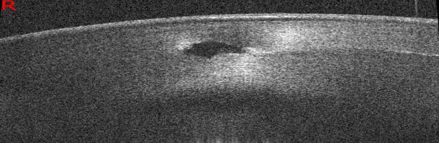
17 Anterior Segment Imaging 2µm Ultra High Digital Resolution Cornea scan Epithelium Bowman s membrane Stroma Endothelium

18 Anterior Segment Imaging Anterior Angle scan Epithelium Schlemm s canal Schwalbe s line Endothelium Scleral spur Iris

19 Anterior Segment Imaging Back of the crystalline Lens Epithelium Posterior capsule Berger space Vitreous body membrane

20 Wide Anterior Segment Imaging Sclera and Angle Structrure Sclera Epithelium Schwalbe s line Schlemm s canal Iris Cornea

21 Wide Anterior Segment Imaging Enhance mode - Sclera and Angle Structrure Sclera Blood vessel Endothelium Pars plana Ciliary body Sclerar spur

22 Wide Anterior Segment Imaging Angle to Angle Sclerar Spur Iris Posterior pigment epithelium Lens Sclera


23 Wide Anterior Segment Imaging Wide scan with lens defect IOL observation

24 Lens fitting Contact lens on the cornea 4 mm scan Contact lens on the Cornea and the Sclera 10 mm


25 3 x 3 mm 3 x 3 mm OCT Angiography. Coming soon Depth coded view Superficial capillary plexus Deep capillary plexus
26 CLINICAL CAPABILITIES - Detect pathologies at earlier stages (e.g. detection small lipofuiscin changes in RPE as FAF does) - Elevate your practice profile - Raise diagnostic level by a broader range of services: - Manage a broader range of diseases - Glaucoma - Anterior part of the eye - Manage more advanced diseases - Verify periphery - Optimised practice workflow: - Technician performs the exam for the doctor to review and analyse the result (Thereby saving the doctors time and increasing the number of diagnostic reviews) - Networking is free of charge and does not require extra hardware. - Extremely easy and intuitive for operators at any level - Fast workflow to minimise patient chair time
27 CLINICAL CAPABILITIES - Visualise retinal conditions with ultra high-definition imaging (3µm digital) - Excellent B-scans quality - Wide scan of retina - Visualisation of choroid (new research confirms usability of verifying choroid thickness on enhanced scans similiar to swept source) - Qualitative and quantitative tools for clearer assessments of your patient's condition - Identity and monitor at-risk patients for glaucoma with comprehensive reports - Ganglion analysis allows detectection of glaucoma at an earlier stage - Hemisphere method for early stages of glaucoma and non typical cases. - Full ONH Analysis with DDLS factor - RNFL analysis - Non contact gonioscopy for every patient - Pachymetry - Advanced OCT technology in REAL compact solution (other compact requires floor footprint as regular devices.) - Widest refraction range allows you to scan higher groups of patients - Report Manager enables pre-define desired reports for rapid printing - Enhanced patient comfort by easy education. - Ability to scan the smallest pupil without dilating.
28 KEY FEATURES IN THIS PRICE SHELF - Automatic positioning (Full Auto, Auto) - Touch control* - Voice assistance support patient through examination - Excellent scan quality, Ultra High digital resolution - Real Small floor footprint (patient-operator one side) - Only USB connection cable (allows to use any type of efficient PC) - Central 12x12 mm Retina scan - Peripherial Scanning - Anterior scan without Adapter (only narrow scan) - White to white wide Anterior scan with Adapter (no foreahead support required) - Both Anterior option included in the price (all others charge extra) - Moveable & changeable shape of fixation target realised on the OLED display - Refraction range +/-25 D - Networking (Same functionality on review station, free of charge licences) * - when provided PC with touch screen

29 SOCT Copernic REVO technical data Technology: Scanning speed: Axial resolution: Resolution (digital) Lightsource Scan range Scan program Refraction range: Fixation target: Fundus observation PC interface Dimensions: Weight: Spectral domain OCT A-scan/sec 5 µm 2 µm SLED 830 nm, 50nm width Retina 5 12mm Anterior 3 16 mm 3D, Raster, B-scan, Radial, Cross -25 D to 25 D OLED display (moveable positon, changeable shape) OCT live reconstruction USB x 551 x 450 mm (WxDxH) 23 kg
The World s fastest OCT. As simple as pressing. the start button
 The World s fastest OCT As simple as pressing the start button lution continues Optopol engineering team, designers of the first commercially available Spectral Domain OCT in the world, are proud to present
PRIMUS 200 from ZEISS The essential OCT
 PRIMUS 200 from ZEISS The essential OCT Seeing beyond the surface. ZEISS PRIMUS 200 // INNOVATION MADE BY ZEISS Clear Visualization. Advanced Technology. Reliability. Essential elements of your first OCT.
Visualize. Analyze. Personalize. OCT + OCTA. with
 Visualize. Analyze. Personalize. OCT + OCTA with Avanti Widefield OCT with AngioVue OCTA Imaging Comprehensive Structural and Functional Imaging in a Single Imaging Platform Comprehensive OCT Imaging The
Visualize. Analyze. Personalize. OCT + OCTA
 Visualize. Analyze. Personalize. OCT + OCTA A New Approach to Protecting Vision AngioVue OCT Angiography brings valuable new information to clinical practice. Non-invasive visualization of retinal vasculature.
PRIMUS 200 from ZEISS The essential OCT
 EN 00_00I The contents of the brochure may differ from the current status of approval of the product in your country. Please contact your regional representative for more information. Subject to change
3/16/2018. Ultrasound Biomicroscopy in Glaucoma By Ahmed Salah Abdel Rehim. Prof. of Ophthalmology Al-Azhar University
 Ultrasound Biomicroscopy in Glaucoma By Ahmed Salah Abdel Rehim Prof. of Ophthalmology Al-Azhar University 1 Ultrasound biomicroscopy (UBM) is a recent technique to visualize anterior segment with the
Mark Dunbar: Disclosure
 Important Things to Understand About OCT Mark T. Dunbar, O.D., F.A.A.O. Bascom Palmer Eye Institute University of Miami, School of Medicine Mark Dunbar: Disclosure Optometry Advisory Board for: Allergan
Cirrus TM HD-OCT. Details define your decisions
 Cirrus TM HD-OCT Details define your decisions 2 With high-definition OCT Carl Zeiss Meditec takes you beyond standard spectral domain Built on 10 years experience at the vanguard of innovation, Carl Zeiss
Cirrus TM HD-OCT. Details defi ne your decisions
 Cirrus TM HD-OCT Details defi ne your decisions 2 With high-defi nition OCT Carl Zeiss Meditec takes you beyond standard spectral domain Built on 10 years experience at the vanguard of innovation, Carl
HOCT-1I 1F All-in-One Optical Coherence Tomography with Fundus
 HOCT-1I 1F All-in-One Optical Coherence Tomography with Fundus Specification Type Resolution(in Tissue) A scan Rate Scan Range SD-OCT / Fundus Z :6~7um, XY:20um 68,000 A-scan/sec. [Fundus] X:6-12mm, Y:6-9mm,
Introducing ANGIOVUE ESSENTIAL. Built on the Avanti Widefield OCT Platform. OCT Angiography for Primary Eye Care
 Introducing ANGIOVUE ESSENTIAL Built on the Avanti Widefield OCT Platform OCT Angiography for Primary Eye Care Transform Your View of the Retina OCT Angiography (OCTA) is a quick non-invasive test that
3/23/2016. Diagnostic Services Taylor Pannell CRA, OCT-C. Services Available. Important info for the Tech to know. Visual Fields
 Services Available Diagnostic Services Taylor Pannell CRA, OCT-C Static and Kinetic Visual Fields Pachymetry Anterior and Posterior Segment OCT Fundus Photos FAF,FA,ICG Slit Lamp Photography Confocal HRT
DRI OCT Triton Series A Multimodal Swept Source OCT
 DRI OCT Triton Series A Multimodal Swept Source OCT Color Red-Free FA FAF Posterior Anterior See what others can t see. A Multimodal Swept Source OCT DEEP RANGE IMAGING Swept Source OCT imaging massively
Histology of the Eye
 Histology of the Eye Objectives By the end of this lecture, the student should be able to describe: The general structure of the eye. The microscopic structure of:»cornea.»retina. EYE BULB Three coats
THE EYE: RETINA AND GLOBE
 Neuroanatomy Suzanne Stensaas February 24, 2011, 10:00-12:00 p.m. Reading: Waxman Ch. 15. Your histology and gross anatomy books should be useful. Reading: Histology of the Eye from any histology book
Advances in OCT Murray Fingeret, OD
 Disclosures Advances in OCT Murray Fingeret, OD Consultant Alcon, Allergan, Bausch & Lomb, Carl Zeiss Meditec, Diopsys, Heidelberg Engineering, Reichert, Topcon Currently Approved OCT Devices OCT Devices
XUE HUI Department of Histology& Embryology, Basic Medicine College of Jilin University
 SENSE ORGAN XUE HUI Department of Histology& Embryology, Basic Medicine College of Jilin University EYE fibrous globe lens photosensitive cells a system of cells and nerves concentric layers the sclera
PLEX Elite 9000 from ZEISS Swept-Source OCT
 PLEX Elite 9000 from ZEISS Swept-Source OCT Uncovering the undiscovered. ZEISS PLEX Elite 9000 // INNOVATION MADE BY ZEISS 2 Ultra-wide angiography En face montage Image courtesy of Prof. G. Querques,
Experience Spectacular Retinal Imaging with the new NIDEK F-10 Digital Ophthalmoscope
 Experience Spectacular Retinal Imaging with the new NIDEK F-10 Digital Ophthalmoscope The F-10 was developed to give Ophthalmologists a high definition (HD) diagnostic imaging system. Designed to provide
Structural examina.on: Imaging
 ManaMa: Glaucoma Structural examina.on: Imaging Luís Abegão Pinto, MD, PhD Department of Ophthalmology CHLC Lisbon Faculty of Medicine, Lisbon University 1 11-10- 2013 Structural changes Qualitative changes
_ Assessment of the anterior chamber. Review of anatomy of the angle
 Assessment of the anterior chamber Dr Simon Barnard PhD BSc FCOptom FAAO DCLP Department of Optometry & Visual Science City University London, UK Review of anatomy of the angle Figure 1. Anatomical section
OCT Image Analysis System for Grading and Diagnosis of Retinal Diseases and its Integration in i-hospital
 Progress Report for1 st Quarter, May-July 2017 OCT Image Analysis System for Grading and Diagnosis of Retinal Diseases and its Integration in i-hospital Milestone 1: Designing Annotation tool extraction
Unit VIII Problem 8 Anatomy: Orbit and Eyeball
 Unit VIII Problem 8 Anatomy: Orbit and Eyeball - The bony orbit: it is protecting our eyeball and resembling a pyramid: With a base directed: anterolaterally. And an apex directed: posteromedially. Notes:
5/18/2014. Fundamentals of Gonioscopy Workshop Aaron McNulty, OD, FAAO Walt Whitley, OD, MBA, FAAO
 1 Fundamentals of Gonioscopy Workshop Aaron McNulty, OD, FAAO Walt Whitley, OD, MBA, FAAO 2 3 4 5 6 Optometry s Meeting 2014 The Most Valuable Glaucoma Tool Glaucoma Diagnosis Gonioscopy Central corneal
EasyScan: Smart Retinal Imaging
 easyscan EasyScan: Smart Retinal Imaging Superior Imaging Enjoy the benefits of SLO technology and capture high-quality images easily for accurate diagnosis. Never Dilate Reduce examination time, capture
Medical School Histology Basics. VIBS 289 lab. Eye
 Medical School Histology Basics VIBS 289 lab Eye Larry Johnson Texas A&M University Aqueous humor OUTLINE OVERVIEW CELLULAR STRUCTURES THROUGH WHICH LIGHT PASSES A. CORNEA B. LENS C. RETINA STRUCTURES
Cornea/Anterior Segment OCT. User Experience
 Cornea/Anterior Segment OCT User Experience User Experience Case#1 Post Penetrating Keratoplasty Tokyo Medical University / Kohsei Chuo General Hospital Hideki Mori MD, PhD Almost eight years have passed
Evolving glaucoma management True diagnostic integration for the preservation of vision
 Evolving glaucoma management True diagnostic integration for the preservation of vision // GLAUCOMA MANAGEMENT MADE BY ZEISS The moment you are certain it is glaucoma. This is the moment we work for. There
Vision I. Steven McLoon Department of Neuroscience University of Minnesota
 Vision I Steven McLoon Department of Neuroscience University of Minnesota 1 Eye Cornea Sclera Conjunctiva 2 Eye The conjunctiva lines the inner surface of the eyelids and outer surface of the sclera. 3
Simply the best OCT & OCTA image quality.
 Avanti Widefield OCT with AngioVue OCT Angiography Simply the best OCT & OCTA image quality. Dear Friends of Optovue, Since introducing Spectral Domain OCT to the ophthalmology market in 2006, Optovue
Moving forward with a different perspective
 Moving forward with a different perspective The Leader In Vision Diagnostics Offers A New Perspective Marco has served the eyecare community by offering exceptional lane products and automated high tech
C a t a r a c t G l a u c o m a R e t i n a R e f r a c t i v e. The GDxVCC Early answers and ongoing assessment for glaucoma
 C a t a r a c t G l a u c o m a R e t i n a R e f r a c t i v e The GDxVCC Early answers and ongoing assessment for glaucoma The quantifiable approach to quality care Only Humphrey GPA software Early insight
Corporate Medical Policy
 Corporate Medical Policy Optical Coherence Tomography (OCT) Anterior Segment of the Eye File Name: Origination: Last CAP Review: Next CAP Review: Last Review: optical_coherence_tomography_(oct)_anterior_segment_of_the_eye
A LITTLE ANATOMY. three layers of eye: 1. outer: corneosclera. 2. middle - uvea. anterior - iris,ciliary body. posterior - choroid
 GLAUCOMA A LITTLE ANATOMY three layers of eye: 1. outer: corneosclera 2. middle - uvea anterior - iris,ciliary body posterior - choroid connection at the pars plana between post and ant uvea 3. retina
Glaucoma. Cornea. Iris
 Glaucoma Introduction Glaucoma is a group of eye diseases that can lead to blindness if not treated. Openangle glaucoma, the most common form of glaucoma, affects about 3 million Americans. Half of those
4/22/16. Eye. External Anatomy of Eye. Accessory Structures. Bio 40B Dr. Kandula
 Eye Bio 40B Dr. Kandula External Anatomy of Eye Accessory Structures l Eyebrows l Levator Palpebrae Superioris - opens eye l Eyelashes l Ciliary glands modified sweat glands l Small sebaceous glands l
2/26/2017. Sameh Galal. M.D, FRCS Glasgow. Lecturer of Ophthalmology Research Institute of Ophthalmology
 Sameh Galal M.D, FRCS Glasgow Lecturer of Ophthalmology Research Institute of Ophthalmology No financial interest in the subject presented 1 Managing cataracts in children remains a challenge. Treatment
The Measure of Confidence
 Heidelberg_936357.qxd:Layout 1 5/9/08 12:01 PM 12:02 Page 1 (Cyan (Magenta (Yellow (Black (UV Five Powerful Solutions to Fit Your Practice PowerCheck Glaucoma FastCheck+ GPS Software and Retina Edema Index
Goals. Glaucoma PARA PEARL TO DO. Vision Loss with Glaucoma
 Glaucoma Janet R. Fett, OD Drs. Kincaid, Fett and Tharp So Sioux City, NE eyewear21@hotmail.com Goals Understand Glaucoma Disease process Understand how your data (objective and subjective) assists in
The Orbit. The Orbit OCULAR ANATOMY AND DISSECTION 9/25/2014. The eye is a 23 mm organ...how difficult can this be? Openings in the orbit
 The eye is a 23 mm organ...how difficult can this be? OCULAR ANATOMY AND DISSECTION JEFFREY M. GAMBLE, OD COLUMBIA EYE CONSULTANTS OPTOMETRY & UNIVERSITY OF MISSOURI DEPARTMENT OF OPHTHALMOLOGY CLINICAL
WORKSHOP B Ophthalmic Imaging: All Hands on Tech! COPE Course PS
 WORKSHOP B Ophthalmic Imaging: All Hands on Tech! COPE Course 44334-PS Ophthalmic Imaging: All Hands on Tech! Southern College of Optometry April 17, 2015 COPE #44334-PS Faculty Dr. Michael Gerstner Dr.
Course # Getting to Know Your OCT
 Course # 140 Getting to Know Your OCT Course Title: Lecturer: Getting to Know Your OCT Brad Sutton, OD, FAAO IU School of Optometry Financial Disclosures No financial disclosures Optical Coherence Tomography-OCT
Ocular Anatomy for the Paraoptometric
 Ocular Anatomy for the Paraoptometric Minnesota Optometric Association Paraoptometric CE Friday September 30, 2016 Lindsay A. Sicks, OD, FAAO Assistant Professor, Illinois College of Optometry lsicks@ico.edu
Overview. Macular OCT Artifact Study
 Imaging Artifacts Sarah Moyer, CRA, OCT-C Director, Ophthalmic Imaging Kittner Eye Center University of North Carolina Chapel Hill, NC Disclose financial interest now Overview Sarah s Thoughts on Artifacts
The Sense Organs 10/13/2016. The Human Eye. 1. Sclera 2. Choroid 3. Retina. The eye is made up of three layers:
 The human body gathers information from the outside world by using the five senses of: The Sense Organs 12.3 Sight Hearing Taste Smell Touch This information is essential in helping the body maintain homeostasis.
THE CHRONIC GLAUCOMAS
 THE CHRONIC GLAUCOMAS WHAT IS GLAUCOMA? People with glaucoma have lost some of their field of all round vision. It is often the edge or periphery that is lost. That is why the condition can be missed until
Optical Coherence Tomography: Pearls for the Anterior Segment Surgeon Basic Science Michael Stewart, M.D.
 Optical Coherence Tomography: Pearls for the Anterior Segment Surgeon Basic Science Michael Stewart, M.D. Disclosure OCT Optical Coherence Tomography No relevant financial relationships I will refer to
Swept-Source OCT Angiography: SS OCT Angio TM
 Swept-Source OCT Angiography: SS OCT Angio TM Not available in all countries, please check with your distributor. 2015.09 Swept-Source OCT Angiography: SS OCT Angio TM Introduction Optical coherence tomography
Technicians & Nurses Program
 ASCRS ASOA Symposium & Congress Technicians & Nurses Program April 17-21, 2015 San Diego, California Optical Coherence Tomography: Essentials in Anterior and Posterior Segment Imaging Michael Stewart,
Macular Ganglion Cell Complex Measurement Using Spectral Domain Optical Coherence Tomography in Glaucoma
 Med. J. Cairo Univ., Vol. 83, No. 2, September: 67-72, 2015 www.medicaljournalofcairouniversity.net Macular Ganglion Cell Complex Measurement Using Spectral Domain Optical Coherence Tomography in Glaucoma
Diagnosis and Therapy in Ophthalmology Hand-held optical coherence tomography imaging in children with anterior segment dysgenesis
 Diagnosis and Therapy in Ophthalmology Hand-held optical coherence tomography imaging in children with anterior segment dysgenesis Anastasia V. Pilat, 1,2 Viral Sheth, 1,2 Ravi Purohit, 1,2 Frank A. Proudlock,
Routine OCT and UBM of the anterior segment
 Reprinted from No. 160 december 2012 Volume 17 Routine OCT and UBM of the anterior segment Michel Puech ISSN : 1274-5243 R e p r i n t e d w i t h t h e s u p p o r t o f t h e L a b o r a t o r y Q u
OCT Interpretation in Retinal Disease
 OCT Interpretation in Retinal Disease Jay M. Haynie, OD, FAAO Financial Disclosure I have received honoraria or am on the advisory board for the following companies: Carl Zeiss Meditec Advanced Ocular
Wallace L.M. Alward, M.D. Frederick C. Blodi Chair Department of Ophthalmology
 5/13/17 Gonioscopy Still State of the Art After 100 Years Principles of Gonioscopy I have no conflicts to report regarding this lecture. Wallace L.M. Alward, M.D. Frederick C. Blodi Chair Wallace L.M.
THE CHRONIC GLAUCOMAS
 THE CHRONIC GLAUCOMAS WHAT IS GLAUCOMA People with glaucoma have lost some of their field of all round vision. It is often the edge or periphery that is lost. That is why the condition can be missed until
UNDERSTAND MORE ABOUT UVEITIS UVEITIS
 UNDERSTAND MORE ABOUT UVEITIS UVEITIS Uveitis What is uveitis? Uveitis is inflammation of the uvea, the middle layer of your eye. The eye is shaped much like a tennis ball, with three different layers
Learn Connect Succeed. JCAHPO Regional Meetings 2016
 Learn Connect Succeed JCAHPO Regional Meetings 2016 pearls and pitfalls of ophthalmic imaging JCHAPO 2016 Conference Vikas Chopra, M.D. Medical Director, UCLA Doheny Eye Centers Pasadena Principal Investigator,
Glaucoma Evaluation. OCT Pearls for Glaucoma. OCT: Retinal Nerve Fiber Layer. Financial Disclosures. OCT: Macula. Case Example
 OCT Pearls for Glaucoma using OCT of the macula for glaucoma Glaucoma Evaluation Right eye Visual Acuity 20/25 20/25 IOP 13 13 Central corneal 530 530 thickness Anterior exam Normal with PCIOL Normal with
he Role of UBM and Anterior Segment OCT in Anterior Segment Imaging
 Ophthalmic Instrumentation T he Role of UBM and Anterior Segment OCT in Anterior Segment Imaging M. Chockalingam DNB FRCS PGDHM N. V. Arulmozhi Varman MS Since its development and usage, Ultrasound biomicroscopy
Fundus Autofluorescence. Jonathan A. Micieli, MD Valérie Biousse, MD
 Fundus Autofluorescence Jonathan A. Micieli, MD Valérie Biousse, MD The retinal pigment epithelium (RPE) has many important functions including phagocytosis of the photoreceptor outer segments Cone Rod
STRUCTURE & FUNCTION An Integrated Approach for the Detection and Follow-up of Glaucoma. Module 3a GDx
 STRUCTURE & FUNCTION An Integrated Approach for the Detection and Follow-up of Glaucoma Module 3a GDx Educational Slide Deck Carl Zeiss Meditec, Inc. November 2005 1 Structure & Function Modules Module
Continuing Femtosecond Laser Innovation
 VICTUS Femtosecond Laser Platform Continuing Femtosecond Laser Innovation See better. Live better. VICTUS Femtosecond Laser Platform The VICTUS Femtosecond Laser combines sophisticated high-speed OCT technology
02/03/2014. Average Length: 23mm (Infant ~16mm) Approximately the size of a quarter Volume: ~5mL
 Approximately the size of a quarter Volume: ~5mL") Identify the anatomy of the eye. Explain the basic physiology of the parts of the eye. Briefly discuss various surgeries related to different parts of the anatomy. Average Length: 23mm (Infant ~16mm) Approximately
Identify the anatomy of the eye. Explain the basic physiology of the parts of the eye. Briefly discuss various surgeries related to different parts of the anatomy. Average Length: 23mm (Infant ~16mm) Approximately
Optical Coherence Tomography Imaging for Glaucoma Today and Tomorrow. Christopher Kai-Shun Leung
 REVIEW ARTICLE Optical Coherence Tomography Imaging for Glaucoma Today and Tomorrow Christopher Kai-Shun Leung Abstract: Digital imaging technologies for glaucoma diagnostics have evolved rapidly over
New Technologies in Glaucoma Management: From ERG to OCT
 What s New and What s Next in Glaucoma New Technologies in Glaucoma Management: From ERG to OCT Ben Gaddie, OD FAAO Murray Fingeret, OD FAAO IOP 24- Hour IOP Role of hysteresis in glaucoma risk Cerebrospinal
Management of Angle Closure Glaucoma Hospital Authority Convention 18 May 2015
 Management of Angle Closure Glaucoma Hospital Authority Convention 18 May 2015 Jimmy Lai Clinical Professor Department of Ophthalmology The University of Hong Kong 1 Primary Angle Closure Glaucoma PACG
Gonioscopy and Slit Lamp Exam for the Glaucoma Suspect. Disclosure GONIOSCOPY: Gonioscopy Why?? What should I look for? GONIOSCOPY
 Gonioscopy and Slit Lamp Exam for the Glaucoma Suspect Disclosure Michael Chaglasian has the following disclosures:» 1. Advisory Board: Alcon, Allergan, Bausch+Lomb, Carl Zeiss Meditec, Merck, Sucampo»
Written by Administrator Wednesday, 13 January :27 - Last Updated Thursday, 21 January :34
 angle closure glaucoma A type of glaucoma caused by a sudden and severe rise in eye pressure. Occurs when the pupil enlarges too much or too quickly, and the outer edge of the iris blocks the eye s drainage
OCT in the Diagnosis and Follow-up of Glaucoma
 OCT in the Diagnosis and Follow-up of Glaucoma Karim A Raafat MD. Professor Of Ophthalmology Cairo University Hmmmm! Do I have Glaucoma or not?! 1 Visual Function 100% - N Gl Structure : - 5000 axon /
Optical Coherence Tomography (OCT)
") Understanding and Interpreting OCT Mark Dunbar: Disclosure The Swiss Army Pocket Knife of Eye Care Mark T. Dunbar, O.D., F.A.A.O. Bascom Palmer Eye Institute University of Miami, School of Medicine Consultant
Understanding and Interpreting OCT Mark Dunbar: Disclosure The Swiss Army Pocket Knife of Eye Care Mark T. Dunbar, O.D., F.A.A.O. Bascom Palmer Eye Institute University of Miami, School of Medicine Consultant
Systems for Anterior Chamber Angle Evaluation 長庚紀念醫院青光眼科吳秀琛
 Systems for Anterior Chamber Angle Evaluation 長庚紀念醫院青光眼科吳秀琛 Clinical Techniques for Assessing Angle Width A light from the side showing physiological iris bombe Slit lamp-grading of peripheral AC depth
Ganglion cell analysis by optical coherence tomography (OCT) Jonathan A. Micieli, MD Valérie Biousse, MD
 Jonathan A. Micieli, MD Valérie Biousse, MD") Ganglion cell analysis by optical coherence tomography (OCT) Jonathan A. Micieli, MD Valérie Biousse, MD Figure 1. Normal OCT of the macula (cross section through the line indicated on the fundus photo)
Ganglion cell analysis by optical coherence tomography (OCT) Jonathan A. Micieli, MD Valérie Biousse, MD Figure 1. Normal OCT of the macula (cross section through the line indicated on the fundus photo)
Widefield Retinal Imaging with Auto Fluorescence Technology in the Optometric Practice
 Widefield Retinal Imaging with Auto Fluorescence Technology in the Optometric Practice This course will define ultra-widefield retinal imaging and autofluorescence for the attendee. Will show how it is
SOUTH-EAST EUROPEAN JOURNAL of OPHTHALMOLOGY 2015; 1 (1) 34 40
 34 40") Review article SOUTH-EAST EUROPEAN JOURNAL of OPHTHALMOLOGY 2015; 1 (1) 34 40 Retinal nerve fiber layer versus peripapillary capillary density assessment A powerful tool for detecting optic nerve head
Review article SOUTH-EAST EUROPEAN JOURNAL of OPHTHALMOLOGY 2015; 1 (1) 34 40 Retinal nerve fiber layer versus peripapillary capillary density assessment A powerful tool for detecting optic nerve head
Corporate Medical Policy
 Corporate Medical Policy Glaucoma, Evaluation by Ophthalmologic Techniques File Name: Origination: Last CAP Review: Next CAP Review: Last Review: glaucoma_evaluation_by_ophthalmologic_techniques 3/2001
Anterior segment imaging
 CET CONTINUING Sponsored by 1 CET POINT Anterior segment imaging Sundeep Vaswani, BSc (Hons), MCOptom 39 The anterior segment of the eye encompasses all structures from the front surface of the cornea
OCT Angiography in Primary Eye Care
 OCT Angiography in Primary Eye Care An Image Interpretation Primer Julie Rodman, OD, MS, FAAO and Nadia Waheed, MD, MPH Table of Contents Diabetic Retinopathy 3-6 Choroidal Neovascularization 7-9 Central
The Special Senses: Part A
 PowerPoint Lecture Slides prepared by Janice Meeking, Mount Royal College CHAPTER 15 The Special Senses: Part A Warm Up What is the function of the eyeball? List any structures of the eyeball that you
Interpretation of corneal tomography
 Interpretation of corneal tomography Presented by Chameen Samarawickrama - Westmead Hospital - Liverpool Hospital - University of Sydney - University of New South Wales The University of Sydney Page 1
Distinction layer by layer. HRT II Rostock Cornea Module
 Distinction layer by layer HRT II Rostock Cornea Module Homogenously illuminated, undistorted images Movie capture Manual Pachymetry Epithelial and intra-corneal pachymetry Full corneal thickness Post-LASIK
What is the Value of Swept Source oct Technology in Biometry? Experts discussed the IOLMaster 700 at the ESCRS ebook. Content provided by:
 ebook Content provided by: What is the Value of Swept Source oct Technology in Biometry? Experts discussed the IOLMaster 700 at the ESCRS 2016 Participating experts: G. Barrett, MD, Australia; D. Chang,
Yasser R. Serag, MD Tamer Wasfi, MD El- Saied El-Dessoukey, MD Magdi S. Moussa, MD Anselm Kampik, MD
 Microperimetric Evaluation of Brilliant Blue G- assisted Internal Limiting Membrane Peeling By Yasser R. Serag, MD Tamer Wasfi, MD El- Saied El-Dessoukey, MD Magdi S. Moussa, MD Anselm Kampik, MD The internal
Around The Globe in 60 Minutes
 Around The Globe in 60 Minutes Around the GLOBE in Sixty Minutes Basic Ocular Anatomy, Examination, and Diagnostic Techniques Introduction Focusing on canine and feline ocular anatomy and basic examination
The ideal tool for early detection and monitoring of AMD.
 The ideal tool for early detection and monitoring of AMD. presenting maia 1 MAIA, the new frontier of Fundus Perimetry (microperimetry) assesses the function of the macula representing an effective clinical
SAFE, PERMANENT EYE-COLOR CHANGE
 SAFE, PERMANENT EYE-COLOR CHANGE Prepared by Gregg Homer JSD (PhD) February 1, 2012 THE PIGMENTARY GLAUCOMA ISSUE Glaucoma Defined Glaucoma is currently defined as a disturbance of the structural or functional
Glaucoma Diagnosis. Definition of Glaucoma. Diagnosing Glaucoma. Vision Institute Annual Fall Conference
 Glaucoma Diagnosis Vision Institute Annual Fall Conference Mitchell W. Dul, OD, MS, FAAO mdul@sunyopt.edu Richard J. Madonna, MA, OD, FAAO rmadonna@sunyopt.edu Definition of Glaucoma Glaucoma can be regarded
The Visual System. Retinal Anatomy Dr. Casagrande February 2, Phone: Office: T2302 MCN
 The Visual System Retinal Anatomy Dr. Casagrande February 2, 2004 Phone: 343-4538 Email: vivien.casagrande@mcmail.vanderbilt.edu Office: T2302 MCN Reading assignments and Good Web Sites Chapter 2 in Tovée,
FACING YOUR FUNDIC FEARS: EXAMINATION OF THE OCULAR FUNDUS J. Seth Eaton, VMD, DACVO Cornell University Veterinary Specialists
 FACING YOUR FUNDIC FEARS: EXAMINATION OF THE OCULAR FUNDUS J. Seth Eaton, VMD, DACVO Cornell University Veterinary Specialists The goal of a thorough fundus examination is to clinically evaluate the structures
Is this glaucoma? Leo Semes, OD Michael Chaglasian, OD Danica Marrelli, OD. Optometry s Meeting 2015 Seattle, WA
 Is this glaucoma? Leo Semes, OD Michael Chaglasian, OD Danica Marrelli, OD Optometry s Meeting 2015 Seattle, WA Case 1. 54 WM Engineer is referred to UAB Eye Care as a glaucoma suspect. Mild myopic refractive
The Eye. The Orbit. The EYE What a Trip!!! - The Anterior Segment 5/12/2015. Jill J Luebbert, CPOT, ABOC
 The EYE What a Trip!!! - The Anterior Segment Jill J Luebbert, CPOT, ABOC The Eye The Orbit Bony socket containing the eye and most of its accessory organs consisting of 7 bones 1 The Seven Bones of the
INTRODUCTION J. DAWCZYNSKI, E. KOENIGSDOERFFER, R. AUGSTEN, J. STROBEL. Department of Ophthalmology, University Hospital Jena, Jena - Germany
 European Journal of Ophthalmology / Vol. 17 no. 3, 2007 / pp. 363-367 Anterior segment optical coherence tomography for evaluation of changes in anterior chamber angle and depth after intraocular lens
When optical coherence tomography (OCT)
") Macular Imaging: SD-OCT in nterior Segment Surgical Practice Many pathologic processes of the macula can be visualized or quantified only with this modality. y Steven G. Safran, MD When optical coherence
Macular Imaging: SD-OCT in nterior Segment Surgical Practice Many pathologic processes of the macula can be visualized or quantified only with this modality. y Steven G. Safran, MD When optical coherence
SPECIAL SENSES. Anatomy & Physiology
 SPECIAL SENSES Anatomy & Physiology BELL WORK: DEFINE LACRIMAL ACHROMATIC OTOSCOPE TENNITIS VERTIGO STANDARD 25) Define key terms associated with vision disorders, ear disorders, nose disorders, and mouth
The Evolution of Fundus Perimetry
 The Evolution of Fundus Perimetry Company Profile CenterVue designs and manufactures highly automated medical devices for the diagnosis and management of ocular pathologies, including those that represent
A Curious Case of Bilateral Optic Disc Edema Brittney Dautremont, DO, MPH
 A Curious Case of Bilateral Optic Disc Edema Brittney Dautremont, DO, MPH PGY2 Ophthalmology Resident Grandview Medical Center Dayton, OH CASE PRESENTATION 51 year old white female presenting with blurred
Differential diagnosis of the red eye. Carol Slight Nurse Practitioner Ophthalmology
 Differential diagnosis of the red eye Carol Slight Nurse Practitioner Ophthalmology The red eye Conjunctivitis HSV Keratitis Acute angle closure glaucoma Anterior Uveitis Red eye Scleritis Subconjunctival
Multifunctional 1050 nm Spectral Domain Oct System at 147 khz for Posterior Eye Imaging
 Multifunctional 1050 nm Spectral Domain Oct System at 147 khz for Posterior Eye Imaging DOI 10.17691/stm2015.7.1.01 Received October 30, 2014 Anqi Zhang, PhD, Postdoctoral Fellow, Department of Bioengineering;
Learn Connect Succeed. JCAHPO Regional Meetings 2017
 Learn Connect Succeed JCAHPO Regional Meetings 2017 STANDARDIZING MEASUREMENTS FOR CLINICAL RESEARCH AND OUTCOMES STUDIES AMY JOST, BS, COMT, CCRC, OSC FINANCIAL DISCLOSURE I have no financial interests
Surgical Anatomy Ear and Eye. Presenters: Dr. Jim Hurrell and Dr. Dennis McCurnin
 Surgical Anatomy Ear and Eye Presenters: Dr. Jim Hurrell and Dr. Dennis McCurnin A Warm Welcome from My Faculty TEAM and Me!!! 2 The Pledge of Allegiance 3 The Senses 4 Hearing 3 Layers of Ear EXTERNAL