UNDERTAKING MACULAR DISORDERS STUDIED WITH OPTICAL COHERENCE TOMOGRAPHY. taking full responsibility for the originality of the material submitted &
|
|
|
- Warren Mathews
- 6 years ago
- Views:
Transcription
1 UNDERTAKING This is to state that I, DR. HUZEIFA M. HUSEIN have completed this dissertation on MACULAR DISORDERS STUDIED WITH OPTICAL COHERENCE TOMOGRAPHY for the degree of M.Ch. in Ophthalmology, taking full responsibility for the originality of the material submitted & the methodology of presentation. The study was conducted with the help and guidance of, Dr. Ajay I. Dudani & Dr. M. S. Husein at their respective clinics. DR. HUZEIFA M. HUSEIN M.B.B.S., D.O.M.S, F.C.P.S., M.S. (USAIM)
2 DISSERTATION MACULAR DISORDERS STUDIED WITH OPTICAL COHERENCE TOMOGRAPHY University of Seychelles American Institute of Medicine (USAIM) M.Ch. Ophthalmology Batch: October 2008 Candidate: PG Guide: Dr. Huzeifa M. Husein Dr. Ajay I. Dudani 1
3 ABSTRACT / SCOPE The study was carried out to obtain diagnosis of Macular Disorders using Optical Coherence Tomography for the following conditions 1. Diabetic Maculopathy 2. Age Related Macular Degeneration (ARMD) 3. Central Serous Retinopathy 4. Macular Hole 5. Cystoid Macular Oedema 6. Epiretinal Membrane 7. Macular Scar 8. Idiopathic Polypoidal Choroidal Vasculopathy Diagnosis was aimed at providing conclusive evidence, for initial observations made by direct & indirect ophthalmoscopy. These OCT interpretations were helpful in providing an insight into the probable course of the disease, modality of treatment (Laser / Anti VEGF) required, effect of treatment (Eg: Decrease in CSME with focal laser) as also effective tool for patient counseling. Early lesions not visible by Direct / Indirect / 90D examinations or lesions without quantitative data (How big or how thick) even disputed lesions (Present or absent) can be distinguished out easily. 2
4 MATERIALS & METHODS 40 cases with Macular pathology were studied from April 2006 to September All consecutive cases with positive clinical findings on Direct Ophthalmoscope and Indirect Ophthalmoscope, showing pathological involvement of macula were selected. Age / Sex exclusion were not a criteria. On screening, those patients having macular lesions were subjected to detailed evaluation. History: Elicited from the patient onset, duration and vision reduction was noted. Symptoms: Macropsia Micropsia Metamorphopsia Positive Central Scotoma (inquired) Family History: Specific family history was asked (if related). Systemic History: History of Tuberculosis with treatment, Diabetes Mellitus, and Hypertension was ascertained. Past History: History of any ocular surgery or treatment was asked. History of any ocular laser was also asked. Drug History: Chlorpromazine Chloroquine AKT drugs 3
5 Examination: Distance Vision noted on Snellen s Chart Near Vision noted on Jagger s Colour Vision Ishihara s Chart External Examination on Slit Lamp: Cornea Anterior Chamber Iris Pattern Pupil Lens The above were examined to rule out any pathology. Intraocular pressure was noted with Keeler Pulsair (Intellipuff). AMSLER S GRID All the patients in the study were also subjected to Amsler s Grid. Each eye was tested separately, with reading glasses where prescribed. The central dot was to be focused. Any distortion, waviness of lines, burred areas or scotomas were questioned. After this, all the patients were examined with dilated pupils. Initially, Direct Ophthalmoscope. Subsequently, Indirect Ophthalmoscopy was done. Lastly, Slit Lamp +90D was used. The presence of Macular Lesion was noted as follows: o Size o Shape o Oedema o Hemorrhage o Exudate o Scarring 4
6 o Pigmentation Fundus Fluorscein angiography was performed in few patients with suspect clinical findings. Lab Investigations Complete Blood Count, Haemoglobin, ESR, Blood Sugar Fasting & Post Parandial, HIV, VDRL, Torch Titers, X ray Chest After the above tests, the OCT was performed in all the patients. Procedure / Patient Experience The patient s experience is normally brief and comfortable. Each eye scan be done roughly in 5 7 minutes. The patient is seated in front of the OCT camera with chin on chin rest & forehead touching the headrest. The central ring is brought into focus, after which retinal vessels & optic disc are brought into focus. A laser beam, directed by the onboard computer scans various parts of the retina. The patient sees a rectangular field of red punctuated by green lights while the test is in progress. OBSERVATIONS & STATISTICS TABLE I: PATIENTS ACCORDING TO AGE Age in Years No. Of Patients % >
7 TABLE II: SEX DISTRIBUTION TOTAL MALE FEMALE (70%) 12 (30%) TABLE III: DIFFERENT MACULAR DISORDERS (No. OF CASES) MACULAR DISORDER No. OF PATIENTS % Diabetic Maculopathy Age Related Macular Degeneration 8 20 Central Serous Retinopathy 6 15 Macular Hole Cystoid Macular Oedema Epiretinal Membrane Macular Scar 2 5 Idiopathic Polypoidal Choroidal Vasculopathy 2 5 TABLE IV: CASES WITH POSITIVE MACULAR SYMPTOMS SYMPTOMS No. OF CASES % MICROPSIA 2 5 MACROPSIA 4 10 METAMORPHOPSIA POSITIVE CENTRAL SCOTOMA 2 5 6
8 TABLE V: CASES WITH DEFECTIVE COLOR VISION COLOR VISION No. OF CASES % NORMAL DEFECTIVE INDISTINCT TABLE VI: CASES WITH AMSLER GRID CHANGES CHANGE No. OF CASES % NEGATIVE POSITIVE TABLE VII: MACULAR DISORDER & SYSTEMIC DISEASE SYSTEMIC DISEASE No. OF CASES % MALARIA TUBERCULOSIS TOXOPLASMOSIS DIABETES MELLITUS TABLE VIII: CASES WITH ABNORMAL FLUORSCEIN ON ANGIOGRAM ANGIOGRAM No. OF CASES % HYPO FLUORESCENCE HYPER FLUORESCENCE ANGIOGRAM NOT DONE
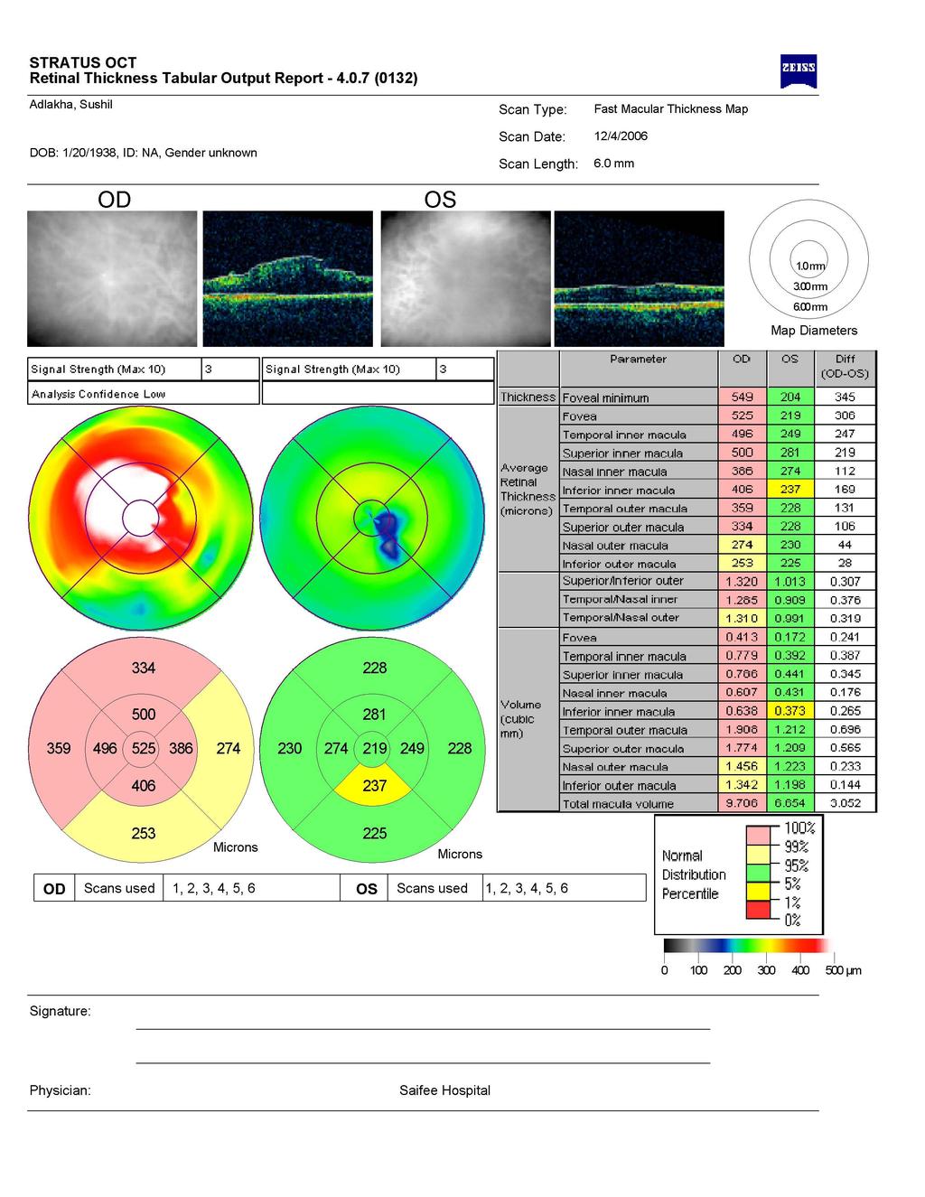
9 DISCUSSION I. DIABETIC MACULOPATHY 11 cases (out of total 40) (27.5%) were of diabetic maculopathy. After routine examination, fundus photography, FFA was done to rule out Clinically Significant Macular Oedema (CSME). In all the cases, it was difficult to diagnose with certainty CSME. (By Indirect Ophthalmoscopy, fundus photograph) CSME being defined by the ETDRS study - Thickening of retina within 500μm from the center of fovea - Hard exudates at or within 500μm from the center of fovea, associated with adjacent retinal thickening - Retinal thickening of 1 Disk Diameter or more, any part of which maybe within 1 Disk Diameter from the center of fovea In our (above) patients with Diabetic Maculopathy, 4 cases (10%) showed focal leak and 3 cases (7.5%) diffuse oedema on FFA. OCT picture showed retinal thickening >500μm at the center of the fovea, corresponding to CSME. Focal grid laser was given to 4 (10%) of the patients with focal leaks and 3 (7.5%) with diffuse leaks were given grid laser. 4 (10%) remaining patients had retinal thickening with Neovascularization. I.e. Proliferative Diabetic Retinopathy (PDR), which was treated with Pan Retinal Photocoagulation and also grid laser. Good visual recovery was seen in 7 out of 11 patients. Repeat OCT could be done in only 2 patients due to poor patient follow up and cost factor. Repeat OCT in these patients showed that retinal thickening was decreased to <225μm with good visual acuity improvement. 8
10 Diabetic CSME has been described in four patterns by OCT i. Sponge Like Thickening Thickening of the retina like a sponge is seen here Treatment Focal Laser ii. Cystoid Macular Oedema Cyst like spaces are seen between the internal nuclear layer and outer plexiform layer Treatment Intravitreal Kenacort then, Focal Laser iii. Serous, Sub foveal Retinal Detachment An elevation is seen, corresponding to a Retinal Detachment just above the RPE chorio capillary layer Treatment Intravitreal Kenacort iv. Taut Posterior Hyaloid Membrane The outermost layer of the retina appears to be pulled taut by the hyaloid, giving rise to traction Treatment Para Planna Vitrectomy II. AGE RELATED MACULAR DEGENERATION (ARMD) Total 8 patients (20%) were diagnosed with ARMD. 5 had Dry ARMD (62.5%) and 3 had Wet ARMD (37.5%). The 3 patients with Wet ARMD underwent FFA and only one was seen to have classic Choroidal Neovascularization (CNV). On OCT imaging the Dry ARMD patients showed a) Local modulation of the RPE layer b) Thinning of neurosensory retina like a geographic atrophic patch On OCT imaging the Wet ARMD patients showed a) Classic CNV: Fusiform thickening and localized disruption of the RPE Chorio Capillary layer. Retinal oedema and sub retinal fluid also helped in diagnosis 9
11 b) Occult CNV: Seen as an area of increased Choroidal reflectivity due to increased penetration of the RPE c) Fibrovascular PED: Seen as elevated RPE layer above an area of mild back scattering region, corresponding to fibrovascular proliferation All the patients (3) of Wet ARMD had metamorphopsia. These patients were referred for PDT or TTT to a private practioner. The patients being poor didn t go there and thus had severe scarring with vision loss. Active Stage Highly reflective, many layered area was seen protruding into the sub retinal space. Intermediate Stage CNV reflectivity became stronger and its margin in the sub retinal space became smooth. Cicatricial Stage Moderately high reflective are covered by a dome shaped highly reflective area which corresponded to RPE was seen. III. CENTRAL SEROUS RETINOPATHY (CSR) CSR was seen in 6 patients (15%) in our study. 2 of these (33.3%) were females and 4 (66.6%) were males. Thus confirming that the incidence was more in males than females. 5 patients were in the age group of years and one was between years. All the patients were subjected to FFA first then OCT. 4 patients (66.6%) on FFA showed smoke stack appearance and 2 patients (33.3%) showed non specific leakage of dye. OCT was diagnostic in 2 of these non specific FFA patients. (8) 10
12 3 of the 6 patients (50%) had metamorphopsia with a positive Amsler Grid test. OCT imaging showed a characteristic elevation of the neurosensory retina. 3 of the 6 patients of CSR had Visual Acuity of 6/12 (Corrected by +1.5D). 3 of these patients followed up with us and had final visual acuity of 6/6 without any treatment. OCT was possible to be repeated in only one patient, it was seen as the characteristic settling of neurosensory retina. IV. MACULAR HOLE Macular holes were contributory by 5 cases (12.5%). From these 3 cases (60%) were diagnosed to be lamellar holes and 2 (40%) to be full thickness holes. (1 being post trauma). Of the total 5 cases, 3 patients had metamorphopsia with positive Amsler Grid. 2 patients with full thickness holes (50%) had positive central scotoma. Watzake (9) sign was positive in 2 patients with full thickness macular hole. In 3 of the patients with partial thickness holes, OCT was the ultimate diagnostic tool. Differentiation: Lamellar Hole: Diagnosed by evidence of perifoveal vitreous detachment with no loss of whole retinal tissue Full Thickness Hole: Diagnosed by the clear loss if retinal tissue at the fovea extending up o the RPE. Importance of OCT in Macular Hole - Surgical Planning & post surgery follow up - Screening & monitoring of other eye - Vitro retinal interface assessment. Demonstration of Posterior Vitreous Detachment (PVD), Vitreo macular traction & surrounding fluid cuff (2) 11
13 V. CYSTOID MACULAR OEDEMA (CME) 3 patients (7.5%) had poor postoperative vision after routine cataract surgery I.e. < 6/36. Fundus photograph showed a dull foveal reflex. On FFA there was absence of the characteristic flower petal appearance. On comparison of FFA with OCT sensitivity, in detecting CME is 96% & specificity being 100% (10) However in all these patients, OCT showed characteristic cyst like spaces in the retinal layer. Local NSAIDS were prescribed for 3 months period. VI. EPIRETINAL MEMBRANE (ERM) A total of 3 patients (7.5%) had ERM. 2 were following Retinal Detachment surgery (66.6%). 1 patient with no discernable cause (33.3%). All the 3 patients had metamorphopsia. OCT imaging showed thickened membrane with a cleavage point seen in all 3 patients. Using OCT it was possible to study characteristics of ERM - Membrane presence with thickness - Retinal fluid accumulation - Cystic Changes - Presence of focal verses global retinal surface adherence The above increased the quality of preoperative assessment, helping in giving a postoperative visual prognosis. (11) Advantage of OCT being Depending upon the site of cleavage point, the surgeon can enter the vitrectomy scissor for ERM peeling. VII. MACULAR SCAR In total 2 of the patients (5%) had a macular scar. 1 after toxoplasma and the other idiopathic. 12
14 VIII. IDIOPATHIC POLYPOIDAL CHOROIDAL VASCULOPATHY (IPCV) Idiopathic polypoidal choroidal vasculopathy (IPCV) was diagnosed in only 3 patients (5%). They were initially diagnosed, erroneously as CNV. Later on Indo Cyanine Green (ICG), they showed polypoidal mass with complete wash out after ICG. OCT in both patients showed serous PED, seen as RPE chorio capillary layer elevation. 13 patients (32.5%) showed metamorphopsia. Some patients who didn t have symptoms of metamorphopsia had a positive Amsler Grid test, as seen in 15 patients (37.5%). CONCLUSION Our study consisted of 40 cases of macular disorders who underwent OCT. The following conclusions were drawn 1. Diabetic Maculopathy the most common. 11 cases (27.5%) 2. a) 4 cases (36.3%) of the 11 cases of Diabetic Maculopathy had Proliferative Diabetic Retinopathy with Taut Posterior Hyaloid Membrane, in which cases par planna vitrectomy is indicated. b) 7 cases (63.7%) remaining, had focal as well as diffuse leaks on FFA, in whom focal or grid laser was given. 3. Age Related Macular Degeneration (ARMD) was seen in 8 cases (20%). 3 cases (37.5%) were of Wet ARMD & 5 cases (62.5%) were of Dry ARMD. OCT helped determine the depth & spread of Choroidal Neovascularization (CNV). 4. Central Serous Retinopathy (CSR) was seen in 6 cases (15%). FFA showed no characteristic appearance in 3 patients (50%) and was thus diagnosed on OCT by elevation of neurosensory retina. 13
15 5. Macular Holes were seen in 5 cases (12.5%). 3 being lamellar hole (60%) & 2 full thickness holes (40%). OCT determined the grade of macular hole as well as the line of treatment. 6. Cystoid Macular Oedema (CME) was present in 3 cases (7.5%), which was difficult to diagnose on FFA. OCT revealed characteristic Cystoid Spaces with increased thickness of the layers. 7. Epiretinal Membrane (ERM) present in 3 cases (7.5%). 2 cases (66.6%) were following Retinal Detachment Surgery and 1 was idiopathic (33.3%). In all 3 cases, cleavage line was seen, which was useful during surgery for ERM peeling. 8. In 3 cases of CSR (7.5%), 2 cases (5%) of Idiopathic Polypoidal Choroidal Vasculopathy (IPCV) and 3 cases of CME (7.5%), in which FFA was not characteristic, OCT gave ultimate diagnosis. Thus OCT was diagnostic in all these above cases. 14
16 CSME 15
17 PATIENTS ACCORDING TO AGE >60 Age in Years No. of Patients No. of Patients DIFFERENT MACULAR DISORDERS (No. OF CASES) Central Serous Retinopathy Diabetic Maculopathy Cystoid Macular Edema Macular Scar Age Related Macular Degeneration Macular hole Epiretinal Membrane Idiopathic Polypoidal Choroidal Vasculopathy
18 BIBLIOGRAPHY 1. Hee MR, Izatt JA, Swanson EA, et al. Optical coherence tomography of the human retina. Arch Ophthalmol 113: , Puliafito CA, Hee MR, Lin CP, Reichel E, Schuman JS, et al. Imaging of macular diseases with optical coherence tomography. Ophthalmology 102: , Swanson EA, Huang D, Fujimoto JG, Puliafito CA. Method & apparatus for optical imaging with means for controlling longitudinal range of sample vs. patent. 14 June 1994, #3321, Youngquist RL, Carr S, Danies Den. Optical coherence domain reflectometery. A new optical evaluation technique. Opt. Lit. 1987; 12: Jakada K, Yokohoma I, Chida K, Noda J. New measurement system for fault location in optical wave guide devices, based on interferometric technique; Appl. Opt 1987; 26: Werner W, Fercher AF, Mengednot K. Eye length measurement by interferometery with partially coherent light. Opt. Lit. 1988; 13: Takahashi K, Ida H, Fukuchi T, Mallumura M. Staging of CNV by OCT. Schepens retina associates, Boston, MA 02114, USA. 8. Puliafito CA, Hee MR et al. OCT of Central Serous Retinopathy. Am J Ophthalmol 1995; 120 (1): Drener West, De Bistros, Guyner DR. Natural History of Idiopathic Macular Hole & Cysts. Arch Ophthal. 110: ; Stanford MR, Anticliff RJ et al. Comparison between OCT & FFA for detection of Cystoid Macular Oedema. Ophthal. 2000: 107 (3): Puliafito CA, Wilkins JR et al. Characterization of Epiretinal Membranes using OCT. Ophthalmology 1996; 103 (12): Abrams GW, Blumenkraz MS, Lewis H, Campo RV. Vitrectomy for Diabetic Macular Traction and Oedema associated with Posterior Hyaloid Traction. Ophthalmology 1992; 99 (5): Palestine AG, Davis MD, Nussenblatt RB, Kuafman SC, Ferris FL. Macular Thickening & Visual Acuity Measurement in patients with Cystoid Macular Oedema. Ophthalmology 1987; 94 (9): Reichel E, Puliafito C, George A, Sulkes D, Rivellese M. optical Cherence Tomography after Laser Photo Coagulation for Clinically Significant Macular Oedema. Ophthal Surg Lasers 2000;31 (3):
19 MASTER CHART Sr. No. OPD Age Sex V/A Symptoms Amsler Systemic Fundus Provisional FFA No. in History Diagnosis Yrs Metamorphopsia Micropsia Positive Scotoma /06 58 M 6/ Hard Drusen Dry ARMD Not /06 65 F 6/ Drusen Dry ARMD Not /06 54 F 6/ DM Hard Exudates/ Diabetic Focal CSME Maculopathy Leak CSR /06 42 M 6/ Diffuse Elevation of Macula /06 13 M FC12-14ft Toxoplasma CR Atrophic Scar at Macula Macular Scar Not /06 48 M 6/ DM FR Dull? CME No Flower Petal Pattern /06 71 M FC6-8ft TB Drusen + Haemorrhage Wet ARMD /06 14 M 6/ FR Dull Lamellar Hole /06 39 M 6/ Irreg. ERM Membrane on Macula /06 18 F 6/ DM CSME Diabetic Maculopathy /06 63 M FC16-18ft FR Dull Lamellar Hole /06 33 F 6/ Elevation of Macula /06 49 F 6/ Elevation of Macula CSR IPCV Smoke Stack Irreg. Leak Not Not Focal Leak Hypofluores cence Smoke Stack ICG done OCT Localized Disruption of RPE Localized Disruption of RPE Sponge Like Thickening Elevation of Neurosensory Retina Thinning of Retina Cystic Spaces in Retina Disruption of RPE- CC Layer No Loss of Whole Retinal Tissue Cleavage Line Seen Sponge Like Thickening No Loss of Whole Retinal Tissue Elevation of Neurosensory Retina Polypoidal Elevation, Serous PED
20 MASTER CHART Sr. No. OPD No. Age Yrs Sex V/A Symptoms Amsler Systemic History Fundus Provisional Diagnosis Metamorphopsia Micropsia Positive Scotoma /07 48 F FC1-2ft FR Dull Full Thickness Macular Hole FFA /07 17 M <6/ FR Dull CME Diffuse Leak /07 62 M 6/ Hard Drusen Dry ARMD Not /07 34 F 6/ DM Exudate / Dot Diabetic Diffuse Haemorrhage Maculopathy Leak /07 74 M FC10-12ft DM NVD Diabetic Not Maculopathy /07 39 M 6/ DM CSME Diabetic Diffuse Maculopathy Leak /07 31 M 6/ Elevation of CSR Smoke Macula Stack /07 71 M 6/36p DM NVE + Inferior Vitreous Haemorrhage Diabetic Maculopathy (PDR) /07 39 M 6/ DM CSME Diabetic Maculopathy /07 45 M 6/ FR Dull Full Thickness Macula Hole /07 39 M 6/ Serous Elevation of Macula /07 51 F 6/24p Cellophane Maculopathy /07 78 F FC14-16ft Irreg. Membrane over Macula CSR ERM Wet ARMD NVE+ Hypofluoresc ence Focal Leak Hypofluoresc ence Ink Blot Appear ance Not Not OCT Loss of Retinal Tissue Cystic Spaces in Retina Thickening of RPE Sponge Like Thickening Sponge Like Thickening Sponge Like Thickening Elevation of Neurosensory Retina Taut Post. Hyaloid Memb. Sponge Like Thickening Loss of Retinal Tissue Elevation of Neurosensory retina Elevated Retina Localized Disruption of RPE
21 MASTER CHART Sr. No. OPD No. Age Yrs Sex V/A Symptoms Amsler Systemic History Fundus Metamorphopsia Micropsia Positive Scotoma /07 54 M FC8-10ft TB Haemorrhage Below Fovea /07 44 F 6/ Elevation of Retina Provisional Diagnosis Wet ARMD /07 37 M 6/ DM FR Dull Lamellar Hole CSR FFA Not Diffuse Leak Hypofluoresc ence ICG /07 62 M 6/ Irreg. Membrane over Macula IPCV /07 59 F 6/ Hard Drusen Dry ARMD Not /07 55 M 6/ Subretinal CSR No e/o Precipitates Smoke /07 49 M FC 4-6ft DM NVD + NVE Diabetic Maculopathy with PDR /07 26 M 6/ Epiretinal Membrane + Cryo marks ERM (Post RD Sx) /07 51 M 6/ DM FR Dull + Drusen Dry ARMD Not /07 57 M 6/ DM CSME Diabetic Diffuse Maculopathy Leak /07 46 M 6/ DM CSME Diabetic Diffuse Maculopathy Leak /07 52 M 6/ DM / TB NVD + Diabetic Not Maculopathy + PDR /07 68 M FC14-16ft Stack Not Not FR Dull CME Not OCT Disruption of RPE Elevation of Neurosensory Retina Full Thickness Retinal Break Absent Serous PED Localized Disruption of RPE Elevation of Neurosensory Retina Taut Post. Hyaloid Memb. Elevation over Retina Thickening of RPE Thickening of Retina Sponge Like Thickening Taut Post. Hyaloid Memb. Cystoid Spaces
Optical Coherence Tomography in Diabetic Retinopathy. Mrs Samantha Mann Consultant Ophthalmologist Clinical Lead of SEL-DESP
 Optical Coherence Tomography in Diabetic Retinopathy Mrs Samantha Mann Consultant Ophthalmologist Clinical Lead of SEL-DESP Content OCT imaging Retinal layers OCT features in Diabetes Some NON DR features
Study of clinical significance of optical coherence tomography in diagnosis & management of diabetic macular edema
 Original Research Article Study of clinical significance of optical coherence tomography in diagnosis & management of diabetic macular edema Neha Kantilal Desai 1,*, Somesh Vedprakash Aggarwal 2, Sonali
OCT Assessment of the Vitreoretinal Relationship in CSME
 December 2007 Sonia Rani John et al. - IFIS 375 ORIGINAL ARTICLE OCT Assessment of the Vitreoretinal Relationship in CSME Dr. Manoj S. DNB FRCS, Dr. Unnikrishnan Nair MS DO FRCS, Dr. Gargi Sathish MS Introduction
ZEISS AngioPlex OCT Angiography. Clinical Case Reports
 Clinical Case Reports Proliferative Diabetic Retinopathy (PDR) Case Report 969 PROLIFERATIVE DIABETIC RETINOPATHY 1 1-year-old diabetic female presents for follow-up of proliferative diabetic retinopathy
PART 1: GENERAL RETINAL ANATOMY
 PART 1: GENERAL RETINAL ANATOMY General Anatomy At Ora Serrata At Optic Nerve Head Fundoscopic View Of Normal Retina What Is So Special About Diabetic Retinopathy? The WHO definition of blindness is
EPIRETINAL MEMBRANE & VITREOMACULAR TRACTION
 EPIRETINAL MEMBRANE & VITREOMACULAR TRACTION Management of ERM and VMT K.V.Chalam,MD,PhD,MBA,FACS Professor and Director of Retina Loma Linda Eye Institute Los Angeles, USA REVIEW ANATOMY The vitreous
The Human Eye. Cornea Iris. Pupil. Lens. Retina
 The Retina Thin layer of light-sensitive tissue at the back of the eye (the film of the camera). Light rays are focused on the retina then transmitted to the brain. The macula is the very small area in
Optical Coherence Tomograpic Features in Idiopathic Retinitis, Vasculitis, Aneurysms and Neuroretinitis (IRVAN)
") Columbia International Publishing Journal of Ophthalmic Research (2014) Research Article Optical Coherence Tomograpic Features in Idiopathic Retinitis, Vasculitis, Aneurysms and Neuroretinitis (IRVAN)
Columbia International Publishing Journal of Ophthalmic Research (2014) Research Article Optical Coherence Tomograpic Features in Idiopathic Retinitis, Vasculitis, Aneurysms and Neuroretinitis (IRVAN)
The College of Optometrists - Learning outcomes for the Professional Certificate in Medical Retina
 Learning outcomes for the Professional Certificate in Medical Retina, incorporating diabetic retinopathy screening and age related macular degeneration The professional certificate is a prerequisite to
Yasser R. Serag, MD Tamer Wasfi, MD El- Saied El-Dessoukey, MD Magdi S. Moussa, MD Anselm Kampik, MD
 Microperimetric Evaluation of Brilliant Blue G- assisted Internal Limiting Membrane Peeling By Yasser R. Serag, MD Tamer Wasfi, MD El- Saied El-Dessoukey, MD Magdi S. Moussa, MD Anselm Kampik, MD The internal
Clinically Significant Macular Edema (CSME)
") Clinically Significant Macular Edema (CSME) 1 Clinically Significant Macular Edema (CSME) Sadrina T. Shaw OMT I Student July 26, 2014 Advisor: Dr. Uwaydat Clinically Significant Macular Edema (CSME) 2
Clinically Significant Macular Edema (CSME) 1 Clinically Significant Macular Edema (CSME) Sadrina T. Shaw OMT I Student July 26, 2014 Advisor: Dr. Uwaydat Clinically Significant Macular Edema (CSME) 2
A retrospective nonrandomized study was conducted at 3
 Department of Ophthalmology, Kangbuk Samsung Hospital, Sungkyunkwan University College of Medicine 1, Seoul, Korea Hangil Eye Hospital 2, Incheon, Korea Seoul National University Bundang Hospital 3, Seongnam,
International Journal of Health Sciences and Research ISSN:
 International Journal of Health Sciences and Research www.ijhsr.org ISSN: 2249-9571 Original Research Article A Multivariate Analysis of Intravitreal Injection of Anti-VEGF Bevacizumab in the Treatment
RETINAL CONDITIONS RETINAL CONDITIONS
 GENERAL INFORMATION RETINAL CONDITIONS RETINAL CONDITIONS WHAT ARE RETINAL CONDITIONS? Retinal conditions affect the light-sensitive tissue at the back of eye known as the retina. They include diseases
 measure of your overall performance. An isolated glucose test is helpful to let you know what your sugar level is at one moment, but it doesn t tell you whether or not your diabetes is under adequate control
Diabetic Retinopathy
 Diabetic Retinopathy Diabetes can be classified into type 1 diabetes mellitus and type 2 diabetes mellitus, formerly known as insulin-dependent diabetes mellitus, and non-insulin diabetes mellitus, respectively.
Vitrectomy for Diabetic Cystoid Macular Edema
 Vitrectomy for Diabetic Cystoid Macular Edema Yukihiro Sato, Zeon Lee and Hiroyuki Shimada Department of Ophthalmology, Nihon University School of Medicine, Tokyo, Japan Purpose: We evaluated visual outcomes
R&M Solutions
 Mohamed Hosny El-Bradey, MD., Assistant Professor of Ophthalmology, Tanta University. Wael El Haig, MD., Professor of Ophthalmology. Zagazeeg University. 1 Myopic CNV is considered the most common vision
Case report 12/10/2014. Delphine Lam ; Dr Mayer Srour Service d ophtalmologie Professeur E.Souied Université Paris Est
 Case report 12/10/2014 Delphine Lam ; Dr Mayer Srour Service d ophtalmologie Professeur E.Souied Medical history Man, 75 years old Complaint: Vision loss in left eye in June 2014 Past ophthalmologic history:
Role of OCT in the diagnosis and follow up of diabetic macular edema
 Seminars in Ophthalmology 0882-0538/02/1701-019$16.00 2002, Vol. 17, No. 1, pp. Swets & Zeitlinger Role of OCT in the diagnosis and follow up of diabetic macular edema Giacomo Panozzo, Elena Gusson, arbara
THE ROLE OF anti-vegf IN DIABETIC RETINOPATHY AND AGE RELATED MACULAR DEGENERATION
 THE ROLE OF anti-vegf IN DIABETIC RETINOPATHY AND AGE RELATED MACULAR DEGENERATION MOESTIDJAB DEPARTMENT OF OPHTHALMOLOGY SCHOOL OF MEDICINE AIRLANGGA UNIVERSITY DR SOETOMO HOSPITAL SURABAYA INTRODUCTION
Andrew J. Barkmeier, MD; Benjamin P. Nicholson, MA; Levent Akduman, MD
 c l i n i c a l s c i e n c e Effectiveness of Laser Photocoagulation in Clinically Significant Macular Edema With Focal Versus Diffuse Parafoveal Thickening on Optical Coherence Tomography Andrew J. Barkmeier,
DOME SHAPED MACULOPATHY. Ιωάννης Ν. Βαγγελόπουλος Χειρ. Οφθαλμίατρος - Βόλος
 DOME SHAPED MACULOPATHY Ιωάννης Ν. Βαγγελόπουλος Χειρ. Οφθαλμίατρος - Βόλος DOME SHAPED MACULOPATHY-DEFINITIONS The entity Dome Shaped Macula ( DSM ) was first described by Gaucher and associates in 2008
OPTIC DISC PIT Pathogenesis and Management OPTIC DISC PIT
 OPTIC DISC PIT Pathogenesis and Management Abdel-Latif Siam Ain Shams University Cairo Egypt OPTIC DISC PIT Congenital pit is an atypical coloboma usually located on the temporal edge of the disc, associated
Visual and Anatomical Outcomes of Vitreous Surgery for Large Macular Holes
 March 2009 Raju K.V. et al. - Closed Globe Injuries 31 ORIGINAL ARTICLE Visual and Anatomical Outcomes of Vitreous Surgery for Large Macular Holes Dr. Mahesh G. MS DO DNB FRCSEd, Dr. A. Giridhar MS, Dr.
Recalcitrant Diabetic Macular Oedema: Therapeutic Options
 December 2007 A. Giridhar et al. - Recalcitrant DME 451 CONSULTATION S E C T I O N Recalcitrant Diabetic Macular Oedema: Therapeutic Options Dr. Cyrus M Shroff 1, Dr. N S Muralidhar 2, Dr. R Narayanan
The Quick Guide to OCT Mastery 50 Real Cases with Expert Analysis
 OPTICAL COHERENCE TOMOGRAPHY The Quick Guide to OCT Mastery 50 Real Cases with Expert Analysis VOL 1 Sanjay Sharma, MD, FRCS, MSc (Epid), MBA Ophthalmologist, Epidemiologist Queen s University, Canada
Is OCT-A Needed As An Investigative Tool During The Management Of Diabetic Macular Edema
 Is OCT-A Needed As An Investigative Tool During The Management Of Diabetic Macular Edema Ayman M Khattab MD, FRCS Professor of Ophthalmology Cairo University Diabetic Macular Edema (DME) Diabetic macular
Moncef Khairallah, MD
 Moncef Khairallah, MD Department of Ophthalmology, Fattouma Bourguiba University Hospital Faculty of Medicine, University of Monastir Monastir, Tunisia INTRODUCTION IU: anatomic form of uveitis involving
ATLAS OF OCT. Retinal Anatomy in Health & Pathology by Neal A. Adams, MD. Provided to you by:
 ATLAS OF OCT Retinal Anatomy in Health & Pathology by Neal A. Adams, MD Provided to you by: Atlas of OCT The OCT Atlas is written by Neal A. Adams, MD, and produced by Heidelberg Engineering, Inc. to help
Michael P. Blair, MD Retina Consultants, Ltd Libertyville/Des Plaines, Illinois Clinical Associate University of Chicago 17 October 2015
 Michael P. Blair, MD Retina Consultants, Ltd Libertyville/Des Plaines, Illinois Clinical Associate University of Chicago 17 October 2015 So What Parts of the Eye Retina are Affected by VHL Neural tissue
Audit of Macular Hole Surgery, Visual Outcome Prediction on OCT Appearance of Macular Hole
 International Journal of Ophthalmology & Visual Science 2017; 2(4): 93-97 http://www.sciencepublishinggroup.com/j/ijovs doi: 10.11648/j.ijovs.20170204.13 Audit of Macular Hole Surgery, Visual Outcome Prediction
A Patient s Guide to Diabetic Retinopathy
 Diabetic Retinopathy A Patient s Guide to Diabetic Retinopathy 840 Walnut Street, Philadelphia PA 19107 www.willseye.org Diabetic Retinopathy 1. Definition Diabetic retinopathy is a complication of diabetes
Fundus Fluorescein Angiography in Diabetic Retinopathy: Correlation of Angiographic Findings to the Clinical Maculopathy Abstract: Purpose:
 IOSR Journal of Dental and Medical Sciences (IOSR-JDMS) e-issn: 2279-0853, p-issn: 2279-0861.Volume 15, Issue 2 Ver. XII (Feb. 2016), PP 80-88 www.iosrjournals.org Fundus Fluorescein Angiography in Diabetic
Retina Conference. Janelle Fassbender, MD, PhD University of Louisville Department of Ophthalmology and Visual Sciences 09/04/2014
 Retina Conference Janelle Fassbender, MD, PhD University of Louisville Department of Ophthalmology and Visual Sciences 09/04/2014 Subjective CC/HPI: 64 year old Caucasian female referred by outside ophthalmologist
Diagnosis and treatment of diabetic retinopathy. Blake Cooper MD Ophthalmologist Vitreoretinal Surgeon Retina Associates Kansas City
 Diagnosis and treatment of diabetic retinopathy Blake Cooper MD Ophthalmologist Vitreoretinal Surgeon Retina Associates Kansas City Disclosures Consulted for Novo Nordisk 2017,2018. Will be discussing
Vitreomacular interface disorders. Ghanbari MD 1393:10:25
 Vitreomacular interface disorders Ghanbari MD 1393:10:25 Human vitreous after dissection of the sclera, choroid, and retina. Lamellar structure of the posterior vitreous cortex (PVC) in the monkey. V =
OCT Interpretation in Retinal Disease
 OCT Interpretation in Retinal Disease Jay M. Haynie, OD, FAAO Financial Disclosure I have received honoraria or am on the advisory board for the following companies: Carl Zeiss Meditec Advanced Ocular
Long-term Management of AMD. Motasem Al-latayfeh, MD Assistant Prof. Ophthalmology Hashemite University Jordan
 Long-term Management of AMD Motasem Al-latayfeh, MD Assistant Prof. Ophthalmology Hashemite University Jordan DEFINITION 1 Age-related macular degeneration (AMD) is a disorder of the macula characterized
OCT in Diabetic Macular Edema and its Correlation with Flourescein Angiography
 Uvea OCT in Diabetic Macular Edema and its Correlation with Flourescein Angiography Kirti Jaisingh MS Kirti Jaisingh MS, Yashpal Goel* MS, Kshitij Aditya** DO * Guru Nanak Eye Centre, New Delhi ** Baba
When optical coherence tomography (OCT)
") Macular Imaging: SD-OCT in nterior Segment Surgical Practice Many pathologic processes of the macula can be visualized or quantified only with this modality. y Steven G. Safran, MD When optical coherence
Macular Imaging: SD-OCT in nterior Segment Surgical Practice Many pathologic processes of the macula can be visualized or quantified only with this modality. y Steven G. Safran, MD When optical coherence
OCT Angiography in Primary Eye Care
 OCT Angiography in Primary Eye Care An Image Interpretation Primer Julie Rodman, OD, MS, FAAO and Nadia Waheed, MD, MPH Table of Contents Diabetic Retinopathy 3-6 Choroidal Neovascularization 7-9 Central
Role of Fluorescein angiography in evaluation of posterior segment disorders
 Original Article Role of Fluorescein angiography in evaluation of posterior segment disorders Arvind R, Surendar S 2, Ch. Jagan Mohan Rao 3 Associate Professor, 2 Postgraduate student, 3 Senior resident,
The Foundation WHAT IS THE RETINA? continued next page. RETINA HEALTH SERIES Facts from the ASRS
 The Foundation American Society of Retina Specialists Committed to improving the quality of life of all people with retinal disease. Vitreomacular Traction Syndrome The vitreous humor is a transparent,
Posterior Segment Update
 Posterior Segment Update Featured Speaker: Dr. Kyle Cheatham, FAAO, DIP ABO DISCLOSURE STATEMENT We have no direct financial or proprietary interest in any companies, products or services mentioned in
Published on Points de Vue International Review of Ophthalmic Optics (http://www.pointsdevue.com)
") Published on Points de Vue International Review of Ophthalmic Optics (http://www.pointsdevue.com) Home > OCT and retinal pathologies OCT and retinal pathologies Sylvain AURIOL, Véronique PAGOT-MATHIS e-mail
Published on Points de Vue International Review of Ophthalmic Optics (http://www.pointsdevue.com) Home > OCT and retinal pathologies OCT and retinal pathologies Sylvain AURIOL, Véronique PAGOT-MATHIS e-mail
JMSCR Vol 06 Issue 12 Page December 2018
 www.jmscr.igmpublication.org Impact Factor (SJIF): 6.379 Index Copernicus Value: 79.54 ISSN (e)-2347-176x ISSN (p) 2455-0450 DOI: https://dx.doi.org/10.18535/jmscr/v6i12.39 Central Macular Thickness analysis
OPHTHALMOLOGICAL DISORDERS
 Telephone No.: 24622495 Telegraphic Address: Aeronautical: VIDDYAYX Commercial: AIRCIVIL NEW DELHI E Mail: dri@dgca.nic.in Fax:01124629211 GOVERNMENT OF INDIA AERONAUTICAL INFORMATION SERVICE DIRECTOR
The Common Clinical Competency Framework for Non-medical Ophthalmic Healthcare Professionals in Secondary Care
 The Common Clinical Competency Framework for Non-medical Ophthalmic Healthcare Professionals in Secondary Care Medical Retina November 2016 Association of Health Professions in Ophthalmology General basic
ROLE OF LASER PHOTOCOAGULATION VERSUS INTRAVITREAL TRIAMCINOLONE ACETONIDE IN ANGIOGRAPHIC MACULAR EDEMA IN DIABETES MELLITUS
 ORIGINAL ARTICLE ROLE OF LASER PHOTOCOAGULATION VERSUS INTRAVITREAL TRIAMCINOLONE ACETONIDE IN ANGIOGRAPHIC MACULAR EDEMA IN DIABETES MELLITUS Aggarwal Somesh VP 1, Shah Sonali N 2, Bharwada Rekha M 3,
Diabetic Retinopathy
 Diabetic Retinopathy Diabetes mellitus is one of the leading causes of irreversible blindness worldwide. In the United States, it is the most common cause of blindness in people younger than 65 years.
Diabetic Retinopathy A Presentation for the Public
 Diabetic Retinopathy A Presentation for the Public Ray M. Balyeat, MD The Eye Institute Tulsa, Oklahoma The Healthy Eye Light rays enter the eye through the cornea, pupil and lens. These light rays are
Ophthalmology Macular Pathways
 Ophthalmology Macular Pathways Age related Macular Degeneration Diabetic Macular Oedema Macular Oedema secondary to Central Retinal Macular Oedema secondary to Branch Retinal CNV associated with pathological
Do You See What I See!!! Shane R. Kannarr, OD
 Do You See What I See!!! Shane R. Kannarr, OD skannarr@kannarreyecare.com Define Specialty Testing Additional Test to: Prove/Disprove Diagnosis To monitor progression of a condition To document a condition
8/6/17. Disclosures Aerie Pharmaceuticals Alcon BioTissue Diopsys Optovue Shire
 Nathan Lighthizer, O.D., F.A.A.O. Associate Professor Assistant Dean for Clinical Care Director of Continuing Education Chief of Specialty Care Clinics Oklahoma College of Optometry Tahlequah, OK lighthiz@nsuok.edu
Understanding Diabetic Retinopathy
 Understanding Diabetic Retinopathy What Is Diabetic Retinopathy? Diabetes damages blood vessels in the rear of the eye. This condition is called diabetic retinopathy. It can lead to vision loss or blindness.
Perspectives on Screening for Diabetic Retinopathy. Dr. Dan Samaha, Optometrist, MSc Clinical Lecturer School of Optometry, Université de Montréal
 Perspectives on Screening for Diabetic Retinopathy 1 Dr. Dan Samaha, Optometrist, MSc Clinical Lecturer School of Optometry, Université de Montréal Current standards 2 According to the Canadian Diabetes
Macular Hole Associated with Vogt-Koyanagi-Harada Disease at the Acute Uveitic Stage
 Published online: September 15, 2015 2015 The Author(s) Published by S. Karger AG, Basel 1663 2699/15/0063 0328$39.50/0 This article is licensed under the Creative Commons Attribution-NonCommercial 4.0
VISUAL OUTCOME IN DIABETIC MACULAR EDEMA AFTER GRID LASER TREATMENT
 The Professional Medical Journal DOI: 10.17957/TPMJ/16.2856 ORIGINAL PROF-2856 VISUAL OUTCOME IN DIABETIC MACULAR EDEMA AFTER GRID LASER TREATMENT 1. MBBS.FCPS Assistant Professor Ophthalmology Independent
Eyes on Diabetics: How to Avoid Blindness in Diabetic Patient
 Eyes on Diabetics: How to Avoid Blindness in Diabetic Patient Rova Virgana FK Unpad Pusat Mata Nasional RS Mata Cicendo Bandung Eye Center (Hospital and Clinic) PIT IDI Jabar 2018 Keys Facts from WHO
3/6/2014. Hoda MH Mostafa MD Associate Professor of Ophthalmology Cairo University. The author has no proprietary interest. Today s Objectives
 Hoda MH Mostafa MD Associate Professor of Ophthalmology Cairo University The author has no proprietary interest Today s Objectives Identify the CLINICAL SCENARIOS IN MACULAR EDEMA where OCT plays a MAJOR
FRANZCO, MD, MBBS. Royal Darwin Hospital
 Diabetes and Eye By Dr. Nishantha Wijesinghe FRANZCO, MD, MBBS Consultant Ophthalmologist Royal Darwin Hospital 98% of Diabetics do not need to suffer from severe visual loss Yet Diabetic eye disease is
Key words: Choroidal neovascularisation, Laser coagulation, Retinal imaging
 420 Mini Review The Role of Optical Coherence Tomography (OCT) in the Diagnosis and Management of Retinal Angiomatous Proliferation (RAP) in Patients with Age-related Macular Degeneration Antonio Polito,
Amber Priority. Image Library
 Amber Priority Image Library Amber flag Diabetic Maculopathy (M1) Pre-proliferative Diabetic Retinopathy (R2) Old, treated and now inactive DR (R1/M0/P1or R0/M0/P1) Where only partial or incomplete images
Fluorescein Angiography
 Last revision: October 2011 by Luis Arias Fluorescein Angiography Authors: Luis Arias, MD Hospital Universitari de Bellvitge - University of Barcelona. Spain Jordi Monés, MD Institut de la Màcula i de
Moving forward with a different perspective
 Moving forward with a different perspective The Leader In Vision Diagnostics Offers A New Perspective Marco has served the eyecare community by offering exceptional lane products and automated high tech
11/29/2016 MACULAR MALADIES: TYPICAL & ATYPICAL CASES
 MACULAR MALADIES: TYPICAL & ATYPICAL CASES Dawn Pewitt, OD, FAAO Triad Eye Institute, Grove, OK Dpewitt@triadeye.com Disclosure Statement: No financial disclosures COPE 51218-PS Please silence all mobile
OCT Interpretation. Financial Disclosure. Jay M. Haynie, OD, FAAO. OCT Image Layers 7/21/2014
 OCT Interpretation Jay M. Haynie, OD, FAAO Financial Disclosure I have received honoraria or am on the advisory board for the following companies: Olympia Tacoma Renton Kennewick - Washington Carl Zeiss
Case Report Inherent Challenges in Managing Long Standing Refractory Diabetic Macular Edema
 Cronicon OPEN ACCESS EC OPHTHALMOLOGY Case Report Inherent Challenges in Managing Long Standing Refractory Diabetic Macular Edema V Swetha E Jeganathan 1,2 * and Karen Madill 3 1 Department of Ophthalmology,
Optical Coherence Tomography: Pearls for the Anterior Segment Surgeon Basic Science Michael Stewart, M.D.
 Optical Coherence Tomography: Pearls for the Anterior Segment Surgeon Basic Science Michael Stewart, M.D. Disclosure OCT Optical Coherence Tomography No relevant financial relationships I will refer to
Facts About Diabetic Eye Disease
 Facts About Diabetic Eye Disease Points to Remember 1. Diabetic eye disease comprises a group of eye conditions that affect people with diabetes. These conditions include diabetic retinopathy, diabetic
Course # Getting to Know Your OCT
 Course # 140 Getting to Know Your OCT Course Title: Lecturer: Getting to Know Your OCT Brad Sutton, OD, FAAO IU School of Optometry Financial Disclosures No financial disclosures Optical Coherence Tomography-OCT
Atrophic AMD with Vitreomacular Traction Syndrome.
 Case Record 5 Atrophic AMD with Vitreomacular Traction Syndrome. June 2012 Dr Peter Frampton DOptom MSc FCOptom BAppSc(Optom)(AUS) DipTp(AS) DipTp(SP) DipTp(IP) Introduction : New Diagnostic Technologies
MANAGING DIABETIC RETINOPATHY. <Your Hospital Name> <Your Logo>
 MANAGING DIABETIC RETINOPATHY It s difficult living with Diabetes Mellitus. Ask any diabetic... Their lives are centered around meal plans, glucose levels, and insulin
Macular conditions. by Louise Stainer BSc(Hons)
") 4 dispensingoptics December 2013 Macular conditions by Louise Stainer BSc(Hons) CompetencIes covered: Dispensing opticians: Ocular Examination, Ocular Abnormalities, Low Vision Optometrists: Ocular Disease
4 dispensingoptics December 2013 Macular conditions by Louise Stainer BSc(Hons) CompetencIes covered: Dispensing opticians: Ocular Examination, Ocular Abnormalities, Low Vision Optometrists: Ocular Disease
Optical coherence tomography in diabetic macular edema: patterns and related risk factors
 Original article Optical coherence tomography in diabetic macular edema: patterns and related risk factors Mohammadreza Ahmadpour-Baghdadabad Masoudreza Manaviat Ahmad Shojaoddiny-Ardekani Yazd Diabetes
CASE PRESENTATION. DR.Sravani 1 st yr PG Dept of Ophthalmology
 CASE PRESENTATION DR.Sravani 1 st yr PG Dept of Ophthalmology Name : X X X X X Age : 50yrs Sex : male Occupation : Farmer Residence : Mothkur CHIEF COMPLAINTS : - Diminision of vision in Right Eye since
Optical Coherence Tomography (OCT) in Uveitis Piergiorgio Neri, BMedSc, MD, PhD Head Ocular Immunology Unit
 in Uveitis Piergiorgio Neri, BMedSc, MD, PhD Head Ocular Immunology Unit") The Eye Clinic Polytechnic University of Marche Head: Prof Alfonso Giovannini November, 1991 Optical Coherence Tomography (OCT) in Uveitis Piergiorgio Neri, BMedSc, MD, PhD Head Ocular Immunology Unit
The Eye Clinic Polytechnic University of Marche Head: Prof Alfonso Giovannini November, 1991 Optical Coherence Tomography (OCT) in Uveitis Piergiorgio Neri, BMedSc, MD, PhD Head Ocular Immunology Unit
OCCLUSIVE VASCULAR DISORDERS OF THE RETINA
 OCCLUSIVE VASCULAR DISORDERS OF THE RETINA Learning outcomes By the end of this lecture the students would be able to Classify occlusive vascular disorders (OVD) of the retina. Correlate the clinical features
Oishi A, Miyamoto K, Yoshimura N. Etiology of carotid cavernous fistula in Japanese. Jpn J Ophthalmol. 2009;53:40-43.
 Kimura T, Takagi H, Miyamoto K, Kita M, Watanabe D, Yoshimura N. Macular hole with epiretinal membrane after triamcinolone-assisted vitrectomy for proliferative diabetic retinopathy. Retinal Cases Brief
Authors. Introduction. Introduction. Materials and Methods. Objective 10/27/2015
 Idiopathic Polypoidal Choroidal Vasculopathy (IPCV) in Thai Population Presenting with Choroidal Neovascularization (CNV) A multicenter study Authors Yonrawee Piyacomn 1, Chavakij Bhoomibunchoo 1, Yosanan
Optical coherence tomography-guided classification of epiretinal membranes
 DOI 10.1007/s10792-014-9975-z ORIGINAL PAPER Optical coherence tomography-guided classification of epiretinal membranes Vasileios Konidaris Sofia Androudi Alexandros Alexandridis Anna Dastiridou Periklis
Often asymptomatic but can cause a reduction in BCVA and distortion of vision.
 Christopher Wolfe, OD, FAAO, Dipl. ABO Epiretinal Membrane (ERM) and Vitreomacular Traction (VMT) Epiretinal membrane (macular pucker, cellophane maculopathy, premacular fibrosis) consists of a layer of
Dr/ Marwa Abdellah EOS /16/2018. Dr/ Marwa Abdellah EOS When do you ask Fluorescein angiography for optic disc diseases???
 When do you ask Fluorescein angiography for optic disc diseases??? 1 NORMAL OPTIC DISC The normal optic disc on fluorescein angiography is fluorescent due to filling of vessels arising from the posterior
A population based study of macular choroidal neovascularization using optical coherence tomography in Eastern China
 EXPERIMENTAL AND THERAPEUTIC MEDICINE 8: 371-376, 2014 A population based study of macular choroidal neovascularization using optical coherence tomography in Eastern China JIE ZHAO, JUN HU, HAO LU and
World Sight Day Case Studies. Mark Frost Screening Manager South East London DESP
 World Sight Day 2015 Case Studies Mark Frost Screening Manager South East London DESP Introduction All of the following cases have been identified in our screening programme over the last 3 years. The
Widefield Retinal Imaging with Auto Fluorescence Technology in the Optometric Practice
 Widefield Retinal Imaging with Auto Fluorescence Technology in the Optometric Practice This course will define ultra-widefield retinal imaging and autofluorescence for the attendee. Will show how it is
COMPARISON OF INTRAVITREAL TRIAMCINOLONE INJECTION VS LASER PHOTOCOAGULATION IN ANGIOGRAPHIC MACULAR EDEMA IN DIABETIC RETINOPATHY
 Original Article COMPARISON OF INTRAVITREAL TRIAMCINOLONE INJECTION VS LASER PHOTOCOAGULATION IN ANGIOGRAPHIC MACULAR EDEMA IN DIABETIC RETINOPATHY Aggarwal Somesh V 1, Shah Sonali N 2, Bharwada Rekha
Use of Scanning Laser Ophthalmoscope Microperimetry in Clinically Significant Macular Edema in Type 2 Diabetes Mellitus
 Use of Scanning Laser Ophthalmoscope Microperimetry in Clinically Significant Macular Edema in Type 2 Diabetes Mellitus Fumihiko Mori, Satoshi Ishiko, Norihiko Kitaya, Taiichi Hikichi, Eiichi Sato, Akira
Fundus Autofluorescence. Jonathan A. Micieli, MD Valérie Biousse, MD
 Fundus Autofluorescence Jonathan A. Micieli, MD Valérie Biousse, MD The retinal pigment epithelium (RPE) has many important functions including phagocytosis of the photoreceptor outer segments Cone Rod
Disease-Specific Fluorescein Angiography
 Ruth E. Picchiottino, CRA Disease-Specific Fluorescein Angiography 15 Disease-Specific Fluorescein Angiography Recommendations for tailoring retinal fluorescein angiography to diabetic retinopathy, macular
Year 2 MBChB Clinical Skills Session Ophthalmoscopy. Reviewed & ratified by: Mr M Batterbury Consultant Ophthalmologist
 Year 2 MBChB Clinical Skills Session Ophthalmoscopy Reviewed & ratified by: o Mr M Batterbury Consultant Ophthalmologist Learning objectives o To understand the anatomy and physiology of the external and
Diabetic Retinopathy Screening in Hong Kong. Dr. Rita Gangwani M.S, FRCS (Ophth), FCOphth(HK), FHKAM Eye Institute, The University of Hong Kong
, FCOphth(HK), FHKAM Eye Institute, The University of Hong Kong") Diabetic Retinopathy Screening in Hong Kong Dr. Rita Gangwani M.S, FRCS (Ophth), FCOphth(HK), FHKAM Eye Institute, The University of Hong Kong Co-Investigators Prof. David Wong Prof. Sarah McGhee Dr. Wico
Diabetic Retinopathy Screening in Hong Kong Dr. Rita Gangwani M.S, FRCS (Ophth), FCOphth(HK), FHKAM Eye Institute, The University of Hong Kong Co-Investigators Prof. David Wong Prof. Sarah McGhee Dr. Wico
History/principles of the OCT What does the normal retinal OCT look like Vitreal disorders Retinal/RPE disorders Choroidal disorders
 Nathan Lighthizer, O.D., F.A.A.O. Assistant Professor Assistant Dean for Clinical Care Director of Continuing Education Chief of Specialty Care Clinics Chief of Electrodiagnostics Clinic Oklahoma College
Manual. Manual Welsh Eye Care Initiative. A Welsh Eye Care Initiative. Protocol. The Assessment and Management of Age-related Macular Degeneration
 A Protocol 1.0 Definitions The following terms are important in this text: Wet Macular Degeneration Condition caused by the growth of abnormal blood vessels under the retina. Symptoms appear suddenly and
Contents. Chapter 1 Common Pitfalls in the Use of Optical Coherence Tomography for Macular Diseases Lihteh Wu and Teodoro Evans
 Contents Chapter 1 Common Pitfalls in the Use of Optical Coherence Tomography for Macular Diseases Lihteh Wu and Teodoro Evans 1.1 Introduction... 1 1.2 Limitations of Time-Domain OCT... 2 1.2.1 Acquisition
Diabetes and Eye Health more than meets the eye Vision Initiative - in association with PSA
 Diabetes and Eye Health more than meets the eye Vision Initiative - in association with PSA Vision 2020 Australia Vision Initiative RANZCO & OAA (Vic) Proud members of Vision 2020 Australia Outline Vision
M acular microholes have been described in patients
 189 EXTENDED REPORT Macular microholes: pathogenesis and natural history H J Zambarakji, P Schlottmann, V Tanner, A Assi, Z J Gregor... See end of article for authors affiliations... Correspondence to:
RETINAL THICKENING MAY BE PRESENT IN A NUMBER. B-scan Ultrasonography for the Detection of Macular Thickening METHODS
 B-scan Ultrasonography for the Detection of Macular Thickening JAMES C. LAI, MD, SANDRA S. STINNETT, DRPH, AND GLENN J. JAFFE, MD PURPOSE: To report the sensitivity and specificity of B-scan ultrasonography
Study of 189 Cases of Diabetic Retinopathy at CMC Larkana
 Original Article Study of 189 Cases of Diabetic Retinopathy at CMC Larkana Shahid Jamal Siddiqui, Sayed Imtiaz Ali Shah, Abdul Qadir Shaikh, Mohammed Yousuf Depar, Safder Ali Abbassi Pak J Ophthalmol 2007,