Dry AMD: the regulatory view
|
|
|
- Ethelbert Johnson
- 6 years ago
- Views:
Transcription
1 EMA Ophthalmology Workshop 2011 Dry AMD: the regulatory view Marco Coassin, MD PhD University of Rome Campus Bio-Medico - Italy
2 In this presentation Personal views Previous scientific advices No currently approved drugs No validated surrogate endpoints Case by case evaluation Risk to benefit ratio considerations The views presented are those of the individual and may not be understood or quoted as being made on behalf of the EMA or reflecting the position of EMA or one of its committees or working parties
3 The FDA currently recommends that clinical study sponsors use change in visual function as a primary endpoint and also considers anatomic markers such as the extent of spread of geographic atrophy (GA). Csaky et al. IOVS 2008
4 Endpoints in clinical trials Primary endpoints: clinical relevance for the patient = functional (preservation of visual acuity) Surrogate endpoints (anatomic and biomarkers): reasonably likely to predict clinical benefit An anatomical feature detected by imaging technology needs to be validated in order to be used in the clinical trials, requiring a very high correlation between the endpoint and visual function (i.e., in patients with diabetic retinopathy a three-step change on the EDTRS scale correlated with a three-line vision loss)
5 Endpoints for Dry AMD BCVA change Progression of geographic atrophy Progression to severe visual loss Progression to wet AMD Drusen regression Contrast sensitivity Low luminance VA Perimetric measures Multifocal ERG Near acuity Reading speed Quality of life
6 Peculiarity of Dry AMD GA lesions may have a relatively late effect on distance BCVA (foveolar involvement) BCVA might not be sensitive enough to assess GA progression within a reasonable time frame Disease progression may not be evident until late in the course of the disease at which time an interventional treatment could have little benefit due to the extent of irreversible retina damage
7 The antecedent: antivirals for CMV retinitis Anatomical marker as primary endpoint BCVA as secondary endpoint Regulatory R/B consideration: Useful anatomical marker Not always able to quantify the magnitude of effect Not always predictor of VA changes Compared to GA: - GA is not an infection - GA progression is slower than retinitis - to treat an infectious diseases with an antiinfective agent is a clear pharmacological rationale

8 GA may progress more rapidly than visual loss 20/20 20/80 5 yrs

9 ...but not always 73 yo 20/80 63 yo 20/80
10 Evaluating GA progression No primary outcome for efficacy has yet been validated for dry AMD in a licensing procedure Early phase evidence based on enlargement of GA as endpoint is lacking The mean change of growth is area size dependent Poor correlation of BCVA loss with: baseline lesion size, change in lesion area over 12 months and foveal/non foveal localization Difficult to measure all the areas of atrophy and to include the most peripheral lesions Different patterns of GA, likelihood of progression, etc
11 Evaluation of GA: CFP Traditional method: Colour Fundus Photographs CFP has been shown to be reproducible in several clinical trials, including AREDS Poor discrimination between dead/nonfunctioning RPE, living but depigmented RPE and yellowish coloration caused by large drusen Poor predictive sensitivity - capable of identifying only 6% of eyes that progress to late stages of AMD in 5 years (Klein et al. 1997)
12 Evaluation of GA: FAF Fundus Auto-Fluorescence (FAF) imaging seem to provide more info on GA than CFP Ongoing trials on GA have FAF as clinical endpoint Hyperfluorescent regions in peri-lesional areas at FAF were linked to increased rates of lesion growth Several studies indicate that the lesion areas quantified with CFP and FAF are significantly correlated, but not equivalent


13 Smaller lesions can be measured with FAF but their influence on the sensitivity of the method and their prognostic value is not clear With FAF some of the smaller areas in the periphery are left out and others of similar size are included Not clear algorithm in defining boundaries of GA in CFP vs. FAF vs. OCT
14 Evaluation of GA Is FAF showing clinical relevant retinal changes? Standard FAF seems overestimate foveal GA and underestimate extrafoveal GA when compared to Near InfraRed FAF (Pilotto et al. 2011) The loss of NIA precedes loss of FAF (Kellner et al. 2010) Irreversible degeneration precedes changes shown by FAF and CFP? SD-OCT showed early photoreceptor loss, while FAF corresponded to the linear disruption of choroidal hyperreflectivity (Schmitz-Vandenberg et al. only 21 eyes, no data on progression) OCT has good reproducibility (Yehoshua et al. 2011)
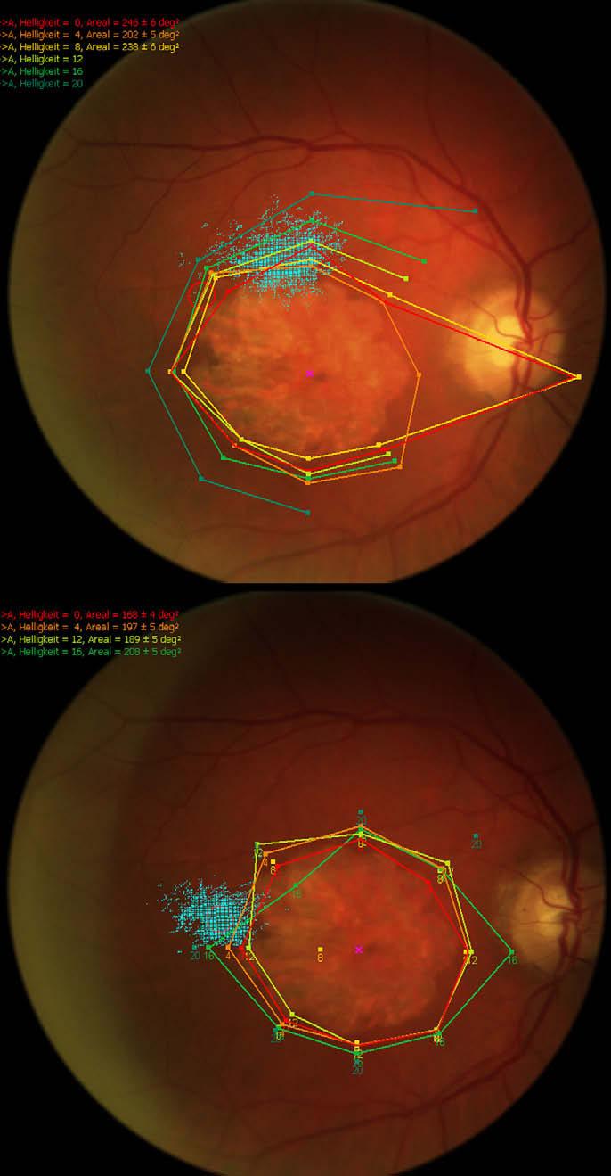
15 Microperimetry
16 Microperimetry Database of normal values available for the MP1 (Midena et al. 2011) Early reduction in macular mean sensitivity and mean defect in intermediate AMD can be detected before VA changes (Dinc et al. 2008; Chen et al. 2011) Changes in retinal sensitivity in GA progression described by NIH in 18 eyes followed for 24 months (Meleth et al. 2011) Mean number of scotomatous points increased significantly with time (P = 0.004) at a rate of 4.4 points/year. Mean retinal sensitivities of all points, all responding points, and all perilesional points all decreased significantly with time (P < 0.003), as did fixation quality within the 2 and 4 circles (P < 0.002) The growth of GA lesion area was associated with the changes in the number of scotomatous points (P = 0.01) but not with changes in the other microperimetric parameters.
17 Multifocal ERG 2011 ISCEV standard for clinical multifocal electroretinography now available before only guidelines Few studies on AMD included mferg evaluation Several different types of ERG electrodes are commercially available and not all produce comparable results (differences in ERG amplitude are based on electrode placement to the cornea) Normal values: each laboratory must develop its own normative data. Variations in recording equipment and parameters make the use of data from other sources inappropriate. Because electrophysiologic data are not necessarily described by a normal distribution, laboratories should report median values rather than means, and determine boundaries of normality. The mferg, like the full-field ERG, is smaller in amplitude in older individuals and in those with highly myopic eyes so that age and refractive error may be important in the evaluation of some patients. In any case, ageadjusted normative data is recommended. (Hood et al. 2011)
18 Contrast sensitivity CS tests thought to be more sensitive to early eye disease than VA Lacks of specificity to distinguish between eye diseases Useful for describing the difficulties in everyday visual tasks and linked to QoL (Rubin et al. 1994) CS loss is disabling for mobility tasks if >0.9 log U and for reading if >1.4 log U (West et al. 2002) Consensus that a halving of CS (six letters on the Pelli- Robson chart) has an impact on task performance and quality of life comparable to a doubling of the visual angle (Rubin et al. 2001)
19 Near acuity tests/reading speed Sloan's M notation is more standardized than Jaeger notation for measuring near acuity. The height of a lowercase 1M letter subtends 5 minarc at a 1 m viewing distance and corresponds roughly to the size of ordinary newsprint. Viewing distance should be specified (1M print read at a distance of 40 cm would be recorded as 0.40/1.00M) The MNREAD Test is composed of 19 standardized sentences in a logarithmic progression of sizes. The test can be used to measure reading acuity (the smallest print size that can be read), maximum reading speed, and critical print size (the smallest print size for maximum reading speed). Reading speed correlates with GA size (Sunness 1996)
20 Near acuity tests/reading speed Invest Ophthalmol Vis Sci Jun 1;52(6): Test-retest variability of reading performance metrics using MNREAD in patients with age-related macular degeneration. Patel PJ, Chen FK, Da Cruz L, Rubin GS, Tufail A. PURPOSE: To determine the test-retest variability of reading ability using the MNREAD charts in patients with stable age-related macular degeneration (AMD). METHODS: In this prospective study, reading ability was measured at two visits in 124 nontreated eyes of 124 patients with AMD, who were enrolled in an ongoing clinical trial using a standardized MNREAD protocol. Only patients with stable AMD who could perform the reading test at 40 cm at both visits were included in the analysis. Different scoring rules were applied to calculate critical print size and maximum reading speed. RESULTS: Data from the 59 patients with a mean (SD) age of 78 (7.6) years who met the study criteria were analyzed at a mean (SD) interval of 43 (6) days between measurements. The 95% coefficient of repeatability (CR) was 0.30 logmar for reading acuity. The CR for critical print size and maximum reading speed varied depending on the analysis method applied. CONCLUSIONS: This is a report of estimates of the intersession test-retest variability of reading performance metrics in patients with stable AMD. The results are helpful both in defining end points in clinical trials for AMD and in distinguishing clinical change from measurement variability in clinical practice.
21 Drusen regression Cochrane Database Syst Rev Jul 8;(3):CD Laser treatment of drusen to prevent progression to advanced age-related macular degeneration Parodi MB, Virgili G, Evans JR. OBJECTIVES: To examine the effectiveness and adverse effects of laser photocoagulation of drusen in AMD. SELECTION CRITERIA: Randomised controlled trials (RCTs) of laser treatment of drusen in AMD in which laser treatment had been compared with no intervention or sham treatment. Two types of trials were included. Some trials studied one eye of each patient (unilateral studies); other studies recruited patients with bilateral drusen and randomised one eye to photocoagulation or control and the fellow eye to the other group. MAIN RESULTS: We found nine studies which randomised 2216 people: four unilateral trials, three bilateral trials and two trials that included both a unilateral and a bilateral study arm. Overall, the studies were of moderate quality. Only half of the trials reported adequate allocation sequence generation, allocation concealment and masking of visual acuity outcome assessors. Although two (of the nine) studies reported significant drusen disappearance at two years, photocoagulation did not appear to affect the development of CNV at two years follow up (nine studies, 1767 people followed up, odds ratio (OR) 1.04, 95% CI 0.71 to 1.51) or the loss of three or more lines of visual acuity (six studies, 1628 people followed up, OR 1.17, 95% CI 0.75 to 1.82). AUTHORS' CONCLUSIONS: The trials included in this review confirm the clinical observation that laser photocoagulation of drusen leads to their disappearance. However, there is no evidence that this subsequently results in a reduction in the risk of developing CNV, geographic atrophy or visual acuity loss.
22 Low luminance VA Ophthalmology Sep;115(9): Low luminance visual dysfunction as a predictor of subsequent visual acuity loss from geographic atrophy in age-related macular degeneration. Sunness JS, Rubin GS, Broman A, Applegate CA, Bressler NM, Hawkins BS. METHODS: Annual examinations included measurement of best-corrected VA, low luminance VA, Pelli-Robson contrast sensitivity, reading speed, examination, and fundus photography. The total GA area was quantified, as was the GA within a mm(2) circle centered on the fovea. Ninety-nine patients with GA; 2 years follow up. RESULTS: Participants with baseline VA of 20/50 or more had a 40% 2-year rate of VA loss of 3 lines or more, compared with 13% for the participants with worse baseline acuities. The baseline low-luminance deficit (LLD) in VA was a strong predictor of subsequent VA loss for all levels of baseline VA. Within the good baseline VA group, the relative risk (RR) of 3-line loss for the worse LLD group compared with the better LLD group was 2.88 (95% confidence interval [CI], ). The LLD is a stable and reproducible measure. Other significant visual function predictors of subsequent VA loss in eyes with good baseline VA included foveal dark-adapted sensitivity (RR, 4.20; 95% CI, ) and reduced reading rate (RR, 2.43; 95% CI, ). The rate of VA loss within the good acuity group was higher when the GA included 25% to 75% of the central 10.2 mm(2) than in eyes with GA including less than 25% or more than 75% of the central 10.2 mm(2). The following were not significant predictors of subsequent VA loss among these participants: age, gender, fellow eye diagnosis, fellow eye VA, baseline GA area, and GA ER. CONCLUSIONS: Visual function measures can predict the risk of future VA loss in subjects with GA and good baseline VA. They may allow identification of the highest risk group for VA loss, enabling more efficient design of clinical trials. They also may be appropriate surrogate measures of foveal health in short-term treatment trials.
23 Follow-up in Dry AMD trials Doubling of the visual angle in 2 years (reported in the studies on the natural course of dry AMD) To detect a 25% reduction in GA rate (α=0.05; power=0.80; losses to FU=15%), 153 patients per arm should be followed for 2 years (Sunness 1997) Advisable to have all patients continued until the last patient has the final visit at month 24
24 Is BCVA so useless in trials? Phase II trial on a topical agent for Dry AMD 10 patients, TID for 2 years, FE as control Primary outcome: BCVA change at 24 mos Secondary outcome: changes in area of GA, contrast sensitivity, microperimetry and total drusen area from baseline Results: statistically significant change in BCVA but not for all the other secondary outcomes! IOVS 2010
25 Conclusions Visual function is essential in describing outcomes from trials Relevant secondary clinical/functional outcomes available Functional outcomes could be correlated with parallel imaging outcomes to increase evidence (sensitivity of the respective endpoints, correlation to functional loss, effective dosage) which could be taken to support the use of imaging endpoints from a risk benefit perspective Feasible to select a specified GA population in order to use visual loss as primary outcome (vision falls most rapidly in eyes with better baseline VA i.e., VA>=20/50 had a 40% two-year rate of >=3 line VA loss [Sunness 2008]) Include responder analyses Including patients at greatest risk of progression of GA into a separate trial would allow planning a shorter study with a possible functional outcome.
26 Conclusions No primary outcome for efficacy has yet been validated for this condition in a licensing procedure Mean change from baseline in GA area might be an acceptable primary endpoint if the limits of the method (CFP, FAF, OCT) are taken into account and relevant complementary data/secondary clinical/functional outcomes provide supportive evidence It may be difficult to determine whether central spared areas are present and correlation with visual acuity and other functional tests may be helpful A parallel monitoring of lesion growth with SD-OCT might be added to support the data collected with FAF and/or CFP By potentially stratifying risk and enrolling patients at high risk of progression, the use of FAF may optimise the clinical trial design A subgroup analysis of different phenotypes of GA might demonstrate different progression rate for some subtypes Use stratification methods or percentage of GA area change Include good numbers of patients with different lesion sizes to allow generalisability Sponsors developing products in this therapeutic area are encouraged to seek EMA scientific advice
The Role of Phenotype in Selectively Enriching Patients for Clinical Studies
 The Role of Phenotype in Selectively Enriching Patients for Clinical Studies Developing Treatments for Dry Age-Related Macular Degeneration (AMD) Workshop November 15, 2014 National Academy of Sciences
Clinical Trial Endpoints for Macular Diseases
 Clinical Trial Endpoints for Macular Diseases Developed in collaboration Learning Objective Upon completion, participants should be able to: Summarize types of biomarkers of progression and treatment response
Original Policy Date
 MP 9.03.08 Photocoagulation of Macular Drusen Medical Policy Section Miscellaneous Policies Issue 12/2013 Original Policy Date 12/2013 Last Review Status/Date Reviewed with literature search/12/2013 Return
Secondary Analysis of Clinical Trials Data A Biostatistician s Experience
 Secondary Analysis of Clinical Trials Data A Biostatistician s Experience Gui-shuang Ying, PhD Center for Preventive Ophthalmology and Biostatistics Perelman School of Medicine University of Pennsylvania
Clinical Study Synopsis
 Clinical Study Synopsis This Clinical Study Synopsis is provided for patients and healthcare professionals to increase the transparency of Bayer's clinical research. This document is not intended to replace
Fundus Autofluorescence. Jonathan A. Micieli, MD Valérie Biousse, MD
 Fundus Autofluorescence Jonathan A. Micieli, MD Valérie Biousse, MD The retinal pigment epithelium (RPE) has many important functions including phagocytosis of the photoreceptor outer segments Cone Rod
CLINICAL SCIENCES. Consistency Between Visual Acuity Scores Obtained at Different Test Distances
 CLINICAL SCIENCES Consistency Between Visual Acuity Scores Obtained at Different Test Distances Theory vs Observations in Multiple Studies Li Ming Dong, PhD; Barbara S. Hawkins, PhD; Marta J. Marsh, MS
Clinical Trials Related to Age Related Macular Degeneration
 Clinical Trials Related to Age Related Macular Degeneration Kirti Singh MD, DNB, FRCS Kirti Singh MD, DNB, FRCS, Pooja Jain MBBS, Nitasha Ahir MBBS, Divya Jain MD, DNB Guru Nanak Eye Centre, Maulana Azad
CENTENE PHARMACY AND THERAPEUTICS NEW DRUG REVIEW 2Q17 April May
 BRAND NAME Lucentis GENERIC NAME ranibizumab MANUFACTURER Genentech, Inc. DATE OF APPROVAL June 30, 2006 PRODUCT LAUNCH DATE July 13, 2006 REVIEW TYPE Review type 1 (RT1): New Drug Review Full review of
Contrast Sensitivity and Reading: Assessment and Reliability with the Reading Explorer (REX) Test
 Test") Contrast Sensitivity and Reading: Assessment and Reliability with the Reading Explorer (REX) Test Giacomelli G 1, Volpe R, Virgili G 1, Farini A 2, Arrighi R 2,Barbieri C, Menchini U 1 1. Department of
Contrast Sensitivity and Reading: Assessment and Reliability with the Reading Explorer (REX) Test Giacomelli G 1, Volpe R, Virgili G 1, Farini A 2, Arrighi R 2,Barbieri C, Menchini U 1 1. Department of
London Medicines Evaluation Network Review
 London Medicines Evaluation Network Review Evidence for initiating intravitreal bevacizumab for the management of wet age-related macular degeneration (wet-amd) in eyes with vision better than 6/12 November
Geographic atrophy (GA) is a significant cause of progressive. Predictive Value of Outer Retina En Face OCT Imaging for Geographic Atrophy Progression
 is a significant cause of progressive. Predictive Value of Outer Retina En Face OCT Imaging for Geographic Atrophy Progression") Retina Predictive Value of Outer Retina En Face OCT Imaging for Geographic Atrophy Progression Audrey Giocanti-Auregan, 1,2 Ramin Tadayoni, 2,3 Franck Fajnkuchen, 1,2,4 Pauline Dourmad, 4 Stéphanie Magazzeni,
Retina Predictive Value of Outer Retina En Face OCT Imaging for Geographic Atrophy Progression Audrey Giocanti-Auregan, 1,2 Ramin Tadayoni, 2,3 Franck Fajnkuchen, 1,2,4 Pauline Dourmad, 4 Stéphanie Magazzeni,
Figure 1. Illustration of the progression of visual acuity in RESCUE
 Press Release GenSight Biologics reports topline results at Week 48 of the RESCUE Phase III clinical trial of GS010 in subjects within six months of visual loss onset due to Leber Hereditary Optic Neuropathy
Efficacy of Anti-VEGF Agents in the Treatment of Age-Related Macular Degeneration
 Efficacy of Anti-VEGF Agents in the Treatment of Age-Related Macular Degeneration Marilita M. Moschos Abstract- Purpose: To evaluate by OCT and mf-erg the macular function in eyes with CNV due to ARMD
Regulation of products for Macular Oedema EYE 2011, EMA
 Safeguarding public health Regulation of products for, EMA David Silverman, Clinical Assessor, MHRA 27 October 2011 Points to be covered Length & number of studies Trial population Endpoints Comparators
These issues are covered in more detail below.
 26.3.07 Comments from Novartis on the Assessment Report for the Health Technology Appraisal of Pegaptinib and Ranibizumab for the treatment of age-related macular degeneration In general we feel that the
Maximum Reading Speed in Patients With Geographic Atrophy Secondary to Age-Related Macular Degeneration
 Special Issue Maximum Reading Speed in Patients With Geographic Atrophy Secondary to Age-Related Macular Degeneration Rohit Varma, 1 Eric H. Souied, 2 Adnan Tufail, 3 Elizabeth Tschosik, 4 Daniela Ferrara,
VERTEPORFIN IN PHOTODYNAMIC THERAPY STUDY GROUP
 Verteporfin Therapy of Subfoveal Choroidal Neovascularization in Age-related Macular Degeneration: Two-year Results of a Randomized Clinical Trial Including Lesions With Occult With No Classic Choroidal
FEP Medical Policy Manual
 FEP Medical Policy Manual Last Review: September 2016 Next Review: September 2017 Related Policies 9.03.08 Photodynamic Therapy for Choroidal Neovascularization 9.03.20 Intraocular Radiation Therapy for
A Comparative Study of Age Related Macular Degeneration In Relation To SD-OCTand Fundus Photography.
 IOSR Journal of Dental and Medical Sciences (IOSR-JDMS) e-issn: 2279-0853, p-issn: 2279-0861.Volume 14, Issue 11 Ver. III (Nov. 2015), PP 33-37 www.iosrjournals.org A Comparative Study of Age Related Macular
Year 4 Results For a Phase 1 Trial of Voretigene Neparvovec in Biallelic RPE65- Mediated Inherited Retinal Disease
 8:00 AM Year 4 Results For a Phase 1 Trial of Voretigene Neparvovec in Biallelic RPE65- Mediated Inherited Retinal Disease Albert M. Maguire, MD OBJECTIVE Assess maintenance of functional vision/visual
Laser treatment of drusen to prevent progression to advanced age-related macular degeneration(review)
") Cochrane Database of Systematic Reviews Laser treatment of drusen to prevent progression to advanced age-related macular degeneration(review) Virgili G, Michelessi M, Parodi MB, Bacherini D, Evans JR Virgili
Cochrane Database of Systematic Reviews Laser treatment of drusen to prevent progression to advanced age-related macular degeneration(review) Virgili G, Michelessi M, Parodi MB, Bacherini D, Evans JR Virgili
Subgroup Analysis of the MARINA Study of Ranibizumab in Neovascular Age-Related Macular Degeneration
 Subgroup Analysis of the MARINA Study of in Neovascular Age-Related Macular Degeneration David S. Boyer, MD, 1 Andrew N. Antoszyk, MD, 2 Carl C. Awh, MD, 3 Robert B. Bhisitkul, MD, PhD, 4 Howard Shapiro,
Long-term Management of AMD. Motasem Al-latayfeh, MD Assistant Prof. Ophthalmology Hashemite University Jordan
 Long-term Management of AMD Motasem Al-latayfeh, MD Assistant Prof. Ophthalmology Hashemite University Jordan DEFINITION 1 Age-related macular degeneration (AMD) is a disorder of the macula characterized
The New Frontier of Microperimetry
 Macular Integrity Assessment The New Frontier of Microperimetry Index 4 Company Profile Microperimetry is attracting our attention more and more as a method that is superior to standard automated perimetry
The New Frontier of Microperimetry
 Macular Integrity Assessment The New Frontier of Microperimetry Microperimetry is attracting our attention more and more as a method that is superior to standard automated perimetry for visual function
EU Regulatory workshop Ophthalmology clinical development and scientific advice. Industry view on DME and macular edema secondary to RVO
 EU Regulatory workshop Ophthalmology clinical development and scientific advice. Industry view on DME and macular edema secondary to RVO Yehia Hashad, M.D. Vice President and Global Therapeutic Area Head
Electrodiagnostics Alphabet Soup
 Nathan Lighthizer, O.D., F.A.A.O Assistant Professor, NSUOCO Chief of Specialty Care Clinics Chief of Electrodiagnostics Clinic What is electrodiagnostics testing? Visual Pathway Basic Understanding VEP
Intrasession Test Retest Variability of Microperimetry in Age-Related Macular Degeneration
 Retina Intrasession Test Retest Variability of Microperimetry in Age-Related Macular Degeneration Zhichao Wu, Lauren N. Ayton, Robyn H. Guymer, and Chi D. Luu Centre for Eye Research Australia, University
FEP Medical Policy Manual
 FEP Medical Policy Manual Last Review: September 2016 Next Review: September 2017 Related Policies 9.03.20 Intraocular Radiation Therapy for Age-Related Macular Degeneration Photodynamic Therapy for Choroidal
Geographic atrophy (GA) is the advanced atrophic form of
 is the advanced atrophic form of") Clinical Trials Treatment of Geographic Atrophy by the Topical Administration of OT-551: Results of a Phase II Clinical Trial Wai T. Wong, 1,2 Waynekid Kam, 2 Denise Cunningham, 3 Molly Harrington, 4 Keri
Clinical Trials Treatment of Geographic Atrophy by the Topical Administration of OT-551: Results of a Phase II Clinical Trial Wai T. Wong, 1,2 Waynekid Kam, 2 Denise Cunningham, 3 Molly Harrington, 4 Keri
o White dot syndromes pattern recognition o Activity and damage o Quality of life o Key points o Idiopathic o Sarcoidosis o Multiple sclerosis
 Introduction Clinical Assessment of Posterior Uveitis Philip I. Murray Centre for Translational Inflammation Research University of Birmingham Birmingham and Midland Eye Centre o Classification of uveitis
Hydroxychloroquine and Chloroquine Retinopathy: Recommendations on Screening
 linical Guidelines Hydroxychloroquine and hloroquine Retinopathy: Recommendations on Screening February 2018 - Review date: February 2021 Executive Summary Recent data have highlighted that hydroxychloroquine
HTA Systematic review of treatment of dry age-related macular degeneration and Stargardt disease.
 HTA 16.09.10 Systematic review of treatment of dry age-related macular degeneration and Stargardt disease. Supplementary file 3. Cell therapies Schwartz et al See Appendi 2 (Stargardt s disease) Song et
FEP Medical Policy Manual
 FEP Medical Policy Manual Last Review: September 2016 Next Review: September 2017 Related Policies 6.01.10 Stereotactic Radiosurgery and Stereotactic Body Radiotherapy 8.01.10 Charged-Particle (Proton
R&M Solutions
 Mohamed Hosny El-Bradey, MD., Assistant Professor of Ophthalmology, Tanta University. Wael El Haig, MD., Professor of Ophthalmology. Zagazeeg University. 1 Myopic CNV is considered the most common vision
In office electrodiagnostics: what can it do for you
 9/6/6 In office electrodiagnostics: what can it do for you Nathan Lighthizer, O.D., F.A.A.O Assistant Professor, NSUOCO Chief of Specialty Care Clinics Chief of Electrodiagnostics Clinic Course Outline/Objective
Dr Dianne Sharp Ophthalmologist Retina Specialists, Parnell Greenlane Clinical Centre
 Dr Dianne Sharp Ophthalmologist Retina Specialists, Parnell Greenlane Clinical Centre 11:00-11:55 WS #115: The Revolution in Macular Degeneration Management 12:05-13:00 WS #127: The Revolution in Macular
Fundus autofluorescence in exudative age-related macular degeneration
 Fundus autofluorescence in exudative age-related macular degeneration Q. Peng*, Y. Dong* and P.Q. Zhao Department of Ophthalmology, Xinhua Hospital Affiliated to Shanghai JiaoTong University School of
Supplementary Appendix
 This appendix has been provided by the authors to give readers additional information about their work. Supplement to: Edwards TL, Jolly JK, MacLaren RE, et al.. N Engl J Med 206;374:996-8. DOI: 0.056/NEJMc50950
ZEISS AngioPlex OCT Angiography. Clinical Case Reports
 Clinical Case Reports Proliferative Diabetic Retinopathy (PDR) Case Report 969 PROLIFERATIVE DIABETIC RETINOPATHY 1 1-year-old diabetic female presents for follow-up of proliferative diabetic retinopathy
The beneficial effects of focal photocoagulation for clinically
 Retinal Function in Diabetic Macular Edema after Focal Laser Photocoagulation Vivienne C. Greenstein, 1 Haifan Chen, 1 Donald C. Hood, 2 Karen Holopigian, 1 William Seiple, 1 and Ronald E. Carr 1 PURPOSE.
OCT and muti-focal ERG findings in spontaneous closure of bilateral traumatic macular holes
 Doc Ophthalmol (2008) 116:159 164 DOI 10.1007/s10633-008-9113-1 CASE REPORT OCT and muti-focal ERG findings in spontaneous closure of bilateral traumatic macular holes Hongling Chen Æ Mingzhi Zhang Æ Shizhou
Intraocular Radiation Therapy for Age-Related Macular Degeneration
 Medical Policy Manual Medicine, Policy No. 134 Intraocular Radiation Therapy for Age-Related Macular Degeneration Next Review: April 2019 Last Review: June 2018 Effective: August 1, 2018 IMPORTANT REMINDER
VMA at the macula resulting in VMT
 Ocriplasmina for pharmacologic treatment in VMT Teresio Avitabile 1 Introduction PVD is a normal, physiologic process that occurs with aging; however, in some cases, PVD is incomplete Incomplete PVD localized
Spectral-domain Optical Coherence Tomography Imaging of Age-related Macular Degeneration
 Imaging Spectral-domain Optical Coherence Tomography Imaging of Age-related Macular egeneration Carlos Alexandre de Amorim Garcia Filho, 1 Philip J Rosenfeld, 2 Zohar Yehoshua 3 and Giovanni Gregori 3
Spontaneous Large Serous Retinal Pigment Epithelial Tear
 This is an Open Access article licensed under the terms of the Creative Commons Attribution-NonCommercial-NoDerivs 3.0 License (www.karger.com/oa-license), applicable to the online version of the article
High Resolution Imaging in Patients with Retinal Dystrophies
 High Resolution Imaging in Patients with Retinal Dystrophies Ophthalmic Photographers Society Annual Midyear Meeting April 2, 213 Jacque Duncan, M.D. UCSF Department of Ophthalmology How can retinal imaging
2018 OPTIONS FOR INDIVIDUAL MEASURES: REGISTRY ONLY. MEASURE TYPE: Process
 Quality ID #14 (NQF 0087): Age-Related Macular Degeneration (AMD): Dilated Macular Examination National Quality Strategy Domain: Effective Clinical Care 2018 OPTIONS FOR INDIVIDUAL MEASURES: REGISTRY ONLY
Clinical Policy: Implantable Miniature Telescope for Age Related Macular Degeneration Reference Number: CP.MP.517
 Clinical Policy: Implantable Miniature Telescope for Age Related Macular Reference Number: CP.MP.517 Effective Date: 11/16 Last Review Date: 11/17 See Important Reminder at the end of this policy for important
Opthea Initiates OPT-302 Diabetic Macular Edema Clinical Trial
 ASX and Media Release 3 January 2018 Opthea Initiates OPT-302 Diabetic Macular Edema Clinical Trial Melbourne, Australia; January 3 2018 Opthea Limited (ASX:OPT), a late stage biopharmaceutical company
SUMMARY. Heather Casparis, MD,* and Neil M. Bressler, MD MARINA AND ANCHOR
 The following are summaries of selected presentations and posters from the American Society of Retina Specialists and European VitreoRetinal Society Annual Meeting held September 9 13, 2006, in Cannes,
NEOVASCULAR AGE-RELATED MACULAR DEGENERation
 Randomized, Double-Masked, -Controlled Trial of Ranibizumab for Neovascular Age-Related Macular Degeneration: PIER Study Year 2 PREMA ABRAHAM, HUIBIN YUE, AND LAURA WILSON PURPOSE: To evaluate efficacy
Diabetic Retinopathy: Recent Advances in Treatment and Treatment Approaches
 Diabetic Retinopathy: Recent Advances in Treatment and Treatment Approaches Dr. David Wong Associate Professor Retina Specialist, Department of Ophthalmology & Vision Sciences, University of Toronto, Canada
Choroidal neovascularization (CNV) secondary to age-related
 secondary to age-related") Retina Multifocal Pupillography Identifies Ranibizumab-Induced Changes in Retinal Function for Exudative Age-Related Macular Degeneration Faran Sabeti, 1 Ted Maddess, 1 Rohan W. Essex, 1,2 and Andrew C.
Retina Multifocal Pupillography Identifies Ranibizumab-Induced Changes in Retinal Function for Exudative Age-Related Macular Degeneration Faran Sabeti, 1 Ted Maddess, 1 Rohan W. Essex, 1,2 and Andrew C.
OCT Angiography The Next Frontier
 Choroid Retina avascular 5/13/2017 OCT Angiography The Next Frontier Pierce Kenworthy OD, FAAO June 9, 2017 OCT Angiography (OCTA) 2016 Non-invasive, motion contrast imaging Represents erythrocyte movement
8 NeuroMeeting Riparare il cervello: nuove frontiere terapeutiche. Napoli 12 e 13 Maggio La terapia genica. Francesca Simonelli
 8 NeuroMeeting Riparare il cervello: nuove frontiere terapeutiche Napoli 12 e 13 Maggio 2016 La terapia genica Francesca Simonelli Direttore clinica oculistica Seconda Universita degli Studi di Napoli
The limited number of currently approved
 CLINICAL TRIALS OF VERTEPORFIN AND PEGAPTANIB: WHAT ARE THE RESULTS? * William F. Mieler, MD ABSTRACT Currently available treatment options for the management of choroidal neovascularization (CNV) in age-related
DOME SHAPED MACULOPATHY. Ιωάννης Ν. Βαγγελόπουλος Χειρ. Οφθαλμίατρος - Βόλος
 DOME SHAPED MACULOPATHY Ιωάννης Ν. Βαγγελόπουλος Χειρ. Οφθαλμίατρος - Βόλος DOME SHAPED MACULOPATHY-DEFINITIONS The entity Dome Shaped Macula ( DSM ) was first described by Gaucher and associates in 2008
Fundus Autofluorescence
 Brittany Bateman, BS Fundus autofluorescence imaging is used to record fluorescence that may occur naturally in ocular structures or as a byproduct of a disease process. This technique allows the topographic
Widefield Retinal Imaging with Auto Fluorescence Technology in the Optometric Practice
 Widefield Retinal Imaging with Auto Fluorescence Technology in the Optometric Practice This course will define ultra-widefield retinal imaging and autofluorescence for the attendee. Will show how it is
Cirrus TM HD-OCT. Details defi ne your decisions
 Cirrus TM HD-OCT Details defi ne your decisions 2 With high-defi nition OCT Carl Zeiss Meditec takes you beyond standard spectral domain Built on 10 years experience at the vanguard of innovation, Carl
FROM OUTDATED TO UPDATED Eminence-Based Medicine
 FROM OUTDATED TO UPDATED Eminence-Based Medicine Evidence-Based Medicine A REVIEW OF KEY CLINICAL TRIALS Anthony DeWilde, OD FAAO 1 EMINENCE BASED MEDICINE 2 EVIDENCE BASED MEDICINE 3 4 CLINICAL TRIALS
CLINICAL SCIENCES. Verteporfin Therapy for Subfoveal Choroidal Neovascularization in Age-Related Macular Degeneration
 CLINICAL SCIENCES Verteporfin Therapy for Subfoveal Choroidal Neovascularization in Age-Related Macular Degeneration Three-Year Results of an Open-Label Extension of 2 Randomized Clinical Trials TAP Report
In its initial report, the Early Treatment Diabetic Retinopathy. A Severity Scale for Diabetic Macular Edema Developed from ETDRS Data
 A Severity Scale for Diabetic Macular Edema Developed from ETDRS Data Ronald E. Gangnon, 1,2 Matthew D. Davis, 3 Larry D. Hubbard, 3 Lloyd M. Aiello, 4 Emily Y. Chew, 5 Frederick L. Ferris III, 5 Marian
Ophthalmology Macular Pathways
 Ophthalmology Macular Pathways Age related Macular Degeneration Diabetic Macular Oedema Macular Oedema secondary to Central Retinal Macular Oedema secondary to Branch Retinal CNV associated with pathological
Yasser R. Serag, MD Tamer Wasfi, MD El- Saied El-Dessoukey, MD Magdi S. Moussa, MD Anselm Kampik, MD
 Microperimetric Evaluation of Brilliant Blue G- assisted Internal Limiting Membrane Peeling By Yasser R. Serag, MD Tamer Wasfi, MD El- Saied El-Dessoukey, MD Magdi S. Moussa, MD Anselm Kampik, MD The internal
Macular pseudoholes (MPHs) are well-demarcated, DEVELOPMENT OF MACULAR PSEUDOHOLES. A 36-Month Period of Follow-up
 are well-demarcated, DEVELOPMENT OF MACULAR PSEUDOHOLES. A 36-Month Period of Follow-up") DEVELOPMENT OF MACULAR PSEUDOHOLES A 36-Month Period of Follow-up MONICA VARANO, MD,* CECILIA SCASSA, MD,* NICOLETTA CAPALDO, MD,* MARTA SCIAMANNA, MD,* VINCENZO PARISI, MD* Purpose: To assess the changes
DEVELOPMENT OF MACULAR PSEUDOHOLES A 36-Month Period of Follow-up MONICA VARANO, MD,* CECILIA SCASSA, MD,* NICOLETTA CAPALDO, MD,* MARTA SCIAMANNA, MD,* VINCENZO PARISI, MD* Purpose: To assess the changes
10/17/2017. FDA Approved. Zeiss AngioPlex TM Optovue AngioVue TM
 Images retinal microvasculature without dye injection Displays structure and function from a single imaging system Standard of Care-2011 DFE, Fundus Photos, VF 10-2, SD-OCT, FAF, or mferg 2016-AAO Baseline
Optical Coherence Tomography in Diabetic Retinopathy. Mrs Samantha Mann Consultant Ophthalmologist Clinical Lead of SEL-DESP
 Optical Coherence Tomography in Diabetic Retinopathy Mrs Samantha Mann Consultant Ophthalmologist Clinical Lead of SEL-DESP Content OCT imaging Retinal layers OCT features in Diabetes Some NON DR features
OPHTHALMOLOGICAL DISORDERS
 Telephone No.: 24622495 Telegraphic Address: Aeronautical: VIDDYAYX Commercial: AIRCIVIL NEW DELHI E Mail: dri@dgca.nic.in Fax:01124629211 GOVERNMENT OF INDIA AERONAUTICAL INFORMATION SERVICE DIRECTOR
What You Should Know About Acute Macular Neuroretinopathy
 What You Should Know About Acute Macular Neuroretinopathy David J. Browning MD, PhD Chong Lee BS Acute macular neuroretinopathy is a condition characterized by the sudden, painless onset of paracentral
Fundus Autofluorescence and its PRACTICAL applications: Retina Beyond the Color. Start to think about this. Disclosure 5/21/2015
 Fundus Autofluorescence and its PRACTICAL applications: Retina Beyond the Color Jeffry D. Gerson, O.D., F.A.A.O Olathe, KS jgerson@hotmail.com Start to think about this. Disclosure I have worked with/consulted
RETINA 2018 OBJECTIVES OCT VERY USEFUL INFORMATION SAFE AND FRIENDLY 1/11/2018 KELLY MITCHELL
 RETINA 2018 KELLY MITCHELL OBJECTIVES HIGHLIGHT NEW DIAGNOSTIC & TREATMENT OPTIONS REVIEW DIAGNOSTIC KEYS OF SELECT RETINAL DISEASES DISCUSS USE OF IMAGING AND REFERRAL RECOURSES FOR PATIENT BENEFIT OCT
CLINICAL SCIENCES. Laser Burn Intensity and the Risk for Choroidal Neovascularization in the CNVPT Fellow Eye Study
 CLINICAL SCIENCES Laser Burn Intensity and the Risk for Choroidal Neovascularization in the CNVPT Fellow Eye Study Richard S. Kaiser, MD; Jeffrey W. Berger, MD, PhD ; Maureen G. Maguire, PhD; Allen C.
2018 OPTIONS FOR INDIVIDUAL MEASURES: CLAIMS ONLY. MEASURE TYPE: Process
 Quality ID #14 (NQF 0087): Age-Related Macular Degeneration (AMD): Dilated Macular Examination National Quality Strategy Domain: Effective Clinical Care 2018 OPTIONS FOR INDIVIDUAL MEASURES: CLAIMS ONLY
The Evolution of Fundus Perimetry
 The Evolution of Fundus Perimetry Company Profile CenterVue designs and manufactures highly automated medical devices for the diagnosis and management of ocular pathologies, including those that represent
Common Causes of Vision Loss
 Common Causes of Vision Loss Learning Objectives To identify the most common causes of vision loss in the United States To differentiate the most common forms of agerelated macular degeneration and diabetic
Public Health and Eye Care
 Public Health and Eye Care Rohit Varma, MD, MPH Professor and Chair USC Department of Ophthalmology Director, USC Eye Institute Associate Dean, Keck School of Medicine of USC Los Angeles, CA 1 Prevalence
Long-Term Follow-Up of Patient with Diabetic Macular Edema Receiving Fluocinolone Acetonide Intravitreal Implant
 Ophthalmol Ther (2015) 4:51 58 DOI 10.1007/s40123-015-0028-0 CASE REPORT Long-Term Follow-Up of Patient with Diabetic Macular Edema Receiving Fluocinolone Acetonide Intravitreal Implant Thomas Bertelmann
International Journal of Basic and Applied Physiology
 Multifocal Electroretinography in Assessment Of Diseases Of Posterior Pole Of Retina JagdeepKaur S. Dani*, Mitesh M. Sinha**, Archana H. Patel**, Anju B. Mehta ***, Geeta B. Nair**** *Associate Professor,
Applying structure-function to solve clinical cases
 Applying structure-function to solve clinical cases Professor Michael Kalloniatis Centre for Eye Health, and, School of Optometry and Vision Science Acknowledgements Some material prepared by Nayuta Yoshioka
Macular Morphology and Visual Acuity in the Comparison of Age-related Macular Degeneration Treatments Trials
 Macular Morphology and Visual Acuity in the Comparison of Age-related Macular Degeneration Treatments Trials Glenn J. Jaffe, MD, 1 Daniel F. Martin, MD, 2 Cynthia A. Toth, MD, 1 Ebenezer Daniel, MPH, PhD,
Clinical Study Synopsis
 Clinical Study Synopsis This Clinical Study Synopsis is provided for patients and healthcare professionals to increase the transparency of Bayer's clinical research. This document is not intended to replace
Downloaded from:
 Evans, JR; Lawrenson, JG (2013) Dietary interventions for AMD: what do we know and what do we not know? The British journal of ophthalmology. ISSN 0007-1161 DOI: https://doi.org/10.1136/bjophthalmol- 2013-303134
A PROPOSAL FOR THE HEAR SEE HOPE FOUNDATION VISUALIZING A CURE
 A PROPOSAL FOR THE HEAR SEE HOPE FOUNDATION VISUALIZING A CURE SEPTEMBER 2017 VISUALIZING A CURE: UNDERSTANDING CONE DEGENERATION IN USHER SYNDROME AND RETINITIS PIGMENTOSA Within the retina, photoreceptors
Binocular Visual Acuity Summation and Inhibition in an Ocular Epidemiological Study: The Los Angeles Latino Eye Study MATERIALS AND METHODS
 Binocular Visual Acuity Summation and Inhibition in an Ocular Epidemiological Study: The Los Angeles Latino Eye Study Stanley P. Azen, 1,2 Rohit Varma, 1,3 Susan Preston-Martin, 2 Mei Ying-Lai, 2 Denise
The New Pretender: A Large UK Case Series Of Retinal Injuries in Children Secondary to Hand-Held Lasers
 The New Pretender: A Large UK Case Series Of Retinal Injuries in Children Secondary to Hand-Held Lasers Naz Raoof 1, Patrick Bradley 2, Maria Theodorou 2, Anthony T. Moore 2, 3, Michel Michaelides 2,4*
Fluorescein Angiography
 Last revision: October 2011 by Luis Arias Fluorescein Angiography Authors: Luis Arias, MD Hospital Universitari de Bellvitge - University of Barcelona. Spain Jordi Monés, MD Institut de la Màcula i de
Case selection in macular relocation surgery for age related macular degeneration
 186 SCIENTIFIC REPORT Case selection in macular relocation surgery for age related macular degeneration D Wong, P Stanga, M Briggs, P Lenfestey, E Lancaster, K K Li, K S Lim, C Groenewald... Background:
Prior Authorization Review Panel MCO Policy Submission
 Prior Authorization Review Panel MCO Policy Submission A separate copy of this form must accompany each policy submitted for review. Policies submitted without this form will not be considered for review.
Title: NLRP3 plays a protective role during the development of age related macular degeneration through the induction of IL-18 by drusen components.
 Title: NLRP3 plays a protective role during the development of age related macular degeneration through the induction of IL-18 by drusen components. Sarah L. Doyle 1*, Matthew Campbell 2*, Ema Ozaki 2,
An A to Z guide on Epiretinal Membranes (ERMs) Paris Tranos PhD,ICO,FRCS OPHTHALMICA Vitreoretinal & Uveitis Department
 Paris Tranos PhD,ICO,FRCS OPHTHALMICA Vitreoretinal & Uveitis Department") An A to Z guide on Epiretinal Membranes (ERMs) Paris Tranos PhD,ICO,FRCS OPHTHALMICA Vitreoretinal & Uveitis Department Types of ERM Natural history OCT prognostic factors ERM with co-existing pathology
An A to Z guide on Epiretinal Membranes (ERMs) Paris Tranos PhD,ICO,FRCS OPHTHALMICA Vitreoretinal & Uveitis Department Types of ERM Natural history OCT prognostic factors ERM with co-existing pathology
Amblyopia: is visual loss permanent?
 952 Ophthalmology and Vision Science, Queen s University, Royal Victoria Hospital, Belfast BT 12 6BA M K El Mallah U Chakravarthy P M Hart Corrrespondence to: Usha Chakravarthy u.chakravarthy@qub.ac.uk
Photodynamic Therapy for Choroidal Neovascularization
 Photodynamic Therapy for Choroidal Neovascularization Policy Number: 9.03.08 Last Review: 10/2014 Origination: 10/2000 Next Review: 10/2015 Policy Blue Cross and Blue Shield of Kansas City (Blue KC) will
The National Eye Institute (NEI) of the National Institutes of
 of the National Institutes of") RESEARCH OPPORTUNITIES Report from the NEI/FDA Ophthalmic Clinical Trial Design and Endpoints Symposium* Karl G. Csaky, 1 Elaine A. Richman, 2 and Frederick L. Ferris, III 3 The National Eye Institute
RESEARCH OPPORTUNITIES Report from the NEI/FDA Ophthalmic Clinical Trial Design and Endpoints Symposium* Karl G. Csaky, 1 Elaine A. Richman, 2 and Frederick L. Ferris, III 3 The National Eye Institute
AGE-RELATED EYE DISEASE
 CLINICAL SCIENC Responsiveness of the National Eye Institute Visual Function Questionnaire to Progression to Advanced Age-Related Macular Degeneration, Vision Loss, and Lens Opacity AREDS Report No. 14
Visual prognosis after panretinal photocoagulation for. Proliferative diabetic retinopathy (PDR)
") Visual prognosis after panretinal photocoagulation for proliferative diabetic retinopathy Toke Bek 1 and Mogens Erlandsen 2 1 Department of Ophthalmology, A rhus University Hospital, A rhus, Denmark 2
Visual prognosis after panretinal photocoagulation for proliferative diabetic retinopathy Toke Bek 1 and Mogens Erlandsen 2 1 Department of Ophthalmology, A rhus University Hospital, A rhus, Denmark 2
Sorsby's pseudoinflammatory macular dystrophy
 British Journal of Ophthalmology, 1981, 65, 859-865 Sorsby's pseudoinflammatory macular dystrophy A. HOSKIN, K. SEHMI, AND A. C. BIRD From the Department of Clinical Ophthalmology, Institute of Ophthalmology,
Manual. Manual Welsh Eye Care Initiative. A Welsh Eye Care Initiative. Protocol. The Assessment and Management of Age-related Macular Degeneration
 A Protocol 1.0 Definitions The following terms are important in this text: Wet Macular Degeneration Condition caused by the growth of abnormal blood vessels under the retina. Symptoms appear suddenly and