Local Macrophage Proliferation in Adipose Tissue Is a Characteristic of Obesity-Associated Inflammation: A Dissertation
|
|
|
- Norma Wells
- 6 years ago
- Views:
Transcription

1 University of Massachusetts Medical School GSBS Dissertations and Theses Graduate School of Biomedical Sciences Local Macrophage Proliferation in Adipose Tissue Is a Characteristic of Obesity-Associated Inflammation: A Dissertation Shinya U. Amano University of Massachusetts Medical School Follow this and additional works at: Part of the Cellular and Molecular Physiology Commons, Endocrinology Commons, and the Nutritional and Metabolic Diseases Commons Recommended Citation Amano, SU. Local Macrophage Proliferation in Adipose Tissue Is a Characteristic of Obesity-Associated Inflammation: A Dissertation. (2013). University of Massachusetts Medical School. GSBS Dissertations and Theses. Paper 652. DOI: /M2BW2Z. This material is brought to you by escholarship@umms. It has been accepted for inclusion in GSBS Dissertations and Theses by an authorized administrator of escholarship@umms. For more information, please contact Lisa.Palmer@umassmed.edu.
2 LOCAL MACROPHAGE PROLIFERATION IN ADIPOSE TISSUE IS A CHARACTERISTIC OF OBESITY-ASSOCIATED INFLAMMATION A Dissertation Presented By Shinya Ulysses Amano M.A. Submitted to the Faculty of the University of Massachusetts Graduate School of Biomedical Sciences, Worcester in partial fulfillment of the requirements for the degree of DOCTOR OF PHILOSOPHY March 27, 2013 MD/PHD PROGRAM
3 LOCAL MACROPHAGE PROLIFERATION IN ADIPOSE TISSUE IS A CHARACTERISTIC OF OBESITY-ASSOCIATED INFLAMMATION A Dissertation Presented By Shinya Ulysses Amano M.A. The signatures of the Dissertation Defense Committee signifies completion and approval as to the style and content of the Dissertation Michael Czech, Ph.D., Thesis Advisor Hardy Kornfeld, M.D., Member of Committee Stuart Levitz, M.D., Member of Committee John Harris, M.D., Ph.D., Member of Committee Gökhan Hotamisligil, M.D., Ph.D., Member of Committee The signature of the Chair of the Committee signifies that the written dissertation meets the requirements of the Dissertation Committee Dale Greiner, Ph.D., Chair of Committee The signature of the Dean of the Graduate School of Biomedical Sciences signifies that the student has met all graduation requirements of the school. Anthony Carruthers, Ph.D. Dean of the Graduate School of Biomedical Sciences MD/PhD Program March 27, 2013
4 iii Acknowledgements First and foremost, I would like to thank Dr. Michael Czech for bringing me into his lab and supporting me through good times and difficult times. It has been a pleasure working under his scientific and diplomatic wisdom. His unwavering optimism is infectious, and I am very thankful for his kindness. I would also like to thank Dr. Myriam Aouadi for being an excellent mentor, for encouraging me to take up this project, and for providing excellent scientific guidance and support throughout my time in the Czech lab. I have had the pleasure of working with a great team of scientists during my time in the Czech lab. Firstly I must thank Dr. Greg Tesz for taking me under his wing when I first joined the lab. I would like to thank Dr. Jessica Cohen for her crucial and skillful assistance in nearly all aspects of this body of work, as well as Dr. Michaela Tencerova for critical help. I would also like to thank all other members of the GeRP team for their friendship and technical help: Sarah Nicoloro, Pranitha Vangala, Joe Yawe. I would also like to thank our lab guru Joe Virbasius for his scientific advice and his vegetable gardening expertise. Also, I must thank Mengxi Wang, Chang-An Guo, Dr. Adilson Guilherme, and Dr. David Pedersen for being excellent collaborators. Special thanks to Dr. Van Tran and Dr. Sophia Kogan, and thanks to all other lab members, past and present, and of course to Marty and Debbie. I would like to acknowledge my Thesis Research Advisory Committee members, Dr. Dale Greiner, Dr. Stewart Levitz, and especially Dr. Hardy Kornfeld and Dr. John Harris for being my advisors for the past several years. Also, I am grateful to the University of Massachusetts MD/PhD program for their past and future support, and for providing me with excellent classmates who I am proud to call my friends. In particular, I must thank Timothy Chang, John Kaminski, Brian Quattrochi, and Dr. Thomas Flood. I must thank my undergraduate and Master s thesis research advisor, Dr. Rex Pratt, for giving me an excellent foundation in scientific investigation, and my Wesleyan University Scholarship donor Donna S. Morea for sponsoring my work in research labs for all four years of my undergraduate work as well as over summers. Most importantly, none of this would have been possible were it not for the support of my family. My mother and father made many sacrifices to provide their children with the absolute best education available, and I think about that every day. I would like to thank my father, mother, sister, and grandparents for loving and supporting me at all times. I would also like to thank my beautiful, loving wife for being there for me during this experience. Her constant support makes all goals attainable. Thank you all so much.
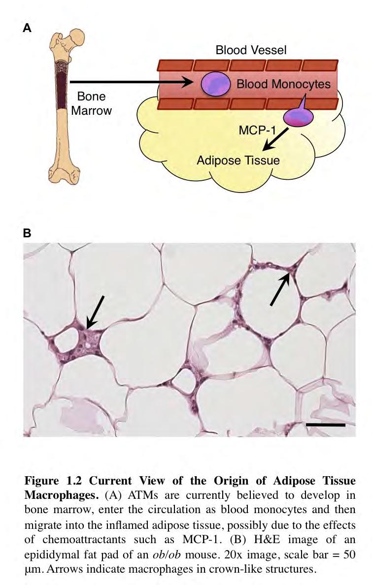
5 iv Abstract Obesity and diabetes are major public health problems facing the world today. Extending our understanding of adipose tissue biology, and how it changes in obesity, will hopefully better equip our society in dealing with the obesity epidemic. Macrophages and other immune cells accumulate in the adipose tissue in obesity and secrete cytokines that can promote insulin resistance. Adipose tissue macrophages (ATMs) are thought to originate from bone marrow-derived monocytes, which infiltrate the tissue from the circulation. Much work has been done to demonstrate that inhibition of monocyte recruitment to the adipose tissue can ameliorate insulin resistance. While monocytes can enter the adipose tissue, we have shown here that local macrophage proliferation may be the predominant mechanism by which macrophages self-renew in the adipose tissue. We demonstrated that two cell proliferation markers, Ki67 and EdU, can be readily detected in macrophages isolated from adipose tissue of both lean and obese mice. These analyses revealed that 2-4% of ATMs in lean and 10-20% of ATMs in obese mice express the proliferation marker Ki67. Importantly, Ki67+ macrophages were identified within the adipose tissue in crown-like structures. Similarly, a 3-hour in vivo pulse with the thymidine analog EdU showed that nearly 5% of macrophages in epididymal adipose tissue of ob/ob mice were in the S-phase of cell division. Interestingly, obesity increased the rate of macrophage proliferation in adipose tissue but did not affect macrophage proliferation in other tissues. We also used clodronate liposomes to deplete circulating monocytes in obese mice. Surprisingly, monocyte depletion for a total of at least 80 hours did not cause a decrease in ATM content in adipose tissue. Prolonged exposure of mice to
6 v EdU in drinking water revealed that approximately half of the ATMs in the epididymal fat pads of ob/ob mice had proliferated locally within 80 hours. Amazingly, these rates were the same with or without monocyte depletion, meaning that the proliferating cells were not freshly recruited monocytes. Overall, these results suggest that local proliferation unexpectedly makes a major contribution to maintaining the large population of macrophages present in the obese adipose tissue in the steady state. This suggests that increased rates of local macrophage proliferation may also be partly responsible for the massive increase in ATM content that occurs in obesity. This information could have implications for future therapeutic strategies in the management of diabetes.
7 vi Table of Contents Signature Page... ii Acknowledgements... iii Abstract... iv Table of Contents... vi List of Figures... ix List of Frequently Used Abbreviations... x CHAPTER I: Introduction... 1 Mechanisms of Insulin Resistance... 4 Adipocyte Dysfunction and Inflammation... 4 The Lipid Spillover Hypothesis... 4 Adipose Tissue Hypoxia... 5 Endoplasmic Reticulum Stress... 6 The progression of insulin resistance to diabetes mellitus... 7 Obesity and Inflammation... 9 Discovery of Inflammation in Obesity... 9 Inflammation and Insulin Resistance... 9 Inflammatory Signals Interfere with Insulin Signaling Inflammatory Signals Also Inhibit Adipocyte Lipogenic Function Immune Cells in Adipose Tissue Adipose Tissue Macrophages Macrophages Accumulate in Adipose Tissue in Obesity Alterations in ATM Content Affect Insulin Sensitivity The Increased Inflammatory Properties of ATMs in Obesity Inhibition of ATM Inflammation Improves Insulin Sensitivity Adipose Tissue Macrophages May Also Play a Protective Role The Origin of ATMs... 21
8 vii Evidence That ATMs are Derived from Monocytes Examples of Local Macrophage Proliferation Selective Ablation of Monocytes Preserves Tissue Macrophages Alveolar Macrophages Langerhans cells Kupffer Cells Kidney Macrophages Atherosclerosis Macrophage Proliferation in Other Tissues Summary and Objectives CHAPTER II: Local Proliferation of Adipose Tissue Macrophages is a Characteristic of Obesity-Associated Inflammation Abstract Introduction Results Adipose tissue macrophages express the cell division marker Ki Macrophages proliferate locally in the adipose tissue Obesity increases macrophage proliferation specifically in adipose tissue Differential effect of obesity on proliferation of diverse immune cell types in adipose tissue Macrophage proliferation decreases with weight loss Discussion Experimental Procedures Acknowledgements Supplementary Figures CHAPTER III: Discussion and Future Directions What is the signal promoting local macrophage proliferation in adipose tissue? What intracellular signaling pathways are required for ATM proliferation?... 91
9 viii What are the relative contributions of local macrophage proliferation and monocyte recruitment to maintaining the total macrophage population in adipose tissue? What is the average macrophage residence time in the adipose tissue and does it change in obesity? What are the metabolic implications of increasing or inhibiting macrophage proliferation specifically in the adipose tissue? References... 95
10 ix List of Figures Figure 1.1 Obesity leads to inflammation and insulin resistance in adipose tissue Figure 1.2 Current view of the origin of adipose tissue macrophages Figure 2.1 Ki67 expression in adipose tissue macrophages of lean and obese mice Figure 2.2 Macrophage cell division in adipose tissue detected by EdU incorporation in lean and obese mice Figure 2.3 Obesity does not stimulate macrophage proliferation in liver or spleen Figure 2.4 Adipose tissue macrophage proliferation occurs independently of blood monocyte recruitment Figure 2.5 Adipose tissue macrophage proliferation decreases with weight loss Supplementary Figure 2.1 Flow cytometry gating scheme for identifying macrophages in adipose tissue SVF Supplementary Figure 2.2 Flow cytometry gating schemes for spleen and liver macrophages and blood monocytes Supplementary Figure 2.3 Adipose tissue macrophage proliferation occurs independently of blood monocyte recruitment Supplementary Figure 2.4 Effect of obesity on proliferation of diverse immune cell types in adipose tissue Supplementary Figure 2.5 Adipose tisse macrophage proliferation decreases with weight loss in mice... 83
11 x List of Frequently Used Abbreviations Abbreviation Term FFA Free fatty acid VEGF Vascular endothelial growth factor HFD High fat diet ND Normal diet ER Endoplasmic reticulum LDL Low-density lipoprotein BMI Body mass index TNFα Tumor necrosis factor alpha IL-1β Interleukin 1 beta IL-4 Interleukin 4 IL-6 Interleukin 6 IRS Insulin receptor substrate JNK c-jun N-terminal kinase NFκB Nuclear factor kappa B IKKβ Inhibitor of NFκB kinase subunit beta PPARγ Peroxisome proliferator-activated receptor-gamma ATM Adipose tissue macrophage SVF Stromal-vascular fraction MCP-1 Monocyte chemoattractant protein 1 CCR2 CC-motif chemokine receptor 2 CLS Crown-like structure WT Wild-type GTT Glucose tolerance test M-CSF Macrophage-colony stimulating factor GM-CSF Granulocyte-macrophage colony stimulating factor EdU 5- ethynyl-2 -deoxyuridine BrdU 5-bromo-2'-deoxyuridine
12 CHAPTER I: Introduction 1
13 2 In order to survive, animals must be able to store excess energy in times of plenty, and release that energy when food sources are absent. The most volume- and weight efficient way to store excess energy is in the form of lipid 1. Lipid is an ideal form in which to store energy due to its high energy density and a hydrophobic nature that reduces obligatory water retention, allowing animals to store as much energy in as little weight as possible. In most animals lipids are stored in distinct adipose depots 2. Over the course of evolutionary history the adipose tissue has developed into an organ that is efficient at the uptake of excess nutrient, the conversion of that nutrient into lipid triglyceride, and the controlled release of lipid during starvation conditions 2,3. For example, adult polar bears must endure seasonal fasting for four or more months each year and lose up to 40% of their total body weight in that time. Depending on the amount of fat stored, nearly all of that weight loss can be due to fat catabolism, while most or all lean body mass is retained 4. Thus, the adipose tissue has the capacity to undergo enormous changes in mass in response to environmental conditions, and its proper function is critical for survival. In humans, adipose tissue is also very efficient at storing excess nutrient and has the capacity to expand tremendously often to a stage where greater than 50% of the total body weight is adipose tissue 5. In the time of our ancestors, this ability to store excess energy was beneficial for survival during times of scarcity, but in the modern civilized world, human exposure to excess nutrient is perpetual. As a result, the global rates of overweight and obesity have reached epidemic levels. Obesity is typically defined as a body mass index (BMI) of over 30 kg/m 2 a calculation based on a person s
14 3 height and weight. According to the World Health Organization more than 1 in 10 of the world s adult population was obese in 2008, and its prevalence is on the rise. Overweight and obesity were the fifth leading cause of global deaths 6. Obesity is associated with many additional health problems, including insulin resistance, cardiovascular disease, fatty liver disease, and cancer, among several others 7,8. Indeed, 44% of the global diabetes burden, 23% of ischemic heart disease, and a large portion of cancer are attributable to obesity 6. In the United States, annual health care expenditures related to overweight and obesity are projected to reach nearly $1 trillion by 2030, which represents nearly 20% of total spending on health care 6,9. In order for our society to deal with this worsening epidemic, we must advance our understanding of adipose tissue physiology and the pathogenesis of diabetes and obesity.
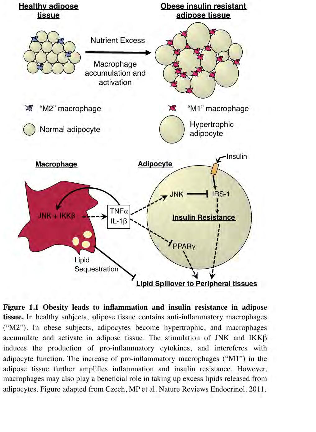
15 4 Mechanisms of Insulin Resistance Adipocyte Dysfunction and Inflammation Obesity and insulin resistance are closely associated with a state of chronic, lowgrade inflammation 3, Adipose tissue plays a central role in the development of insulin resistance. While muscle and liver are the main insulin-sensitive tissues of the body 13, adipose tissue is also highly insulin sensitive and is able to exert profound control over the function of those peripheral tissues 14. Interestingly, the inflammatory response that occurs in obesity seems to originate in and remain within the adipose tissue itself 14. Adipose tissue expansion places tremendous strain on the cells within the tissue, ultimately leading to adipocyte dysfunction, in which adipocytes are unable to synthesize triglyceride and instead release free fatty acid into the circulation 14. Under these conditions, both the hypertrophic adipocytes and resident adipose tissue macrophages produce inflammatory cytokines that act to interfere with insulin signaling in adipocytes and other cells, as well as to recruit additional immune cells to the adipose tissue, which produce more cytokines in a cycle of inflammation 15. These and other topics will be discussed below. The Lipid Spillover Hypothesis One prevailing theory on how obesity leads to insulin resistance is the lipotoxicity theory first proposed in In the early stages of obesity, adipocytes remain capable of storing the increasing load of incoming triglyceride. As adipose tissue expands further, the ability of the adipocytes to store lipid decreases and excess lipid
16 5 spills over into the circulation in a phenomenon known as lipolysis. This increased rate of lipolysis in obesity results in increased plasma levels of free fatty acids (FFAs) 16,17. In obesity, FFAs and lipid metabolites can accumulate in liver and muscle, and cause insulin resistance in these tissues 14,18. Several lines of evidence support this view. Obesity and insulin resistance are strongly associated with high circulating FFA levels 18. Reductions of FFA levels using anti-lipolytic compounds or by enhancement of adipose tissue activity has been shown to improve insulin sensitivity in peripheral tissues 19,20. However, it is clear that insulin resistance can occur in the absence of elevated FFA levels, and that elevated FFA levels do not always cause insulin resistance 21. Nonetheless, there is no doubt that elevated FFA and lipid metabolites in liver and muscle can cause insulin resistance in those tissues, as shown in humans using lipid infusions and in mice using tissue-specific lipoprotein lipase overexpression The importance of the ability of the adipose tissue to sequester lipids away from the rest of the body is highlighted by the hyperlipidemia and insulin resistance observed in lipodystrophic mice 25,26 and humans 27,28 that lack adipose tissue. Lipid sequestration and release represent important ways in which adipose tissue function can affect insulin sensitivity throughout the rest of the body. Adipose Tissue Hypoxia Another consequence of adipose tissue growth is increased oxygen demand. Adipose tissue growth is dependent on new blood vessel formation 29. However, in obesity, the closely coupled processes of adipocyte expansion and neovascularization are
17 6 disrupted, and adipose tissue hypo-perfusion and hypoxia result 30. Improvement of adipose tissue perfusion by overexpression of vascular endothelial growth factor (VEGF) specifically in adipose tissue was shown to improve hypoxia, adipose tissue inflammation and whole body glucose homeostasis 31. Overexpression of VEGF in adipose tissue has also been shown to reduce adiposity 32. A recent study has confirmed that adipose tissue vasculature depletion causes systemic insulin resistance and local adipose tissue inflammation, while overexpression of VEGF decreased adipose tissue inflammation and reduced systemic insulin resistance in a high fat diet (HFD)-fed mouse model 33. These studies again highlight the important role adipose tissue plays in the proper metabolic functioning of the entire body. Adipose tissue hypo-perfusion may be a precipitating factor in the series of events leading to adipocyte dysfunction, adipose tissue inflammation, and total body insulin resistance. Endoplasmic Reticulum Stress As adipose tissue expands, tremendous strain is placed on the adipocytes to accommodate the influx of nutrient, and to process and store that nutrient as triglyceride. The surge in protein and lipid synthesis increases the synthetic demands placed on the endoplasmic reticulum (ER). When protein production exceeds the capacity of the ER to properly fold and modify nascent peptides, a cellular response takes place that slows protein production and increases the expression of protein folding chaperones 34. It has been shown that cellular changes that occur in ER stress can inhibit insulin signaling in adipocytes in mice 35,36 and the ER stress response has been shown to be elevated in the
18 7 adipose tissue of humans with insulin resistance 37. Genetic manipulation of the ER stress pathway can alleviate insulin resistance in mouse models of obesity 36, and treatment of obese mice with small molecules that aid in protein folding also improve glucose homeostasis 38. It should be noted that ER stress can also contribute to insulin resistance in peripheral tissues as well 39,40. ER stress can also initiate inflammatory signaling within the adipocyte 36,41, and may provide a link between excessive adipose tissue growth and the emergence of adipose tissue inflammation. The progression of insulin resistance to diabetes mellitus In the early phases of obesity and in the pre-diabetic state, the insulin resistance of peripheral tissues does not immediately result in hyperglycemia 42. During this time, the pancreatic beta cells are able to maintan normal blood glucose levels despite insulin resistance of the peripheral tissues by increasing their mass, in a process known as beta cell compensation 42. Beta cell mass is increased in obese non-diabetic compared to lean non-diabetic humans 43 and the same is true in rodents 44. During this time, the beta cells also increase their expression of insulin 45. This phase of compensatory hyperinsulinemia is followed by a decline in beta cell function 42. If obesity progresses and insulin resistance worsens, tremendous strain in placed on the beta cells, and the ability of the beta cells to produce the compensatory hyperinsulinemia decreases 46,47. Beta cell mass declines, and it has been shown that beta cells undergo apoptosis 43. The details and mechanisms of beta cell failure in diabetes, which include lipotoxicity from adipose tissue lipid spillover as well as ER stress, are reviewed in Prentki and Nolan, As the beta cells
19 8 progressively fail, the declining insulin levels are no longer able to overcome the insulin resistance of the peripheral tissues, and hyperglycemia ensues 48. Clinically, a fasting plasma glucose level below 100 mg/dl is considered normal, mg/dl is considered impaired fasting glucose or pre-diabetic, and diabetes mellitus is diagnosed when a patient has a fasting plasma glucose of over 126 mg/dl 49,50.
20 9 Obesity and Inflammation Discovery of Inflammation in Obesity Perhaps the most important factor in causing and driving adipose tissue dysfunction is inflammation. Whether induced by ER stress, hypoxia, or other factors, adipose tissue hypertrophy often results in adipocyte dysfunction. Adipocyte dysfunction, in turn, is a critical component in a cycle of inflammation within the adipose tissue 14. Hotamisligil and colleagues and Feinstein and colleagues first discovered in 1993 that the inflammatory cytokine tumor necrosis factor alpha (TNFα) was expressed in adipose tissue and was capable of inducing systemic insulin resistance 10,51. Importantly, TNFα expression was shown to be greatly increased in obese animals compared to lean controls. Since that time, the adipose tissue has been identified as the source of many cytokines and some even consider adipose tissue an endocrine organ 52. Inflammation and Insulin Resistance Since 1993, hundreds of studies have confirmed the link between inflammation and insulin resistance, and numerous excellent reviews on the subject exist 3,12,14,15, but key points will be discussed here. Studies in humans revealed that obese adipose tissue expresses TNFα constitutively, and that its expression in adipose tissue decreases after weight loss 53,54. Similarly, TNFα concentrations were found to be elevated in the plasma of obese patients, and TNFα plasma levels decreased after weight loss 55. Expression of the inflammatory cytokine interleukin 1 beta (IL-1β) and its receptor are increased in the
21 10 visceral adipose tissue of obese humans 56 and IL-1 and TNF have been shown to act synergistically and can increase one another s production 57. Further evidence for obesity being an inflammatory condition came from several studies that confirmed that elevated inflammatory markers in obese patients are predictive of the development of type 2 diabetes Other inflammatory cytokines increased in obesity include interferon gamma, resistin, and interleukin 6 (IL-6) among many others (reviewed in Shoelson et al ). Importantly, TNFα infusion into healthy human subjects can quickly cause insulin resistance 63 and IL-1β has been shown to induce insulin resistance in cultured adipocytes 64. Taken together, these and many other findings support the concept that obesity is an inflammatory condition, and inflammation itself is linked to the development of insulin resistance in humans. Importantly, attenuation of inflammation leads to an improvement in insulin resistance. Neutralization of TNFα using a TNF receptor-igg-fusion protein in a genetically obese rat model caused significant improvement in peripheral glucose uptake 10. Also genetic deletion of TNFα in obese mouse models resulted in improved insulin sensitivity 65. Unfortunately early studies which attempted to neutralize TNFα in human patients did not show a benefit in insulin sensitivity 66. However, neutralization of TNFα has shown improvements in insulin resistance in patients with rheumatoid arthritis and ankylosing spondylitis 67,68. Also, treatment with Interleukin-1-receptor antagonist yielded a mild improved glycaemia in human patients with type 2 diabetes 69 and also in mice 70,71. These studies indicate that inflammatory cytokines play a critical role in the regulation of insulin sensitivity.
22 11 Inflammatory Signals Interfere With Insulin Signaling Inflammatory cytokines interfere with the insulin signaling pathway within insulin target cells 3,12,14. Adipocytes and other insulin target cells express the insulin receptor at their surface. The insulin receptor, when activated by insulin, phosphorylates the insulin receptor substrate (IRS) proteins within the cell on specific tyrosine residues. (For an excellent review on insulin signaling, see Taniguchi, ). These IRS proteins are responsible for conveying the insulin signal to downstream effectors in the cell that produce the cellular response to insulin. Inflammatory signals interfere with the insulin signaling pathway at the level of the IRS proteins 3,12,14. The stress-activated protein kinases c-jun amino-terminal kinase (JNK) and inhibitor of nuclear factor kappa B (NFκB) kinase subunit beta (IKKβ) have proven to be critical molecules that link inflammation and insulin signaling 12,73. Many inflammatory signals, both from extracellular and intracellular sources, produce cellular responses involving these molecules. For example, JNK and IKKβ are both activated by inflammatory cytokine signaling from outside the cell and ER stress signals from within the cell 3,12. TNFα signaling was first shown to inhibit insulin signaling through inhibitory serine phosphorylation of IRS-1 in Since that time it has become clear that TNFα and IL-1β signals activate JNK 75,76 and JNK, in turn, directly inhibits the insulin signaling cascade by inhibiting IRS-1 activity 77. JNK accomplishes this by phosphorylating IRS-1 at Serine 307 which prevents it from being activated by the insulin receptor 77,78. Indeed JNK has been shown to play a central role in the development of obesity and insulin
23 12 resistance in liver, muscle, adipose tissue 79, and more recently in macrophages as well 80, and genetic JNK deficiency protects mice from insulin resistance 79. Similarly, inflammatory cytokines, including TNFα and IL-1β, also activate IKKβ 75,76. Proinflammatory IKKβ activity has also been shown to increase in obesity, and inhibition of IKKβ activity, particularly in macrophages, has been shown to protect mice from the insulin resistance in obesity Unlike JNK however, IKKβ activity inhibits insulin signaling not through direct action on IRS-1, but by activating the transcription of a long list of inflammatory genes 84,85, which subsequently feed back on the cell and inhibit the response to insulin. This is an important way in which inflammatory signaling perpetuates a cycle of inflammation and insulin resistance. Inflammatory Signals Also Inhibit Adipocyte Lipogenic Function Inflammation also plays a role in generating the adipose tissue dysfunction and lipid spillover discussed above. In obesity, adipocytes are less able to sequester lipid and store it as triglyceride 14. This may be partly due to the fact that enzymes responsible for de novo synthesis of fatty acids and the esterification of fatty acids intro triglyceride are significantly reduced in the adipose tissue of obese humans 86 as well as in obese mouse models 87,88. The master regulator of these adipogenic genes, and a necessary factor in the maintenance of mature adipocyte function, is peroxisome proliferator-activated receptor-gamma (PPARγ) 89,90. Several studies have shown that TNFα and IL-1 are able to downregulate PPARγ action in adipocytes Thus, inflammatory cytokines not only inhibit insulin signaling in adipocytes, they also may contribute to the inhibition of
24 13 adipocyte function and the resultant increase in lipid release from adipocytes in the obese state. Immune Cells in Adipose Tissue Numerous immune cell types populate the adipose tissue, and the quantities and proportions of those cell types change in obesity, contributing to inflammatory cytokine production. Macrophages are the largest population of immune cell type in the adipose tissue and produce the majority of the inflammatory cytokines that are increased in obesity 11, and will be discussed extensively below. Another large population of immune cells in adipose tissue is eosinophils, which are very numerous in lean adipose tissue, and decrease their presence in obesity 95. Eosinophils have been shown to secrete interleukin 4 (IL-4) in the adipose tissue, which modulates macrophage function and adipose tissue insulin sensitivity in a beneficial way 95. Similarly, regulatory T cell number in lean adipose tissue is very high, and decreases in obesity 96. Concurrent with the decrease in regulatory T cells, an increase in CD4 T H 1 cells and CD8 T cells occurs in the adipose tissue in obesity 97,98. Importantly, the influx of these T cells has been shown to precede macrophage accumulation in the adipose tissue, and depletion of CD8 T cells decreases macrophage content in obese adipose tissue, suggesting that T cells play a role in controlling adipose tissue macrophage content 97. The various subsets of natural killer T cells also appear to be involved in adipose tissue inflammation and metabolism, although there are conflicting reports as to what that role may be B cells have also been shown to increase in their presence in adipose tissue in obesity and produce pathogenic
25 14 antibodies in the adipose tissue 98. Interestingly, mast cells, which are normally associated with allergic responses, increase in the adipose tissue in obesity, and deficiency or stabilization of mast cells has been shown to improve insulin sensitivity in obese mice 104. Thus, the adipose tissue is host to a variety of immune cells whose populations change with obesity and immune cell recruitment contributes to adipose tissue inflammation and obesity.
26 15 Adipose Tissue Macrophages Macrophages Accumulate in Adipose Tissue in Obesity Analysis of gene expression in cells isolated from obese adipose tissue has revealed that adipose tissue macrophages (ATMs) are the predominant source of the inflammatory cytokines elevated in obesity 11. Macrophages accumulate in the adipose tissue in huge numbers in the obese state, a phenomenon first described by Weisberg et. al. and Xu et. al. in ,105. In a lean individual, of all non-adipocyte cell types in the adipose tissue (called the stromal-vascular fraction, or SVF) only 5-10% may consist of macrophages, while macrophages can be as high as 40% of all stromal cells in obese human adipose tissue, and as high as 50% in morbidly obese mouse adipose tissue 11. Importantly, ATM content is closely correlated with adipocyte size and body weight in mice and in humans 11, Furthermore, macrophage accumulation in adipose tissue is temporally associated with the onset of insulin resistance 105. Also, it is very interesting to note that in humans, not all obese individuals have insulin resistance, and inflammatory gene expression and ATM content in visceral adipose tissue has been shown to be a far better predictor of insulin resistance than simple measures of adiposity 109. Taken together, these studies provide good evidence that under physiological conditions, macrophage content within adipose tissue is increased in obesity and is linked to the development of insulin resistance. Alterations in Adipose Tissue Macrophage Content Affect Insulin Sensitivity
27 16 Since the initial discovery of macrophages in adipose tissue, many attempts have been made to either decrease or increase ATM content, in an attempt to determine if doing so would alter insulin sensitivity. Several groups have succeeded in demonstrating improved insulin sensitivity associated with a decrease in ATM content. For example, treatment of obese mice with the insulin-sensitizing drug rosiglitazone has been shown to decrease ATM content 105. Surgery-induced weight loss in morbidly obese human subjects has been shown to be associated with decreased macrophage content in white adipose tissue and decreased inflammatory gene expression 110. Diet and exercise-induced weight loss has also been found to decrease ATM content 111. Diptheria toxin-induced cell death of macrophages within the adipose tissue in obese mice was associated with improved insulin sensitivity as well 112. Depletion of ATMs by intraperitoneal clodronate liposome injection also improved insulin sensitivity in obese mouse models 113. Inhibition of monocyte recruitment to the adipose tissue has, in certain cases, been found to produce a modest decrease in ATM content and was associated with improved insulin sensitivity in those cases 108,114,115. This topic will be discussed in further detail below. Conversely, adipose tissue-specific overexpression of monocyte chemoattractant protein 1 (MCP-1) increased ATM content and decreased insulin sensitivity in both lean and obese mouse models 115,116. Taken together, these studies support the concept that ATM content is closely correlated with insulin resistance. The Increased Inflammatory Properties of ATMs in Obesity. Not only do macrophage numbers increase in adipose tissue in obesity, but their
28 17 activation state is also polarized towards a pro-inflammatory phenotype 117,118. Macrophages are a very versatile cell type, able to attain a broad range of functional states based on the cues present in their environment 119. In lean mice, ATMs are generally characterized by an anti-inflammatory M2-like phenotype, as determined by their surface marker and gene expression profiles 117. Anti-inflammatory macrophages are characterized by anti-inflammatory cytokines rather than pro-inflammatory cytokines, and are thought to prevent inflammation and promote tissue repair 120. Anti-inflammatory M2-like macrophages are believed to be generated by exposure to anti-inflammatory signals such as IL-4, IL-10, IL-13, and glucocorticoids, whereas pro-inflammatory M1- like macrophage phenotype is believed to be a response to exposure to inflammatory cytokines such as interferon gamma and TNF-α 120. In obesity, a significant fraction of ATMs lose that anti-inflammatory phenotype and become more pro-inflammatory, or M1-like, expressing inflammatory cytokines that inhibit the ability of the surrounding adipocytes to respond to insulin 118. While macrophage sub-populations in the adipose tissue may have overlapping marker expression profiles, it is generally thought that CD11c expression is characteristic of pro-inflammatory macrophage subtypes 112,118,121. Interestingly, preventing ATMs from attaining an anti-inflammatory phenotype in obese mouse adipose tissue predisposes mice to diet-induced obesity and insulin resistance 122. Furthermore, a recent publication reported that systemic helminth infection, a canonical M2-polarizing condition, was associated with a shift in ATM phenotype in obese mice to an entirely anti-inflammatory state, and insulin sensitivity was normalized in these mice 95. Taken together, these reports provide strong evidence that not only increased
29 18 macrophage number, but also the increased inflammatory ATM phenotype that occurs in obesity plays an important role in the development of insulin resistance. Inhibition of Adipose Tissue Macrophage Inflammation Improves Insulin Sensitivity In addition to absolute decreases in macrophage number in adipose tissue, inhibition of inflammatory signaling within macrophages can also improve insulin resistance. Genetic deletion of JNK specifically in hematopoietic cells, accomplished by bone marrow transplant, was sufficient to protect mice from diet-induced insulin resistance even though the mice became just as obese as control mice 123. This study was confirmed and extended by a recent publication showing deletion of both JNK1 and JNK2 specifically in macrophages protects mice from obesity-induced insulin resistance 80. It is important to note that both of these studies also observed a decreased macrophage content in the adipose tissue in the JNK-deficient macrophage condition. Similarly to macrophage-specific JNK deletion, macrophage-specific IKKβ deletion also protects mice from diet-induced adipose tissue inflammation and insulin resistance without protecting from diet-induced weight gain 83. While these studies emphasize the role of inflammatory signaling within macrophages in obesity, they have all been limited to studying macrophages throughout the body, while the question of the role of macrophages specifically within adipose tissue has been unresolved. Our group recently developed sirna delivery technology able to specifically target macrophages within obese adipose tissue 124. Using this technology, we were able to silence production of two inflammatory cytokines, TNFα and osteopontin, specifically in adipose tissue
30 19 macrophages, and show improved whole body glucose tolerance. This study confirms the role of inflammatory signaling specifically in macrophages, specifically in adipose tissue, as being of central importance in the development of insulin resistance in obesity. Adipose Tissue Macrophages May Also Play a Protective Role Despite the extensive evidence that ATMs are inflammatory and act to exacerbate insulin resistance, there is also evidence for a protective role of macrophages. Kosteli et. al. have shown that acute fasting-induced lipolysis or drug-induced lipolysis is associated with rapid recruitment of macrophages to the adipose tissue 125. Furthermore, they showed that depletion of macrophages was associated with increased lipid release from adipose tissue during fasting conditions. These findings were confirmed by another group that directly observed that ATMs accumulate lipid in obese mice and become triglyceride foam cells even in the basal obese state 126. Additional evidence supporting this hypothesis is that macrophages in the obese adipose tissue localize around dead and dying adipocytes, forming structures termed crown-like structures (CLS) 106,107. When examined by electron microscopy, the macrophages in these structures were found to contain lipidfilled phagolysosomes, suggesting they sequester the lipid being released from the dying adipocytes and prevent it from entering the circulation 106. These reports support a role for ATMs in taking up excess lipid in order to protect the rest of the body from the toxic effects of free fatty acids being released from adipocytes.
31 20
32 21 The Origin of ATMs Evidence That ATMs Are Derived From Monocytes Much effort has been focused on determining the factors that attract macrophages to the adipose tissue in obesity. It is generally believed that myeloid precursors develop in the bone marrow, enter the circulation as blood monocytes and subsequently migrate into tissues where they terminally differentiate into macrophages 127. Consistent with this view, early studies using total body lethal irradiation followed by bone marrow transplant concluded that ATMs were mostly bone-marrow derived 11. Subsequent studies aimed to reduce monocyte recruitment to the adipose tissue in obesity. Monocyte chemoattractant protein 1 (MCP-1) was identified in several studies as one of the inflammatory cytokines whose expression increases in obese adipose tissue 11,128,129. Whole body genetic deletion of MCP-1 was found to produce a modest but significant decrease in ATM content in mice fed a high-fat diet for weeks 115. Similarly, whole body genetic deletion of the MCP-1 receptor, CCR2, produced a modest but significant reduction in ATM content in mice fed a high fat diet 108. This study also employed a short term drug-induced inhibition of CCR2 and observed a small but significant difference in ATM content in obese mice after 17 days of daily injection with a small molecule inhibitor of CCR It is important to note that three other studies using MCP-1 and CCR2 knockout animal models and employing nearly identical experimental conditions failed to demonstrate any difference in ATM content in obesity Inhibition of osteopontin, an inflammatory cytokine and extracellular matrix protein
33 22 elevated in obesity, by either genetic deletion or injection of neutralizing antibody, was also associated with decreased macrophage content in adipose tissue in obese mice and an improvement in insulin sensitivity 133,134. Another study employed genetic deletion of alpha-4 integrin, which is required for macrophage adhesion to endothelial cells during transmigration into tissues 114. Myeloid-specific absence of this adhesion marker was also associated with a small but significant decrease in ATM content in diet-induced obese mouse models 114. Likewise, a myeloid-specific knockout of Cbl-associated protein, a protein involved in macrophage migration, produced a decreased ATM content in obese adipose tissue 135. It is critical to note that both of these studies employed total body lethal irradiation followed by bone marrow transplant in order to generate the myeloid-specific knockout models, and a recent study has shown that irradiation dramatically reduces the lifespan of resident macrophages 136. On the other hand, as with most any tissue, monocytes can be induced to enter adipose tissue. As described above, overexpression of MCP-1 specifically in the adipose tissue was associated with an increased macrophage content in the adipose tissue, presumably due to increased monocyte recruitment from the blood 115,116. Also described above, induction of lipolysis has been shown to attract monocytes to adipose tissue in lean mice 125. However, a recent paper showed that the cells which infiltrate that adipose tissue during lipolysis may actually be neutrophils 137. It is interesting to note that, unlike in lean animals, induction of lipolysis in adipose tissue of obese mice by fasting did not stimulate the recruitment of monocytes to adipose tissue 138. However, a recent paper demonstrated that monocytes labeled with dye and adoptively transferred into obese
34 23 recipient mice do appear within the adipose tissue within 2-3 days following transfer, and more of the dye-labeled monocytes are recruited to adipose tissue in obese mice as compared to lean mice 139. Importantly, they demonstrated that the recruitment of labeled monocytes to adipose tissue was partially dependent on MCP-1 and CCR Overall, there is substantial evidence that monocytes can be induced to enter adipose tissue by various factors, and that the absence of certain chemokines and adhesion and migrationrelated molecules can attenuate the increase of macrophages in adipose tissue that occurs in obesity. However, the chemokine studies have reported varied results, and the adhesion and migration molecule knockout studies may also be interpreted as attenuation of the ability of knockout monocytes to repopulate of the adipose tissue following the destruction of resident adipose tissue macrophages by total body lethal irradiation. Therefore, we hypothesized that, in the absence of total body lethal irradiation, ATMs arise not from circulating monocytes, but from local proliferation in situ.
35 24
36 25 Examples of Local Macrophage Proliferation Selective Ablation of Monocytes Preserves Tissue Macrophages Monocytes are developed from myeloid precursor cells in the bone marrow, and mature monocytes enter the bloodstream and enter the tissues where they terminally differentiate into macrophages 127,140. It is generally thought that terminally differentiated cells are not capable of re-entering the cell cycle to proliferate. However, macrophages have long been known to be capable of proliferating in situ and those examples will be reviewed below. The best early evidence that tissue macrophages could proliferate locally came from experiments that involved using radioactive strontium to deplete bone marrow cells 141. Strontium is in the same chemical group as calcium, and is stably incorporated into hydroxyapatite crystals in bone in a similar fashion to calcium 142. Radioactive strontium-89 is a beta particle emitter with a half life of over 50 days 142. Therefore, simply injecting mice with radioactive strontium provided an easy, targeted method to deplete bone marrow cells without affecting proliferating cells in other tissues 141. This represents a key difference from modern day bone marrow depletion studies that employ total-body lethal irradiation, which can destroy proliferating cells not only in the bone marrow, but in many tissues of the body 136. In the strontium study, monocytes were depleted to undetectable levels for the duration of the day study periods 141. Despite this profound and prolonged monocytopenia, there were no changes in total number of resident macrophages in the lung or peritoneal cavity 141. When assayed for proliferation markers, 3% of peritoneal and 1.5% of alveolar macrophages were found to be actively
37 26 synthesizing DNA during a one hour pulse of radioactive thymidine 141. These results suggested that resident macrophage population renewal under normal circumstances does not depend heavily on influx of circulating monocytes. In the sections below, I will review the recent studies focused on resident macrophage proliferation which have shown that, in several tissues, local proliferation plays a predominant role in maintaining tissue macrophage populations, with recruitment of monocytes playing little or no role. Alveolar Macrophages As described above, the 1982 radioactive strontium bone marrow depletion studies first identified that alveolar macrophages could proliferate locally. Prior to that time alveolar macrophages were believed to be derived from circulating monocytes in both mouse 143,144 and human 145,146. However, these early studies employed total body irradiation, which inhibits the proliferation of host cells. Subsequent bone marrow transplant studies elucidated that a more gentle, fractionated irradiation regimen prevented host macrophages from destruction, and resulted in alveolar macrophages remaining of host origin for at least 45 weeks following bone marrow transplant 147. Protection of the lungs from lethal irradiation with a lead shield prior to bone marrow transplant resulted in alveolar macrophages remaining of host origin for the lifetime of the mouse, despite reconstitution of resident macrophage populations with donor cells in tissues that received irradiation 136. Also, in parabiotic mice which are surgically joined to share a circulatory system, alveolar macrophages were shown to self-renew with little involvement of incoming monocytes 148. In humans who received allogeneic bone marrow
38 27 transplant and whose lungs were repopulated with macrophages of donor origin, donor macrophages recruited to lungs also proliferate in situ, resulting in a three-fold increase in alveolar macrophage content seven weeks after transplant 149. In addition to proliferating locally to maintain steady-state macrophage density, alveolar macrophages also proliferate in response to inflammatory insults. Instillation of carbon particles into the lungs of mice was found to stimulate local alveolar macrophage proliferation, which accounted for an increase in macrophage content even in mice depleted of monocytes by strontium Similarly, a recent study demonstrated that the massive increase in macrophage content in the lung that occurs during helminth infection can arise even in the complete absence of circulating blood monocytes 151. They also showed that this surprising result was due entirely to the local proliferation of macrophages within the lung 151. Thus, in the lung, local macrophage proliferation is well documented and may be the predominant mechanism for macrophage population renewal in the steady state, as well as the mechanism by which macrophage populations increase during inflammatory insult. Langerhans Cells One of the phagocytic, antigen-presenting phagocytic cell type in the epidermis of the skin is the Langerhans cell. These cells were originally thought to originate from bone marrow-derived cells 152 but in 2002 they were shown to be self-renewing within the skin 153. Using both bone marrow transplant and surgically joined parabiotic mouse models, Langerhans cells remained of host origin for the duration of the studies 18 and
39 28 6 months, respectively 153. Furthermore, this study demonstrated that Langerhans cells actively synthesize DNA in the skin, as up to 30% of the Langerhans cells incorporated BrdU after a 30 day BrdU exposure 153. Only when resident Langerhans cells were depleted with UV irradiation or toxic chemicals did replacement of resident Langerhans cells with circulating cells of donor origin occur 153. However, a follow-up study showed that even during the repopulation phenomenon that occurs in damaged skin, circulating monocytes enter the inflamed skin and undergo several rounds of proliferation, forming clusters of Langerhans cells 154. Evidence in humans also supports a role for Langerhans cell self-renewal in the skin, as a case study of a human hand transplant showed that skin Langerhans cells remained of donor origin even 4.5 years after transplantation 155. Taken together, these results indicate that under steady state conditions, skin Langerhans cells replenish themselves almost entirely through local proliferation in situ, and monocyte recruitment only occurs in response to inflammatory insult. Furthermore, even under inflammatory conditions, local proliferation of the freshly recruited monocytes plays an important role in populating the skin with Langerhans cells. Kupffer Cells Liver Kupffer cells also are widely believed to be derived from monocytes due to a study which demonstrated that Kupffer cells can be derived from blood monocytes following total body lethal irradiation 156. However, several early publications implicated a possible role for local proliferation in the maintenance of the Kupffer cell population in mice Kupffer cells were subsequently shown to indeed have proliferative capacity
40 29 but that, under steady state conditions, monocyte recruitment was the predominant force in maintaining the Kupffer cell population 161. On the contrary, one study came to the conclusion that under physiological steady state conditions, Kupffer cells are long lived and mostly self-renewing 162. Interestingly, several groups have reported the Kupffer cells proliferate in response to inflammatory insult. Intravenous injection of zymosan has been found to stimulate robust local Kupffer cell proliferation within the liver 161. This result was later confirmed and expanded to show that local proliferation is indeed the predominant force behind Kupffer cell hyperplasia in response to zyomsan insult 163,164. Similarly, liver injury induced by carbon tetrachloride is accompanied by an expansion in the liver macrophage population, which has been shown to be due to both local proliferation and monocyte recruitment 165. Further studies will be required to clarify these seemingly conflicting results, but it is clear that to some degree or other, liver macrophages are capable of proliferating in situ under both steady state and inflammatory conditions. Kidney Macrophages accumulate in the kidney in various disease states, and there they contribute to inflammatory processes that exacerbate kidney injury 166. A surprising number of studies have reported local macrophage proliferation in kidney inflammation. For example, in rat anti-glomerular basement membrane glomerulonephropathy, local macrophage proliferation has proven to be the major mechanism by which macrophage numbers increase in the glomerulus 167. Indeed, measurements of proliferation markers in
41 30 the macrophages in this and another related study were astoundingly high, with over 60% of all macrophages expressing the proliferation marker PCNA at any given time, and the 30-40% of those cells staining positive for BrdU incorporation after a 3-hour BrdU pulse 167,168. High levels of local macrophage proliferation have also been documented in glomerulonephritis in human patients 169. Importantly, proliferating macrophages were found to be concentrated in regions of severe tissue damage, suggesting that proliferating macs are an important mediator of kidney injury 169. Local macrophage proliferation has been noted in non-immune-mediated kidney injury models as well, and the association of macrophage proliferation with tissue injury and fibrosis was clear in these models also 170. Importantly, local macrophage proliferation is also the mechanism by which macrophages accumulate in the kidney in diabetic nephropathy in obese mice 171. Furthermore, macrophage accumulation associated with organ rejection in allogeneic kidney transplant studies has been shown to be due to initial recruitment of circulating monocytes which then undergo local proliferation in situ to increase the macrophage content 172. Taken together, there is substantial evidence that local macrophage proliferation in the kidney represents an important mechanism by which macrophage populations increase in kidney diseases. Atherosclerosis Macrophages are an integral cell type in the formation of atherosclerotic plaques 173, and an impressive number of studies support a role for local proliferation in the accumulation of macrophages in the growing plaque. This body of work has even
42 31 warranted recent reviews on the topic of macrophage proliferation in atherosclerosis 174,175. Proliferating macrophages within atherosclerotic plaques were first observed in rabbit models, using microscopic examination of plaques following pulses of radiolabeled thymidine analog or growth-arresting colchicine treatment 176,177. Around the same time, expression of genes associated with cell proliferation were found to be increased in human atherosclerotic plaques, in which macrophages were suggested to be the predominant proliferating cell type 178,179. Myeloid-specific deletions of the tumorsuppressor genes p27 and retinoblastoma caused increased intimal macrophage proliferation and accelerated atherosclerosis development in apolipoprotein E knockout animals 180,181. Similarly, mutation of the cyclin-dependent kinase inhibitor 2A gene in myeloid cells in low-density-lipoprotein (LDL) receptor knockout animals caused increased macrophage proliferation and exacerbated atherosclerosis 182. While several other reports using the tumor suppressor gene p53 yielded both positive and negative 186,187 results in regards to macrophage proliferation, it is clear that macrophage proliferation in situ is involved in plaque formation and the maintenance of macrophage populations in atherosclerotic plaques. Macrophage Proliferation in Other tissues Macrophages in several other tissues have been documented to proliferate locally to maintain macrophage populations in the steady state. For example, the spleen is one of the largest repositories of macrophages in the body, and local proliferation may account for up to half of the macrophage content in the spleen 188,189. Microglia, a subtype of
43 32 macrophages in the central nervous system, have been shown by several groups to proliferate locally Peritoneal macrophages have also been shown to have proliferative capacity in vitro as well as in vivo Local macrophage proliferation has been documented in various other disease models including arthritis 196,197, and finally in macrophages associated with tumors Indeed, locally proliferating macrophages have recently been identified in high-grade breast cancers and indicate a poor prognosis 202,203.
44 33 Summary and Objectives The adipose tissues in obese, insulin resistant mice and humans accumulate inflammatory cells, including macrophages, which secrete cytokines and other factors that can modulate adipocyte function. These adipose macrophages are thought to originate from bone marrow-derived monocytes, which infiltrate the tissue from the circulation. However, genetic deletion of molecules thought to be important in the recruitment of monocytes to adipose tissue produced either a modest decrease in adipose tissue macrophage content compared to controls, or no decrease in adipose tissue macrophage content at all. Despite the fact that macrophages are generally thought to be a terminally differentiated cell type, there are many examples of macrophage proliferation locally within tissues. Local proliferation can be the predominant way in which tissue macrophages maintain normal density under basal conditions, and local proliferation can account for the massive increases in macrophage tissue density that occur in certain inflammatory conditions. However, the role of local macrophage proliferation in adipose tissue inflammation in the obese state has not been thoroughly investigated. Therefore, we sought to elucidate the role of local macrophage proliferation in maintaining macrophage populations in the adipose tissue of lean and obese mice. Using both genetic and diet-induced obese mouse models, we examined macrophage populations for proliferative markers in the adipose tissue under steady-state lean and obese conditions, as well as during weight loss. To clarify the origin of adipose tissue macrophages, we studied adipose tissue macrophage population dynamics in the setting
45 34 of monocyte depletion. Finally, we made several initial efforts into elucidating the factors that may be stimulating local macrophage proliferation in the adipose tissue.
46 35 Chapter II: Local proliferation of adipose tissue macrophages in obesity-associated inflammation
47 36 Abstract The adipose tissues in obese, insulin resistant mice and humans accumulate inflammatory cells, including macrophages, which secrete cytokines and other factors that can modulate adipocyte function. These adipose macrophages are thought to originate from bone marrow-derived monocytes, which infiltrate the tissue from the circulation. Here we show that a significant fraction of macrophages unexpectedly undergo cell division locally within adipose tissue, as detected by Ki67 expression and 5- ethynyl-2 -deoxyuridine incorporation. Adipose macrophages, but not those in other tissues, displayed dramatically increased proliferation in both genetic and diet-induced mouse models of obesity. Importantly, depletion of blood monocytes over several days had no impact on adipose tissue macrophage content. Conversely, weight loss and lipolysis specifically inhibited macrophage proliferation in the adipose tissue of obese animals. These results reveal that proliferation in situ is an important process by which macrophages accumulate in inflamed adipose tissue in obesity, and could have therapeutic implications for strategies to improve insulin sensitivity.
48 37 Introduction The prevalence of insulin resistance and type 2 diabetes mellitus (T2D) has been rapidly rising worldwide over the past three decades, particularly in developing countries 204. Obesity can induce an insulin-resistant state in adipose tissue, liver, and muscle and is a strong risk factor for the development of T2D 14,15, In obese patients, the inability to appropriately expand subcutaneous adipose tissue leads to ectopic lipid deposition in non-adipose tissues (e.g. liver and muscle) and may be an underlying cause of insulin resistance Immune cells, including macrophages, accumulate in massive numbers in the adipose tissues of obese mice and human subjects 11, It is increasingly appreciated that accumulation of macrophages in adipose tissue correlates with a chronic inflammatory state that ultimately impairs adipocyte function and may contribute to the development of insulin resistance 3,11,15,81,217,218. Consistent with this concept, macrophage numbers in adipose tissue correlate with systemic insulin resistance even in obese human subjects that are matched for BMI 109, Macrophages and other immune cells in adipose tissues also have beneficial effects of removing cellular debris 107 and controlling lipolytic responses in adipocytes 125. Macrophages infiltrating into adipose tissue have been reported to be mostly derived from the bone marrow and there is evidence that monocyte recruitment to the adipose tissue increases in obesity 11,139. Adipocytes have been shown to produce a wide range of factors, such as the chemokine monocyte chemoattractant protein 1 (MCP-1) that may recruit monocytes to adipose tissue. Both the production of MCP-1 and the expression of
49 38 its receptor CC-motif chemokine receptor-2 (CCR2) are enhanced by obesity 116,130. However, genetic deletion of MCP-1 and CCR2 in mice produced either a modest decrease in adipose tissue macrophage (ATM) content compared to controls 108,115, or no effect that could be detected 131,132. Perhaps this is due to other pathways of macrophage migration that operate independently of the MCP-1 pathway. However, these results might also be explained by the possibility that resident macrophages have a prolonged lifespan or proliferate in situ in the adipose tissue. Although terminal differentiation of mammalian cells typically results in reduced proliferative capacity, there is evidence that cells of the mononuclear phagocyte system, including macrophages, can self-replicate in the periphery both during development and in the adult Indeed, local macrophage proliferation has been demonstrated in kidney, atherosclerotic plaques, skin and lung in animals and humans 151,169,176,182,225. The present study was designed to test the hypothesis that significant macrophage cell division also occurs within adipose tissue in mice, since no data are available on this important question. Here we show that a fraction of ATMs in obese mice display the proliferation marker Ki67, a protein expressed during all active phases of the cell cycle but not in non-dividing cells 226. Genetic and diet-induced obesity greatly increased macrophage proliferation in adipose tissue, but not in other tissues studied. Furthermore, approximately 5% of macrophages within epididymal adipose tissue of obese mice were found to be in S-phase during a three-hour pulse of the more stringent cell division marker, 5-ethynyl-2 -deoxyuridine (EdU), and nearly 50% of all macrophages had
50 39 proliferated over the course of an 80 hour EdU exposure, even when circulating monocytes had been depleted. These results suggest the unexpected concept that local macrophage proliferation contributes to the overall macrophage accumulation and adipose tissue inflammation in the insulin resistant state.
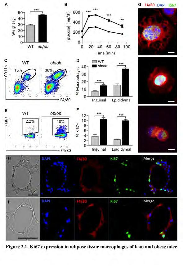
51 40 Results Adipose tissue macrophages express the cell division marker Ki67. To study macrophage accumulation in adipose tissue of obese mice, we used 8 to 12- week old genetically obese (ob/ob) male mice and their lean control wild-type (WT) littermates. To confirm that the ob/ob mice were insulin resistant and obese, we measured body weights and performed glucose tolerance tests (GTT) (Figure 2.1 A-B). The average body weight of the ob/ob mice was approximately 15 g, or 50% higher than that of WT mice, and this was associated with significantly decreased glucose tolerance (Figure 2.1 A-B). We isolated the stromal-vascular fraction (SVF) from subcutaneous (inguinal) and visceral (epididymal) fat pads, which contains all cells of the adipose tissue except adipocytes, and analyzed it using flow cytometry. The SVF was stained with antibodies against two macrophage markers, F4/80 and CD11b. However, these proteins are also expressed in other immune cells, such as eosinophils and neutrophils 95,227. Therefore, we also stained the SVF with antibodies against the eosinophil marker, Siglec-f, and a neutrophil marker, Gr-1. The macrophage population in the adipose tissue was defined as F4/80 + /CD11b + /Siglec-f /Gr-1 (for the complete gating scheme see Supplementary Figure 2.1). Consistent with published studies 11,105, we observed a significant 2-3 fold increase in macrophage content in the adipose tissue of ob/ob compared to WT mice in both subcutaneous and visceral fat pads (Figure 2.1 C- D). To test whether local macrophage proliferation also contributes to macrophage accumulation in the adipose tissue, we stained the SVF cells with an antibody against the
52 41 proliferation marker Ki67, which is a protein expressed during all active phases of the cell cycle 226. Surprisingly, Ki67 signal was detected in approximately 2.3% of epididymal ATMs of lean mice, and in 10% of ATMs of ob/ob mice (Figure 2.1 E-F). Similar percentages of macrophages from subcutaneous adipose tissue were Ki67 + as well (Figure 1F). We then performed immunofluorescence microscopy on plated SVF cells from epididymal fat pads of ob/ob mice. Figure 2.1 also shows images of macrophages (F4/80, red) expressing Ki67 (green) in their nuclei (DAPI, blue) (Figure 2.1 G). Microscopy revealed a similar percentage (approximately 10%) of Ki67 + macrophages as seen with flow cytometry (data not shown). High magnification images reveal macrophages in various stages of division (Figure 2.1 G). Furthermore, Ki67 was often found in multinucleated macrophages (Figure 2.1 G). Next, AT sections from ob/ob mice were stained with antibodies against F4/80 (red) and Ki67 (green) and analyzed by microscopy (Figure 2.1 H-I). Figure 2.1 G and H show Ki67 in F4/80 positive cells in a region of the epididymal AT rich in macrophages termed crown-like structures (CLS). Taken together, these results show by both flow cytometry and microscopic analysis that macrophages express the proliferation marker Ki67 in the AT and to a higher degree in response to obesity in mice caused by either genetic mutation or a diet high in fat.
53 42
54 43 Figure 2.1. Ki67 expression in adipose tissue macrophages of lean and obese mice. (A) Body weights and (B) GTT of WT ( ) and ob/ob ( ) mice, n=5. (C-F) SVF from visceral and subcutaneous adipose tissue of WT and ob/ob mice was isolated and analyzed by flow cytometry. (C) Representative flow cytometry dot plots of SVF from epididymal adipose tissue. (D) Percentage of macrophages in SVF. (E) Representative flow cytometry dot plots of macrophages stained with Ki67. (F) Percentage of macrophages expressing Ki67. n=30-31 from 6 independent experiments for epididymal and n=10 from 2 independent experiments for inguinal fat pads. All graphs are expressed as mean ± s.e.m. Statistical significance was determined by Student s t-test. For GTT, statistical significance was determined by ANOVA and Tukey post test. **p<0.01; ***p< (G-I) Microscopy of plated SVF (G) and adipose tissue sections (H-I) stained with antibodies against F4/80 (red) and Ki67 (green). Nuclei were stained with DAPI (Blue). (G) 63x magnification image of macrophages stained with Ki67 (arrows). Scale bar = 5 μm. (H-I) 20x magnification images of crown-like structures from ob/ob mice. Scale bar = 40 μm.
55 44 Macrophages proliferate locally in adipose tissue. To test whether macrophages undergo cell division within adipose tissue, we injected WT and ob/ob mice with the nucleoside analog to thymidine, EdU. While Ki67 is expressed during all active phases of the cell cycle, EdU is only incorporated into DNA during the S-phase. Three hours following EdU injection in mice, the SVF cells of epididymal fat pads were isolated and analyzed by flow cytometry. Approximately 1% of ATMs in lean mice and about 5% of the macrophages in the obese mice had gone through S-phase as measured during a 3-hour pulse of EdU (Figure 2.2 A-B). Mice were also injected with another nucleoside analog of thymidine, 5-bromo-2'-deoxyuridine (BrdU) and immunohistochemistry on epididymal adipose tissue of ob/ob mice was performed (Figure 2.2 C). As shown in Figure 2.2C, ob/ob adipose tissue contains regions known to be enriched with macrophages called crown-like structures (CLS; denoted by arrows). BrdU staining (denoted by arrowheads) was observed in these structures (Figure 2.2 C). To test whether EdU + macrophages in adipose tissue derived from EdU + macrophages that infiltrated from the blood rather than cells propagating within the tissue, we also examined EdU incorporation in blood monocytes (Figure 2.2 D-E). In these same mice, blood was collected via cardiac puncture and analyzed by flow cytometry. Monocytes were positively identified by expression of CD11b, F4/80, and Ly6C (for complete blood monocyte gating scheme see Supplementary Figure 2.2 A). We failed to detect any EdU incorporation in blood monocytes within the 3-hour pulse, suggesting that EdU +
56 45 macrophages seen in the adipose tissue are not recently recruited blood monocytes (Figure 2.2 D-E). To further ensure that the proliferating macrophages in the adipose tissue were not EdU + blood monocytes, we studied the capacity of ATMs to proliferate ex vivo. SVF cells isolated from epididymal adipose tissue of lean and obese mice were plated and treated with EdU for three hours. The cells were then stained and analyzed by flow cytometry. Approximately 0.3% of the ATMs from lean mice were EdU +, while greater than 2% were positive in macrophages from obese mice (Figure 2.2 F-G). These results suggest that the ATMs have the inherent capacity to proliferate ex vivo, independently of blood monocyte recruitment. Taken together, these results suggest that proliferating macrophages present in adipose tissue are not recently recruited blood monocytes that were already dividing.
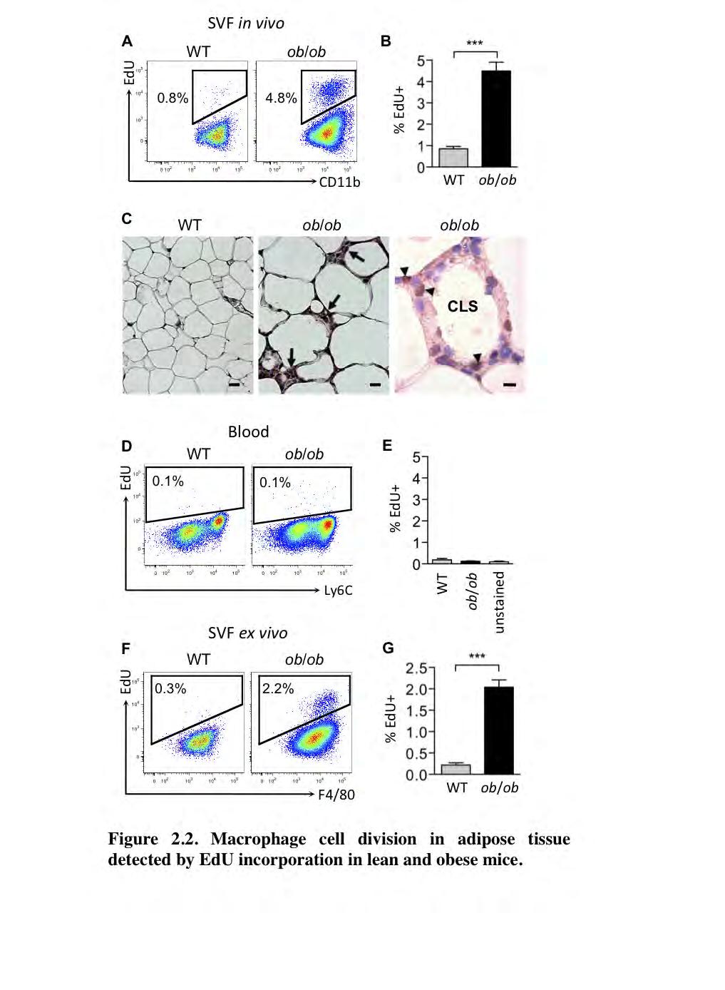
57 46
58 47 Figure 2.2. Macrophage cell division in adipose tissue detected by EdU incorporation in lean and obese mice. Mice were treated with EdU and epididymal adipose tissue SVF and blood were collected 3 hours after treatment and analyzed by flow cytometry. (A) Representative flow cytometry dot plots of adipose tissue macrophages and (D) blood monocytes, and the corresponding bar graphs (B) and (E). (C) Left images, 20x magnification of epididymal adipose tissue of WT and ob/ob mice. Arrows denote crown-like structures (CLS). Scale bar = 50 μm. Right image, 100x magnification image of a CLS in ob/ob epididymal adipose tissue containing cells staining positive for BrdU (arrowheads). Scale bar = 10 μm. BrdU was injected 3 hours before tissue isolation. (F-G) Adipose tissue SVF was isolated and plated in media containing 10 μm EdU. Three hours later cells were harvested and analyzed by flow cytometry. (F) Representative dot plots of ex vivo macrophage EdU incorporation and (G) corresponding rates. All graphs depict mean of EdU + cells ± s.e.m. Statistical significance was determined by Student s t-test. For both sets of experiments n=14-15 mice/group from 3 independent experiments. ***p<0.001.
59 48 Obesity increases macrophage proliferation specifically in adipose tissue. To test whether obesity stimulates macrophage proliferation in tissues other than adipose tissue, we analyzed the proliferation rate of macrophages in spleen and liver following a 3-hour pulse of EdU. The tissues were digested and, as expected, mononuclear cells analyzed by flow cytometry exhibited a higher rate of proliferation in the epididymal adipose tissue of ob/ob compared to WT mice (Figure 2.3 A). Interestingly, in addition to macrophages, non-macrophage (F4/80 ) cells in the adipose tissue also showed an increased rate of proliferation with obesity. The overall rate of EdU incorporation during the pulse into all adipose tissue SVF cells was approximately 0.5% in lean and 4.5% in obese mice (Figure 2.3 A). Conversely, we observed that less than 2% of all cells were EdU + in spleen and liver, and there was no difference in EdU + cells between lean and obese mice (Figure 2.3 B-C). Although macrophage content increased with obesity in adipose tissue (Figure 2.1 C), macrophage content did not increase in spleen or liver in ob/ob mice compared to WT mice (data not shown; for spleen and liver macrophage gating schemes see Supplementary Figure 2.3 B-C). Figure 2.3D shows the rate of proliferation exclusively for macrophages in each tissue in lean and obese states. Importantly, while obesity was associated with a higher rate of macrophage proliferation in adipose tissue, it did not affect the EdU incorporation rate in spleen, liver or blood macrophages. These results suggest that proliferation may contribute to the increased macrophage accumulation in
60 49 the adipose tissue in obesity. Furthermore, something unique about the adipose tissue microenvironment may stimulate macrophage proliferation during obesity.
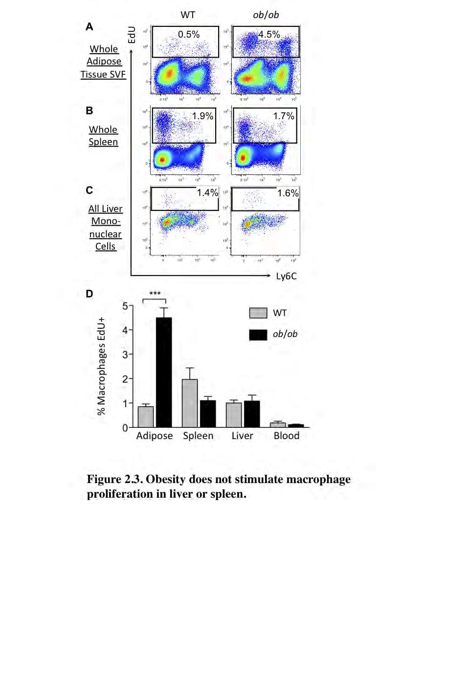
61 50
ACTIVATION AND EFFECTOR FUNCTIONS OF CELL-MEDIATED IMMUNITY AND NK CELLS. Choompone Sakonwasun, MD (Hons), FRCPT
, FRCPT") ACTIVATION AND EFFECTOR FUNCTIONS OF CELL-MEDIATED IMMUNITY AND NK CELLS Choompone Sakonwasun, MD (Hons), FRCPT Types of Adaptive Immunity Types of T Cell-mediated Immune Reactions CTLs = cytotoxic T lymphocytes
ACTIVATION AND EFFECTOR FUNCTIONS OF CELL-MEDIATED IMMUNITY AND NK CELLS Choompone Sakonwasun, MD (Hons), FRCPT Types of Adaptive Immunity Types of T Cell-mediated Immune Reactions CTLs = cytotoxic T lymphocytes
Metabolic Syndrome. DOPE amines COGS 163
 Metabolic Syndrome DOPE amines COGS 163 Overview - M etabolic Syndrome - General definition and criteria - Importance of diagnosis - Glucose Homeostasis - Type 2 Diabetes Mellitus - Insulin Resistance
Question 1. Kupffer cells, microglial cells and osteoclasts are all examples of what type of immune system cell?
 Abbas Chapter 2: Sarah Spriet February 8, 2015 Question 1. Kupffer cells, microglial cells and osteoclasts are all examples of what type of immune system cell? a. Dendritic cells b. Macrophages c. Monocytes
Epigenetic regulation of macrophage polarization and inflammation by DNA methylation in obesity
 Downloaded from http:// on December 17, 2017. https://doi.org/10.1172/jci.insight.87748 Epigenetic regulation of macrophage polarization and inflammation by DNA methylation in obesity Xianfeng Wang, 1
Effector T Cells and
 1 Effector T Cells and Cytokines Andrew Lichtman, MD PhD Brigham and Women's Hospital Harvard Medical School 2 Lecture outline Cytokines Subsets of CD4+ T cells: definitions, functions, development New
Basis of Immunology and
 Basis of Immunology and Immunophysiopathology of Infectious Diseases Jointly organized by Institut Pasteur in Ho Chi Minh City and Institut Pasteur with kind support from ANRS & Université Pierre et Marie
ΦΛΕΓΜΟΝΗ ΚΑΙ ΔΙΑΒΗΤΗΣ
 ΦΛΕΓΜΟΝΗ ΚΑΙ ΔΙΑΒΗΤΗΣ ΘΩΜΑΣ ΠΑΠΑΔΟΠΟΥΛΟΣ, MD, PHD ΕΠΕΜΒΑΤΙΚΟΣ ΚΑΡΔΙΟΛΟΓΟΣ ΙΑΤΡΙΚΟ ΔΙΑΒΑΛΚΑΝΙΚΟ ΚΕΝΤΡΟ Inflammation as a cause of disease has entered the popular imagination. Diet ( macronutrients )
DOWNLOAD PDF ADIPOSE TISSUE AND ADIPOKINES IN HEALTH AND DISEASE (NUTRITION AND HEALTH)
") Chapter 1 : Adiposity, Adipokines, and Adiposopathy - Sick Fat Explained Adipose Tissue and Adipokines in Health and Disease, Second Edition is a useful resource for physicians interested in adipose tissue
Chapter 1 : Adiposity, Adipokines, and Adiposopathy - Sick Fat Explained Adipose Tissue and Adipokines in Health and Disease, Second Edition is a useful resource for physicians interested in adipose tissue
MBB317. Dr D MANGNALL OBESITY. Lecture 2
 MBB317 Dr D MANGNALL OBESITY Lecture 2 When the structure of the insulin receptor was first discovered it was assumed that the active beta subunit tyrosine kinase would phosphorylate some intracellular
Adipose Tissue as an Endocrine Organ. Abdel Moniem Ibrahim, MD Professor of Physiology Cairo University
 Adipose Tissue as an Endocrine Organ Abdel Moniem Ibrahim, MD Professor of Physiology Cairo University Functions of Adipose Tissue Adipose tissue expresses and secretes a variety of bioactive peptides,
LIPOCALIN 2 DEFICIENCY INFLUENCES TRANSFORMING GROWTH FACTOR-β EFFECT ON INFLAMMATION AND EXTRACELLULAR MATRIX REMODELING IN INGUINAL ADIPOCYTES
 LIPOCALIN 2 DEFICIENCY INFLUENCES TRANSFORMING GROWTH FACTOR-β EFFECT ON INFLAMMATION AND EXTRACELLULAR MATRIX REMODELING IN INGUINAL ADIPOCYTES A THESIS SUBMITTED TO THE FACULTY OF THE UNIVERSITY OF MINNESOTA
Brian Francis Zamarron
 Metabolic Abnormalities and Adipose Tissue Leukocyte Dynamics in a Murine Model of Obesity, Weight Loss, and Weight Regain By Brian Francis Zamarron A dissertation submitted in partial fulfillment of the
Cytokines modulate the functional activities of individual cells and tissues both under normal and pathologic conditions Interleukins,
 Cytokines http://highered.mcgraw-hill.com/sites/0072507470/student_view0/chapter22/animation the_immune_response.html Cytokines modulate the functional activities of individual cells and tissues both under
HSN301 Diet and Disease Entire Note Summary
 Topic 1: Metabolic Syndrome Learning Objectives: 1. Define obesity 2. Factors causing obesity 3. Obesity as a risk factor for metabolic syndrome 4. The pathogenesis of metabolic syndrome 5. Treatment of
Pathogenesis of Diabetes Mellitus
 Pathogenesis of Diabetes Mellitus Young-Bum Kim, Ph.D. Associate Professor of Medicine Harvard Medical School Definition of Diabetes Mellitus a group of metabolic diseases characterized by hyperglycemia
Cytokines, adhesion molecules and apoptosis markers. A comprehensive product line for human and veterinary ELISAs
 Cytokines, adhesion molecules and apoptosis markers A comprehensive product line for human and veterinary ELISAs IBL International s cytokine product line... is extremely comprehensive. The assays are
Cell Quality Control. Peter Takizawa Department of Cell Biology
 Cell Quality Control Peter Takizawa Department of Cell Biology Cellular quality control reduces production of defective proteins. Cells have many quality control systems to ensure that cell does not build
Supplementary Figure 1
 Supplementary Figure 1 how HFD how HFD Epi WT p p Hypothalamus p p Inguinal WT T Liver Lean mouse adipocytes p p p p p p Obese mouse adipocytes Kidney Muscle Spleen Heart p p p p p p p p Extracellular
Quantitative Real-Time PCR was performed as same as Materials and Methods.
 Supplemental Material Quantitative Real-Time PCR Quantitative Real-Time PCR was performed as same as Materials and Methods. Expression levels in the aorta were normalized to peptidylprolyl isomerase B
New therapeutic targets for T2DM
 New therapeutic targets for T2DM Targeting inflammation: NF- B, salsalate Gwanpyo Koh Department of Internal Medicine Jeju National University School of Medicine Introduction Obesity is occurring at epidemic
CYTOKINE RECEPTORS AND SIGNAL TRANSDUCTION
 CYTOKINE RECEPTORS AND SIGNAL TRANSDUCTION What is Cytokine? Secreted popypeptide (protein) involved in cell-to-cell signaling. Acts in paracrine or autocrine fashion through specific cellular receptors.
In The Name Of God. In The Name Of. EMRI Modeling Group
 In The Name Of God In The Name Of God EMRI Modeling Group Cells work together in functionally related groups called tissues Types of tissues: Epithelial lining and covering Connective support Muscle movement
Potency Assays Throughout Product Development: Perspectives of an FDA Reviewer
 Potency Assays Throughout Product Development: Perspectives of an FDA Reviewer Chana Fuchs, Ph.D. Division of Monoclonal Antibodies Office of Biotechnology Products Center for Drug Evaluation and Research
Mechanisms of inflammatory responses and development of insulin resistance: how are they interlinked?
 Rehman and Akash Journal of Biomedical Science (2016) 23:87 DOI 10.1186/s12929-016-0303-y RESEARCH Mechanisms of inflammatory responses and development of insulin resistance: how are they interlinked?
Chapter 24 The Immune System
 Chapter 24 The Immune System The Immune System Layered defense system The skin and chemical barriers The innate and adaptive immune systems Immunity The body s ability to recognize and destroy specific
Inflammasome-mediated caspase-1 activity Gatekeeper of inflammation in the adipose tissue. Rinke Stienstra
 Inflammasome-mediated caspase-1 activity Gatekeeper of inflammation in the adipose tissue Rinke Stienstra Obesity promotes the development of insulin resistance and type 2 diabetes County-level Estimates
Summary and concluding remarks
 Summary and concluding remarks This thesis is focused on the role and interaction of different cholesterol and phospholipid transporters. Cholesterol homeostasis is accomplished via a tightly regulated
M.Sc. III Semester Biotechnology End Semester Examination, 2013 Model Answer LBTM: 302 Advanced Immunology
 Code : AS-2246 M.Sc. III Semester Biotechnology End Semester Examination, 2013 Model Answer LBTM: 302 Advanced Immunology A. Select one correct option for each of the following questions:- 2X10=10 1. (b)
Adaptive immune responses: T cell-mediated immunity
 MICR2209 Adaptive immune responses: T cell-mediated immunity Dr Allison Imrie allison.imrie@uwa.edu.au 1 Synopsis: In this lecture we will discuss the T-cell mediated immune response, how it is activated,
T-cell activation T cells migrate to secondary lymphoid tissues where they interact with antigen, antigen-presenting cells, and other lymphocytes:
 Interactions between innate immunity & adaptive immunity What happens to T cells after they leave the thymus? Naïve T cells exit the thymus and enter the bloodstream. If they remain in the bloodstream,
Chapter 11 CYTOKINES
 Chapter 11 CYTOKINES group of low molecular weight regulatory proteins secreted by leukocytes as well as a variety of other cells in the body (8~30kD) regulate the intensity and duration of the immune
renoprotection therapy goals 208, 209
 Subject Index Aldosterone, plasminogen activator inhibitor-1 induction 163, 164, 168 Aminopeptidases angiotensin II processing 64 66, 214 diabetic expression 214, 215 Angiotensin I intrarenal compartmentalization
18. PANCREATIC FUNCTION AND METABOLISM. Pancreatic secretions ISLETS OF LANGERHANS. Insulin
 18. PANCREATIC FUNCTION AND METABOLISM ISLETS OF LANGERHANS Some pancreatic functions have already been discussed in the digestion section. In this one, the emphasis will be placed on the endocrine function
Principles of Adaptive Immunity
 Principles of Adaptive Immunity Chapter 3 Parham Hans de Haard 17 th of May 2010 Agenda Recognition molecules of adaptive immune system Features adaptive immune system Immunoglobulins and T-cell receptors
Subject Index. Bcl-2, apoptosis regulation Bone marrow, polymorphonuclear neutrophil release 24, 26
 Subject Index A1, apoptosis regulation 217, 218 Adaptive immunity, polymorphonuclear neutrophil role 31 33 Angiogenesis cancer 178 endometrium remodeling 172 HIV Tat induction mechanism 176 inflammatory
ACTIVATION OF T LYMPHOCYTES AND CELL MEDIATED IMMUNITY
 ACTIVATION OF T LYMPHOCYTES AND CELL MEDIATED IMMUNITY The recognition of specific antigen by naïve T cell induces its own activation and effector phases. T helper cells recognize peptide antigens through
Chapter 3, Part A (Pages 37-45): Leukocyte Migration into Tissues
: Leukocyte Migration into Tissues") Allergy and Immunology Review Corner: Chapter 3, Part A (pages 37-45) of Cellular and Molecular Immunology (Seventh Edition), by Abul K. Abbas, Andrew H. Lichtman and Shiv Pillai. Chapter 3, Part A (Pages
Allergy and Immunology Review Corner: Chapter 3, Part A (pages 37-45) of Cellular and Molecular Immunology (Seventh Edition), by Abul K. Abbas, Andrew H. Lichtman and Shiv Pillai. Chapter 3, Part A (Pages
Eligibility The NCSF online quizzes are open to any currently certified fitness professional, 18 years or older.
 Eligibility The NCSF online quizzes are open to any currently certified fitness professional, 18 years or older. Deadlines Course completion deadlines correspond with the NCSF Certified Professionals certification
Effects of growth hormone secretagogue receptor agonist and antagonist in nonobese type 2 diabetic MKR mice
 Effects of growth hormone secretagogue receptor agonist and antagonist in nonobese type 2 diabetic MKR mice Rasha Mosa (MBCHC, M.D, PhD candidate) School of Biomedical Sciences University of Queensland
ulcer healing role 118 Bicarbonate, prostaglandins in duodenal cytoprotection 235, 236
 Subject Index Actin cellular forms 48, 49 epidermal growth factor, cytoskeletal change induction in mucosal repair 22, 23 wound repair 64, 65 polyamine effects on cytoskeleton 49 51 S-Adenosylmethionine
Hematopoiesis. - Process of generation of mature blood cells. - Daily turnover of blood cells (70 kg human)
") Hematopoiesis - Process of generation of mature blood cells - Daily turnover of blood cells (70 kg human) 1,000,000,000,000 total cells 200,000,000,000 red blood cells 70,000,000,000 neutrophils Hematopoiesis
Hematopoiesis - Process of generation of mature blood cells - Daily turnover of blood cells (70 kg human) 1,000,000,000,000 total cells 200,000,000,000 red blood cells 70,000,000,000 neutrophils Hematopoiesis
2013 W. H. Freeman and Company. 12 Signal Transduction
 2013 W. H. Freeman and Company 12 Signal Transduction CHAPTER 12 Signal Transduction Key topics: General features of signal transduction Structure and function of G protein coupled receptors Structure
University of California, San Diego La Jolla CA 92093
 AD Award Number: W81XWH-11-1-0131 TITLE: Role of Inflammation and Insulin Resistance in Mouse Models of Breast Cancer PRINCIPAL INVESTIGATOR: Jerrold Olefsky, M.D. CONTRACTING ORGANIZATION: University
Haematopoietic stem cells
 Haematopoietic stem cells Neil P. Rodrigues, DPhil NIH Centre for Biomedical Research Excellence in Stem Cell Biology Boston University School of Medicine neil.rodrigues@imm.ox.ac.uk Haematopoiesis: An
Antigen Presentation and T Lymphocyte Activation. Abul K. Abbas UCSF. FOCiS
 1 Antigen Presentation and T Lymphocyte Activation Abul K. Abbas UCSF FOCiS 2 Lecture outline Dendritic cells and antigen presentation The role of the MHC T cell activation Costimulation, the B7:CD28 family
Production of Exosomes in a Hollow Fiber Bioreactor
 Production of Exosomes in a Hollow Fiber Bioreactor John J S Cadwell, President and CEO, FiberCell Systems Inc INTRODUCTION Exosomes are small lipid membrane vesicles (80-120 nm) of endocytic origin generated
Hematopoiesis. Hematopoiesis. Hematopoiesis
 Chapter. Cells and Organs of the Immune System Hematopoiesis Hematopoiesis- formation and development of WBC and RBC bone marrow. Hematopoietic stem cell- give rise to any blood cells (constant number,
NUTRITION & MALIGNANCY: An Overview
 NUTRITION & MALIGNANCY: An Overview UNIVERSITY OF PNG SCHOOL OF MEDICINE AND HEALTH SCIENCES DISCIPLINE OF BIOCHEMISTRY & MOLECULAR BIOLOGY PBL MBBS II SEMINAR VJ Temple 1 Malignancy and Weight loss (Cachexia)
Blood. Biol 105 Lecture 14 Chapter 11
 Blood Biol 105 Lecture 14 Chapter 11 Outline I. Overview of blood II. Functions of blood III. Composition of blood IV. Composition of plasma V. Composition of formed elements VI. Platelets VII. White blood
ENERGY FROM INGESTED NUTREINTS MAY BE USED IMMEDIATELY OR STORED
 QUIZ/TEST REVIEW NOTES SECTION 1 SHORT TERM METABOLISM [METABOLISM] Learning Objectives: Identify primary energy stores of the body Differentiate the metabolic processes of the fed and fasted states Explain
Insulin sensitivity: modulation by nutrients and inflammation
 Personal perspective Insulin sensitivity: modulation by nutrients and inflammation Simon Schenk, Maziyar Saberi, and Jerrold M. Olefsky Department of Medicine, Division of Endocrinology and Metabolism,
Chapter 13: Cytokines
 Chapter 13: Cytokines Definition: secreted, low-molecular-weight proteins that regulate the nature, intensity and duration of the immune response by exerting a variety of effects on lymphocytes and/or
Campbell's Biology: Concepts and Connections, 7e (Reece et al.) Chapter 24 The Immune System Multiple-Choice Questions
 Chapter 24 The Immune System Multiple-Choice Questions") Campbell's Biology: Concepts and Connections, 7e (Reece et al.) Chapter 24 The Immune System 24.1 Multiple-Choice Questions 1) The body's innate defenses against infection include A) several nonspecific
Campbell's Biology: Concepts and Connections, 7e (Reece et al.) Chapter 24 The Immune System 24.1 Multiple-Choice Questions 1) The body's innate defenses against infection include A) several nonspecific
Prepared by Cyrus H. Nozad, MD, University of Tennessee and John Seyerle, MD, Ohio State University
 Allergy and Immunology Review Corner: Chapter 21 of Middleton s Allergy Principles and Practice, Seventh Edition, edited by N. Franklin Adkinson, et al. Chapter 21: Antigen-Presenting Dendritic Cells (Pages
Potential Atheroprotective Effects of Ixmyelocel-T Cellular Therapy. Kelly J. Ledford, Nikki Murphy, Frank Zeigler, Ronnda L.
 Potential Atheroprotective Effects of Ixmyelocel-T Cellular Therapy Kelly J. Ledford, Nikki Murphy, Frank Zeigler, Ronnda L. Bartel 1 Ixmyelocel-T, an expanded, autologous multicellular therapy cultured
Supplemental Table 1. Primer sequences for transcript analysis
 Supplemental Table 1. Primer sequences for transcript analysis Primer Sequence (5 3 ) Primer Sequence (5 3 ) Mmp2 Forward CCCGTGTGGCCCTC Mmp15 Forward CGGGGCTGGCT Reverse GCTCTCCCGGTTTC Reverse CCTGGTGTGCCTGCTC
There are 2 major lines of defense: Non-specific (Innate Immunity) and. Specific. (Adaptive Immunity) Photo of macrophage cell
 and. Specific. (Adaptive Immunity) Photo of macrophage cell") There are 2 major lines of defense: Non-specific (Innate Immunity) and Specific (Adaptive Immunity) Photo of macrophage cell Development of the Immune System ery pl neu mφ nk CD8 + CTL CD4 + thy TH1 mye
There are 2 major lines of defense: Non-specific (Innate Immunity) and Specific (Adaptive Immunity) Photo of macrophage cell Development of the Immune System ery pl neu mφ nk CD8 + CTL CD4 + thy TH1 mye
Targeting tumour associated macrophages in anti-cancer therapies. Annamaria Gal Seminar Series on Drug Discovery Budapest 5 January 2018
 Targeting tumour associated macrophages in anti-cancer therapies Annamaria Gal Seminar Series on Drug Discovery Budapest 5 January 2018 Macrophages: Professional phagocytes of the myeloid lineage APC,
Overview of the Lymphoid System
 Overview of the Lymphoid System The Lymphoid System Protects us against disease Lymphoid system cells respond to Environmental pathogens Toxins Abnormal body cells, such as cancers Overview of the Lymphoid
Immunology of Asthma. Kenneth J. Goodrum,Ph. Ph.D. Ohio University College of Osteopathic Medicine
 Immunology of Asthma Kenneth J. Goodrum,Ph Ph.D. Ohio University College of Osteopathic Medicine Outline! Consensus characteristics! Allergens:role in asthma! Immune/inflammatory basis! Genetic basis!
DNA vaccine, peripheral T-cell tolerance modulation 185
 Subject Index Airway hyperresponsiveness (AHR) animal models 41 43 asthma inhibition 45 overview 41 mast cell modulation of T-cells 62 64 respiratory tolerance 40, 41 Tregs inhibition role 44 respiratory
Immunology for the Rheumatologist
 Immunology for the Rheumatologist Rheumatologists frequently deal with the immune system gone awry, rarely studying normal immunology. This program is an overview and discussion of the function of the
GENERAL SUMMARY Corpus luteum is a transient endocrine structure formed from the ruptured ovarian follicle. Its main function is to secrete P 4, a pro
 Corpus luteum is a transient endocrine structure formed from the ruptured ovarian follicle. Its main function is to secrete P 4, a pro-gestational hormone, essential for establishment and maintenance of
Week 3 The Pancreas: Pancreatic ph buffering:
 Week 3 The Pancreas: A gland with both endocrine (secretion of substances into the bloodstream) & exocrine (secretion of substances to the outside of the body or another surface within the body) functions
Effect of High-fat or High-glucose Diet on Obesity and Visceral Adipose Tissue in Mice
 ACTA ACADEMIAE MEDICINAE SINICAE 410011 0731-85295846 lixia2014@vip. 163. com 4 C57BL /6 20-1 mrna 20 P < 0. 05 O 20 P > 0. 05 M2 P < 0. 05-1 mrna P < 0. 05 P > 0. 05 R589. 2 DOI 10. 3881 /j. issn. 1000-503X.
The Skinny of the Immune System
 The Skinny of the Immune System Robert Hostoffer, DO, FACOP, FAAP Associate Professor of Pediatrics Case Western Reserve University, Cleveland, Ohio Overview 1. Immune system of the skin 2. Immune Players
Insulin Resistance. Biol 405 Molecular Medicine
 Insulin Resistance Biol 405 Molecular Medicine Insulin resistance: a subnormal biological response to insulin. Defects of either insulin secretion or insulin action can cause diabetes mellitus. Insulin-dependent
The Adaptive Immune Response. B-cells
 The Adaptive Immune Response B-cells The innate immune system provides immediate protection. The adaptive response takes time to develop and is antigen specific. Activation of B and T lymphocytes Naive
Diseases-causing agents, pathogens, can produce infections within the body.
 BIO 212: ANATOMY & PHYSIOLOGY II 1 CHAPTER 16 Lecture: Dr. Lawrence G. Altman www.lawrencegaltman.com Some illustrations are courtesy of McGraw-Hill. LYMPHATIC and IMMUNE Systems Body Defenses Against
Hormonal regulation of. Physiology Department Medical School, University of Sumatera Utara
 Hormonal regulation of nutrient metabolism Physiology Department Medical School, University of Sumatera Utara Homeostasis & Controls Successful compensation Homeostasis reestablished Failure to compensate
Oxidative Stress and Diabetic Complications Epigenetics: Mechanisms and Implications for Diabetic Complications
 This Review is part of a thematic series on Cardiovascular Complications of Diabetes and Obesity, which includes the following articles: The Impact of Macrophage Insulin Resistance on Advanced Atherosclerotic
3/20/2011. Body Mass Index (kg/[m 2 ]) Age at Issue (*BMI > 30, or ~ 30 lbs overweight for 5 4 woman) Mokdad A.H.
![3/20/2011. Body Mass Index (kg/[m 2 ]) Age at Issue (*BMI > 30, or ~ 30 lbs overweight for 5 4 woman) Mokdad A.H.](/thumbs/82/84935173.jpg "3/20/2011. Body Mass Index (kg/[m 2 ]) Age at Issue (*BMI > 30, or ~ 30 lbs overweight for 5 4 woman) Mokdad A.H.") U.S. Adults: 1988 Nineteen states with 10-14% 14% Prevalence of Obesity (*BMI > 30, or ~ 30 lbs overweight for 5 4 woman) Metabolic John P. Cello, MD Professor of Medicine and Surgery, University of California,
U.S. Adults: 1988 Nineteen states with 10-14% 14% Prevalence of Obesity (*BMI > 30, or ~ 30 lbs overweight for 5 4 woman) Metabolic John P. Cello, MD Professor of Medicine and Surgery, University of California,
Supplemental Table I.
 Supplemental Table I Male / Mean ± SEM n Mean ± SEM n Body weight, g 29.2±0.4 17 29.7±0.5 17 Total cholesterol, mg/dl 534.0±30.8 17 561.6±26.1 17 HDL-cholesterol, mg/dl 9.6±0.8 17 10.1±0.7 17 Triglycerides,
Role of Perivascular and Visceral Adipose Tissues in Murine Models of Obesity and Atherosclerosis: A Dissertation
 University of Massachusetts Medical School escholarship@umms GSBS Dissertations and Theses Graduate School of Biomedical Sciences 7-31-2012 Role of Perivascular and Visceral Adipose Tissues in Murine Models
UNIVERSITY OF PNG SCHOOL OF MEDICINE AND HEALTH SCIENCES DIVISION OF BASIC MEDICAL SCIENCES DISCIPLINE OF BIOCHEMISTRY AND MOLECULAR BIOLOGY
 1 UNIVERSITY OF PNG SCHOOL OF MEDICINE AND HEALTH SCIENCES DIVISION OF BASIC MEDICAL SCIENCES DISCIPLINE OF BIOCHEMISTRY AND MOLECULAR BIOLOGY GLUCOSE HOMEOSTASIS An Overview WHAT IS HOMEOSTASIS? Homeostasis
The Importance of Glutamine and Antioxidant Vitamin Supplementation in HIV
 The Importance of Glutamine and Antioxidant Vitamin Supplementation in HIV An Introduction to Glutamine Glutamine is the most abundant amino acid in the human body, and plays extremely important role in
Effector mechanisms of cell-mediated immunity: Properties of effector, memory and regulatory T cells
 ICI Basic Immunology course Effector mechanisms of cell-mediated immunity: Properties of effector, memory and regulatory T cells Abul K. Abbas, MD UCSF Stages in the development of T cell responses: induction
BIOL212 Biochemistry of Disease. Metabolic Disorders - Obesity
 BIOL212 Biochemistry of Disease Metabolic Disorders - Obesity Obesity Approx. 23% of adults are obese in the U.K. The number of obese children has tripled in 20 years. 10% of six year olds are obese, rising
4/5/17. Blood. Blood. Outline. Blood: An Overview. Functions of Blood
 Outline Blood Biol 105 Chapter 11 I. Overview of blood II. Functions of blood III. Composition of blood IV. Composition of plasma V. Composition of formed elements VI. Platelets VII. White blood cells
Supplementary Figure 1.
 Supplementary Figure 1. Increased expression of cell cycle pathway genes in insulin + Glut2 low cells of STZ-induced diabetic islets. A) random blood glucose measuers of STZ and vehicle treated MIP-GFP
Immunology lecture: 14. Cytokines: Main source: Fibroblast, but actually it can be produced by other types of cells
 Immunology lecture: 14 Cytokines: 1)Interferons"IFN" : 2 types Type 1 : IFN-Alpha : Main source: Macrophages IFN-Beta: Main source: Fibroblast, but actually it can be produced by other types of cells **There
Intrinsic cellular defenses against virus infection
 Intrinsic cellular defenses against virus infection Detection of virus infection Host cell response to virus infection Interferons: structure and synthesis Induction of antiviral activity Viral defenses
The Adaptive Immune Responses
 The Adaptive Immune Responses The two arms of the immune responses are; 1) the cell mediated, and 2) the humoral responses. In this chapter we will discuss the two responses in detail and we will start
Cell Adaptation, Cell Injury and Cell Death
 Cell Adaptation, Cell Injury and Cell Death Pathology:- is the study of structural and functional abnormalities that are expressed as diseases of organs and systems. Modern pathology, proposed that injury
5.0 HORMONAL CONTROL OF CARBOHYDRATE METABOLISM
 5.0 HORMONAL CONTROL OF CARBOHYDRATE METABOLISM Introduction: Variety of hormones and other molecules regulate the carbohydrates metabolism. Some of these have already been cited in previous sections.
Supplemental Table 1: Demographics and characteristics of study participants. Male, n (%) 3 (20%) 6 (50%) Age, years [mean ± SD] 33.3 ± ± 9.
![Supplemental Table 1: Demographics and characteristics of study participants. Male, n (%) 3 (20%) 6 (50%) Age, years [mean ± SD] 33.3 ± ± 9.](/thumbs/90/103696086.jpg "Supplemental Table 1: Demographics and characteristics of study participants. Male, n (%) 3 (20%) 6 (50%) Age, years [mean ± SD] 33.3 ± ± 9.") SUPPLEMENTAL DATA Supplemental Table 1: Demographics and characteristics of study participants Lean (n=15) Obese (n=12) Male, n (%) 3 (20%) 6 (50%) Age, years [mean ± SD] 33.3 ± 9.5 44.8 ± 9.1 White, n
SUPPLEMENTAL DATA Supplemental Table 1: Demographics and characteristics of study participants Lean (n=15) Obese (n=12) Male, n (%) 3 (20%) 6 (50%) Age, years [mean ± SD] 33.3 ± 9.5 44.8 ± 9.1 White, n
Obesity, Metabolic Syndrome, and Diabetes: Making the Connections
 Obesity, Metabolic Syndrome, and Diabetes: Making the Connections Alka M. Kanaya, M.D. Associate Professor of Medicine & Epi/Biostats University of California, San Francisco February 26, 21 Roadmap 1.
902 Biomed Environ Sci, 2014; 27(11):
:") 902 Biomed Environ Sci, 2014; 27(11): 902-906 Letter to the Editor Curcuminoids Target Decreasing Serum Adipocyte-fatty Acid Binding Protein Levels in Their Glucose-lowering Effect in Patients with Type
902 Biomed Environ Sci, 2014; 27(11): 902-906 Letter to the Editor Curcuminoids Target Decreasing Serum Adipocyte-fatty Acid Binding Protein Levels in Their Glucose-lowering Effect in Patients with Type
Metabolic integration and Regulation
 Metabolic integration and Regulation 109700: Graduate Biochemistry Trimester 2/2016 Assistant Prof. Dr. Panida Khunkaewla kpanida@sut.ac.th School of Chemistry Suranaree University of Technology 1 Overview
NASH Bench to Bedside
 NASH Bench to Bedside October 2006 Anna Mae Diehl, M.D. Gastroenterology Division Duke University NonAlcoholic Fatty Liver Disease Common ~1/4-1/3 1/3 US adults Outcome highly variable Course indolent
Cutaneous Immunology: Innate Immune Responses. Skin Biology Lecture Series
 Cutaneous Immunology: Innate Immune Responses Skin Biology Lecture Series The Immune Response: Innate and Adaptive Components Source: Wolff, Goldsmith, Katz, Gilchrest, Paller, Leffell. Fitzpatrick s Dermatology
T Lymphocyte Activation and Costimulation. FOCiS. Lecture outline
 1 T Lymphocyte Activation and Costimulation Abul K. Abbas, MD UCSF FOCiS 2 Lecture outline T cell activation Costimulation, the B7:CD28 family Inhibitory receptors of T cells Targeting costimulators for
Adipose Tissue Dysfunction and Diabetic Cardiovascular Disease
 Adipose Tissue Dysfunction and Diabetic Cardiovascular Disease Xin-Liang Ma, MD, PhD Thomas Jefferson University Philadelphia, PA USA CVD As The #1 Causes of Death In USA Male Female Heart Disease and
* Author to whom correspondence should be addressed; Tel.: ; Fax:
 Int. J. Mol. Sci. 2014, 15, 6184-6223; doi:10.3390/ijms15046184 Review OPEN ACCESS International Journal of Molecular Sciences ISSN 1422-0067 www.mdpi.com/journal/ijms Obesity and Its Metabolic Complications:
Supplementary Figures
 Supplementary Figures mir-150 regulates obesityassociated insulin resistance by controlling B cell functions Wei Ying, Alexander Tseng, Richard Cheng-An Chang, Haiqing Wang, Yu-lieh Lin, Srikanth Kanameni,
I. Lines of Defense Pathogen: Table 1: Types of Immune Mechanisms. Table 2: Innate Immunity: First Lines of Defense
 I. Lines of Defense Pathogen: Table 1: Types of Immune Mechanisms Table 2: Innate Immunity: First Lines of Defense Innate Immunity involves nonspecific physical & chemical barriers that are adapted for
Effects of biological response modifiers in psoriasis and psoriatic arthritis Goedkoop, A.Y.
 UvA-DARE (Digital Academic Repository) Effects of biological response modifiers in psoriasis and psoriatic arthritis Goedkoop, A.Y. Link to publication Citation for published version (APA): Goedkoop, A.
Muscle-Macrophage & Macrophage-Macrophage Crosstalk in the Diabetogenic Environment
 Muscle-Macrophage & Macrophage-Macrophage Crosstalk in the Diabetogenic Environment M. Constantine Samaan A thesis submitted in conformity with the requirements for the degree of Masters of Science Institute
Crosstalk between Adiponectin and IGF-IR in breast cancer. Prof. Young Jin Suh Department of Surgery The Catholic University of Korea
 Crosstalk between Adiponectin and IGF-IR in breast cancer Prof. Young Jin Suh Department of Surgery The Catholic University of Korea Obesity Chronic, multifactorial disorder Hypertrophy and hyperplasia