Unexplained visual loss in seven easy steps
|
|
|
- Randall Lewis
- 6 years ago
- Views:
Transcription

 Seven steps in unexplained visual loss 1. Insure visual loss = actual chief complaint 2.")

1 Unexplained visual loss in seven easy steps Andrew G. Lee, MD Chair Ophthalmology, Houston Methodist Hospital, Professor, Weill Cornell MC; Adjunct Professor, Baylor COM, U Iowa, UTMB Galveston, UT MD Anderson Cancer Center, U. Buffalo (SUNY) Seven steps in unexplained visual loss 1. Insure visual loss = actual chief complaint 2. Complete eye exam every time (no shortcuts) 3. Special effort to detect subtle causes of visual loss 4. Formal visual field if unexplained symptoms 5. Special tests (e.g., MERG, OCT, fluorescein angiography, neuroimaging if indicated) 6. Rule out optic neuropathy or hemianopsia 7. Rule out ORGANIC and prove non-organic BEFORE labeling someone as such
2 Step 1: Chief complaint = blurred vision is not sufficient! What do you mean by blurred? One eye or both? Central or side vision or both? Double vision? Jumping eyes? (nystagmus) Processing of visual information? Step 2: Complete eye exam By complete I mean.complete (don t use short cuts in your neuro-op patients!) Check relative afferent pupillary defect yourself Check color vision & visual field Ophthalmoscopy High magnification & high clinical suspicion
 Media (cataract, refractive) Making it up (non-organic) Missed it (look again!")
3 Don t take the shortcut Main Causes For No APD In Unilateral Visual Loss Macular disease (e.g. macular hole) Media (cataract, refractive) Making it up (non-organic) Missed it (look again!) *Bilateral optic neuropathy & retrogeniculate etiologies = normal pupil


4 Complete eye exam Slit lamp biomicroscopy Look after dilation Beware oil droplet cataract Look for posterior subcapsular cataract Match lens opacity to visual acuity Retroillumination Look at lens & grade opacities ( NSC/PSC = 20/30 or 20/30 )
5 Step 3: Rule out things you don t want to end up sending to your neuro-ophthalmologist Oil droplet cataract or subtle posterior subcapsular cataract Refractive error, keratoconus Epiretinal membrane, cystoid macular edema, macular hole, geographic atrophy of retinal pigment epithelium Step 4: Formal visual field Unreliable visual field is the same information as NO visual field performed Confrontation visual field = minimum Media & refractive etiologies rarely produce field defects Any respect of vertical meridian significant


6


7
8 Full Eight Point Exam Formal visual field (even if 20/20) Homonymous & bitemporal hemianopsia may have 20/20 acuity Retrochiasmal disease will have NORMAL structural eye exam (no RAPD, no optic atrophy) Normal eye exam does not r/o pathology Look At The Macula Subtle macular lesions can be missed without high magnification and high suspicion (e.g. macular hole, cystoid macular edema) WNL should mean within normal limits NOT WE NEVER LOOKED

9

10 Post op!


11 Step 5: OCT in Unexplained visual loss? Is it retina or optic nerve? Macular edema or macular hole Epiretinal membrane Cystoid macular edema or subretinal fluid Vitreous traction on macula or optic nerve OCT can see better than me Epiretinal membrane Macular hole Vitreomacular traction Serous fluid under retina Cystoid macular edema


12 Determination of Pallor vs No Pallor OCT can see better than me


13 Consider Ancillary Testing Fluorescein angiography/oct If I see something funny in the macula Electrophysiology if it smells like retina Big blind spot with normal peripapillary retina Ring scotomas Photopsias Diffuse retinal arteriolar narrowing 20 y/o WF with acute loss of visual field RE & photopsias


14 Is this optic neuritis?

 Focal electrical response")

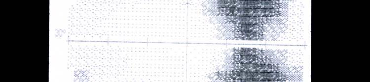
15 What does a ring scotoma look like on HVF? Multifocal Electroretinogram (MERG ) Focal electrical response of photorecepters and bipolar cells Does not detect ganglion cell or axonal response (at present)


16


17 Step 6: Rule out optic neuropathy Look for subtle signs of optic neuropathy Decreased color vision Relative afferent pupillary defect OCT abnormal Mild disc pallor or disc edema Abnormal visual field If you miss a non-optic nerve cause for visual loss (PSC, ERM, refractive) it is no big deal If you miss an optic neuropathy it could be a big deal (compressive optic neuropathy)
18 Step 7: Prove non-organic before labeling patient non-organic Non-organic = preferred term Outdated terms or terms which imply psychologic motivation (hysterical, malingerer) Do you really know they are faking? Do you know their motivation? They might be organic with overlay! Seven steps in unexplained visual loss 1. Insure visual loss = actual chief complaint 2. Complete eye exam every time (no shortcuts) 3. Special effort to detect subtle causes of visual loss 4. Formal visual field if unexplained symptoms 5. Special tests (e.g., MERG, OCT, fluorescein angiography, neuroimaging if indicated) 6. Rule out optic neuropathy or hemianopsia 7. Rule out ORGANIC and prove non-organic BEFORE labeling someone as such

19 Follow up 2010 Pt: You don t remember me do you Dr. Lee? Me: Well, I um.sure maybe Pt: I had lung cancer & you found it thru my eye Me: Really Pt: Yeah, you wrote it up in a journal Me: Oh, yeah, sure, now I remember. How are you, why are you coming today? Pt: I just wanted to tell you that I was still alive and it is been 14 years, so thanks. Longest known survivor


20 Thank you for your time & attention
Five diagnoses you cannot afford to miss. I will not be discussing any off label uses of drugs
 Five diagnoses you cannot afford to miss Andrew G. Lee, MD Chair Ophthalmology, Houston Methodist Hospital, Professor of Ophthalmology, Neurology, & Neurosurgery, Weill Cornell Medical College; Adjunct
3/16/2018. Optic Nerve Examination. Hassan Eisa Swify FRCS Ed (Ophthalmology) Air Force Hospital
 Air Force Hospital") Optic Nerve Examination Hassan Eisa Swify FRCS Ed (Ophthalmology) Air Force Hospital 1 Examination Structure ( optic disc) Function Examination of the optic disc The only cranial nerve (brain tract) which
Optic Nerve Examination Hassan Eisa Swify FRCS Ed (Ophthalmology) Air Force Hospital 1 Examination Structure ( optic disc) Function Examination of the optic disc The only cranial nerve (brain tract) which
Five steps: Overview
 Optic atrophy is not a diagnosis Andrew G. Lee, MD Professor of Ophthalmology, Neurology and Neurosurgery, Weill Cornell Medical College Chair, Department of Ophthalmology, Houston Methodist Hospital,
Objectives. Unexplained Vision Loss: Where Do I Go From Here. History. History. Drug Induced Vision Loss
 Objectives Unexplained Vision Loss: Where Do I Go From Here Denise Goodwin, OD, FAAO Coordinator, Neuro-ophthalmic Disease Clinic Pacific University College of Optometry goodwin@pacificu.edu Know the importance
Diabetic retinopathy damage to the blood vessels in the retina. Cataract clouding of the eye s lens. Cataracts develop at an earlier age in people
 Diabetic Retinopathy What is diabetic eye disease? Diabetic eye disease refers to a group of eye problems that people with diabetes may face as a complication of diabetes. All can cause severe vision loss
Fundus Autofluorescence. Jonathan A. Micieli, MD Valérie Biousse, MD
 Fundus Autofluorescence Jonathan A. Micieli, MD Valérie Biousse, MD The retinal pigment epithelium (RPE) has many important functions including phagocytosis of the photoreceptor outer segments Cone Rod
Dr/ Marwa Abdellah EOS /16/2018. Dr/ Marwa Abdellah EOS When do you ask Fluorescein angiography for optic disc diseases???
 When do you ask Fluorescein angiography for optic disc diseases??? 1 NORMAL OPTIC DISC The normal optic disc on fluorescein angiography is fluorescent due to filling of vessels arising from the posterior
ISPUB.COM. Photopsia post flu: A case of MEWDS. S Baisakhiya, S Dulani, S Lele INTRODUCTION CASE HISTORY
 ISPUB.COM The Internet Journal of Ophthalmology and Visual Science Volume 8 Number 1 Photopsia post flu: A case of MEWDS S Baisakhiya, S Dulani, S Lele Citation S Baisakhiya, S Dulani, S Lele. Photopsia
When optical coherence tomography (OCT)
") Macular Imaging: SD-OCT in nterior Segment Surgical Practice Many pathologic processes of the macula can be visualized or quantified only with this modality. y Steven G. Safran, MD When optical coherence
Macular Imaging: SD-OCT in nterior Segment Surgical Practice Many pathologic processes of the macula can be visualized or quantified only with this modality. y Steven G. Safran, MD When optical coherence
Neuro-Ophthalmic Masqueraders
 Neuro-Ophthalmic Masqueraders Leonid Skorin, Jr., OD, DO, MS, FAAO, FAOCO Mayo Clinic Health System in Albert Lea Denise Goodwin, OD, FAAO Pacific University College of Optometry Please silence all mobile
 measure of your overall performance. An isolated glucose test is helpful to let you know what your sugar level is at one moment, but it doesn t tell you whether or not your diabetes is under adequate control
tracking progression we can better manage our patients. Like any tool, any instrument you ve got to
 EIYESS ALBEINUTI, MD 1 As we know OCT has become very instrumental in taking care of glaucoma patients whether we have the ability to objectively image the RNFL and therefore pickup earlier signs of damage
UC SF. g h. Eye Trauma. Martha Neighbor, MD Emergency Services San Francisco General Hospital University of California
 UC SF Eye Trauma sf g h Martha Neighbor, MD Emergency Services San Francisco General Hospital University of California Goals Recognize vision threatening eye emergencies Treat them when we can Know when
Non-arteritic anterior ischemic optic neuropathy (NAION) with segmental optic disc edema. Jonathan A. Micieli, MD Valérie Biousse, MD
 with segmental optic disc edema. Jonathan A. Micieli, MD Valérie Biousse, MD") Non-arteritic anterior ischemic optic neuropathy (NAION) with segmental optic disc edema Jonathan A. Micieli, MD Valérie Biousse, MD A 75 year old white woman lost vision in the inferior part of her visual
Non-arteritic anterior ischemic optic neuropathy (NAION) with segmental optic disc edema Jonathan A. Micieli, MD Valérie Biousse, MD A 75 year old white woman lost vision in the inferior part of her visual
EPIRETINAL MEMBRANE & VITREOMACULAR TRACTION
 EPIRETINAL MEMBRANE & VITREOMACULAR TRACTION Management of ERM and VMT K.V.Chalam,MD,PhD,MBA,FACS Professor and Director of Retina Loma Linda Eye Institute Los Angeles, USA REVIEW ANATOMY The vitreous
Neuro-imaging for the Ophthalmologist. Karl C. Golnik, MD, MEd University of Cincinnati & The Cincinnati Eye Institute
 Neuro-imaging for the Ophthalmologist Karl C. Golnik, MD, MEd University of Cincinnati & The Cincinnati Eye Institute Neuro-ophthalmology is that subspecialty where the diagnosis is made upon reinterpretation
The Human Eye. Cornea Iris. Pupil. Lens. Retina
 The Retina Thin layer of light-sensitive tissue at the back of the eye (the film of the camera). Light rays are focused on the retina then transmitted to the brain. The macula is the very small area in
Learn Connect Succeed. JCAHPO Regional Meetings 2015
 Learn Connect Succeed JCAHPO Regional Meetings 2015 OPTIC NEUROPATHY AS EASY AS 1,2,3,4 OPTIC NERVE ANATOMY M. Tariq Bhatti, MD Departments of Ophthalmology and Neurology Duke Eye Center and Duke University
Recurrent intraocular hemorrhage secondary to cataract wound neovascularization (Swan Syndrome)
") Recurrent intraocular hemorrhage secondary to cataract wound neovascularization (Swan Syndrome) John J. Chen MD, PhD; Young H. Kwon MD, PhD August 6, 2012 Chief complaint: Recurrent vitreous hemorrhage,
Recurrent intraocular hemorrhage secondary to cataract wound neovascularization (Swan Syndrome) John J. Chen MD, PhD; Young H. Kwon MD, PhD August 6, 2012 Chief complaint: Recurrent vitreous hemorrhage,
OCT : retinal layers. Extraocular muscles. History. Central vs Peripheral vision. History: Temporal course. Optical Coherence Tomography (OCT)
") Optical Coherence Tomography (OCT) OCT : retinal layers 7 Central vs Peripheral vision Extraocular muscles RPE E Peripheral Vision: Rods (95 million) 30% Ganglion cells Central Vision: Cones (5 million)
Optical Coherence Tomography (OCT) OCT : retinal layers 7 Central vs Peripheral vision Extraocular muscles RPE E Peripheral Vision: Rods (95 million) 30% Ganglion cells Central Vision: Cones (5 million)
Ganglion cell analysis by optical coherence tomography (OCT) Jonathan A. Micieli, MD Valérie Biousse, MD
 Jonathan A. Micieli, MD Valérie Biousse, MD") Ganglion cell analysis by optical coherence tomography (OCT) Jonathan A. Micieli, MD Valérie Biousse, MD Figure 1. Normal OCT of the macula (cross section through the line indicated on the fundus photo)
Ganglion cell analysis by optical coherence tomography (OCT) Jonathan A. Micieli, MD Valérie Biousse, MD Figure 1. Normal OCT of the macula (cross section through the line indicated on the fundus photo)
Yasser R. Serag, MD Tamer Wasfi, MD El- Saied El-Dessoukey, MD Magdi S. Moussa, MD Anselm Kampik, MD
 Microperimetric Evaluation of Brilliant Blue G- assisted Internal Limiting Membrane Peeling By Yasser R. Serag, MD Tamer Wasfi, MD El- Saied El-Dessoukey, MD Magdi S. Moussa, MD Anselm Kampik, MD The internal
Diabetes & Your Eyes
 Diabetes & Your Eyes Diabetes is a disease that occurs when the pancreas does not secrete enough insulin or the body is unable to process it properly. Insulin is the hormone that regulates the level of
Five Things You re Missing with Your Fundus Camera
 ebook Five Things You re Missing with Your Fundus Camera By Donald J. Siegel, OD, Sun City West Eye Care Sponsored by: Before I began incorporating EIDON true-color imaging into my practice, my retinal
PART 1: GENERAL RETINAL ANATOMY
 PART 1: GENERAL RETINAL ANATOMY General Anatomy At Ora Serrata At Optic Nerve Head Fundoscopic View Of Normal Retina What Is So Special About Diabetic Retinopathy? The WHO definition of blindness is
CHAPTER 13 CLINICAL CASES INTRODUCTION
 2 CHAPTER 3 CLINICAL CASES INTRODUCTION The previous chapters of this book have systematically presented various aspects of visual field testing and is now put into a clinical context. In this chapter,
Study of clinical significance of optical coherence tomography in diagnosis & management of diabetic macular edema
 Original Research Article Study of clinical significance of optical coherence tomography in diagnosis & management of diabetic macular edema Neha Kantilal Desai 1,*, Somesh Vedprakash Aggarwal 2, Sonali
Abstract title: Vision loss from myelinated retinal nerve fiber layer with maculopathy. Authors: Man Kin (Eric) Chow, OD Lori Vollmer, OD, FAAO
 Chow, OD Lori Vollmer, OD, FAAO") Abstract title: Vision loss from myelinated retinal nerve fiber layer with maculopathy. Authors: Man Kin (Eric) Chow, OD Lori Vollmer, OD, FAAO Joseph Sowka, OD, FAAO General Topic: Ocular Disease Primary
Abstract title: Vision loss from myelinated retinal nerve fiber layer with maculopathy. Authors: Man Kin (Eric) Chow, OD Lori Vollmer, OD, FAAO Joseph Sowka, OD, FAAO General Topic: Ocular Disease Primary
Optical Coherence Tomograpic Features in Idiopathic Retinitis, Vasculitis, Aneurysms and Neuroretinitis (IRVAN)
") Columbia International Publishing Journal of Ophthalmic Research (2014) Research Article Optical Coherence Tomograpic Features in Idiopathic Retinitis, Vasculitis, Aneurysms and Neuroretinitis (IRVAN)
Columbia International Publishing Journal of Ophthalmic Research (2014) Research Article Optical Coherence Tomograpic Features in Idiopathic Retinitis, Vasculitis, Aneurysms and Neuroretinitis (IRVAN)
Ocular Pathology I 6234_16385 Rm HBSB 203-E 1:00-3:00pm. Tonya G. Ketcham, OD, PhD , RM 2113
 Ocular Pathology I 6234_16385 Rm HBSB 203-E 1:00-3:00pm Tonya G. Ketcham, OD, PhD tketcham@optometry.uh.edu 3-1799, RM 2113 Course Syllabus Course Description To describe normal anomalies and pathologic
Optic Nerve Disorders: Structure and Function and Causes
 Optic Nerve Disorders: Structure and Function and Causes Using Visual Fields, OCT and B-scan Ultrasound to Diagnose and Follow Optic Nerve Visual Losses Ohio Ophthalmological Society and Ophthalmic Tech
Case Follow Up. Sepi Jooniani PGY-1
 Case Follow Up Sepi Jooniani PGY-1 Triage 54 year old M Pt presents to prelim states noticed today he had reddness to eyes, states worse in R eye. Pt denies any pain or itching. No further complaints.
Do You See What I See!!! Shane R. Kannarr, OD
 Do You See What I See!!! Shane R. Kannarr, OD skannarr@kannarreyecare.com Define Specialty Testing Additional Test to: Prove/Disprove Diagnosis To monitor progression of a condition To document a condition
Pearls, Pitfalls and Advances in Neuro-Ophthalmology
 Pearls, Pitfalls and Advances in Neuro-Ophthalmology Nancy J. Newman, MD Emory University Atlanta, GA Consultant for Gensight Biologics, Santhera Data Safety Monitoring Board for Quark AION Study Medical-legal
Syllabus-Ophthalmology Rotation Course: Objectives & Goals LOYOLA UNIVERSITY CHICAGO STRITCH SCHOOL OF MEDICINE
 Syllabus-Ophthalmology Rotation Course: Objectives & Goals LOYOLA UNIVERSITY CHICAGO STRITCH SCHOOL OF MEDICINE Department of Ophthalmology Course Objectives: By Core Competencies GENERAL INFORMATION:
A Patient s Guide to Diabetic Retinopathy
 Diabetic Retinopathy A Patient s Guide to Diabetic Retinopathy 840 Walnut Street, Philadelphia PA 19107 www.willseye.org Diabetic Retinopathy 1. Definition Diabetic retinopathy is a complication of diabetes
Retina Conference. Janelle Fassbender, MD, PhD University of Louisville Department of Ophthalmology and Visual Sciences 09/04/2014
 Retina Conference Janelle Fassbender, MD, PhD University of Louisville Department of Ophthalmology and Visual Sciences 09/04/2014 Subjective CC/HPI: 64 year old Caucasian female referred by outside ophthalmologist
What You Should Know About Acute Macular Neuroretinopathy
 What You Should Know About Acute Macular Neuroretinopathy David J. Browning MD, PhD Chong Lee BS Acute macular neuroretinopathy is a condition characterized by the sudden, painless onset of paracentral
CNS 2 Physiology lab
 It should be noted that the doctor emphasized that this material is also considered as continuation of the theory material and is INCLUDED IN THE THEORY EXAM. Presbiopia: is decrease in accommodation of
Moncef Khairallah, MD
 Moncef Khairallah, MD Department of Ophthalmology, Fattouma Bourguiba University Hospital Faculty of Medicine, University of Monastir Monastir, Tunisia INTRODUCTION IU: anatomic form of uveitis involving
ADULT-ONSET FOVEOMACULAR VITELLIFORM DYSTROPHY. By: Chris Munnerlyn, OMT Student University of Arkansas for Medical Sciences
 ADULT-ONSET FOVEOMACULAR VITELLIFORM DYSTROPHY By: Chris Munnerlyn, OMT Student University of Arkansas for Medical Sciences ADULT-ONSET FOVEOMACULAR VITELLIFORM DYSTROPHY (AOFVD) AOFVD is a condition that
Widefield Retinal Imaging with Auto Fluorescence Technology in the Optometric Practice
 Widefield Retinal Imaging with Auto Fluorescence Technology in the Optometric Practice This course will define ultra-widefield retinal imaging and autofluorescence for the attendee. Will show how it is
The effect of intravitreal bevacizumab in a rare case of retinal dystrophy with secondary cystoid macular edema
 Romanian Journal of Ophthalmology, Volume 61, Issue 2, April-June 2017. pp:123-127 CASE REPORT The effect of intravitreal bevacizumab in a rare case of retinal dystrophy with secondary cystoid macular
Communicating with patients about alternative therapies: A case of optic nerve hypoplasia
 Communicating with patients about alternative therapies: A case of optic nerve hypoplasia Chief Complaint:14 year old male with poor vision since birth History of Present Illness: An otherwise healthy
Neuro-Ocular Grand Rounds Anthony B. Litwak,OD, FAAO VA Medical Center Baltimore, Maryland
 Neuro-Ocular Grand Rounds Anthony B. Litwak,OD, FAAO VA Medical Center Baltimore, Maryland Dr. Litwak is on the speaker and advisory boards for Alcon and Zeiss Meditek COMMON OPTIC NEUROPATHIES THAT CAN
OPHTHALMOLOGICAL DISORDERS
 Telephone No.: 24622495 Telegraphic Address: Aeronautical: VIDDYAYX Commercial: AIRCIVIL NEW DELHI E Mail: dri@dgca.nic.in Fax:01124629211 GOVERNMENT OF INDIA AERONAUTICAL INFORMATION SERVICE DIRECTOR
Evaluation of ONH Pallor in Glaucoma Patients and Suspects. Leticia Rousso, O.D. Joseph Sowka, O.D
 Evaluation of ONH Pallor in Glaucoma Patients and Suspects Leticia Rousso, O.D Joseph Sowka, O.D I. Abstract This case report will evaluate a young glaucoma suspect with unilateral sectoral optic nerve
SUMMARY BENCHMARKS FOR PREFERRED PRACTICE PATTERN GUIDELINES
 SUMMARY BENCHMARKS FOR PREFERRED PRACTICE PATTERN GUIDELINES TABLE OF CONTENTS Summary Benchmarks for Preferred Practice Pattern Guidelines Introduction... 1 Glaucoma Primary Open-Angle Glaucoma (Initial
OCT Assessment of the Vitreoretinal Relationship in CSME
 December 2007 Sonia Rani John et al. - IFIS 375 ORIGINAL ARTICLE OCT Assessment of the Vitreoretinal Relationship in CSME Dr. Manoj S. DNB FRCS, Dr. Unnikrishnan Nair MS DO FRCS, Dr. Gargi Sathish MS Introduction
OCCLUSIVE VASCULAR DISORDERS OF THE RETINA
 OCCLUSIVE VASCULAR DISORDERS OF THE RETINA Learning outcomes By the end of this lecture the students would be able to Classify occlusive vascular disorders (OVD) of the retina. Correlate the clinical features
DIABETIC RETINOPATHY
 THE UK GUIDE DIABETIC RETINOPATHY Everything you need to know about diabetic retinopathy Jaheed Khan BSc (Hons) MBBS MD FRCOphth Fellow of the Royal College of Ophthalmologists Association for Research
Sudden Vision Loss. Brendan Girschek, MD, FRCSC, FACS Vitreoretinal Surgery Cedar Valley Medical Specialists
 Sudden Vision Loss Brendan Girschek, MD, FRCSC, FACS Vitreoretinal Surgery Cedar Valley Medical Specialists My Credentials -Residency in Ophthalmology at the LSU Eye Center in New Orleans, LA -Fellowship
53 year old woman attends your practice for routine exam. She has no past medical history or family history of note.
 Case 1 Normal Tension Glaucoma 53 year old woman attends your practice for routine exam. She has no past medical history or family history of note. Table 1. Right Eye Left Eye Visual acuity 6/6 6/6 Ishihara
IMAGE OF THE MOMENT PRACTICAL NEUROLOGY
 178 PRACTICAL NEUROLOGY IMAGE OF THE MOMENT Gawn G. McIlwaine*, James H. Vallance* and Christian J. Lueck *Princess Alexandra Eye Pavilion, Chalmers Street, Edinburgh UK; The Canberra Hospital, P.O. Box
OPTIC DISC PIT Pathogenesis and Management OPTIC DISC PIT
 OPTIC DISC PIT Pathogenesis and Management Abdel-Latif Siam Ain Shams University Cairo Egypt OPTIC DISC PIT Congenital pit is an atypical coloboma usually located on the temporal edge of the disc, associated
GRADUAL LOSS OF VISION. Teresa Anthony Consultant Ophthalmologist Emersons Green NHS Treatment Centre Bristol
 GRADUAL LOSS OF VISION Teresa Anthony Consultant Ophthalmologist Emersons Green NHS Treatment Centre Bristol CVI Registration Low Vision: 6/60 Sight impaired: 3/60 Severe sight impaired:
Course # Flashes and Floaters and Curtains, Oh My!
 Course # 132 Flashes and Floaters and Curtains, Oh My! FLASHES and FLOATERS and CURTAINS, OH MY!!! FLASHES OF LIGHT Vitreous is the villain Retinal traction Retinal hole Retinal tear Migraine Classic migraine
Course # Flashes and Floaters and Curtains, Oh My!
 Course # 132 Flashes and Floaters and Curtains, Oh My! FLASHES and FLOATERS and CURTAINS, OH MY!!! FLASHES OF LIGHT Vitreous is the villain Retinal traction Retinal hole Retinal tear Migraine Classic migraine
Andrew G. Lee, MD. 1. Optic Neuropathy 2. Retinopathy 3. Electrophysiology 4. Optical Coherence Tomography 5. Macular Photostress Test
 How to distinguish retinal disorders from causes of optic nerve dysfunction? Andrew G. Lee, MD The Methodist Hospital Houston, TX Learning Objectives 1. To define the overlapping clinical presentations
Cirrus TM HD-OCT. Details defi ne your decisions
 Cirrus TM HD-OCT Details defi ne your decisions 2 With high-defi nition OCT Carl Zeiss Meditec takes you beyond standard spectral domain Built on 10 years experience at the vanguard of innovation, Carl
OCT Interpretation in Retinal Disease
 OCT Interpretation in Retinal Disease Jay M. Haynie, OD, FAAO Financial Disclosure I have received honoraria or am on the advisory board for the following companies: Carl Zeiss Meditec Advanced Ocular
Electrodiagnostics Alphabet Soup
 Nathan Lighthizer, O.D., F.A.A.O Assistant Professor, NSUOCO Chief of Specialty Care Clinics Chief of Electrodiagnostics Clinic What is electrodiagnostics testing? Visual Pathway Basic Understanding VEP
Macular Hole Associated with Vogt-Koyanagi-Harada Disease at the Acute Uveitic Stage
 Published online: September 15, 2015 2015 The Author(s) Published by S. Karger AG, Basel 1663 2699/15/0063 0328$39.50/0 This article is licensed under the Creative Commons Attribution-NonCommercial 4.0
NEPTUNE RED BANK BRICK
 NEPTUNE RED BANK BRICK Diabetes & The Eye Diabetics are more likely to develop Cataracts at a younger age. Diabetics are twice as likely to develop Glaucoma when compared to non-diabetics. The primary
10/27/2013. Optic Red Herrings
 Optic Red Herrings 1 Optic neuropathy Compressive Inflammatory Toxic Glaucomatous Ischemic Post traumatic GLAUCOMATOUS OPTIC NEUROPATHY Glaucoma: Traditionally defined as a progressive optic neuropathy
Technologies and Methods for Visualizing the Retina
 Transcript Details This is a transcript of an educational program accessible on the ReachMD network. Details about the program and additional media formats for the program are accessible by visiting: https://reachmd.com/programs/revealing-retina/technologies-and-methods-for-visualizing-theretina/3663/
Clinically Significant Macular Edema (CSME)
") Clinically Significant Macular Edema (CSME) 1 Clinically Significant Macular Edema (CSME) Sadrina T. Shaw OMT I Student July 26, 2014 Advisor: Dr. Uwaydat Clinically Significant Macular Edema (CSME) 2
Clinically Significant Macular Edema (CSME) 1 Clinically Significant Macular Edema (CSME) Sadrina T. Shaw OMT I Student July 26, 2014 Advisor: Dr. Uwaydat Clinically Significant Macular Edema (CSME) 2
RETINAL CONDITIONS RETINAL CONDITIONS
 GENERAL INFORMATION RETINAL CONDITIONS RETINAL CONDITIONS WHAT ARE RETINAL CONDITIONS? Retinal conditions affect the light-sensitive tissue at the back of eye known as the retina. They include diseases
COEXISTENCE OF OPTIC NERVE HEAD DRUSEN
 COEXISTENCE OF OPTIC NERVE HEAD DRUSEN AND COMBINED HAMARTOMA OF THE RETINA AND RETINAL PIGMENT EPITHELIUM IN A TAIWANESE MALE Yo-Chen Chang 1 and Rong-Kung Tsai 2,3 1 Department of Ophthalmology, Kaohsiung
Case report 12/10/2014. Delphine Lam ; Dr Mayer Srour Service d ophtalmologie Professeur E.Souied Université Paris Est
 Case report 12/10/2014 Delphine Lam ; Dr Mayer Srour Service d ophtalmologie Professeur E.Souied Medical history Man, 75 years old Complaint: Vision loss in left eye in June 2014 Past ophthalmologic history:
Visual loss and foveal lesions in Usher's syndrome
 British Journal of Ophthalmology, 1979, 63, 484-488 Visual loss and foveal lesions in Usher's syndrome GERALD FISHMAN, VICTORIA VASQUEZ, MARLENE FISHMAN, AND BERGER' From the Department of Ophthalmology,
What Is O.C.T. and Why Should I Give A Rip? OCT & Me How Optical Coherence Tomography Changed the Life of a Small Town Optometrist 5/19/2014
 OCT & Me How Optical Coherence Tomography Changed the Life of a Small Town Optometrist Email: myoder@wcoil.com Mark A. Yoder, O.D. 107 N. Main Street PO Box 123 Bluffton, OH 45817 @yoderod 115.02 Histoplasma
Understanding Diabetic Retinopathy
 Understanding Diabetic Retinopathy What Is Diabetic Retinopathy? Diabetes damages blood vessels in the rear of the eye. This condition is called diabetic retinopathy. It can lead to vision loss or blindness.
The Quick Guide to OCT Mastery 50 Real Cases with Expert Analysis
 OPTICAL COHERENCE TOMOGRAPHY The Quick Guide to OCT Mastery 50 Real Cases with Expert Analysis VOL 1 Sanjay Sharma, MD, FRCS, MSc (Epid), MBA Ophthalmologist, Epidemiologist Queen s University, Canada
Diagnosis and treatment of diabetic retinopathy. Blake Cooper MD Ophthalmologist Vitreoretinal Surgeon Retina Associates Kansas City
 Diagnosis and treatment of diabetic retinopathy Blake Cooper MD Ophthalmologist Vitreoretinal Surgeon Retina Associates Kansas City Disclosures Consulted for Novo Nordisk 2017,2018. Will be discussing
Moving forward with a different perspective
 Moving forward with a different perspective The Leader In Vision Diagnostics Offers A New Perspective Marco has served the eyecare community by offering exceptional lane products and automated high tech
Cirrus TM HD-OCT. Details define your decisions
 Cirrus TM HD-OCT Details define your decisions 2 With high-definition OCT Carl Zeiss Meditec takes you beyond standard spectral domain Built on 10 years experience at the vanguard of innovation, Carl Zeiss
Applying structure-function to solve clinical cases
 Applying structure-function to solve clinical cases Professor Michael Kalloniatis Centre for Eye Health, and, School of Optometry and Vision Science Acknowledgements Some material prepared by Nayuta Yoshioka
VISUAL EVOKED POTENTIAL PATTERN ELECTRORETINOGRAM ASSESSMENT OF NEURO VISUAL FUNCTION. Lee Shettle, D.O. Lee Shettle Eye & Hearing
 VISUAL EVOKED POTENTIAL PATTERN ELECTRORETINOGRAM ASSESSMENT OF NEURO VISUAL FUNCTION Lee Shettle, D.O. Lee Shettle Eye & Hearing Overview of VEP / PERG Why should I Use this Test? How does it work? Clinical
Diabetic Retinopathy Information
 http://www.midwestretina.com Phone: (614)-339-8500 Toll Free: (866)-373-8462 Sugat S. Patel, M.D. Louis J. Chorich III, M.D. Dino D. Klisovic, M.D. Lisa M. Borkowski, M.D. Dominic M. Buzzacco, M.D. Johnstone
Audit of Macular Hole Surgery, Visual Outcome Prediction on OCT Appearance of Macular Hole
 International Journal of Ophthalmology & Visual Science 2017; 2(4): 93-97 http://www.sciencepublishinggroup.com/j/ijovs doi: 10.11648/j.ijovs.20170204.13 Audit of Macular Hole Surgery, Visual Outcome Prediction
R&M Solutions
 Mohamed Hosny El-Bradey, MD., Assistant Professor of Ophthalmology, Tanta University. Wael El Haig, MD., Professor of Ophthalmology. Zagazeeg University. 1 Myopic CNV is considered the most common vision
Is OCT-A Needed As An Investigative Tool During The Management Of Diabetic Macular Edema
 Is OCT-A Needed As An Investigative Tool During The Management Of Diabetic Macular Edema Ayman M Khattab MD, FRCS Professor of Ophthalmology Cairo University Diabetic Macular Edema (DME) Diabetic macular
JMSCR Volume 03 Issue 01 Page January 2015
 www.jmscr.igmpublication.org Impact Factor 3.79 ISSN (e)-2347-176x Clinical Evaluation of Patients Presenting With Rhegmatogenous Retinal Detachment Authors Dr Rani Sujatha.M.A 1, Dr Sridevi Prakash T
Glaucoma. Cornea. Iris
 Glaucoma Introduction Glaucoma is a group of eye diseases that can lead to blindness if not treated. Openangle glaucoma, the most common form of glaucoma, affects about 3 million Americans. Half of those
Alan G. Kabat, OD, FAAO (901)
") THE SWOLLEN OPTIC DISC: EMERGENCY OR ANOMALY? Alan G. Kabat, OD, FAAO (901) 252-3691 Memphis, Tennessee alan.kabat@alankabat.com Course description: The swollen disc presents a diagnostic dilemma. While
THE SWOLLEN OPTIC DISC: EMERGENCY OR ANOMALY? Alan G. Kabat, OD, FAAO (901) 252-3691 Memphis, Tennessee alan.kabat@alankabat.com Course description: The swollen disc presents a diagnostic dilemma. While
Brampton Hurontario Street Brampton, ON L6Y 0P6
 Diabetic Retinopathy What is Diabetic Retinopathy Diabetic retinopathy is one of the leading causes of blindness world-wide. Diabetes damages blood vessels in many organs of the body including the eyes.
Perspectives on Screening for Diabetic Retinopathy. Dr. Dan Samaha, Optometrist, MSc Clinical Lecturer School of Optometry, Université de Montréal
 Perspectives on Screening for Diabetic Retinopathy 1 Dr. Dan Samaha, Optometrist, MSc Clinical Lecturer School of Optometry, Université de Montréal Current standards 2 According to the Canadian Diabetes
Champlain LHIN. Estimated that 55,563 people over age 18 live with diabetes
 Champlain LHIN Estimated that 55,563 people over age 18 live with diabetes Healthy, caring communities supported by health services of choice that achieve results- today and for the future Impact of Diabetes
Role Of Various Factors In The Treatment Of Optic Neuritis----A Study Abstract Aim: Materials & Methods Discussion: Conclusion: Key words
 IOSR Journal of Dental and Medical Sciences (IOSR-JDMS) e-issn: 2279-0853, p-issn: 2279-0861.Volume 15, Issue 9 Ver. X (September). 2016), PP 51-57 www.iosrjournals.org Role Of Various Factors In The Treatment
THE NATURAL HISTORY OF TRACTIONAL CYSTOID MACULAR EDEMA
 THE NATURAL HISTORY OF TRACTIONAL CYSTOID MACULAR EDEMA SOFIA CHARALAMPIDOU, MRCOPHTH,* JOHN NOLAN, PHD, STEPHEN BEATTY, FRCOPHTH* Background: To describe clinical outcomes in a series of patients with
University Hospital Basel. Optical Coherence Tomography Emerging Role in the Assessment of MS PD Dr. Konstantin Gugleta
 University Hospital Basel Optical Coherence Tomography Emerging Role in the Assessment of MS PD Dr. Konstantin Gugleta 15th State of the Art SMSS, Lucerne January 2013 Retinal Nerve Fiber Layer 1.200.000
Learn Connect Succeed. JCAHPO Regional Meetings 2015
 Learn Connect Succeed JCAHPO Regional Meetings 2015 VISUAL FIELDS No financial conflicks Florida Society of Ophthalmology 2015 Gary Schemmer, MD Definition of Visual Field The area in space perceived by
The Common Clinical Competency Framework for Non-medical Ophthalmic Healthcare Professionals in Secondary Care
 The Common Clinical Competency Framework for Non-medical Ophthalmic Healthcare Professionals in Secondary Care Medical Retina November 2016 Association of Health Professions in Ophthalmology General basic
04/06/2015. Documentation Do s and Don ts In The Retina Practice. Financial Disclosure. Documentation Dos and Don ts
 Documentation Do s and Don ts In The Retina Practice William T. Koch, COA, COE, CPC Administrative Director Director of Billing Operations The Retina Institute St. Louis, Missouri Advisory Boards Allergan
MANAGING DIABETIC RETINOPATHY. <Your Hospital Name> <Your Logo>
 MANAGING DIABETIC RETINOPATHY It s difficult living with Diabetes Mellitus. Ask any diabetic... Their lives are centered around meal plans, glucose levels, and insulin
Papilledema. Golnaz Javey, M.D. and Jeffrey J. Zuravleff, M.D.
 Papilledema Golnaz Javey, M.D. and Jeffrey J. Zuravleff, M.D. Papilledema specifically refers to optic nerve head swelling secondary to increased intracranial pressure (IICP). Optic nerve swelling from
Interferon-Associated Retinopathy: Communicating with Internal Medicine Ari Wes, Esther S. Hong, MD, and Thomas A. Oetting, MS, MD
 Interferon-Associated Retinopathy: Communicating with Internal Medicine Ari Wes, Esther S. Hong, MD, and Thomas A. Oetting, MS, MD July 26, 2010 Chief Complaint: New floaters in both eyes. History of Present
The Common Clinical Competency Framework for Non-medical Ophthalmic Healthcare Professionals in Secondary Care
 The Common Clinical Competency Framework for Non-medical Ophthalmic Healthcare Professionals in Secondary Care Cataract November 2016 Association of Health Professions in Ophthalmology General basic competences
Misdiagnosed Vogt-Koyanagi-Harada (VKH) disease and atypical central serous chorioretinopathy (CSC)
 disease and atypical central serous chorioretinopathy (CSC)") HPTER 12 Misdiagnosed Vogt-Koyanagi-Harada (VKH) disease and atypical central serous chorioretinopathy (S) linical Features VKH disease is a bilateral granulomatous panuveitis often associated with exudative
HPTER 12 Misdiagnosed Vogt-Koyanagi-Harada (VKH) disease and atypical central serous chorioretinopathy (S) linical Features VKH disease is a bilateral granulomatous panuveitis often associated with exudative