Optical Coherence Tomography (OCT) in Uveitis Piergiorgio Neri, BMedSc, MD, PhD Head Ocular Immunology Unit
|
|
|
- Eugene Tyler
- 6 years ago
- Views:
Transcription
 in Uveitis Piergiorgio Neri, BMedSc, MD, PhD Head Ocular Immunology Unit How OCT changed 1994: A specialized test")


1 The Eye Clinic Polytechnic University of Marche Head: Prof Alfonso Giovannini November, 1991 Optical Coherence Tomography (OCT) in Uveitis Piergiorgio Neri, BMedSc, MD, PhD Head Ocular Immunology Unit How OCT changed 1994: A specialized test for retinal specialists 2013: A broad based tool for comprehensive ophthalmologists OCT main features Similar principle to B Scan ultrasonography Non invasive, non contact transpupillary imaging Can image retinal structures in vivo Resolution (longitudinal) of 5-17 microns (10 x superior to ultrasound B-Scan) The anatomic layers within the retina can be differentiated and retinal thickness can be measured. Difficulties with opacified media (cataract, corneal edema, band kerathopathy ) 1


2 OCT Types Why SD-OCT changes the view of Ophthalmlogy Time Stratus Domain Time Domain OCT OCT Spectral Domain OCT Time domain Spectral domain Jean Baptiste Fourier OCT in evaluation of inflammatory 2

3 OCT in evaluation of inflammatory OCT in evaluation of inflammatory OCT in evaluation of inflammatory OCT in evaluation of inflammatory 3
4 OCT in evaluation of inflammatory Can You differentiate it? Toxoplasmosis Guagnini et al: Graefe s 2007; 245:158 Lymphoma Courtesy Prof N Cassoux 4


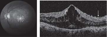
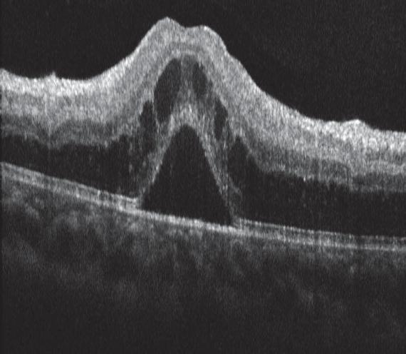
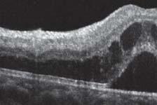
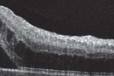
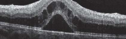
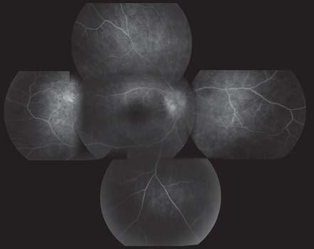
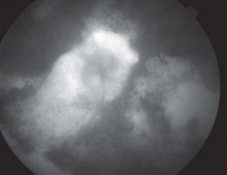
5 OCT in evaluation of inflammatory Neri P et Al. Clin Exp Rheumetol 2013 OCT in evaluation of inflammatory involvement in uveitis can help to detect: 1. Increased or decreased retinal thickness 2. Cystoid changes 3. Subretinal fluid 4. Vitreous traction 5. Epiretinal membrane 6. Choroidal exploration Neri P et Al. Clin Exp Rheumetol


:361-2.")
6 Behçet disease Central Foveal Thickness Table CFT (µm) Baseline 3 Months 12 Months Last follow-up Mean Media 441,3 167,4 167,2 162,7 Ozdemir H, Mudun B, Karacorlu M, Karacorlu S. Serous detachment of macula in Behçet disease.retina Apr-May;25(3): SD 48,6 12,8 14,3 5,6 OCT-Pregnancy Before After 6


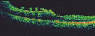
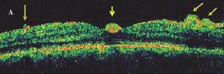
7 7-yr old After steroids+mmf POHS Failed surgical removal Days after OCT in evaluation of inflammatory involvement in uveitis can help to detect: 1. Increased or decreased retinal thickness 2. Cystoid changes 3. Subretinal fluid 4. Vitreous traction 5. Epiretinal membrane 6. Choroidal exploration Steroids+MMF+anti-VEGF 7


 device closer to the eye so that an inverted image is obtained Deeper")
8 OCT in evaluation of inflammatory involvement in uveitis can help to detect: 1. Increased or decreased retinal thickness 2. Cystoid changes 3. Subretinal fluid 4. Vitreous traction 5. Epiretinal membrane 6. Choroidal exploration Exploring choroid by the OCT Enhanced depth imaging OCT Swept Source OCT Enhanced depth imaging (EDI)-OCT Enhanced depth imaging (EDI)-OCT Placing the objective lens of the Spectralis SDOCT (SD-OCT) device closer to the eye so that an inverted image is obtained Deeper structures placed closer to the zero delay, Better visualization of the choroid Spaide et Al. Am J Ophthalmol


9 Courtesy of Professor Paulo E. Stanga, the Royal Eye Hospital & University of Manchester Professor Jose Maria Ruiz Moreno, University of Albacete, Spain Department of Ophthalmology, Fukushima Medical University. Take the slice! 9


10 Prognosis Atrophy Fibrosis Loss of Layers Figure 2. Representative High-Definition Optical Coherence Tomography Image of a Normal Macula The right eye of patient 22 had normal macular thickness with a preserved photoreceptor layer. White arrow indicates hypereflective line corresponding to the photoreceptor layer, while the blue arrow indicates the hypereflective line corresponding to the RPE. The hyporeflective space in between these lines, denoted by the red arrow, corresponds to the photoreceptor outer segments. Image represents horizontal line scan through center of fovea. Forooghian F, Yeh S, Faia LJ, Nussenblatt RB Uveitic foveal atrophy: clinical features and associations. Arch Ophthalmol Feb;127(2): Figure 1. Relationship Between Photoreceptor Layer Status and Visual Acuity Visual acuities of eyes with intact, partially intact, or absent photoreceptor layers were significantly different from each other (p < ). All pairwise comparisons between these groups were also significantly different (p < 0.001). Horizontal lines indicate mean values. Forooghian F, Yeh S, Faia LJ, Nussenblatt RB Uveitic foveal atrophy: clinical features and associations. Arch Ophthalmol Feb;127(2): Forooghian F, Yeh S, Faia LJ, Nussenblatt RB Uveitic foveal atrophy: clinical features and associations. Arch Ophthalmol Feb;127(2):
 is a non-invasive method that gives additional")


11 Limits 1. Opacified media: AS anomalies (cataract, posterior synechiae, etc ) Vitreous opacities 2. Scarce collaboration Conclusions In patients with uveitis, optical coherence tomography (OCT) is a non-invasive method that gives additional accuracy in the assessment and follow-up of the disease In clinical studies, OCT is unavoidable as is it the case for all methods that give objective & quantifiable data The OCT technology is still undergoing gradual improvement of its performance and has not reached yet the limits of its possibilities higher resolution OCT, higher speed of acquisition, spectral domain OCT, swept source OCT Conclusions Combined with other methods such as indocyanine green angiography, laser flare photometry and UBM, OCT contributes to improve the diagnosis and management of uveitis. Piergiorgio Neri Secretary General p.neri@univpm.it 11
Optical Coherence Tomography in Diabetic Retinopathy. Mrs Samantha Mann Consultant Ophthalmologist Clinical Lead of SEL-DESP
 Optical Coherence Tomography in Diabetic Retinopathy Mrs Samantha Mann Consultant Ophthalmologist Clinical Lead of SEL-DESP Content OCT imaging Retinal layers OCT features in Diabetes Some NON DR features
Advances in OCT Murray Fingeret, OD
 Disclosures Advances in OCT Murray Fingeret, OD Consultant Alcon, Allergan, Bausch & Lomb, Carl Zeiss Meditec, Diopsys, Heidelberg Engineering, Reichert, Topcon Currently Approved OCT Devices OCT Devices
OCT Interpretation in Retinal Disease
 OCT Interpretation in Retinal Disease Jay M. Haynie, OD, FAAO Financial Disclosure I have received honoraria or am on the advisory board for the following companies: Carl Zeiss Meditec Advanced Ocular
History/principles of the OCT What does the normal retinal OCT look like Vitreal disorders Retinal/RPE disorders Choroidal disorders
 Nathan Lighthizer, O.D., F.A.A.O. Assistant Professor Assistant Dean for Clinical Care Director of Continuing Education Chief of Specialty Care Clinics Chief of Electrodiagnostics Clinic Oklahoma College
Moncef Khairallah, MD
 Moncef Khairallah, MD Department of Ophthalmology, Fattouma Bourguiba University Hospital Faculty of Medicine, University of Monastir Monastir, Tunisia INTRODUCTION IU: anatomic form of uveitis involving
DRI OCT Triton Series A Multimodal Swept Source OCT
 DRI OCT Triton Series A Multimodal Swept Source OCT Color Red-Free FA FAF Posterior Anterior See what others can t see. A Multimodal Swept Source OCT DEEP RANGE IMAGING Swept Source OCT imaging massively
The Quick Guide to OCT Mastery 50 Real Cases with Expert Analysis
 OPTICAL COHERENCE TOMOGRAPHY The Quick Guide to OCT Mastery 50 Real Cases with Expert Analysis VOL 1 Sanjay Sharma, MD, FRCS, MSc (Epid), MBA Ophthalmologist, Epidemiologist Queen s University, Canada
OCT Assessment of the Vitreoretinal Relationship in CSME
 December 2007 Sonia Rani John et al. - IFIS 375 ORIGINAL ARTICLE OCT Assessment of the Vitreoretinal Relationship in CSME Dr. Manoj S. DNB FRCS, Dr. Unnikrishnan Nair MS DO FRCS, Dr. Gargi Sathish MS Introduction
DOME SHAPED MACULOPATHY. Ιωάννης Ν. Βαγγελόπουλος Χειρ. Οφθαλμίατρος - Βόλος
 DOME SHAPED MACULOPATHY Ιωάννης Ν. Βαγγελόπουλος Χειρ. Οφθαλμίατρος - Βόλος DOME SHAPED MACULOPATHY-DEFINITIONS The entity Dome Shaped Macula ( DSM ) was first described by Gaucher and associates in 2008
Reproducibility of Choroidal Thickness Measurements Across Three Spectral Domain Optical Coherence Tomography Systems
 Reproducibility of Choroidal Thickness Measurements Across Three Spectral Domain Optical Coherence Tomography Systems The MIT Faculty has made this article openly available. Please share how this access
Study of clinical significance of optical coherence tomography in diagnosis & management of diabetic macular edema
 Original Research Article Study of clinical significance of optical coherence tomography in diagnosis & management of diabetic macular edema Neha Kantilal Desai 1,*, Somesh Vedprakash Aggarwal 2, Sonali
Is OCT-A Needed As An Investigative Tool During The Management Of Diabetic Macular Edema
 Is OCT-A Needed As An Investigative Tool During The Management Of Diabetic Macular Edema Ayman M Khattab MD, FRCS Professor of Ophthalmology Cairo University Diabetic Macular Edema (DME) Diabetic macular
When optical coherence tomography (OCT)
") Macular Imaging: SD-OCT in nterior Segment Surgical Practice Many pathologic processes of the macula can be visualized or quantified only with this modality. y Steven G. Safran, MD When optical coherence
Macular Imaging: SD-OCT in nterior Segment Surgical Practice Many pathologic processes of the macula can be visualized or quantified only with this modality. y Steven G. Safran, MD When optical coherence
R&M Solutions
 Mohamed Hosny El-Bradey, MD., Assistant Professor of Ophthalmology, Tanta University. Wael El Haig, MD., Professor of Ophthalmology. Zagazeeg University. 1 Myopic CNV is considered the most common vision
Why Is Imaging Critical in My Uveitis Practice?
 Why Is Imaging Critical in My Uveitis Practice? Dilraj S. Grewal, MD Developed in collaboration Imaging Is the Backbone of Uveitis Workup and Monitoring Treatment Response FP FAF B- scan Multimodal Imaging
Title: OCT Analysis Workshop: Interpretation of OCT printouts
 Title: OCT Analysis Workshop: Interpretation of OCT printouts Authors: David Yang, OD, FAAO Staff Optometrist, VA Palo Alto Health Care System Associate Clinical Professor, UC Berkeley School of Optometry
Cirrus TM HD-OCT. Details define your decisions
 Cirrus TM HD-OCT Details define your decisions 2 With high-definition OCT Carl Zeiss Meditec takes you beyond standard spectral domain Built on 10 years experience at the vanguard of innovation, Carl Zeiss
Macular Hole Associated with Vogt-Koyanagi-Harada Disease at the Acute Uveitic Stage
 Published online: September 15, 2015 2015 The Author(s) Published by S. Karger AG, Basel 1663 2699/15/0063 0328$39.50/0 This article is licensed under the Creative Commons Attribution-NonCommercial 4.0
Citation BioMed Research International, 2015, v. 2015, article no Creative Commons: Attribution 3.0 Hong Kong License
 Title Relationship between Outer Retinal Layers Thickness and Visual Acuity in Diabetic Macular Edema Author(s) Wong, RLM; Lee, JWY; Yau, GSK; Wong, IYH Citation BioMed Research International, 2015, v.
Choroidal Mapping; a Novel Approach for Evaluating Choroidal Thickness and Volume
 Imaging Technique Choroidal Mapping; a Novel Approach for Evaluating Choroidal Thickness and Volume Jila Noori 1, MD; Mohammad Riazi Esfahani 1,2, MD Fedra Hajizadeh 2, MD; Mohammad-Mehdi Zaferani 1, MD
Measurement of Choroidal Thickness in Normal Eyes Using 3D OCT-1000 Spectral Domain Optical Coherence Tomography
 pissn: 111-8942 eissn: 292-9382 Korean J Ophthalmol 212;26(4):255-259 http://dx.doi.org/1.3341/kjo.212.26.4.255 Original Article Measurement of Choroidal Thickness in Normal Eyes Using 3D OCT-1 Spectral
VMA at the macula resulting in VMT
 Ocriplasmina for pharmacologic treatment in VMT Teresio Avitabile 1 Introduction PVD is a normal, physiologic process that occurs with aging; however, in some cases, PVD is incomplete Incomplete PVD localized
An A to Z guide on Epiretinal Membranes (ERMs) Paris Tranos PhD,ICO,FRCS OPHTHALMICA Vitreoretinal & Uveitis Department
 Paris Tranos PhD,ICO,FRCS OPHTHALMICA Vitreoretinal & Uveitis Department") An A to Z guide on Epiretinal Membranes (ERMs) Paris Tranos PhD,ICO,FRCS OPHTHALMICA Vitreoretinal & Uveitis Department Types of ERM Natural history OCT prognostic factors ERM with co-existing pathology
An A to Z guide on Epiretinal Membranes (ERMs) Paris Tranos PhD,ICO,FRCS OPHTHALMICA Vitreoretinal & Uveitis Department Types of ERM Natural history OCT prognostic factors ERM with co-existing pathology
8/6/17. Disclosures Aerie Pharmaceuticals Alcon BioTissue Diopsys Optovue Shire
 Nathan Lighthizer, O.D., F.A.A.O. Associate Professor Assistant Dean for Clinical Care Director of Continuing Education Chief of Specialty Care Clinics Oklahoma College of Optometry Tahlequah, OK lighthiz@nsuok.edu
Mark Dunbar: Disclosure
 Important Things to Understand About OCT Mark T. Dunbar, O.D., F.A.A.O. Bascom Palmer Eye Institute University of Miami, School of Medicine Mark Dunbar: Disclosure Optometry Advisory Board for: Allergan
OCT Interpretation. Financial Disclosure. Jay M. Haynie, OD, FAAO. OCT Image Layers 7/21/2014
 OCT Interpretation Jay M. Haynie, OD, FAAO Financial Disclosure I have received honoraria or am on the advisory board for the following companies: Olympia Tacoma Renton Kennewick - Washington Carl Zeiss
Swept-Source OCT Angiography: SS OCT Angio TM
 Swept-Source OCT Angiography: SS OCT Angio TM Not available in all countries, please check with your distributor. 2015.09 Swept-Source OCT Angiography: SS OCT Angio TM Introduction Optical coherence tomography
In 1990 our group first described reticular pseudodrusen as a. Choroidal Changes Associated with Reticular Pseudodrusen. Retina
 Retina Choroidal Changes Associated with Reticular Pseudodrusen Giuseppe Querques, 1,2 Lea Querques, 1,2 Raimondo Forte, 1 Nathalie Massamba, 1 Florence Coscas, 1 and Eric H. Souied 1 PURPOSE. To analyze
Clinical Study Spectral Domain OCT: An Aid to Diagnosis and Surgical Planning of Retinal Detachments
 Ophthalmology Volume 2011, Article ID 725362, 4 pages doi:10.1155/2011/725362 Clinical Study Spectral Domain OCT: An Aid to Diagnosis and Surgical Planning of Retinal Detachments Graham Auger and Stephen
Course # Getting to Know Your OCT
 Course # 140 Getting to Know Your OCT Course Title: Lecturer: Getting to Know Your OCT Brad Sutton, OD, FAAO IU School of Optometry Financial Disclosures No financial disclosures Optical Coherence Tomography-OCT
CENTRAL SEROUS CHORIORETINOPATHY (CSC) IS
 IS") Association Between the Efficacy of Half-Dose Photodynamic Therapy With Indocyanine Green Angiography and Optical Coherence Tomography Findings in the Treatment of Central Serous Chorioretinopathy MASSIMO
Association Between the Efficacy of Half-Dose Photodynamic Therapy With Indocyanine Green Angiography and Optical Coherence Tomography Findings in the Treatment of Central Serous Chorioretinopathy MASSIMO
Mariam Raouf Fadel M.B., B.Ch. M.Sc., Cairo University. A thesis. Submitted by. For partial fulfillment of. MD Degree in Ophthalmology
 Correlation of fundus autofluorescence and spectral domain OCT findings of the macula with visual outcome after successful repair of rhegmatogenous retinal detachment A thesis Submitted by Mariam Raouf
Introducing ANGIOVUE ESSENTIAL. Built on the Avanti Widefield OCT Platform. OCT Angiography for Primary Eye Care
 Introducing ANGIOVUE ESSENTIAL Built on the Avanti Widefield OCT Platform OCT Angiography for Primary Eye Care Transform Your View of the Retina OCT Angiography (OCTA) is a quick non-invasive test that
Characterization of serous retinal detachments in uveitis patients with optical coherence tomography
 Characterization of serous retinal detachments in uveitis patients with optical coherence tomography Annamieka Simmons-Rear, Oregon Health and Science University Steven Yeh, Emory University Brian T. Chan-Kai,
Yasser R. Serag, MD Tamer Wasfi, MD El- Saied El-Dessoukey, MD Magdi S. Moussa, MD Anselm Kampik, MD
 Microperimetric Evaluation of Brilliant Blue G- assisted Internal Limiting Membrane Peeling By Yasser R. Serag, MD Tamer Wasfi, MD El- Saied El-Dessoukey, MD Magdi S. Moussa, MD Anselm Kampik, MD The internal
OPTIC DISC PIT Pathogenesis and Management OPTIC DISC PIT
 OPTIC DISC PIT Pathogenesis and Management Abdel-Latif Siam Ain Shams University Cairo Egypt OPTIC DISC PIT Congenital pit is an atypical coloboma usually located on the temporal edge of the disc, associated
Optical Coherence Tomograpic Features in Idiopathic Retinitis, Vasculitis, Aneurysms and Neuroretinitis (IRVAN)
") Columbia International Publishing Journal of Ophthalmic Research (2014) Research Article Optical Coherence Tomograpic Features in Idiopathic Retinitis, Vasculitis, Aneurysms and Neuroretinitis (IRVAN)
Columbia International Publishing Journal of Ophthalmic Research (2014) Research Article Optical Coherence Tomograpic Features in Idiopathic Retinitis, Vasculitis, Aneurysms and Neuroretinitis (IRVAN)
We are IntechOpen, the world s leading publisher of Open Access books Built by scientists, for scientists. International authors and editors
 We are IntechOpen, the world s leading publisher of Open Access books Built by scientists, for scientists 3,700 108,500 1.7 M Open access books available International authors and editors Downloads Our
Key words: Choroidal neovascularisation, Laser coagulation, Retinal imaging
 420 Mini Review The Role of Optical Coherence Tomography (OCT) in the Diagnosis and Management of Retinal Angiomatous Proliferation (RAP) in Patients with Age-related Macular Degeneration Antonio Polito,
You can C-ME after Uveitis
 You can C-ME after Uveitis Abstract: Approximately 50% of uveitis patients will present with vision loss secondary to cystoid macular edema[1]. Two patients with uveitis present with a constant decrease
Case report 12/10/2014. Delphine Lam ; Dr Mayer Srour Service d ophtalmologie Professeur E.Souied Université Paris Est
 Case report 12/10/2014 Delphine Lam ; Dr Mayer Srour Service d ophtalmologie Professeur E.Souied Medical history Man, 75 years old Complaint: Vision loss in left eye in June 2014 Past ophthalmologic history:
Clinical Study Choroidal Thickness in Eyes with Unilateral Ocular Ischemic Syndrome
 Hindawi Publishing Corporation Journal of Ophthalmology Volume 215, Article ID 62372, 5 pages http://dx.doi.org/1.1155/215/62372 Clinical Study Choroidal Thickness in Eyes with Unilateral Ocular Ischemic
Cirrus TM HD-OCT. Details defi ne your decisions
 Cirrus TM HD-OCT Details defi ne your decisions 2 With high-defi nition OCT Carl Zeiss Meditec takes you beyond standard spectral domain Built on 10 years experience at the vanguard of innovation, Carl
EPIRETINAL MEMBRANES IN NEOVASCULAR AGE-RELATED MACULAR DEGENERATION. Effect on Outcomes of Anti-vascular Endothelial Growth Factor Therapy
 EPIRETINAL MEMBRANES IN NEOVASCULAR AGE-RELATED MACULAR DEGENERATION Effect on Outcomes of Anti-vascular Endothelial Growth Factor Therapy EMINE ESRA KARACA, MD,* BURÇIN KEPEZ YILDIZ, MD, MEHMET ÖZGÜR
OCT Angiography in Primary Eye Care
 OCT Angiography in Primary Eye Care An Image Interpretation Primer Julie Rodman, OD, MS, FAAO and Nadia Waheed, MD, MPH Table of Contents Diabetic Retinopathy 3-6 Choroidal Neovascularization 7-9 Central
Diabetic Retinopathy Clinical Research Network
 Diabetic Retinopathy Clinical Research Network Comparison of Time Domain OCT and Spectral Domain OCT Retinal Thickness Measurement in Diabetic Macular Edema Version 1.0 June 16, 2009 comparison of td vs
Optical Coherence Tomography: Pearls for the Anterior Segment Surgeon Basic Science Michael Stewart, M.D.
 Optical Coherence Tomography: Pearls for the Anterior Segment Surgeon Basic Science Michael Stewart, M.D. Disclosure OCT Optical Coherence Tomography No relevant financial relationships I will refer to
Patterns of Macular Edema in Uveitis as Diagnosed by Optical Coherence Tomography in Tertiary Eye Center
 Original Article Patterns of Macular Edema in Uveitis as Diagnosed by Optical Coherence Tomography in Tertiary Eye Center Sharad Gupta 1, Dev Narayan Shah 2, Sagun Narayan Joshi 3, Manoj Aryal 4, Lila
The College of Optometrists - Learning outcomes for the Professional Certificate in Medical Retina
 Learning outcomes for the Professional Certificate in Medical Retina, incorporating diabetic retinopathy screening and age related macular degeneration The professional certificate is a prerequisite to
3/6/2014. Hoda MH Mostafa MD Associate Professor of Ophthalmology Cairo University. The author has no proprietary interest. Today s Objectives
 Hoda MH Mostafa MD Associate Professor of Ophthalmology Cairo University The author has no proprietary interest Today s Objectives Identify the CLINICAL SCENARIOS IN MACULAR EDEMA where OCT plays a MAJOR
Vitreomacular interface disorders. Ghanbari MD 1393:10:25
 Vitreomacular interface disorders Ghanbari MD 1393:10:25 Human vitreous after dissection of the sclera, choroid, and retina. Lamellar structure of the posterior vitreous cortex (PVC) in the monkey. V =
Retina Conference. Janelle Fassbender, MD, PhD University of Louisville Department of Ophthalmology and Visual Sciences 09/04/2014
 Retina Conference Janelle Fassbender, MD, PhD University of Louisville Department of Ophthalmology and Visual Sciences 09/04/2014 Subjective CC/HPI: 64 year old Caucasian female referred by outside ophthalmologist
IN NICU OCT UTILIZES A CONCEPT KNOWN AS INTERFEROMETRY APPLICATIONS FOR OCT THE PRIMARY USE IN THE EYE - RETINA
 2016 25 YEARS OF OPTICAL COHERENCE TOMOGRAPHY OPTICAL COHERENCE TOMOGRAPHY IN NICU Marcin Stopa, MD, PhD, FEBO Department of Ophthalmology, Chair of Ophthalmology and Optometry. Poznan University of Medical
Optical Coherence Tomography of the Retina. New Technology - New Insights
 Bahrain Medical Bulletin, Vol. 27, No. 3, September 2005 Optical Coherence Tomography of the Retina. New Technology - New Insights Mohinder Singh, FRCS,FRCOphth* Optical imaging is an emerging new technology,
EPIRETINAL MEMBRANE & VITREOMACULAR TRACTION
 EPIRETINAL MEMBRANE & VITREOMACULAR TRACTION Management of ERM and VMT K.V.Chalam,MD,PhD,MBA,FACS Professor and Director of Retina Loma Linda Eye Institute Los Angeles, USA REVIEW ANATOMY The vitreous
The Human Eye. Cornea Iris. Pupil. Lens. Retina
 The Retina Thin layer of light-sensitive tissue at the back of the eye (the film of the camera). Light rays are focused on the retina then transmitted to the brain. The macula is the very small area in
AperTO - Archivio Istituzionale Open Access dell'università di Torino
 AperTO - Archivio Istituzionale Open Access dell'università di Torino Artifacts in automatic retinal segmentation using different optical coherence tomography instruments. This is the author's manuscript
Quantitative Reduction in Central Foveal Thickness After First Anti-VEGF Injection as a Predictor of Final Outcome in BRVO Patients
 Original clinical study Quantitative Reduction in Central Foveal Thickness After First Anti-VEGF Injection as a Predictor of Final Outcome in BRVO Patients Rupak Roy, MS, Kumar Saurabh, MS, Avirupa Ghose,
 www.brisbaneeyeclinic.com.au Brisbane Eye Clinic is a modern ophthalmology practice focused on the provision of excellent medical eye care. The Clinic has two convenient consulting locations, our Wickham
Audit of Macular Hole Surgery, Visual Outcome Prediction on OCT Appearance of Macular Hole
 International Journal of Ophthalmology & Visual Science 2017; 2(4): 93-97 http://www.sciencepublishinggroup.com/j/ijovs doi: 10.11648/j.ijovs.20170204.13 Audit of Macular Hole Surgery, Visual Outcome Prediction
Vitreo-retinal interface pathologies and fibrinolytic treatment approaches
 Vitreo-retinal interface pathologies and fibrinolytic treatment approaches Constantin J. Pournaras Memorial A. de Rothschild Clinical Research Group La Colline Ophthalmology Center Vitreoretinal Interface
ZEISS AngioPlex OCT Angiography. Clinical Case Reports
 Clinical Case Reports Proliferative Diabetic Retinopathy (PDR) Case Report 969 PROLIFERATIVE DIABETIC RETINOPATHY 1 1-year-old diabetic female presents for follow-up of proliferative diabetic retinopathy
Fellow Eye Findings of Highly Myopic Subjects Operated for Retinal Detachment Associated with a Macular Hole
 Fellow Eye Findings of Highly Myopic Subjects Operated for Retinal Detachment Associated with a Macular Hole Guido Ripandelli, MD, Andrea Maria Coppé, MD, Vincenzo Parisi, MD, Mario Stirpe, MD Purpose:
What Is O.C.T. and Why Should I Give A Rip? OCT & Me How Optical Coherence Tomography Changed the Life of a Small Town Optometrist 5/19/2014
 OCT & Me How Optical Coherence Tomography Changed the Life of a Small Town Optometrist Email: myoder@wcoil.com Mark A. Yoder, O.D. 107 N. Main Street PO Box 123 Bluffton, OH 45817 @yoderod 115.02 Histoplasma
OCT in Diabetic Macular Edema and its Correlation with Flourescein Angiography
 Uvea OCT in Diabetic Macular Edema and its Correlation with Flourescein Angiography Kirti Jaisingh MS Kirti Jaisingh MS, Yashpal Goel* MS, Kshitij Aditya** DO * Guru Nanak Eye Centre, New Delhi ** Baba
RETINAL THICKENING MAY BE PRESENT IN A NUMBER. B-scan Ultrasonography for the Detection of Macular Thickening METHODS
 B-scan Ultrasonography for the Detection of Macular Thickening JAMES C. LAI, MD, SANDRA S. STINNETT, DRPH, AND GLENN J. JAFFE, MD PURPOSE: To report the sensitivity and specificity of B-scan ultrasonography
MACULAR EDEMA (ME) IS
 IS") CLINICAL SCIENCES Subfoveal Serous Retinal Detachment in Patients With Uveitic Macular Edema Jeannette Ossewaarde van Norel, MD; Elize M. Berg, MD; Karen M. Sijssens, MD, PhD; Aniki Rothova, MD, PhD Objective:
CLINICAL SCIENCES Subfoveal Serous Retinal Detachment in Patients With Uveitic Macular Edema Jeannette Ossewaarde van Norel, MD; Elize M. Berg, MD; Karen M. Sijssens, MD, PhD; Aniki Rothova, MD, PhD Objective:
A retrospective nonrandomized study was conducted at 3
 Department of Ophthalmology, Kangbuk Samsung Hospital, Sungkyunkwan University College of Medicine 1, Seoul, Korea Hangil Eye Hospital 2, Incheon, Korea Seoul National University Bundang Hospital 3, Seongnam,
When Retina is not detached anymore. Alexandra Mouallem, Agnès Glacet-Bernard Service du Professeur Souied Le 19/03/2014
 When Retina is not detached anymore, Agnès Glacet-Bernard Service du Professeur Souied Le 19/03/2014 Medical History Mr V. 57 yo man December 2013 : Bullous superior retinal detachment caused by 2 retinal
A Patient s Guide to Diabetic Retinopathy
 Diabetic Retinopathy A Patient s Guide to Diabetic Retinopathy 840 Walnut Street, Philadelphia PA 19107 www.willseye.org Diabetic Retinopathy 1. Definition Diabetic retinopathy is a complication of diabetes
Measurement of Subfoveal Choroidal Thickness Using Spectral Domain Optical Coherence Tomography
 c l i n i c a l s c i e n c e Measurement of Subfoveal Choroidal Thickness Using Spectral Domain Optical Coherence Tomography Emily A. McCourt, MD; Brian C. Cadena, PhD; Cullen J. Barnett, CRA; Antonio
Flore De Bats, 1 Benjamin Wolff, 2,3 Martine Mauget-Faÿsse, 2 Claire Scemama, 2 and Laurent Kodjikian Introduction
 Case Reports in Medicine Volume 2013, Article ID 260237, 7 pages http://dx.doi.org/10.1155/2013/260237 Case Report B-Scan and En-Face Spectral-Domain Optical Coherence Tomography Imaging for the Diagnosis
Observation of Posterior Precortical Vitreous Pocket Using Swept-Source Optical Coherence Tomography
 Anatomy and Pathology Observation of Posterior Precortical Vitreous Pocket Using Swept-Source Optical Coherence Tomography Hirotaka Itakura, Shoji Kishi, Danjie Li, and Hideo Akiyama Department of Ophthalmology,
Acquired vitelliform detachment in patients with subretinal drusenoid deposits (reticular pseudodrusen)
") Zurich Open Repository and Archive University of Zurich Main Library Strickhofstrasse 39 CH-8057 Zurich www.zora.uzh.ch Year: 2011 Acquired vitelliform detachment in patients with subretinal drusenoid
Zurich Open Repository and Archive University of Zurich Main Library Strickhofstrasse 39 CH-8057 Zurich www.zora.uzh.ch Year: 2011 Acquired vitelliform detachment in patients with subretinal drusenoid
NIH Public Access Author Manuscript JAMA Ophthalmol. Author manuscript; available in PMC 2013 September 10.
 NIH Public Access Author Manuscript Published in final edited form as: JAMA Ophthalmol. 2013 May ; 131(5): 693 694. doi:10.1001/jamaophthalmol.2013.692. Effect of Intravitreous Anti Vascular Endothelial
Clinical Case Presentation. Branch Retinal Vein Occlusion. Sarita M. Registered Nurse Whangarei Base Hospital
 Clinical Case Presentation on Branch Retinal Vein Occlusion Sarita M. Registered Nurse Whangarei Base Hospital Introduction Case Study Pathogenesis Clinical Features Investigations Treatment Follow-up
Fundus Autoflu ores cence Findings Pre and Post Intravitreal Bevacizumab Injection in Patients with Diabetic Macular Edema
 Med. J. Cairo Univ., Vol. 84, No. 1, September: 1093-1100, 2016 www.medicaljournalofcairouniversity.net Fundus Autoflu ores cence Findings Pre and Post Intravitreal Bevacizumab Injection in Patients with
Correspondence should be addressed to Ludovico Iannetti;
 BioMed Research International Volume 2015, Article ID 853728, 5 pages http://dx.doi.org/10.1155/2015/853728 Clinical Study Correlation between Visual Acuity, Inner Segment/Outer Segment Junction, and Cone
Technicians & Nurses Program
 ASCRS ASOA Symposium & Congress Technicians & Nurses Program April 17-21, 2015 San Diego, California Optical Coherence Tomography: Essentials in Anterior and Posterior Segment Imaging Michael Stewart,
ATLAS OF OCT. Retinal Anatomy in Health & Pathology by Neal A. Adams, MD. Provided to you by:
 ATLAS OF OCT Retinal Anatomy in Health & Pathology by Neal A. Adams, MD Provided to you by: Atlas of OCT The OCT Atlas is written by Neal A. Adams, MD, and produced by Heidelberg Engineering, Inc. to help
Seokhyun Bae, Kiwon Jin, Hakyoung Kim and So Hyun Bae *
 Bae et al. BMC Ophthalmology (2015) 15:180 DOI 10.1186/s12886-015-0170-4 RESEARCH ARTICLE Open Access Clinical parameters related to metamorphopsia outcome in patients with resolved central serous chorioretinopathy
Longitudinal spectral domain optical coherence tomography changes in eyes with intraocular lymphoma
 University of Nebraska Medical Center DigitalCommons@UNMC Journal Articles: Ophthalmology Ophthalmology 9-2013 Longitudinal spectral domain optical coherence tomography changes in eyes with intraocular
CORRELATION BETWEEN CENTRAL FOVEAL THICKNESS AND VISUAL ACUITY IN PATIENTS WITH IDIOPATHIC VITREOMACULAR TRACTION
 CORRELATION BETWEEN CENTRAL FOVEAL THICKNESS AND VISUAL ACUITY IN PATIENTS WITH IDIOPATHIC VITREOMACULAR TRACTION MEHMET M. UZEL, MD, MEHMET CITIRIK, MD, CAGRI ILHAN, MD, KEMAL TEKIN, MD Purpose: To evaluate
Oishi A, Miyamoto K, Yoshimura N. Etiology of carotid cavernous fistula in Japanese. Jpn J Ophthalmol. 2009;53:40-43.
 Kimura T, Takagi H, Miyamoto K, Kita M, Watanabe D, Yoshimura N. Macular hole with epiretinal membrane after triamcinolone-assisted vitrectomy for proliferative diabetic retinopathy. Retinal Cases Brief
Role of OCT in the diagnosis and follow up of diabetic macular edema
 Seminars in Ophthalmology 0882-0538/02/1701-019$16.00 2002, Vol. 17, No. 1, pp. Swets & Zeitlinger Role of OCT in the diagnosis and follow up of diabetic macular edema Giacomo Panozzo, Elena Gusson, arbara
 www.brisbaneeyeclinic.com.au Brisbane Eye Clinic is a modern ophthalmology practice focused on the provision of excellent medical eye care. The Clinic has two convenient consulting locations, our Wickham
Clinically Significant Macular Edema (CSME)
") Clinically Significant Macular Edema (CSME) 1 Clinically Significant Macular Edema (CSME) Sadrina T. Shaw OMT I Student July 26, 2014 Advisor: Dr. Uwaydat Clinically Significant Macular Edema (CSME) 2
Clinically Significant Macular Edema (CSME) 1 Clinically Significant Macular Edema (CSME) Sadrina T. Shaw OMT I Student July 26, 2014 Advisor: Dr. Uwaydat Clinically Significant Macular Edema (CSME) 2
Convergence in. Introduction. Case Report: Dr. Piyali SenM.B.B.S, Dr. Abhipsha Saha M.B.B.S, Dr. Anuradha Chandra M.S,FAICO
 Convergence in Dr. Piyali SenM.B.B.S, Dr. Abhipsha Saha M.B.B.S, Dr. Anuradha Chandra M.S,FAICO Introduction non-progressive ophthalmoplegia with or without ptosis affecting part or all of the occulomotor
Learn Connect Succeed. JCAHPO Regional Meetings 2017
 Learn Connect Succeed JCAHPO Regional Meetings 2017 How Retinal Imaging Guides Treatment Odette Margit Houghton MD Question 1 Which OCT has the highest resolution? A: Swept source OCT B: Spectral domain
A spectral-domain OCT study of formerly premature children. Prat Itharat MD May 30, 2008 Vanderbilt Eye Institute Preceptor: Dr.
 A spectral-domain OCT study of formerly premature children. Prat Itharat MD May 30, 2008 Vanderbilt Eye Institute Preceptor: Dr. Recchia Background: Optical coherence tomography (OCT) OCT analogous to
Measuring of the fovea and foveola using line scans and 3D Macular scans obtained with spectral domain optical coherent tomography.
 Measuring of the fovea and foveola using line scans and 3D Macular scans obtained with spectral domain optical coherent tomography. Vakhrameeva O.A. 1, Moiseenko G.A. 1, Maltsev D.S. 2, Sukhinin M.V. 2,
Clinical Trial Endpoints for Macular Diseases
 Clinical Trial Endpoints for Macular Diseases Developed in collaboration Learning Objective Upon completion, participants should be able to: Summarize types of biomarkers of progression and treatment response
Clinical Outcomes After Intravitreal Bevacizumab Injection for Diabetic Macular Edema
 Original Article Clinical Outcomes After Intravitreal Bevacizumab Injection for Diabetic Macular Edema Karen Joyce G. Castro, MD, Marie Joan V. Loy, MD International Eye Institute St. Luke s Medical Center
Correlation of visual acuity and optical coherence tomography in patients with decreased visual acuity after surgery for retinal detachment
 VOL. NO. PHILIPPINE JOURNAL OF Ophthalmology JULY ORIGINAL ARTICLE DECEMBER 9 Ildefonso M. Chan, MD, Darby E. Santiago, MD Rafael E. de Guzman III, MD, Department of Ophthalmology and Visual Sciences Sentro
Visual and Anatomical Outcomes of Vitreous Surgery for Large Macular Holes
 March 2009 Raju K.V. et al. - Closed Globe Injuries 31 ORIGINAL ARTICLE Visual and Anatomical Outcomes of Vitreous Surgery for Large Macular Holes Dr. Mahesh G. MS DO DNB FRCSEd, Dr. A. Giridhar MS, Dr.
Citation. As Published Publisher. Version
 Effect of Intravitreous Anti Vascular Endothelial Growth Factor Therapy on Choroidal Thickness in Neovascular Age-Related Macular Degeneration Using Spectral-Domain The MIT Faculty has made this article
THE OPHTHALMOLOGIST S NEEDS FOR THE ANALYSIS OF THE RETINA
 biophotonics end-users needs THE OPHTHALMOLOGIST S NEEDS FOR THE ANALYSIS OF THE RETINA Dr Matonti Frédéric CHU Nord / INT AMU Marseille ANATOMY OF THE RETINA ANATOMY OF THE RETINA ANATOMY OF THE RETINA
Envisu TM C-Class SD-OCT System
 Bioptigen SDOIS is intended to acquire, process, display and save depth-resolved images of Ocular tissue microstructure using Spectral Domain Optical Coherence Tomography (SDOCT). The SDOIS is indicated
Although photocoagulation and photodynamic PROCEEDINGS PEGAPTANIB SODIUM FOR THE TREATMENT OF AGE-RELATED MACULAR DEGENERATION *
 PEGAPTANIB SODIUM FOR THE TREATMENT OF AGE-RELATED MACULAR DEGENERATION Evangelos S. Gragoudas, MD ABSTRACT In December 24, the US Food and Drug Administration (FDA) approved pegaptanib sodium. Pegaptanib
RETINA 2018 OBJECTIVES OCT VERY USEFUL INFORMATION SAFE AND FRIENDLY 1/11/2018 KELLY MITCHELL
 RETINA 2018 KELLY MITCHELL OBJECTIVES HIGHLIGHT NEW DIAGNOSTIC & TREATMENT OPTIONS REVIEW DIAGNOSTIC KEYS OF SELECT RETINAL DISEASES DISCUSS USE OF IMAGING AND REFERRAL RECOURSES FOR PATIENT BENEFIT OCT