PRIMUS 200 from ZEISS The essential OCT
|
|
|
- Kelly Sanders
- 6 years ago
- Views:
Transcription


1 EN 00_00I The contents of the brochure may differ from the current status of approval of the product in your country. Please contact your regional representative for more information. Subject to change in design and scope of delivery and as a result of ongoing technical development. CIRRUS, Selective Pixel Profiling, AutoCenter, and FoveaFinder are either registered trademarks or trademarks of Carl Zeiss Meditec AG or of other companies of the ZEISS Group in Germany and/or in other countries. 05 by Carl Zeiss Meditec, Inc. All copyrights reserved by Page Size Today, OCT is essential for the daily practice of quality eye care You cannot treat what you cannot see. Today, OCT makes ocular anatomy visible in ways never available before so you can see more and treat more. With ZEISS PRIMUS 00, you can join doctors around the world who have placed their trust in ZEISS, the pioneer in ophthalmic OCT, as their OCT partner. With ZEISS, you can enjoy the support of the extensive ZEISS global service network and rapid response technologies including remote diagnostics to ensure your OCT is performing at its best. Today, you can confidently invest in your practice with the certainty that comes with the all new essential OCT from ZEISS: PRIMUS 00. Carl Zeiss Suzhou Co. Ltd. Carl Zeiss Meditec, Inc. Carl Zeiss Meditec AG Modern Industrial Square -B No. Xing Pu Road, Suzhou Industrial Park, Suzhou China Hacienda Drive Dublin, CA 9568 USA Toll-Free: Phone: Fax: Goeschwitzer Str Jena GERMANY Phone: Fax: Phone: Fax: PRIMUS 00 from ZEISS The essential OCT


2 Clear Visualization. Advanced Technology. Reliability. Essential elements of your first OCT. Reveal the hidden structures in the retina with the new PRIMUS 00 from ZEISS, the pioneer in ophthalmic OCT. Ideal for small practices, PRIMUS 00 delivers the essential clinical applications to help you manage your patients with certainty. ZEISS PRIMUS 00 is the technology engine with the power and performance you need to deliver the optimal level of clinical care and to grow your practice. See beyond the surface Today, optical coherence tomography (OCT) has become as essential to clinical eye care as perimetry or fundus photography. OCT gives clinical insight to better understand your patient s condition and the power to manage a broader range of pathologies within your practice. By incorporating legendary ZEISS optics and proprietary algorithms, in a compact and intuitive design, the ZEISS PRIMUS 00 is both a diagnostic and a patient education tool as it helps you to effectively communicate the essential aspects of a comprehensive treatment plan.


")


 technology is used to display high-quality fundus")

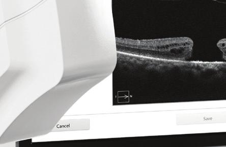
3 Vitreomacular traction syndrome (VMT) Clearer images for deeper insight ZEISS PRIMUS 00 inherits the fundamental quality of world-class ZEISS optics, providing you with clear and compelling OCT and fundus images to help you detect pathologies at the earliest possible moment. High definition B-scans With high SNR (Signal-to-Noise Ratio) and extended integration time, PRIMUS 00 reveals micro-structural details that are critical to effectively manage a broad range of diseases. Selective Pixel ProfilingTM ensures brilliant, high-definition imaging across the B-Scan for accurate diagnostic insights and assured treatment decision making. Clear fundus images Confocal Scanning Laser Ophthalmoscopy (cslo) technology is used to display high-quality fundus images. Clear, sharp fundus images are essential to accurately correlate the B-scans and corresponding fundus image. Central retinal venous occulusion (CRVO) with cystoid macula edema (CME) and subfoveal fluid Age-related macular degeneration (AMD) 5


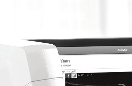
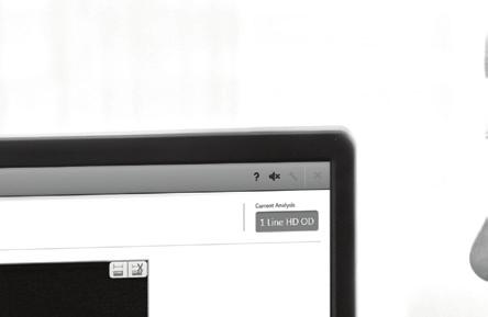


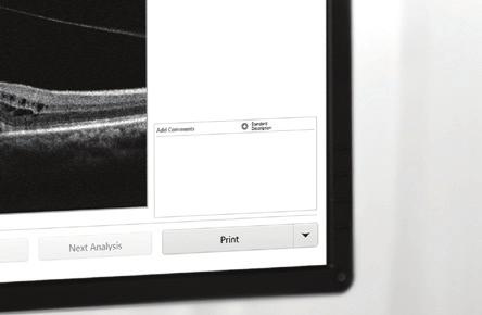

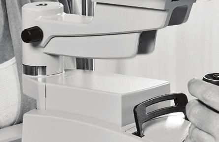


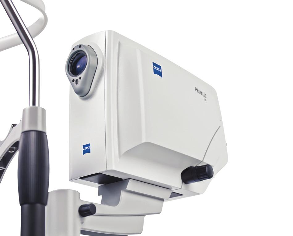
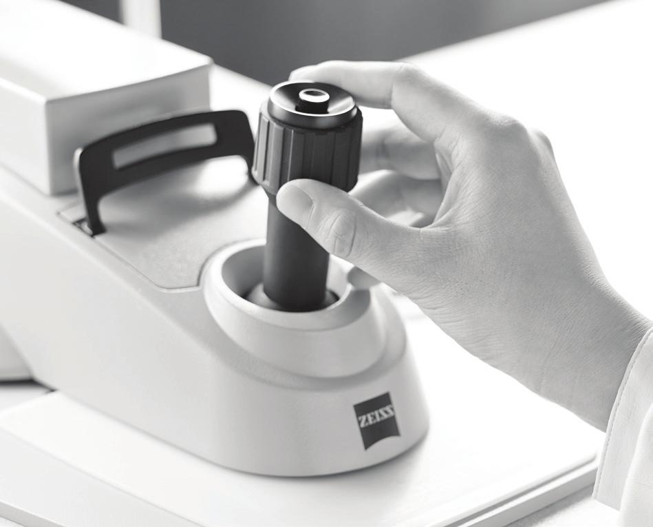

4 Simplicity through smart workflow ZEISS PRIMUS 00 utilizes a simple -step process for capturing all anterior and posterior segment scans. This optimized workflow helps to increase operator efficiency while minimizing patient chair time.. Select Report-driven workflow Enables selection of desired reports and automatically sequences the required scan-protocols for acquisition.. Capture Ease of use through an intuitive design System-guided navigation Easy acquisition protocols prompt and lead you through all selected scans.. Review With its simple and sleek, yet sturdy design, ZEISS PRIMUS 00 offers you a clear view of the patient during scan acquisition. 6 Familiar slit-lamp-style joystick allows you to align and capture images with ease. Pre-selected reports Analyses are automatically prepared for you to scan and select with the click of a button. 7

 reports.")
5 // RETINA ASSESSMENT // GLAUCOMA MANAGEMENT Visualize. Assess. Act. With clear visualization of cross-sectional retinal layers and ZEISS-proven algorithms, ZEISS PRIMUS 00 enables you to make qualitative and quantitative assessments of your patient s retinal condition. Visualize the retina with high definition and 5 Line HD B-scans and measure macular abnormalities using Macular Thickness Analysis. Determine your next steps with confidence. Identify. Monitor. Manage. Identify and monitor at-risk patients for glaucoma with the comprehensive optic nerve and retinal nerve fiber layer (ONH and RNFL) reports. Easy-to-understand graphics can serve as teaching tools for patients and can help improve compliance for better disease management. 5 ETDRS measurement grid with normative data colors uses the Auto FoveaFinder TM to automatically and accurately locate the fovea, providing precise macular thickness values for each visit. Horizontal and vertical B-scans are extracted from the data cube through the center of the disc. RPE layer and disc boundaries are shown in black. ILM and cup boundaries are shown in red. Clear fundus image with options to overlay ILM-RPE thickness map or move the ETDRS to desired foveal location. OCT fundus image of the Optic Nerve Head with AutoCenter TM automatically centers the Optic Disc and RNFL circle. Large B-scan display in color or greyscale helps both with visualizing pathologies quickly and with improving patient communication. Enter or choose from predefined comments for patient report. RNFL thickness map is a topographical display of RNFL thickness. RNFL and Neuro-retinal Rim Thickness are shown in TSNIT graphs. Other key parameters are displayed in a table format with normative data colors. 5 RNFL Quadrant displays patient s RNFL average thickness in each quadrant along the calculation circle with superior and inferior normative data colors. 8 9


6 // ANTERIOR SEGMENT IMAGING Observe. Unveil. Clarify. The ZEISS PRIMUS 00 delivers anterior segment imaging to visualize the fi ne details of the cornea and angles in a fresh and intuitive dimension, providing new clarity for your assessment and for your patient s understanding of their condition. Visualize a clear image of the cornea, iris and sclera, along with the scan-line location. Single-line, high-defi nition angle view B-scan using the Selective Pixel Profi ling TM algorithm. Single-line, high-defi nition cornea B-scan with identifi able epithelium, Bowman s membrane and stroma. 0
, 80 nm A-scan depth.")
, 80 nm Field of view 9º H x º V Transverse resolution 80 μm (in Tissue) Scan Reports Retina analysis Macular thickness using Auto")
 Dimensions of instrument 0L x 80W x 50H (cm) Fixation Internal, External Internal fixation focus adjustment -D to +7D (Diopters) Pupil Size requirement > mm Internal")
7 The moment you see beyond the surface. This is the moment we work for. Technical Specification OCT imaging Methodology Spectral Domain OCT Optical source Super Luminescent Diode (SLD), 80 nm A-scan depth.0 mm (in tissue), 0 Points Axial resolution 5± μm (in Tissue) Transverse resolution 0 μm (in Tissue, FWHM) Fundus imaging Methodology Confocal Scanning Laser Ophthalmoscopy (cslo) Live fundus image During alignment Optical source Super Luminescent Diode (SLD), 80 nm Field of view 9º H x º V Transverse resolution 80 μm (in Tissue) Scan Reports Retina analysis Macular thickness using Auto FoveaFinder 5 Line raster with adjustable orientation Line HD with adjustable orientation Glaucoma analysis ONH and RNFL analysis using AutoCenter Anterior segment Angle view Cornea view Electrical and physical Weight 0 kg (88 lbs) Dimensions of instrument 0L x 80W x 50H (cm) Fixation Internal, External Internal fixation focus adjustment -D to +7D (Diopters) Pupil Size requirement > mm Internal Computer Operating system / processor Windows 7/Intel Core i-0e Memory GB Hard drive / internal storage 500GB/.5inch Display (resolution: 66 x 768) USB ports ports Ethernet ports ports, 0/00 with independent IP address Technical specifications are subject to change. // INSIGHT MADE BY ZEISS
PRIMUS 200 from ZEISS The essential OCT
 PRIMUS 200 from ZEISS The essential OCT Seeing beyond the surface. ZEISS PRIMUS 200 // INNOVATION MADE BY ZEISS Clear Visualization. Advanced Technology. Reliability. Essential elements of your first OCT.
Cirrus TM HD-OCT. Details define your decisions
 Cirrus TM HD-OCT Details define your decisions 2 With high-definition OCT Carl Zeiss Meditec takes you beyond standard spectral domain Built on 10 years experience at the vanguard of innovation, Carl Zeiss
Cirrus TM HD-OCT. Details defi ne your decisions
 Cirrus TM HD-OCT Details defi ne your decisions 2 With high-defi nition OCT Carl Zeiss Meditec takes you beyond standard spectral domain Built on 10 years experience at the vanguard of innovation, Carl
Evolving glaucoma management True diagnostic integration for the preservation of vision
 Evolving glaucoma management True diagnostic integration for the preservation of vision // GLAUCOMA MANAGEMENT MADE BY ZEISS The moment you are certain it is glaucoma. This is the moment we work for. There
PLEX Elite 9000 from ZEISS Swept-Source OCT
 PLEX Elite 9000 from ZEISS Swept-Source OCT Uncovering the undiscovered. ZEISS PLEX Elite 9000 // INNOVATION MADE BY ZEISS 2 Ultra-wide angiography En face montage Image courtesy of Prof. G. Querques,
ZEISS AngioPlex OCT Angiography Making the revolutionary, routine.
 ZEISS AngioPlex OCT Angiography Making the revolutionary, routine. The moment that revolutionary insight becomes routine. // OCT ANGIOGRAPHY MADE BY ZEISS CIRRUS with AngioPlex creates a new era in both
Deeper visualizations for intervening with confidence.
 CIRRUS OCT with AngioPlex from ZEISS Making the revolutionary routine New vascular quantification Deeper visualizations for intervening with confidence. CIRRUS OCT with AngioPlex from ZEISS can be a much
C a t a r a c t G l a u c o m a R e t i n a R e f r a c t i v e. The GDxVCC Early answers and ongoing assessment for glaucoma
 C a t a r a c t G l a u c o m a R e t i n a R e f r a c t i v e The GDxVCC Early answers and ongoing assessment for glaucoma The quantifiable approach to quality care Only Humphrey GPA software Early insight
Moving forward with a different perspective
 Moving forward with a different perspective The Leader In Vision Diagnostics Offers A New Perspective Marco has served the eyecare community by offering exceptional lane products and automated high tech
Structural examina.on: Imaging
 ManaMa: Glaucoma Structural examina.on: Imaging Luís Abegão Pinto, MD, PhD Department of Ophthalmology CHLC Lisbon Faculty of Medicine, Lisbon University 1 11-10- 2013 Structural changes Qualitative changes
SOCT Copernicus REVO. * - Currently import and overlay are avaibale in manual mode only
 SOCT Copernicus REVO Easy Operation (Full auto & Auto mode) Auto alignment (Z-position, C-gate, Focus, Tomogram) Voice guide (support patient through examination) Powerful analysis tools Enhanced tomograms
The Measure of Confidence
 Heidelberg_936357.qxd:Layout 1 5/9/08 12:01 PM 12:02 Page 1 (Cyan (Magenta (Yellow (Black (UV Five Powerful Solutions to Fit Your Practice PowerCheck Glaucoma FastCheck+ GPS Software and Retina Edema Index
Mark Dunbar: Disclosure
 Important Things to Understand About OCT Mark T. Dunbar, O.D., F.A.A.O. Bascom Palmer Eye Institute University of Miami, School of Medicine Mark Dunbar: Disclosure Optometry Advisory Board for: Allergan
The World s fastest OCT. As simple as pressing. the start button
 The World s fastest OCT As simple as pressing the start button lution continues Optopol engineering team, designers of the first commercially available Spectral Domain OCT in the world, are proud to present
HOCT-1I 1F All-in-One Optical Coherence Tomography with Fundus
 HOCT-1I 1F All-in-One Optical Coherence Tomography with Fundus Specification Type Resolution(in Tissue) A scan Rate Scan Range SD-OCT / Fundus Z :6~7um, XY:20um 68,000 A-scan/sec. [Fundus] X:6-12mm, Y:6-9mm,
Advances in OCT Murray Fingeret, OD
 Disclosures Advances in OCT Murray Fingeret, OD Consultant Alcon, Allergan, Bausch & Lomb, Carl Zeiss Meditec, Diopsys, Heidelberg Engineering, Reichert, Topcon Currently Approved OCT Devices OCT Devices
STRUCTURE & FUNCTION An Integrated Approach for the Detection and Follow-up of Glaucoma. Module 3a GDx
 STRUCTURE & FUNCTION An Integrated Approach for the Detection and Follow-up of Glaucoma Module 3a GDx Educational Slide Deck Carl Zeiss Meditec, Inc. November 2005 1 Structure & Function Modules Module
The ideal tool for early detection and monitoring of AMD.
 The ideal tool for early detection and monitoring of AMD. presenting maia 1 MAIA, the new frontier of Fundus Perimetry (microperimetry) assesses the function of the macula representing an effective clinical
Visualize. Analyze. Personalize. OCT + OCTA
 Visualize. Analyze. Personalize. OCT + OCTA A New Approach to Protecting Vision AngioVue OCT Angiography brings valuable new information to clinical practice. Non-invasive visualization of retinal vasculature.
Introducing ANGIOVUE ESSENTIAL. Built on the Avanti Widefield OCT Platform. OCT Angiography for Primary Eye Care
 Introducing ANGIOVUE ESSENTIAL Built on the Avanti Widefield OCT Platform OCT Angiography for Primary Eye Care Transform Your View of the Retina OCT Angiography (OCTA) is a quick non-invasive test that
The only measure of RNFL Integrity: GDxPRO
 The only measure of RNFL Integrity: GDxPRO Look beyond thickness. Underlying structural organization is the key to RNFL At times, stepping onto ice is a risky proposition. Just knowing its thickness might
Visualize. Analyze. Personalize. OCT + OCTA. with
 Visualize. Analyze. Personalize. OCT + OCTA with Avanti Widefield OCT with AngioVue OCTA Imaging Comprehensive Structural and Functional Imaging in a Single Imaging Platform Comprehensive OCT Imaging The
R&M Solutions
 Mohamed Hosny El-Bradey, MD., Assistant Professor of Ophthalmology, Tanta University. Wael El Haig, MD., Professor of Ophthalmology. Zagazeeg University. 1 Myopic CNV is considered the most common vision
Il contributo dell'angio-oct: valutazione integrata della componente nervosa e vascolare della malattia glaucomatosa
 SIMPOSIO G.O.A.L. - LE NUOVE FRONTIERE DIAGNOSTICHE E LE LINEE DI INDIRIZZO AMBULATORIALI DEL GLAUCOMA Coordinatore e moderatore: D. Mazzacane Presidente: L. Rossetti Il contributo dell'angio-oct: valutazione
Optical Coherence Tomography (OCT)
") Understanding and Interpreting OCT Mark Dunbar: Disclosure The Swiss Army Pocket Knife of Eye Care Mark T. Dunbar, O.D., F.A.A.O. Bascom Palmer Eye Institute University of Miami, School of Medicine Consultant
Understanding and Interpreting OCT Mark Dunbar: Disclosure The Swiss Army Pocket Knife of Eye Care Mark T. Dunbar, O.D., F.A.A.O. Bascom Palmer Eye Institute University of Miami, School of Medicine Consultant
The New Frontier of Microperimetry
 Macular Integrity Assessment The New Frontier of Microperimetry Index 4 Company Profile Microperimetry is attracting our attention more and more as a method that is superior to standard automated perimetry
The New Frontier of Microperimetry
 Macular Integrity Assessment The New Frontier of Microperimetry Microperimetry is attracting our attention more and more as a method that is superior to standard automated perimetry for visual function
Comparative evaluation of time domain and spectral domain optical coherence tomography in retinal nerve fiber layer thickness measurements
 Original article Comparative evaluation of time domain and spectral domain optical coherence tomography in retinal nerve fiber layer thickness measurements Dewang Angmo, 1 Shibal Bhartiya, 1 Sanjay K Mishra,
The Evolution of Fundus Perimetry
 The Evolution of Fundus Perimetry Company Profile CenterVue designs and manufactures highly automated medical devices for the diagnosis and management of ocular pathologies, including those that represent
ZEISS AngioPlex OCT Angiography. Clinical Case Reports
 Clinical Case Reports Proliferative Diabetic Retinopathy (PDR) Case Report 969 PROLIFERATIVE DIABETIC RETINOPATHY 1 1-year-old diabetic female presents for follow-up of proliferative diabetic retinopathy
Ganglion cell analysis by optical coherence tomography (OCT) Jonathan A. Micieli, MD Valérie Biousse, MD
 Jonathan A. Micieli, MD Valérie Biousse, MD") Ganglion cell analysis by optical coherence tomography (OCT) Jonathan A. Micieli, MD Valérie Biousse, MD Figure 1. Normal OCT of the macula (cross section through the line indicated on the fundus photo)
Ganglion cell analysis by optical coherence tomography (OCT) Jonathan A. Micieli, MD Valérie Biousse, MD Figure 1. Normal OCT of the macula (cross section through the line indicated on the fundus photo)
tracking progression we can better manage our patients. Like any tool, any instrument you ve got to
 EIYESS ALBEINUTI, MD 1 As we know OCT has become very instrumental in taking care of glaucoma patients whether we have the ability to objectively image the RNFL and therefore pickup earlier signs of damage
Method for comparing visual field defects to local RNFL and RGC damage seen on frequency domain OCT in patients with glaucoma.
 Method for comparing visual field defects to local RNFL and RGC damage seen on frequency domain OCT in patients with glaucoma. Donald C. Hood 1,2,* and Ali S. Raza 1 1 Department of Psychology, Columbia
VisuMax from ZEISS Defining the pulse rate in refractive surgery
 VisuMax from ZEISS Defining the pulse rate in refractive surgery Remarkable precision and detail Defining new trends in modern corneal surgery As a ground-breaking, high-performance femtosecond laser
Seiji T. Takagi, Yoshiyuki Kita, Asuka Takeyama, and Goji Tomita. 1. Introduction. 2. Subjects and Methods
 Ophthalmology Volume 2011, Article ID 914250, 5 pages doi:10.1155/2011/914250 Clinical Study Macular Retinal Ganglion Cell Complex Thickness and Its Relationship to the Optic Nerve Head Topography in Glaucomatous
DRI OCT Triton Series A Multimodal Swept Source OCT
 DRI OCT Triton Series A Multimodal Swept Source OCT Color Red-Free FA FAF Posterior Anterior See what others can t see. A Multimodal Swept Source OCT DEEP RANGE IMAGING Swept Source OCT imaging massively
How to Be Efficient and Effective. Disclosure. Topics CASE CM. Case JF 2007 OHTN / POAG? How to Be Efficient and Effective with. with New Technology
 How to Be Efficient and Effective with Disclosure COPE Course ID: 40750 GL Michael Chaglasian has the following disclosures: 1. Advisory Board: Allergan, Inc., Alcon Labs, B+L Carl Zeiss Meditec 2. Research:
The only measure of RNFL Integrity : GDxPRO
 The ideal glaucoma assessment tool for your practice The only measure of RNFL Integrity : GDxPRO Proven Clinical Performance Numerous studies have validated the diagnostic accuracy and performance of the
Optical Coherence Tomograpic Features in Idiopathic Retinitis, Vasculitis, Aneurysms and Neuroretinitis (IRVAN)
") Columbia International Publishing Journal of Ophthalmic Research (2014) Research Article Optical Coherence Tomograpic Features in Idiopathic Retinitis, Vasculitis, Aneurysms and Neuroretinitis (IRVAN)
Columbia International Publishing Journal of Ophthalmic Research (2014) Research Article Optical Coherence Tomograpic Features in Idiopathic Retinitis, Vasculitis, Aneurysms and Neuroretinitis (IRVAN)
OCT Image Analysis System for Grading and Diagnosis of Retinal Diseases and its Integration in i-hospital
 Progress Report for1 st Quarter, May-July 2017 OCT Image Analysis System for Grading and Diagnosis of Retinal Diseases and its Integration in i-hospital Milestone 1: Designing Annotation tool extraction
Course # Getting to Know Your OCT
 Course # 140 Getting to Know Your OCT Course Title: Lecturer: Getting to Know Your OCT Brad Sutton, OD, FAAO IU School of Optometry Financial Disclosures No financial disclosures Optical Coherence Tomography-OCT
Experience Spectacular Retinal Imaging with the new NIDEK F-10 Digital Ophthalmoscope
 Experience Spectacular Retinal Imaging with the new NIDEK F-10 Digital Ophthalmoscope The F-10 was developed to give Ophthalmologists a high definition (HD) diagnostic imaging system. Designed to provide
Lasers and Imaging PHOTOCOAGULATION PHOTODISRUPTION SLT PHOTOREGENERATION DIAGNOSTIC ULTRASOUND
 Lasers and Imaging PHOTOCOAGULATION PHOTODISRUPTION SLT PHOTOREGENERATION DIAGNOSTIC ULTRASOUND Diagnostic Ultrasound The Eye Cubed delivers highest-quality image resolution and unparalleled sensitivity
Ultrahigh Speed Imaging of the Rat Retina Using Ultrahigh Resolution Spectral/Fourier Domain OCT
 Ultrahigh Speed Imaging of the Rat Retina Using Ultrahigh Resolution Spectral/Fourier Domain OCT The MIT Faculty has made this article openly available. Please share how this access benefits you. Your
Ganglion cell complex scan in the early prediction of glaucoma
 Original article in the early prediction of glaucoma Ganekal S Nayana Super Specialty Eye Hospital and Research Center, Davangere, Karnataka, India Abstract Objective: To compare the macular ganglion cell
Differences between Non-arteritic Anterior Ischemic Optic Neuropathy and Open Angle Glaucoma with Altitudinal Visual Field Defect
 pissn: 1011-8942 eissn: 2092-9382 Korean J Ophthalmol 2015;29(6):418-423 http://dx.doi.org/10.3341/kjo.2015.29.6.418 Original Article Differences between Non-arteritic Anterior Ischemic Optic Neuropathy
Navigated Laser Therapy. A New Era in Retinal Disease Management
 Navigated Laser Therapy A New Era in Retinal Disease Management Bringing Navigation to Retina Treatment Navilas Laser System To unleash the full potential of Retina Navigation, the Navilas Laser System
Lasers and Imaging PHOTOCOAGULATION PHOTODISRUPTION SLT PHOTOREGENERATION DIAGNOSTIC ULTRASOUND
 Lasers and Imaging PHOTOCOAGULATION PHOTODISRUPTION SLT PHOTOREGENERATION DIAGNOSTIC ULTRASOUND One Powerful Vision Photodisruption The tough demands of today s new-generation intraocular lenses (IOLs)
Choroidal Mapping; a Novel Approach for Evaluating Choroidal Thickness and Volume
 Imaging Technique Choroidal Mapping; a Novel Approach for Evaluating Choroidal Thickness and Volume Jila Noori 1, MD; Mohammad Riazi Esfahani 1,2, MD Fedra Hajizadeh 2, MD; Mohammad-Mehdi Zaferani 1, MD
OtticaFisiopatologica
 Anno quindicesimo dicembre 2010 How to assess the retinal nerve fiber layer thickness Antonio Ferreras Miguel Servet University Hospital, Zaragoza. Aragón Health Sciences Institute University of Zaragoza
Linking structure and function in glaucoma
 CET CONTINUING Sponsored by 1 CET POINT Linking structure and function in glaucoma 50 Dr Samantha McGinnigle PhD, BSc (Hons), MCOptom, AHEA This article will give an overview of the latest imaging technology
Do You See What I See!!! Shane R. Kannarr, OD
 Do You See What I See!!! Shane R. Kannarr, OD skannarr@kannarreyecare.com Define Specialty Testing Additional Test to: Prove/Disprove Diagnosis To monitor progression of a condition To document a condition
Peripapillary Retinal Thickness. Maps in the Evaluation of Glaucoma Patients: A Novel Concept.
 Peripapillary Retinal Thickness Maps in the Evaluation of Glaucoma Patients: A Novel Concept The Harvard community has made this article openly available. Please share how this access benefits you. Your
Translating data and measurements from stratus to cirrus OCT in glaucoma patients and healthy subjects
 Romanian Journal of Ophthalmology, Volume 60, Issue 3, July-September 2016. pp:158-164 GENERAL ARTICLE Translating data and measurements from stratus to cirrus OCT in glaucoma patients and healthy subjects
Multi-spot laser coagulation with the VISULAS 532s VITE : A comparative study of 101 procedures
 Multi-spot laser coagulation with the VISULAS 532s VITE : A comparative study of 11 procedures A single-center clinical study comparing treatment workflow and patient comfort of multi-spot laser photocoagulation
CLINICAL SCIENCES. Felipe A. Medeiros, MD; Linda M. Zangwill, PhD; Christopher Bowd, PhD; Robert N. Weinreb, MD
 CLINICAL SCIENCES Comparison of the GDx VCC Scanning Laser Polarimeter, HRT II Confocal Scanning Laser Ophthalmoscope, and Stratus OCT Optical Coherence Tomograph for the Detection of Glaucoma Felipe A.
CLINICAL SCIENCES. Repeatability and Reproducibility of Fast Macular Thickness Mapping With Stratus Optical Coherence Tomography
 CLINICAL SCIENCES Repeatability and Reproducibility of Fast Macular Thickness Mapping With Stratus Optical Coherence Tomography Antonio Polito, MD; Michele Del Borrello, MD; Miriam Isola, MHS; Nicola Zemella,
Research Article Comparison of Central Macular Thickness Measured by Three OCT Models and Study of Interoperator Variability
 The Scientific World Journal Volume 2012, Article ID 842795, 6 pages doi:10.1100/2012/842795 The cientificworldjournal Research Article Comparison of Central Macular Thickness Measured by Three OCT Models
Optical Coherence Tomography in Diabetic Retinopathy. Mrs Samantha Mann Consultant Ophthalmologist Clinical Lead of SEL-DESP
 Optical Coherence Tomography in Diabetic Retinopathy Mrs Samantha Mann Consultant Ophthalmologist Clinical Lead of SEL-DESP Content OCT imaging Retinal layers OCT features in Diabetes Some NON DR features
Fundus Autofluorescence. Jonathan A. Micieli, MD Valérie Biousse, MD
 Fundus Autofluorescence Jonathan A. Micieli, MD Valérie Biousse, MD The retinal pigment epithelium (RPE) has many important functions including phagocytosis of the photoreceptor outer segments Cone Rod
EasyScan: Smart Retinal Imaging
 easyscan EasyScan: Smart Retinal Imaging Superior Imaging Enjoy the benefits of SLO technology and capture high-quality images easily for accurate diagnosis. Never Dilate Reduce examination time, capture
SOUTH-EAST EUROPEAN JOURNAL of OPHTHALMOLOGY 2015; 1 (1) 34 40
 34 40") Review article SOUTH-EAST EUROPEAN JOURNAL of OPHTHALMOLOGY 2015; 1 (1) 34 40 Retinal nerve fiber layer versus peripapillary capillary density assessment A powerful tool for detecting optic nerve head
Review article SOUTH-EAST EUROPEAN JOURNAL of OPHTHALMOLOGY 2015; 1 (1) 34 40 Retinal nerve fiber layer versus peripapillary capillary density assessment A powerful tool for detecting optic nerve head
Retinal nerve fiber layer thickness in Indian eyes with optical coherence tomography
 Original articles in Indian eyes with optical coherence tomography Malik A, Singh M, Arya SK, Sood S, Ichhpujani P Department of Ophthalmology Government Medical College and Hospital, Sector 32, Chandigarh,
Widefield Retinal Imaging with Auto Fluorescence Technology in the Optometric Practice
 Widefield Retinal Imaging with Auto Fluorescence Technology in the Optometric Practice This course will define ultra-widefield retinal imaging and autofluorescence for the attendee. Will show how it is
Analysis of Peripapillary Atrophy Using Spectral Domain Optical Coherence Tomography
 Analysis of Peripapillary Atrophy Using Spectral Domain Optical Coherence Tomography The MIT Faculty has made this article openly available. Please share how this access benefits you. Your story matters.
Retinal Nerve Fiber Layer Measurements in Myopia Using Optical Coherence Tomography
 Original Article Philippine Journal of OPHTHALMOLOGY Retinal Nerve Fiber Layer Measurements in Myopia Using Optical Coherence Tomography Dennis L. del Rosario, MD and Mario M. Yatco, MD University of Santo
OCT in the Diagnosis and Follow-up of Glaucoma
 OCT in the Diagnosis and Follow-up of Glaucoma Karim A Raafat MD. Professor Of Ophthalmology Cairo University Hmmmm! Do I have Glaucoma or not?! 1 Visual Function 100% - N Gl Structure : - 5000 axon /
Corporate Medical Policy
 Corporate Medical Policy Glaucoma, Evaluation by Ophthalmologic Techniques File Name: Origination: Last CAP Review: Next CAP Review: Last Review: glaucoma_evaluation_by_ophthalmologic_techniques 3/2001
Macular Ganglion Cell Complex Measurement Using Spectral Domain Optical Coherence Tomography in Glaucoma
 Med. J. Cairo Univ., Vol. 83, No. 2, September: 67-72, 2015 www.medicaljournalofcairouniversity.net Macular Ganglion Cell Complex Measurement Using Spectral Domain Optical Coherence Tomography in Glaucoma
Research Article Repeatability of Perimacular Ganglion Cell Complex Analysis with Spectral-Domain Optical Coherence Tomography
 Ophthalmology Volume 2015, Article ID 605940, 5 pages http://dx.doi.org/10.1155/2015/605940 Research Article Repeatability of Perimacular Ganglion Cell Complex Analysis with Spectral-Domain Optical Coherence
New Technologies in Glaucoma Management: From ERG to OCT
 What s New and What s Next in Glaucoma New Technologies in Glaucoma Management: From ERG to OCT Ben Gaddie, OD FAAO Murray Fingeret, OD FAAO IOP 24- Hour IOP Role of hysteresis in glaucoma risk Cerebrospinal
Measurement of Choroidal Thickness in Normal Eyes Using 3D OCT-1000 Spectral Domain Optical Coherence Tomography
 pissn: 111-8942 eissn: 292-9382 Korean J Ophthalmol 212;26(4):255-259 http://dx.doi.org/1.3341/kjo.212.26.4.255 Original Article Measurement of Choroidal Thickness in Normal Eyes Using 3D OCT-1 Spectral
The Common Clinical Competency Framework for Non-medical Ophthalmic Healthcare Professionals in Secondary Care
 The Common Clinical Competency Framework for Non-medical Ophthalmic Healthcare Professionals in Secondary Care Medical Retina November 2016 Association of Health Professions in Ophthalmology General basic
FORUM Glaucoma Workplace from ZEISS Clinical Interpretation Guide
 FORUM Glaucoma Workplace from ZEISS Clinical Interpretation Guide ZEISS FORUM Glaucoma Workplace.0 For years, doctors have asked for the operational capability to analyze data from their Humphrey Field
Optical Coherence Tomography: Pearls for the Anterior Segment Surgeon Basic Science Michael Stewart, M.D.
 Optical Coherence Tomography: Pearls for the Anterior Segment Surgeon Basic Science Michael Stewart, M.D. Disclosure OCT Optical Coherence Tomography No relevant financial relationships I will refer to
Learn Connect Succeed. JCAHPO Regional Meetings 2016
 Learn Connect Succeed JCAHPO Regional Meetings 2016 pearls and pitfalls of ophthalmic imaging JCHAPO 2016 Conference Vikas Chopra, M.D. Medical Director, UCLA Doheny Eye Centers Pasadena Principal Investigator,
The Effect of Pupil Dilation on Scanning Laser Polarimetry With Variable Corneal Compensation
 C L I N I C A L S C I E N C E The Effect of Pupil Dilation on Scanning Laser Polarimetry With Variable Corneal Compensation Amjad Horani, MD; Shahar Frenkel, MD, PhD; Eytan Z. Blumenthal, MD BACKGROUND
RETINAL NERVE FIBER LAYER
 CLINICAL SCIENCES The Effect of Scan Diameter on Retinal Nerve Fiber Layer Thickness Measurement Using Stratus Optic Coherence Tomography Giacomo Savini, MD; Piero Barboni, MD; Michele Carbonelli, MD;
NIH Public Access Author Manuscript Br J Ophthalmol. Author manuscript; available in PMC 2010 April 29.
 NIH Public Access Author Manuscript Published in final edited form as: Br J Ophthalmol. 2009 August ; 93(8): 1057 1063. doi:10.1136/bjo.2009.157875. Retinal nerve fibre layer thickness measurement reproducibility
Ophthalmic Imager Role
 MASTER OCT Ophthalmic Photographers Society October 18, 2014 Chicago, IL James B Soque, CRA COA Pamela A Weber, MD Island Retina Shirley, New York Commack, New York Financial Disclosure Genentech Ophthotech
Local Coverage Determination (LCD): Scanning Computerized Ophthalmic Diagnostic Imaging (SCODI) (L34431)
: Scanning Computerized Ophthalmic Diagnostic Imaging (SCODI) (L34431)") Local Coverage Determination (LCD): Scanning Computerized Ophthalmic Diagnostic Imaging (SCODI) (L34431) Links in PDF documents are not guaranteed to work. To follow a web link, please use the MCD Website.
Local Coverage Determination (LCD): Scanning Computerized Ophthalmic Diagnostic Imaging (SCODI) (L34431) Links in PDF documents are not guaranteed to work. To follow a web link, please use the MCD Website.
Diabetic retinopathy accounts for much of the visual impairment
 Diabetic Macular Edema Assessed with Optical Coherence Tomography and Stereo Fundus Photography Charlotte Strøm, 1 Birgit Sander, 1 Nicolai Larsen, 1,2 Michael Larsen, 1 and Henrik Lund-Andersen 1 PURPOSE.
Lasers and Imaging PHOTOCOAGULATION PHOTODISRUPTION SLT PHOTOREGENERATION DIAGNOSTIC ULTRASOUND
 Lasers and Imaging PHOTOCOAGULATION PHOTODISRUPTION SLT PHOTOREGENERATION DIAGNOSTIC ULTRASOUND Diagnostic Ultrasound Innovative Imaging has long been considered the premier name in diagnostic ophthalmic
Overview. Macular OCT Artifact Study
 Imaging Artifacts Sarah Moyer, CRA, OCT-C Director, Ophthalmic Imaging Kittner Eye Center University of North Carolina Chapel Hill, NC Disclose financial interest now Overview Sarah s Thoughts on Artifacts
What Is O.C.T. and Why Should I Give A Rip? OCT & Me How Optical Coherence Tomography Changed the Life of a Small Town Optometrist 5/19/2014
 OCT & Me How Optical Coherence Tomography Changed the Life of a Small Town Optometrist Email: myoder@wcoil.com Mark A. Yoder, O.D. 107 N. Main Street PO Box 123 Bluffton, OH 45817 @yoderod 115.02 Histoplasma
History/principles of the OCT What does the normal retinal OCT look like Vitreal disorders Retinal/RPE disorders Choroidal disorders
 Nathan Lighthizer, O.D., F.A.A.O. Assistant Professor Assistant Dean for Clinical Care Director of Continuing Education Chief of Specialty Care Clinics Chief of Electrodiagnostics Clinic Oklahoma College
CLINICAL SCIENCES. Comparison of Glaucoma Diagnostic Capabilities of Cirrus HD and Stratus Optical Coherence Tomography
 CLINICAL SCIENCES Comparison of Glaucoma Diagnostic Capabilities of Cirrus HD and Stratus Optical Coherence Tomography Seong Bae Park, MD; Kyung Rim Sung, MD, PhD; Sung Yong Kang, MD; Kyung Ri Kim, BS;
Comparison of Spectral/Fourier Domain Optical Coherence Tomography Instruments for Assessment of Normal Macular Thickness
 Comparison of Spectral/Fourier Domain Optical Coherence Tomography Instruments for Assessment of Normal Macular Thickness The MIT Faculty has made this article openly available. Please share how this access
International Journal of Advance Engineering and Research Development EARLY DETECTION OF GLAUCOMA USING EMPIRICAL WAVELET TRANSFORM
 Scientific Journal of Impact Factor (SJIF): 4.72 International Journal of Advance Engineering and Research Development Volume 5, Issue 1, January -218 e-issn (O): 2348-447 p-issn (P): 2348-646 EARLY DETECTION
Study of clinical significance of optical coherence tomography in diagnosis & management of diabetic macular edema
 Original Research Article Study of clinical significance of optical coherence tomography in diagnosis & management of diabetic macular edema Neha Kantilal Desai 1,*, Somesh Vedprakash Aggarwal 2, Sonali
Yasser R. Serag, MD Tamer Wasfi, MD El- Saied El-Dessoukey, MD Magdi S. Moussa, MD Anselm Kampik, MD
 Microperimetric Evaluation of Brilliant Blue G- assisted Internal Limiting Membrane Peeling By Yasser R. Serag, MD Tamer Wasfi, MD El- Saied El-Dessoukey, MD Magdi S. Moussa, MD Anselm Kampik, MD The internal
3/23/2016. Diagnostic Services Taylor Pannell CRA, OCT-C. Services Available. Important info for the Tech to know. Visual Fields
 Services Available Diagnostic Services Taylor Pannell CRA, OCT-C Static and Kinetic Visual Fields Pachymetry Anterior and Posterior Segment OCT Fundus Photos FAF,FA,ICG Slit Lamp Photography Confocal HRT
Clinical Study Choroidal Thickness in Eyes with Unilateral Ocular Ischemic Syndrome
 Hindawi Publishing Corporation Journal of Ophthalmology Volume 215, Article ID 62372, 5 pages http://dx.doi.org/1.1155/215/62372 Clinical Study Choroidal Thickness in Eyes with Unilateral Ocular Ischemic
and at the same patient encounter. Code has been deleted. For scanning computerized ophthalmic diagnostic imaging of optic nerve and retin
 92227: Remote imaging for detection of retinal disease (eg, retinopathy in a patient with diabetes) with analysis and report under physician supervision, unilateral or bilateral. For Medicare, bill only
IN NICU OCT UTILIZES A CONCEPT KNOWN AS INTERFEROMETRY APPLICATIONS FOR OCT THE PRIMARY USE IN THE EYE - RETINA
 2016 25 YEARS OF OPTICAL COHERENCE TOMOGRAPHY OPTICAL COHERENCE TOMOGRAPHY IN NICU Marcin Stopa, MD, PhD, FEBO Department of Ophthalmology, Chair of Ophthalmology and Optometry. Poznan University of Medical
Five Things You re Missing with Your Fundus Camera
 ebook Five Things You re Missing with Your Fundus Camera By Donald J. Siegel, OD, Sun City West Eye Care Sponsored by: Before I began incorporating EIDON true-color imaging into my practice, my retinal
Science & Technologies
 STANDARD COMPUTERIZED PERIMETRY IN FUNCTION OF DIAGNOSTIC GLAUCOMA Iljaz Ismaili, 1 Gazepov Strahil, 2, Goshevska Dashtevska Emilija 1 1 University Eye Clinic,Skopje 2 Clinical Hospital, Shtip Abstract
Exceptional versatility without compromise
 Introducing the VICTUS femtosecond laser platform Exceptional versatility without compromise FEMTOSECOND TECHNOLOGY that empowers Introducing VICTUS the first femtosecond laser capable of exceptional performance
The Common Clinical Competency Framework for Non-medical Ophthalmic Healthcare Professionals in Secondary Care
 The Common Clinical Competency Framework for Non-medical Ophthalmic Healthcare Professionals in Secondary Care Glaucoma November 2016 Association of Health Professions in Ophthalmology General basic competences
Optical Coherence Tomography 3D OCT-1 Maestro
 Optical Coherence Tomography 3D OCT-1 Maestro The OCT World at Your Fingertips 3D OCT-1 Maestro» 50,000 A Scan/sec SD OCT with color fundus camera» Auto alignment, focus and capture of OCT and color fundus