Done by: nisreen obeidat
|
|
|
- Nathan Kelley
- 6 years ago
- Views:
Transcription


1 Sheet: liver and pancreas Done by: nisreen obeidat
 that has the umbilical vein :Fetal circulation 2 Umbilical arteries (from the fetal aorta) that have deoxygenated blood goes to the placenta then")
2 Embryology of the liver The liver develops in the ventral mesentery of the foregut and divides the ventral mesentery :into 1)lesser omentum (between the liver and the stomach) 2)Falciform ligament (between the liver and the anterior abdominal wall) that has the umbilical vein :Fetal circulation 2 Umbilical arteries (from the fetal aorta) that have deoxygenated blood goes to the placenta then it turns back to the fetal by the left umbilical vein and the returning blood is oxygenated and goes to the fetal liver (won't supply it just passing) then continues its way to the ductus venosus then to the IVC then to the right atrium then the left atrium through foramen oval then to the left ventricle then the aorta and what goes to the right ventricle will be pumped to the pulmonary trunk then to the lungs & aorta through ductus arteriosus Remember: right umbilical vein degenerates in the 5th week After birth: 1)Ductus arteriosus becomes ligamentum arteriosum 2)Thrombosis of the umbilical vein so it becomes fibrous tissue that is called "umbilical ligament or ligamentum teres"
 & diaphragmatic (faces the diaphragm) **The liver is fixed by")
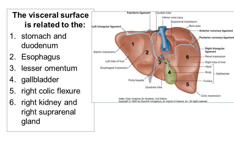
3 Anatomy of the liver **Has a wedge shape and directed downwards & backwards & left **Largest organ in the body, weights 1.5 Kg, located in the right hypochondrial region **The IVC is imbedded in the liver **Has 2 surfaces: visceral (faces the viscera) & diaphragmatic (faces the diaphragm) **The liver is fixed by ligaments that are attached to the anterior abdominal wall & diaphragm **It is divided into 2 main lobes left & right by intralobar fissure Left lobe: small lobe Right lobe: the big lobe; subdivided into smaller lobes which are : Caudate lobe: surrounded by IVC &ligamentum venosus &porta hepatis, and it's connected to right lobe by caudate process Quadrate lobe: surrounded by falciform ligament & porta hepatis & gallbladder **Whole peritoneal covering except one area posteriorly called "BARE AREA" Which is related to the Right suprarenal gland & IVC **Falciform ligament extends superiorly and divides the hypophrenic area into 2 recesses (left & right) and interiorly it's attached with the anterior abdominal wall **There is reflection of the peritoneal to the diaphragm and the result is Right side: upper & lower coronary ligaments meet together and form the right triangular ligament Left side: left triangular ligament
4
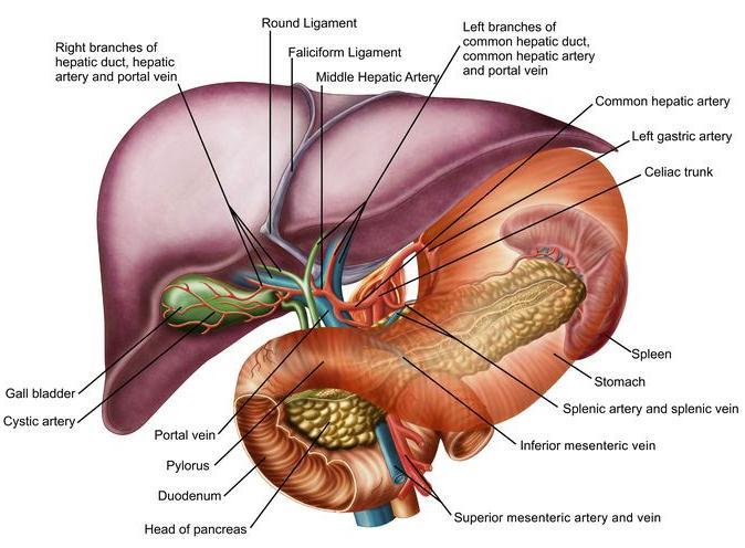
5 RELATION: Convex Diaphragmatic surface: interiorly >> diaphragm & posteriorly there is bare area and I'd mentioned its relation :Flat Visceral surface 1)Stomach & duodenum 2)Esophagus 3)Lesser omentum 4)Gallbladder 5)Right colic flexure 6)Right kidney & right suprarenal gland gallbladder: its sac composed of (fundus &body & neck) its importance is: 1.Collects bile 2.concentrating bile by water absorption 3.secreting mucous *fundus: bulging below the lower margin of liver *body: fixed to the visceral surface of the liver by the peritoneum so the venous drainage between them is continuous *from neck the cystic duct originates and there is a spiral valve to prevent the bending of the neck and closure of it Left hepatic duct + right hepatic duct = common hepatic duct Common hepatic duct + cystic duct= common bile duct Common bile duct + major pancreatic duct = ampulle of vater opens in the midpoint of 2nd part of the duodenum through major papilla Remember: the free margin of the lesser omentum has 3 tubes: common bile duct >> hepatic artery>>portal vein from out to in **The length of common bile duct: 3 inches (7.5 cm) Inch above the duodenum & inch behind the first part of duodenum (portal vein &gastrodoudenal artery) &inch behind the head of pancreas ** :Blood supply )Hepatic arteries (right & left) = 25% of blood supply (oxygenated blood )Portal circulation =75% of blood supply (deoxygenated blood **Venous drainage: hepatic veins (2-4) then IVC

6 Nerve supply: from celiac plexus sympathetic (greatest splanchnic nerves) & ** )parasympathetic (hepatic branches of the vagus nerve Remember: cranial parasympathetic nerves are 3, 7,9,10 ** :Lymphatic drainage Diaphragmatic surface >> anterior mediastinal lymph nodes Visceral surface >> porta hepatis lymph nodes Inside the liver >> posterior mediastinal lymph nodes OR porta hepatis
 separated by a.")
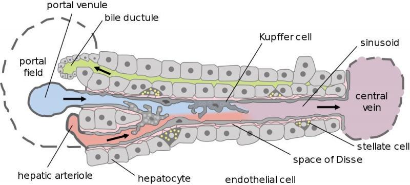
7 Histology of the liver The liver is divided into thousands of small units called lobules by thin layer of connective.tissue.each lobule is about 1mm in diameter and roughly hexagonal in shape The Lobule is composed of radiating double plates of liver cells (Hepatocytes) separated by a.vascular sinusoidal network Each lobule has a central vein in the middle and portal triads at the vertices.each portal triad contains branch of hepatic artery, portal vein and Bile duct Sinusoids have an incomplete lining of highly fenestrated endothelial cells, some of these cells are macrophagic in function (reticulo-endothelial cells), and they are called Kupffer.cells The space between endothelium and hepatocytes is called the Space of Disse. In these spaces, lymph is collected and delivered to lymphatic capillaries. Lymph is collecting in.hepatic duct outside the liver Blood from the branches of hepatic artery and portal vein in the Portal triad, drains into sinusoids and then to the central vein. Central Veins carry the blood to hepatic veins which.end in the Inferior vena cava Bile is formed by liver cells and is discharged into the bile canaliculi within layers of the cell.plates, and then drains into bile duct of the triads

8 Histology of Liver
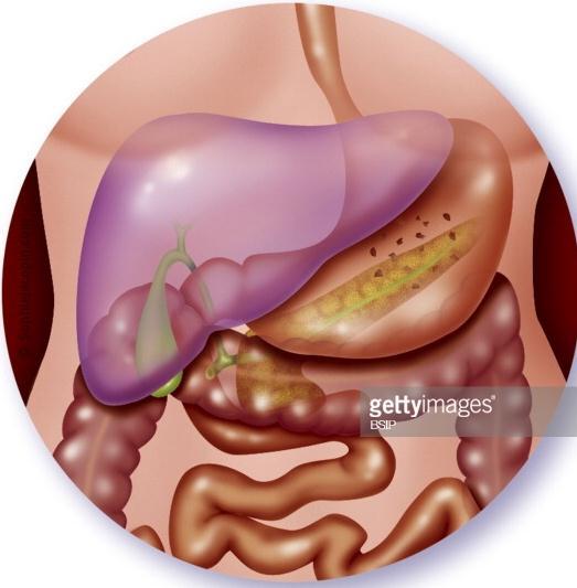
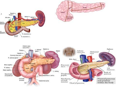
9 Anatomy & histology of pancreas Pancreas is a soft lobulated organ located retroperitoneally across the posterior abdominal.wall, it sits behind the stomach across the back of the abdomen.it is described as an organ having head, neck, body, and tail.the head is disc-shaped and lies within the concavity of the duodenum Part of the head extends to the left behind the superior mesenteric vessels, it is called.uncinate process.the body extends to the left side and ends as a tail near the hilus of the spleen,the pancreas is made up of two types of glands An exocrine gland that secretes digestive enzymes and Sodium bicarbonate into the -1 duodenum through the main and accessory pancreatic ducts. Both ducts are usually.interconnected An endocrine gland, which consists of the islets of Langerhans, secretes hormones into.the bloodstream -2 Islets of Langerhans are named for the German physician Paul Langerhans, who first.described them in The normal human pancreas contains about 1,000,000 islets Cells of islets of Langerhans.Beta cells (?-cells), they make about 65-80% of the cells in the islets and produce Insulin-1 alpha cells (?-cells), 15-20%, they produce an opposing hormone, Glucagon which releases -2.glucose from the liver and fatty acids from fat tissue Delta cells (?-cells), 3-10%, they secrete somatostatin a strong inhibitor of somatotropin, -3 insulin, and glucagon; its role in metabolic regulation is not yet clear. Somatostatin is also produced by the hypothalamus and functions to inhibit secretion of growth hormone by the.pituitary gland **Anterior relation : anterior abdominal wall & transverse colon & mesentery of transverse colon & lesser sac & stomach ** Posterior relation Head >> IVC & Neck : portal vein & Body >>splenich vein & splenich artery & lt kidney & lt suprarenal gland & posterior abdominal wall & Tail : hilum of the spleen **Lienorenal ligament : connects between kidney & spleen **Uncinate process: directed to the left; anterior to it there is superior mesenteric artery & superior mesenteric vein


10 Pancreas P AcinIi Pancreas

11 Histology of gall bladder The gall bladder is a simple muscular sac, lined by a simple columnar epithelium. The inner surface of the gall bladder is covered by the mucosa. The sufrace is made up of a simple columnar epithelium. The epithelial cells have microvilli, and look like absorptive cells in the intestine. Underneath the epithelium is the lamina propria. The wall of the bladder does not have a muscularis mucosae and submucosa. The muscularis externa (muscle layer) contains bundles of smooth muscle cells, collagen and elastic fibres. Underneath this, on the outside of the gall bladder is a thick layer of connective tissue, which contains large blood vessels, nerves and a lymphatic network. Where this layer is attached to the liver, it is called the adventia. In the unattached region, there is an outer layer of mesothelium and loose connective tissue (the serosa). Gall bladder
Anatomy of the liver and pancreas
 Anatomy of the liver and pancreas Prof. Abdulameer Al-Nuaimi E-mail: a.al-nuaimi@sheffield.ac.uk abdulameerh@yahoo.com Liver Aorta Pulm. Trunk Rt. At, Duct. Art. Lt. Ven. Rt. Ven. Internal Posterior
Accessory Glands of Digestive System
 Accessory Glands of Digestive System The liver The liver is soft and pliable and occupies the upper part of the abdominal cavity just beneath the diaphragm. The greater part of the liver is situated under
Pancreas & Biliary System. Dr. Vohra & Dr. Jamila
 Pancreas & Biliary System Dr. Vohra & Dr. Jamila 1 Objectives At the end of the lecture, the student should be able to describe the: Location, surface anatomy, parts, relations & peritoneal reflection
Surface Anatomy. Location Shape Weight Role of Five Surfaces Borders Fissures Lobes Peritoneal Lig
 The Liver Functions Bile production and secretion Detoxification Storage of glycogen Protein synthesis Production of heparin and bile pigments Erythropoiesis (in fetus) Surface Anatomy Location Shape Weight
To describe the liver. To list main structures in porta hepatis.
 GI anatomy Lecture: 6 د. عصام طارق Objectives: To describe the liver. To list main structures in porta hepatis. To define portal system & portosystemic anastomosis. To list parts of biliary system. To
-12. -Renad Habahbeh. -Dr Mohammad mohtasib
 -12 -Renad Habahbeh - -Dr Mohammad mohtasib The Gallbladder -The gallbladder has a body, a fundus (a rounded end), a neck, Hartmann s pouch before the neck and a cystic duct that meets the common hepatic
Lecture 02 Anatomy of the LIVER
 Lecture 02 Anatomy of the LIVER BY Dr Farooq Khan Aurakzai Dated: 02.01.2018 Introduction to Liver Largest gland in the body. 2 nd largest organ of the body. Weight approximately 1500 gm, and is roughly
Duodenum retroperitoneal
 Duodenum retroperitoneal C shaped Initial region out of stomach into small intestine RETROperitoneal viscus Superior 1 st part duodenal cap ; moves upwards and backwards to lie on the R crura medial to
Pancreas and Biliary System
 Pancreas and Biliary System Please view our Editing File before studying this lecture to check for any changes. Color Code Important Doctors Notes Notes/Extra explanation Objectives At the end of the lecture,
د. عصام طارق. Objectives:
 GI anatomy Lecture: 5 د. عصام طارق Objectives: To describe anatomy of stomach, duodenum & pancreas. To list their main relations. To define their blood & nerve supply. To list their lymph drainage. To
In the name ofgod. Abdomen 3. Dr. Zahiri
 In the name ofgod Abdomen 3 Dr. Zahiri Peritoneum Peritoneum It is the serous membrane(a type of loose connective tissue and is covered by mesothelium) that lines the abdominal cavity. Extensions of the
Peritoneum: Def. : It is a thin serous membrane that lines the walls of the abdominal and pelvic cavities and clothes the viscera.
 Peritoneum: Def. : It is a thin serous membrane that lines the walls of the abdominal and pelvic cavities and clothes the viscera. Layers of the peritoneum: 1. Outer Layer ( Parietal Peritoneum) : lines
The abdominal Esophagus, Stomach and the Duodenum. Prof. Oluwadiya KS
 The abdominal Esophagus, Stomach and the Duodenum Prof. Oluwadiya KS www.oluwadiya.com Viscera of the abdomen Abdominal esophagus: Terminal part of the esophagus The stomach Intestines: Small and Large
-Ensherah Mokheemer. -Shatha Al-Jaberi محمد المحتسب- 1 P a g e
 9-9 -Ensherah Mokheemer -Shatha Al-Jaberi محمد المحتسب- 1 P a g e Small intestine has three regions: ( االثني عشر( The duodenum The jejunum The ileum Small intestine Duodenum: -c-shaped -The concavity
Anatomy: Know Your Abdomen
 Anatomy: Know Your Abdomen Glossary Abdomen - part of the body below the thorax (chest cavity); separated by the diaphragm. Anterior - towards the front of the body. For example, the umbilicus is anterior
Block 3: DISSECTION 2 CELIAC TRUNK, JEJUNUM/ILEUM, LARGE INTESTINE, DUODENUM, PANCREAS, PORTAL VEIN; MOBILIZATION OF THE LIVER
 1 Block 3: DISSECTION 2 CELIAC TRUNK, JEJUNUM/ILEUM, LARGE INTESTINE, DUODENUM, PANCREAS, PORTAL VEIN; MOBILIZATION OF THE LIVER Attempt to complete as much as you can of the dissection explained in the
Blood Vessels. Types of Blood Vessels Arteries carry blood away from the heart Capillaries smallest blood vessels. Veins carry blood toward the heart
 C H A P T E R Blood Vessels 20 Types of Blood Vessels Arteries carry blood away from the heart Capillaries smallest blood vessels The site of exchange of molecules between blood and tissue fluid Veins
BLOCK IV: OFFICIAL BODY PARTS LIST FOR ANTERIOR ABDOMINAL WALL AND ABDOMINAL CONTENTS
 BLOCK IV: OFFICIAL BODY PARTS LIST FOR ANTERIOR ABDOMINAL WALL AND ABDOMINAL CONTENTS External oblique muscle Muscular portion Aponeurotic portion Superficial inguinal ring Lateral (inferior) crus Medial
Anatomy of the SMALL INTESTINE. Dr. Noman Ullah Wazir PMC
 Anatomy of the SMALL INTESTINE Dr. Noman Ullah Wazir PMC SMALL INTESTINE The small intestine, consists of the duodenum, jejunum, and illium. It extends from the pylorus to the ileocecal junction were the
Common Bile Duct (CBD)
") Liver Last time we talked about the liver and the doctor started by revising some information about it: It has five surfaces. It reaches the 5 th intercostal space ; some books write that it reaches the
Liver Last time we talked about the liver and the doctor started by revising some information about it: It has five surfaces. It reaches the 5 th intercostal space ; some books write that it reaches the
Liver o The liver is the largest gland in the body and has a wide variety of functions. - It s an accessory organ of GIT
 بسم رلاهللا You don t need to refer to the slides, we included everything here In this lecture we will talk about Liver & Gallbladder Liver o The liver is the largest gland in the body and has a wide variety
Digestive System Module 6: Accessory Organs in Digestion: The Liver, Pancreas, and Gallbladder
 Connexions module: m49293 1 Digestive System Module 6: Accessory Organs in Digestion: The Liver, Pancreas, and Gallbladder Donna Browne Based on Accessory Organs in Digestion: The Liver, Pancreas, and
ANATOMY OF THE DIGESTIVE SYSTEM PART II
 ANATOMY OF THE DIGESTIVE SYSTEM PART II 9.12.2014 Kaan Yücel M.D., Ph.D. http://fhs121.org Dr.Kaan Yücel http://fhs121.org Digestive system Part II 1. LIVER The liver is the largest gland in the body and,
Mousa Salah. Dr. Mohammad Al. Mohtasib. 1 P a g e
 8 Mousa Salah Dr. Mohammad Al. Mohtasib 1 P a g e In the previous lecture we talked about the peritoneum, and we said that the peritonium is a serous sac, and it consists of two layers, visceral and parietal.
Development of the Liver and Pancreas
 Development of the Liver and Pancreas Professor Alfred Cuschieri Department of Anatomy University of Malta Three glandular buds arise from the distal end of the foregut during the fourth week Day 22 -The
Small Plicae Circularis. Short Closely packed together. Sparse, completely absent at distal part Lymphoid Nodule
 Intestines Differences Between Jejunum and Ileum Types Jejunum Ileum Color Deeper red Paler pink Calibre Bigger Smaller Thickness of wall Thick and Heavy Thin and Lighter Vascularity Highly vascularised
Dr. Zahiri. In the name of God
 Dr. Zahiri In the name of God small intestine = small bowel is the part of the gastrointestinal tract Boundaries: Pylorus Ileosecal junction Function: digestion and absorption of food It receives bile
2. capillaries - allow exchange of materials between blood and tissue fluid
 Chapter 19 - Vascular System A. categories and general functions: 1. arteries - carry blood away from heart 2. capillaries - allow exchange of materials between blood and tissue fluid 3. veins - return
The Foregut. At first the esophagus is short. but with descent of the heart and lungs it lengthens rapidly
 GI embryology 2 The Foregut At first the esophagus is short but with descent of the heart and lungs it lengthens rapidly The muscular coat, which is formed by surrounding splanchnic mesenchyme, is striated
The peritoneum. Prof. Oluwadiya KS, MBBS, FMCS(Orthop) Website:
 Website:") The peritoneum Prof. Oluwadiya KS, MBBS, FMCS(Orthop) Website: http://oluwadiya.com The peritoneum Serous membrane that lines the abdominopelvic cavity and invests the viscera The largest serous membrane
The peritoneum Prof. Oluwadiya KS, MBBS, FMCS(Orthop) Website: http://oluwadiya.com The peritoneum Serous membrane that lines the abdominopelvic cavity and invests the viscera The largest serous membrane
Digestive system L 4. Lecturer Dr. Firdous M. Jaafar Department of Anatomy/Histology section
 Digestive system L 4 Lecturer Dr. Firdous M. Jaafar Department of Anatomy/Histology section objectives 1-Describe the structure of liver. 2-Define liver lobule, and identify its zones. 3-Define portal
Fareed Khdair, MD Assistant Professor Chief, Section of Pediatric Gastroenterology, Hepatology, and Nutrition University of Jordan School of Medicine
 Fareed Khdair, MD Assistant Professor Chief, Section of Pediatric Gastroenterology, Hepatology, and Nutrition University of Jordan School of Medicine Outline Lecture one : Gut formation Foregut: esophagus,
Anatomy. Ruba Mahafzah 18/11/2015. Mohammad Allouh. 1 P a g e
 Anatomy Ruba Mahafzah 18/11/2015 Mohammad Allouh 1 P a g e In the previous lecture we started speaking about the liver. We said that the liver is composed of 2 main surfaces: 1. Convex diaphragmatic surface
Laboratory exercises for abdominal organs
 Laboratory exercises for abdominal organs Slide #77 (C007- H- 107A). Pancreas, dog. pancreatic islets CENTROACINAR CELLS ARE THE BEGINNING CELLS OF THE INTERCALATED DUCTS THAT DRAIN THE SECRETORY ACINI
ACTIVITY 11: RESPIRATORY AND DIGESTIVE SYSTEMS RESPIRATORY SYSTEM
 ACTIVITY 11: RESPIRATORY AND DIGESTIVE SYSTEMS OBJECTIVES: 1) How to get ready: Read Chapters 25 and 26, McKinley et al., Human Anatomy, 4e. All text references are for this textbook. 2) Identify structures
Gastric Contrac,le Ac,vity. Regula,on of Gastric Emptying
 Gastric Contrac,le Ac,vity Figure 23.18 Regula,on of Gastric Emptying Gastric emptying is regulated by: Neural enterogastric reflex Hormonal (enterogastrone) mechanisms In the presence of gastric gastrin
Done by: Dina Sawadha & Mohammad Abukabeer
 Done by: Dina Sawadha & Mohammad Abukabeer The stomach *the stomach is a dilated part of the gastro intestinal tract, it's "J" shape. *the lower surface of the stomach ( the greater curvature ) reaches
Digestive Anatomy Lab
 Digestive Anatomy Lab In-Lab Exercises I have included the word list in this document. Any descrepencies between this document and the wordlist, you should default to this document. There is a lot of repetition
RESPIRATORY SYSTEM. described: pp. 744,746 fig. 25.1, described: p. 746 fig described: p. 776 fig. 26.3
 ACTIVITY 11: RESPIRATORY AND DIGESTIVE SYSTEMS OBJECTIVES: 1) How to get ready: Read Chapters 25 and 26, McKinley et al., Human Anatomy, 5e. All text references are for this textbook. 2) Identify structures
Accessory Organs in Digestion: The Liver, Pancreas, and Gallbladder *
 OpenStax-CNX module: m46496 1 Accessory Organs in Digestion: The Liver, Pancreas, and Gallbladder * OpenStax This work is produced by OpenStax-CNX and licensed under the Creative Commons Attribution License
Cardiovascular System Blood Vessels
 Cardiovascular System Blood Vessels Structure of Blood Vessels The three layers (tunics) Tunica intima composed of simple squamous epithelium Tunica media sheets of smooth muscle Contraction vasoconstriction
1 Right & left Hepatic ducts Gastric Impression of spleen
 Pancreatic Model 1 Right & left Hepatic ducts 14 Gastric Impression of spleen 2 Common hepatic duct 15 Renal Impression of spleen 3 Cystic Duct 16 Colic Impression of spleen 4 Common Bile Duct 17 Splenic
The Spleen. Dr Fahad Ullah
 The Spleen BY Dr Fahad Ullah Spleen The spleen is an largest lymphoid organ shaped like a shoe that lies relative to the 9th and 11th ribs and is located in the left hypochondrium. Thus, the spleen is
Jhia Anjela D. Rivera 1 1. BS Biology, Department of Biology, College of Science, Polytechnic University of the Philippines
 DIGESTIVE SYSTEM Jhia Anjela D. Rivera 1 1 BS Biology, Department of Biology, College of Science, Polytechnic University of the Philippines DIGESTIVE SYSTEM Consists of the digestive tract (gastrointestinal
Lab Activity 25. Blood Vessels & Circulation. Portland Community College BI 232
 Lab Activity 25 Blood Vessels & Circulation Portland Community College BI 232 Artery and Vein Histology Walls have 3 layers: Tunica intima Tunica media Tunica externa 2 Tunica Intima Is the innermost layer
Netter's Anatomy Flash Cards Section 4 List 4 th Edition
 Netter's Anatomy Flash Cards Section 4 List 4 th Edition https://www.memrise.com/course/1577335/ Section 4 Abdomen (31 cards) Plate 4-1 Bony Framework of Abdomen 1.1 Costal cartilages 1.2 Iliac crest 1.3
LECTURE 11 & 12: ABDOMINAL VISCERA ABDOMINAL CONTENTS DIVISION. The location of abdominal viscera is divided into 4 quadrants:
 LECTURE 11 & 12: ABDOMINAL VISCERA ABDOMINAL CONTENTS DIVISION The location of abdominal viscera is divided into 4 quadrants: - horizontal line across the umbilicus divides the upper quadrants from the
BY DR NOMAN ULLAH WAZIR
 BY DR NOMAN ULLAH WAZIR The stomach (from ancient Greek word stomachos, stoma means mouth) is a muscular, hollow and the most dilated part of the GIT. It starts from the point where esophagus ends. It
ABDOMEN - GI. Duodenum
 TALA SALEH ABDOMEN - GI Duodenum - Notice the shape of the duodenum, it looks like capital G shape tube which extends from the pyloroduodenal junction to the duodenojejunal junction. - It is 10 inches
MICROSCOPIC STRUCTURE OF LIVER, GALLBLADDER, GALL DUCTS, AND PANCREAS OVERVIEW OF DEVELOPMENT OF THE ALIMENTARY CANAL
 Lecture 2 ESS_3rd semester MICROSCOPIC STRUCTURE OF LIVER, GALLBLADDER, GALL DUCTS, AND PANCREAS OVERVIEW OF DEVELOPMENT OF THE ALIMENTARY CANAL MICROSCOPIC STRUCTURE OF LIVER - is the largest gland of
The cardiovascular system
 The cardiovascular system Components of the Cardiovascular system Heart Vessels: Arteries Capillaries Veins Functions of CVS: Transportation system where blood is the transporting vehicle Carries oxygen,
DIGESTIVE SYSTEM II ACCESSORY DIGESTIVE ORGANS
 DIGESTIVE SYSTEM II ACCESSORY DIGESTIVE ORGANS Dr. Larry Johnson Texas A& M University Objectives Distinguish between the parotid and submandibular salivary glands. Understand and identify the structural
Anatomy of Liver and Spleen
 Anatomy of Liver and Spleen Please view our Editing File before studying this lecture to check for any changes. Color Code Important Doctors Notes Notes/Extra explanation Objectives At the end of the lecture,
Mediastinum and pericardium
 Mediastinum and pericardium Prof. Abdulameer Al-Nuaimi E-mail: a.al-nuaimi@sheffield.ac.uk E. mail: abdulameerh@yahoo.com The mediastinum: is the central compartment of the thoracic cavity surrounded by
Preview from Notesale.co.uk Page 1 of 34
 Abdominal viscera and digestive tract Digestive tract Abdominal viscera comprise majority of the alimentary system o Terminal oesophagus, stomach, pancreas, spleen, liver, gallbladder, kidneys, suprarenal
Slide 154: Pancreas, H&E
 Slide 154: Pancreas, H&E the pancreas, located adjacent to the duodenum, is a mixed exocrine and endocrine gland; it is usually readily identifiable by the presence of the interspersed endocrine pancreatic
Exploring Anatomy: the Human Abdomen
 Exploring Anatomy: the Human Abdomen PERITONEUM AND PERITONEAL CAVITY PERITONEUM The peritoneum is a thin serous membrane that lines the abdominal cavity and covers, in variable amounts, the viscera within
Dissection Lab Manuals: Required Content
 Dissection Lab Manuals: Required Content 1. Introduction a. Basic terminology (directions) b. External features of the cat c. Adaptations to predatory niche d. How to skin a cat e. How to make the incisions
Histology 3. We will continue talking about a few things from last lecture, starting with M cells:
 Histology 3 This is the last Histology lecture in the GI system. Enjoy! There are some extra notes listed as footnotes. We will continue talking about a few things from last lecture, starting with M cells:
The Digestive System
 The Digestive System Identify the Structure and Function. Mesentery of the Large Intestine The mesentery functions to connect the visceral organs to the abdominal wall. Identify the Structure. Nasal Cavity
The stomach is formed of three parts: -
 The stomach is formed of three parts: - (a) CARDIAC STOMACH: - It receives the oesophagus through Cardiac aperture guarded by a cardiac sphincter which prevents regurgitation of food. (b) FUNDIC PART:
Anatomy of the Large Intestine
 Large intestine Anatomy of the Large Intestine 2 Large Intestine Extends from ileocecal valve to anus Length = 1.5-2.5m = 5 feet Regions Cecum = 2.5-3 inch Appendix= 3-5 inch Colon Ascending= 5 inch Transverse=
The Digestive System. Chapter 25
 The Digestive System Chapter 25 Introduction Structure of the digestive system A tube that extends from mouth to anus Accessory organs are attached Functions include Ingestion Movement Digestion Absorption
Midterm 2 is Tuesday 5/28/13
 Business Reminder: No class Monday (Memorial Day) Midterm 2 is Tuesday 5/28/13 Optional review session tomorrow @ 5pm Homework due in Lab 1. PreLab 8 (1pt) 2. Replace a Missing Assignment (4 pts) Homework
Liver. Function of the liver
 Liver & Gallbladder Liver The liver is the largest gland in the body and has a wide variety of functions Weight: 1/50 of body weight in adult & 1/20 of body weight in infant It is exocrine(bile) & endocrine
Paneth Cells. Road Map to the Finish. No Review this Friday. Today 11/29 Finish digestion/accessory organs. Wednesday 12/1 Immune System I
 Road Map to the Finish No Review this Friday Today 11/29 Finish digestion/accessory organs Wednesday 12/1 Immune System I Paneth Cells - base of intestinal glands -! large -! intense acidophilic granules
Biology Human Anatomy Abdominal and Pelvic Cavities
 Biology 351 - Human Anatomy Abdominal and Pelvic Cavities Please place your name and I.D. number on the back of the last page of this exam. You must answer all questions on this exam. Because statistics
DEVELOPMENT OF THE LIVER
 THE LIVER DEVELOPMENT OF THE LIVER THE LIVER The hepa-c diver-culum develops as an outgrowth of the endoderm of the foregut in the region of the second por-on of duodenum. This diver-culum enters the ventral
Respiratory & Digestive Organs of the Head and Neck, Human;
 Name Date Lab Exercise 5: Lab Exercise 6: Lab Exercise 7: Lab Exercise 8: Respiratory & Digestive Organs of the Head and Neck, Human; Histology of the Respiratory System Digestive System Models, Human
بسم هللا الرحمن الرحيم
 بسم هللا الرحمن الرحيم **As we remember from the last lecture: The arterial supply which comes from the single branches of the aorta drains in the portal vein (venous drainage of the gut = portal vein).
Bio 322 Human Anatomy Objectives for the laboratory exercise Digestive System
 Bio 322 Human Anatomy Objectives for the laboratory exercise Digestive System Required reading before beginning this lab: Saladin, KS: Human Anatomy 5 th ed (2017) Chapter 24 For this lab you will use
Overview of Anatomy and Physioloy II Second Year Students
 University of Baghdad College of Nursing Department of Basic Medical Sciences Overview of Anatomy and Physioloy II Second Year Students Asaad Ismail Ahmad, Ph.D. Asaad Ismail Ahmad, Ph.D. Electrolyte and
It passes through the diaphragm at the level of the 10th thoracic vertebra to join the stomach
 The esophagus is a tubular structure (muscular, collapsible tube ) about 10 in. (25 cm) long that is continuous above with the laryngeal part of the pharynx opposite the sixth cervical vertebra The esophagus
Cardiovascular system:
 Cardiovascular system: Mediastinum: The mediastinum: lies between the right and left pleura and lungs. It extends from the sternum in front to the vertebral column behind, and from the root of the neck
DIGESTIVE. CHAPTER 17 Lecture: Part 1 Part 2 BIO 212: ANATOMY & PHYSIOLOGY II
 BIO 212: ANATOMY & PHYSIOLOGY II CHAPTER 17 Lecture: DIGESTIVE Part 1 Part 2 Dr. Lawrence G. Altman www.lawrencegaltman.com Some illustrations are courtesy of McGraw-Hill. SMALL INTESTINE DUODENUM > JEJUNUM
OVARIES URETER FALLOPIAN TUBES BLADDER UROGENITAL OPENINGS (BOTH SEXES) PENIS VAGINA UTERUS
 PENIS VAGINA UTERUS") URETER OVARIES FALLOPIAN TUBES BLADDER UROGENITAL OPENINGS (BOTH SEXES) PENIS VAGINA UTERUS REPRODUCTIVE PRODUCE FEMALE HORMONES EXCRETORY FROM KIDNEY TO BLADDER EXCRETORY STORES URINE REPRODUCTIVE TRANSPORTS
URETER OVARIES FALLOPIAN TUBES BLADDER UROGENITAL OPENINGS (BOTH SEXES) PENIS VAGINA UTERUS REPRODUCTIVE PRODUCE FEMALE HORMONES EXCRETORY FROM KIDNEY TO BLADDER EXCRETORY STORES URINE REPRODUCTIVE TRANSPORTS
ANATOMY AND BASIC FUNCTION OF THE ENDOCRINE GLANDS
 ANATOMY AND BASIC FUNCTION OF THE ENDOCRINE GLANDS Know these endocrine organs of the cat: thymus, thyroid, pancreas, adrenal glands, ovaries, and testes. Review and know microslides, hormones, and structures
- Tamara Wahbeh. - Fareed Khdair. 0 P a g e
 -1 - Tamara Wahbeh - - Fareed Khdair 0 P a g e GI Embryology Note: I included everything in the records and slides; anything in the slide not included in this sheet was not mentioned by the doctor during
Overview of the Digestive
 Overview of the Digestive System Bởi: OpenStaxCollege The function of the digestive system is to break down the foods you eat, release their nutrients, and absorb those nutrients into the body. Although
Chapter 14: The Digestive System
 Chapter 14: The Digestive System Digestive system consists of Muscular tube (digestive tract) alimentary canal Accessory organs teeth, tongue, glandular organs 6 essential activities 1. 2. 3. 4. 5. 6.
- Digestion occurs during periods of low activity - Produces more energy than it uses. - Mucosa
 Introduction Digestive System Chapter 29 Provides processes to break down molecules into a state easily used by cells - A disassembly line: Starts at the mouth and ends at the anus Digestive functions
Digestive System 7/15/2015. Outline Digestive System. Digestive System
 Digestive System Biology 105 Lecture 18 Chapter 15 Outline Digestive System I. Functions II. Layers of the GI tract III. Major parts: mouth, pharynx, esophagus, stomach, small intestine, large intestine,
Anatomy of the spleen. Oluwadiya KS
 Anatomy of the spleen Oluwadiya KS www.oluwadiya.com Introduction The spleen is an ovoid, usually purplish, pulpy mass about the size and shape of one's fist. It is the largest lymphoid tissue in the body
THE ORAL CAVITY
 THE ORAL CAVITY WALL OF ABDOMEN (ANTERIOR) The paraumbilical vein drains into the portal vein and then through the liver. This is an important clinical connection. THE ABDOMINAL VISCERA The small
Chapter 5: Other mediastinal structures. The Large Arteries. The Aorta. Ascending aorta
 Chapter 5: Other mediastinal structures The Large Arteries The Aorta The aorta is the main arterial trunk of the systemic circulation and in the healthy state its wall contain a large amount of yellow
DEVELOPMENT OF THE CIRCULATORY SYSTEM L E C T U R E 5
 DEVELOPMENT OF THE CIRCULATORY SYSTEM L E C T U R E 5 REVIEW OF CARDIAC ANATOMY Heart 4 chambers Base and apex Valves Pericardial sac 3 layers: epi, myo, endo cardium Major blood vessels Aorta and its
The Whipple Operation Illustrations
 The Whipple Operation Illustrations Fig. 1. Illustration of the sixstep pancreaticoduodenectomy (Whipple operation) as described in a number of recent text books by Dr. Evans. The operation is divided
Dr. Weyrich G07: Superior and Posterior Mediastina. Reading: 1. Gray s Anatomy for Students, chapter 3
 Dr. Weyrich G07: Superior and Posterior Mediastina Reading: 1. Gray s Anatomy for Students, chapter 3 Objectives: 1. Subdivisions of mediastinum 2. Structures in Superior mediastinum 3. Structures in Posterior
The Urinary System Pearson Education, Inc.
 26 The Urinary System Introduction The urinary system does more than just get rid of liquid waste. It also: Regulates plasma ion concentrations Regulates blood volume and blood pressure Stabilizes blood
-the stones will obstruct the common bile duct and it might also be precancerous. -so the best treatment is chlolycyctoctomy.
 At the beginning this sheet includes the rest of last lecture s slides liver +gallbladder and the new lecture posterior abdominal wall and its vessels. We will start talking about Cholelithiasis -it means
Lab 8: Digestive System
 BIOL 221 A&P II Lab 8: Digestive System Become familiar with the gross anatomy of the digestive system (Exercise 38) using the models, Fig. 38.1 (Activity 1), and the rat. Recognize and know the functions
The sinus venosus represent the venous end of the heart It receives 3 veins: 1- Common cardinal vein body wall 2- Umbilical vein from placenta 3-
 1 2 The sinus venosus represent the venous end of the heart It receives 3 veins: 1- Common cardinal vein body wall 2- Umbilical vein from placenta 3- Vitelline vein from yolk sac 3 However!!!!! The left
- Digestion occurs during periods of low activity - Produces more energy than it uses. 3 Copyright 2016 by Elsevier Inc. All rights reserved.
 Introduction Digestive System Chapter 29 Provides processes to break down molecules into a state easily used by cells - A disassembly line: Starts at the mouth and ends at the anus Digestive functions
HUMAN HEART. Learn the following structures on the heart models.
 HUMAN HEART Learn the following structures on the heart models. The human heart has four chambers that consist of the right atrium, left atrium, right ventricle, and left ventricle. The atria are smaller
1. Which of the following blood vessels has a thin elastic layer? A. Aorta. B. Pulmonary artery. C. Posterior vena cava. D. Mesenteric capillary.
 CIRCULATORY SYSTEM 1. Which of the following blood vessels has a thin elastic layer? A. Aorta. B. Pulmonary artery. C. Posterior vena cava. D. Mesenteric capillary. 2. Capillary beds are equipped with
Pancreas Quizzes c. Both A and B a. Directly into the blood stream (not using ducts)
") Pancreas Quizzes Quiz 1 1. The pancreas produces hormones. Which type of hormone producing organ is the pancreas? a. Endocrine b. Exocrine c. Both A and B d. Neither A or B 2. Endocrine indicates hormones
Pancreas Quizzes Quiz 1 1. The pancreas produces hormones. Which type of hormone producing organ is the pancreas? a. Endocrine b. Exocrine c. Both A and B d. Neither A or B 2. Endocrine indicates hormones
Figure ) The specific chamber of the heart that is indicated by letter A is called the. Diff: 1 Page Ref: 364
 The specific chamber of the heart that is indicated by letter A is called the. Diff: 1 Page Ref: 364") Essentials of Anatomy and Physiology, 9e (Marieb) Chapter 11 The Cardiovascular System Short Answer Figure 11.1 Using Figure 11.1, identify the following: 1) The Purkinje fibers are indicated by label.
Essentials of Anatomy and Physiology, 9e (Marieb) Chapter 11 The Cardiovascular System Short Answer Figure 11.1 Using Figure 11.1, identify the following: 1) The Purkinje fibers are indicated by label.
The Digestive System and Body Metabolism
 14 PART B The Digestive System and Body Metabolism PowerPoint Lecture Slide Presentation by Jerry L. Cook, Sam Houston University ESSENTIALS OF HUMAN ANATOMY & PHYSIOLOGY EIGHTH EDITION ELAINE N. MARIEB
REVIEW SHEET Anatomy of Blood Vessels
 REVIEW SHEET Anatomy of Blood Vessels Name LabTime/Date Microscopic Structure of the Blood Vessels 1. Cross-sectional views of an aftery of a vein are shown here. ldentify each; on the lines to the sides,
DIGESTIVE SYSTEM. Chapter 25
 DIGESTIVE SYSTEM Chapter 25 DIGESTIVE SYSTEM Digestive Tract Mouth Pharynx Esophagus Stomach Small intestines Large intestines Anus Accessory Organs Teeth Tongue Salivary glands Pancreas Liver Gallbladder