Leo Semes, OD, FAAO UAB Optometry
|
|
|
- Alannah Todd
- 6 years ago
- Views:
Transcription


1 Leo Semes, OD, FAAO UAB Optometry
2 Safe; inert Has long track record - over 45 years Mixes with plasma and highlights blood vessel compromise
3 Using specific exciting (490 nm)and absorption (510 nm) filters B/W film documents vascular changes Eliminated through liver and kidneys in hours Stains skin and mucous membranes (24-36)
4 May contaminate results of urine tests for reducing sugars (up to 4-5 days) Does not diffuse through NORMAL retinal blood vessels Normally leaky choroidal vessels show uniform choroidal flush
5 Mild Nausea, sneezing Pruritis Vomiting Extravasation at injection site
6 Moderate Urticaria (< 2%) Syncope (< 0.2%) Other (< 0.006%) Skin eruptions Thrombophelibitis Pyrexia Focal tissue necrosis Nerve palsy
7 Severe Respiratory events (< %) Laryngeal edema Bronchospasm Anaphylaxis Cardiovascular events (< %) Circulatory shock Cardiac arrest
8 Previous anaphylactic reaction Other previous reactions-weigh risk:benefit Kidney disease (relative contraindication) Pregnancy Avoid,especially during the first trimester
9 Inner Vascular endothelium (integrity) Outer Retinal Pigment Epithelium


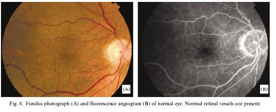
10 Histological analog of a normal angiogram Retinal Vessels RPE Choroid Sclera Thomas Freddo, OD, Ph.D.


11 Inner Blood-Retinal Barrier Retinal capillaries are sealed by tight junctions. Outer Blood-Retinal Barrier Choroidal capillaries are fenestrated and therefore leak fluid, ions & plasma proteins. RPE cells have tight junctions that prevent these substances from diffusing into the retina. Thomas Freddo, OD, Ph.D.


12 Retinal Capillaries Retinal capillaries are non-fenestrated. Using intravascular tracers, that mimic the behavior of plasma proteins, retinal capillaries are impermeable and these proteins do not diffuse into the surrounding retina. Thomas Freddo, OD, Ph.D.


13 Choriocapillaris Choroidal capillaries are fenestrated (arrowheads). Using intravascular tracers, that mimic the behavior of plasma proteins, choroidal capillaries are freely permeable and these proteins diffuse into the surrounding choroidal stroma. Thomas Freddo, OD, Ph.D.


14 Retinal Pigmented Epithelium RPE cells are joined by tight junctions. This histological slide illustrates the use of intravascular tracers, that mimic the behavior of plasma proteins, showing the intercellular spaces between RPE cells to be impermeable to these proteins. Thomas Freddo, OD, Ph.D.
15 Tight junctions between retinal vascular endothelial cells prevent plasma proteins from diffusing into the retina. Tight junctions between RPE cells prevent plasma proteins that have leaked from the fenestrated choroidal capillaries from diffusing into the retina.
16 Oral (shows only late leakage by angioscopy) May be applicable in children or where IV route in impossible Shows only late stage; not circulation phases (angioscopy) Intravenous (IV) - shows transit of fluorescein as blood circulates and recirculates)

17 FA is employed to support diagnoses and direct treatment Examples of conditions that may be amenable to treatment (e.g., photocoagulation) Diabetic macular edema Age-related macular degeneration Other diseases that have neovascularization or macular edema as a consequence
18 Arm to retina [7.5-25] sec (via???) Arterial retinal Venous retinal Total retinal *total transit time can be up to ~ 30 sec
19 Reduced in youth and anemia Prolonged (slow/impaired blood flow) Carotid stenosis Sludging disorders
20 Color fundus photo Red-free photo (reference) Camera ready (SLO, also) Patient prep Injection site Syringe w/5 ml (10% ); injected into the antecubital vein (3-7 sec as bolus)

21 Photographic sequence 2-3 frames/10-12 sec Then every 2 3 sec for next 10 sec Late 5 to 20 minutes
22
23 Choroidal flush (prearterial) Early Phase (arterial) Mid Phase (arteriovenous,venous,laminar) Late Phase (recirculatory)

24
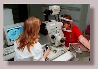
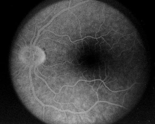
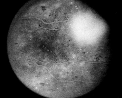
25 Choroidal flush (prearterial) The initial arm-to retina time is recorded as the first appearance of fluorescein in the choroidal vasculature system (10-20 sec) Uniform glow from permeable choroidal circulation Cilioretinal artery fills
26 Early Phase (arterial) The arteries alone in the retinal vascular tree are visibly filled with fluorescein against the choroidal flush Note: a cilioretinal artery fills with the choroidal circulation
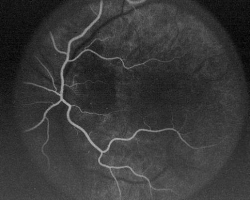
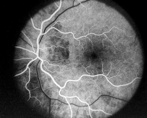
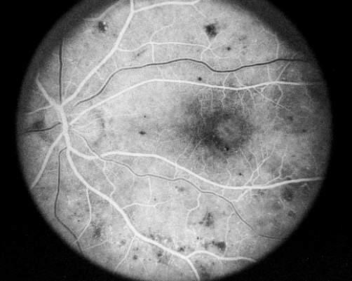
27 Early Phase (arteriovenous) Why is the macula dark? Why are the veins filling at their edges?
28 Early / Mid Phase (arteriovenous,venous; 3-5 min) Early filling of the veins,laminar flow, or border filling of the veins (looks like railroad tracks), followed by Arteries and veins fully filled with fluorescein

29 Early / Mid Phase - venous
30 The Late Phase This phase is documented 5-20 min following injection In normals, no staining or leakage is seen Abnormal results include late leakage not contained by the circulation, or staining

31 Late Phase
32 Hyper fluorescence leakage of neovascularization in (wet) AMD, pooling in central serous choroiretinopathy Hypofluorescence an ischemic territory in vein occlusion (absence) Blocked fluorescence by overlying retinal blood blocking the uniform appearance of the choroidal flush Late staining cystoid macular edema, choroidal malignant melanoma

33 Autofluorescence Natural glow of ocular tissues without fluorescein (e.g., optic nerve drusen, astrocytoma) Pseudofluorescence May be due to unbalanced photographic filter system Also used to describe the margin of scar tissue (e.g., fibrovascular scarring of AMD)
34 FIRST - compare the color and R/F photos NEXT - identify the stage of the angiogram Look for one of the 4 characteristic abnormalities (hyper-, hypo-, blocked or late-phase staining/leakage)

35 Determine if the abnormal fluorescence is changing or remaining the same throughout the angiogram Correlate the anatomical (clinical) and angiographic sites; again refer to the color and R/F photos!!! FA is to confirm diagnoses and guide treatment; not to make a diagnosis
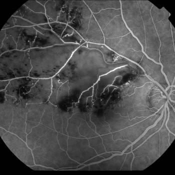
36 DX: BRVO
37 Abnormal examples
38 A: 1. CWS in papillomacular bundle Link to ok/body/ / /1869/i4-u1.0-b f0030.fig?tocnode= Intraretinal hemorrhage B: FA 1. capillary nonperfusion in the area corresponding to the cotton-wool patch; 2. blocked fluorescence caused by the intraretinal hemorrhage Yanoff and Duker in MD consult
39 Healthy 37 YO M 3-hour history of visual loss (VA 20/60). om/das/book/body/ / /1869/I4- u1.0-b f0010.fig?tocnode= A: Retinal whitening is very subtle and the retinal vessels appear normal. (B) FA reveals abnormal arterial filling. (confirming CRAO)
40 FA is employed to support diagnoses and direct treatment Examples of conditions that may be amenable to treatment (e.g., photocoagulation) Diabetic macular edema Age-related macular degeneration Other diseases that have neovascularization or macular edema as a consequence

41
42 ICG (tricarbocyanine) Near IR (780/810 nm) absorption/emission Similar injection protocol / safety spectrum to fluorescein, ex: metabolized in liver contains iodine Advantages Defines CNV better than FA Visualization improved through media opacities, pigment, blood, and scar tissue
43 Most useful for choroidal filling assessment application in AMD Example
 Prolonged choroidal filling (1 min FA) 1 min ICGA 3 min ICGA Reduced diffuse")
44 ICG absorbs and fluoresces in the near IR improving choroidal visibility and diffuses more slowly from the choriocapillaris 68 W/M; 20/30 (color) Prolonged choroidal filling (1 min FA) 1 min ICGA 3 min ICGA Reduced diffuse background fluorescence
 ICGA (1")
45 Prolonged choroidal filling (PCF) more evident on ICGA color FA (1 min) ICGA (1 min)
46 Normal Angiogram Diabetic Retinopathy (NPDR) Proliferative Diabetic Retinopathy (PDR) Age-related Macular Degeneration (AMD)
47


48 Arrowheads = intraretinal hard exudates Arrows on FA = surrounding areas of leaking microaneurysms
49

50
51


52 BLACK Arrow = PDR at ONH, w/ corresponding leakage on FA WHITE arrow = microaneurysms


53 LS Neovascular AMD * = CNVM; hard exudates are also present Black arrow on FA = leakage from NV
Incorporating OCT Angiography Into Patient Care
 Incorporating OCT Angiography Into Patient Care Beth A. Steele, OD, FAAO OCT A: Introduction Isolates microvascular circulation from OCT image data Axial resolution = 5 microns (i.e. fine capillaries visible)
OCT Angiography in Primary Eye Care
 OCT Angiography in Primary Eye Care An Image Interpretation Primer Julie Rodman, OD, MS, FAAO and Nadia Waheed, MD, MPH Table of Contents Diabetic Retinopathy 3-6 Choroidal Neovascularization 7-9 Central
ZEISS AngioPlex OCT Angiography. Clinical Case Reports
 Clinical Case Reports Proliferative Diabetic Retinopathy (PDR) Case Report 969 PROLIFERATIVE DIABETIC RETINOPATHY 1 1-year-old diabetic female presents for follow-up of proliferative diabetic retinopathy
Fluorescein Angiography
 Last revision: October 2011 by Luis Arias Fluorescein Angiography Authors: Luis Arias, MD Hospital Universitari de Bellvitge - University of Barcelona. Spain Jordi Monés, MD Institut de la Màcula i de
Will OCT-Angiography replace FA?
 ASL Roma A PRESIDIO TERRITORIALE NUOVO REGINA MARGHERITA AMBULATORIO PATOLOGIE RETINICHE Resp. Dott.ssa SUSANNA CATALANO CENTRO ITALIANO MACULA Will OCT-Angiography replace FA? Marco Rispoli, Luca di Antonio,
Diabetic Retinopathy
 Diabetic Retinopathy Diabetes can be classified into type 1 diabetes mellitus and type 2 diabetes mellitus, formerly known as insulin-dependent diabetes mellitus, and non-insulin diabetes mellitus, respectively.
Disease-Specific Fluorescein Angiography
 Ruth E. Picchiottino, CRA Disease-Specific Fluorescein Angiography 15 Disease-Specific Fluorescein Angiography Recommendations for tailoring retinal fluorescein angiography to diabetic retinopathy, macular
Dr/ Marwa Abdellah EOS /16/2018. Dr/ Marwa Abdellah EOS When do you ask Fluorescein angiography for optic disc diseases???
 When do you ask Fluorescein angiography for optic disc diseases??? 1 NORMAL OPTIC DISC The normal optic disc on fluorescein angiography is fluorescent due to filling of vessels arising from the posterior
PART 1: GENERAL RETINAL ANATOMY
 PART 1: GENERAL RETINAL ANATOMY General Anatomy At Ora Serrata At Optic Nerve Head Fundoscopic View Of Normal Retina What Is So Special About Diabetic Retinopathy? The WHO definition of blindness is
EyePACS Grading System (Part 2): Detecting Presence and Severity of Background (Non-Proliferative) Diabetic Retinopathy Lesion
: Detecting Presence and Severity of Background (Non-Proliferative) Diabetic Retinopathy Lesion") EyePACS Grading System (Part 2): Detecting Presence and Severity of Background (Non-Proliferative) Diabetic Retinopathy Lesion George Bresnick MD MPA Jorge Cuadros OD PhD Anatomy of the eye: 3 Normal Retina
EyePACS Grading System (Part 2): Detecting Presence and Severity of Background (Non-Proliferative) Diabetic Retinopathy Lesion George Bresnick MD MPA Jorge Cuadros OD PhD Anatomy of the eye: 3 Normal Retina
The Natural History of Diabetic Retinopathy and How Primary Care Makes A Difference
 The Natural History of Diabetic Retinopathy and How Primary Care Makes A Difference We will discuss - How exactly does blood sugar control affect retinopathy? - What are other factors that we measure in
Building The Retina Company
 Building The Retina Company Optos devices produce ultra-widefield (UWF ), high resolution images (optomap ) of approximately 82% (200 ) of the retina. A single optomap can document the retina from the
Diabetic Retinopathy A Presentation for the Public
 Diabetic Retinopathy A Presentation for the Public Ray M. Balyeat, MD The Eye Institute Tulsa, Oklahoma The Healthy Eye Light rays enter the eye through the cornea, pupil and lens. These light rays are
OCT Angiography The Next Frontier
 Choroid Retina avascular 5/13/2017 OCT Angiography The Next Frontier Pierce Kenworthy OD, FAAO June 9, 2017 OCT Angiography (OCTA) 2016 Non-invasive, motion contrast imaging Represents erythrocyte movement
Diagnosis and treatment of diabetic retinopathy. Blake Cooper MD Ophthalmologist Vitreoretinal Surgeon Retina Associates Kansas City
 Diagnosis and treatment of diabetic retinopathy Blake Cooper MD Ophthalmologist Vitreoretinal Surgeon Retina Associates Kansas City Disclosures Consulted for Novo Nordisk 2017,2018. Will be discussing
The Human Eye. Cornea Iris. Pupil. Lens. Retina
 The Retina Thin layer of light-sensitive tissue at the back of the eye (the film of the camera). Light rays are focused on the retina then transmitted to the brain. The macula is the very small area in
FA vs. OCTA? The status of OCTA, today. Fukuoka, JSOS 2016 Gerd Klose. Korobelnik J Fr Ophthalmol (2015)
") FA vs. OCTA? The status of OCTA, today Korobelnik J Fr Ophthalmol (2015) Fukuoka, JSOS 2016 Gerd Klose 1 2 FA / ICGA a well-founded Gold standard! Benefits Useful for many pathologies High contrast, detailed
FA vs. OCTA? The status of OCTA, today Korobelnik J Fr Ophthalmol (2015) Fukuoka, JSOS 2016 Gerd Klose 1 2 FA / ICGA a well-founded Gold standard! Benefits Useful for many pathologies High contrast, detailed
Clinically Significant Macular Edema (CSME)
") Clinically Significant Macular Edema (CSME) 1 Clinically Significant Macular Edema (CSME) Sadrina T. Shaw OMT I Student July 26, 2014 Advisor: Dr. Uwaydat Clinically Significant Macular Edema (CSME) 2
Clinically Significant Macular Edema (CSME) 1 Clinically Significant Macular Edema (CSME) Sadrina T. Shaw OMT I Student July 26, 2014 Advisor: Dr. Uwaydat Clinically Significant Macular Edema (CSME) 2
Case Report: Indocyanine Green Dye Leakage from Retinal Artery in Branch Retinal Vein Occlusion
 Case Report: Indocyanine Green Dye Leakage from Retinal Artery in Branch Retinal Vein Occlusion Hiroki Fujita, Kyoko Ohno-Matsui, Soh Futagami and Takashi Tokoro Department of Visual Science, Tokyo Medical
Amber Priority. Image Library
 Amber Priority Image Library Amber flag Diabetic Maculopathy (M1) Pre-proliferative Diabetic Retinopathy (R2) Old, treated and now inactive DR (R1/M0/P1or R0/M0/P1) Where only partial or incomplete images
Role of Fluorescein angiography in evaluation of posterior segment disorders
 Original Article Role of Fluorescein angiography in evaluation of posterior segment disorders Arvind R, Surendar S 2, Ch. Jagan Mohan Rao 3 Associate Professor, 2 Postgraduate student, 3 Senior resident,
Eyes on Diabetics: How to Avoid Blindness in Diabetic Patient
 Eyes on Diabetics: How to Avoid Blindness in Diabetic Patient Rova Virgana FK Unpad Pusat Mata Nasional RS Mata Cicendo Bandung Eye Center (Hospital and Clinic) PIT IDI Jabar 2018 Keys Facts from WHO
 measure of your overall performance. An isolated glucose test is helpful to let you know what your sugar level is at one moment, but it doesn t tell you whether or not your diabetes is under adequate control
Posterior Segment Update
 Posterior Segment Update Featured Speaker: Dr. Kyle Cheatham, FAAO, DIP ABO DISCLOSURE STATEMENT We have no direct financial or proprietary interest in any companies, products or services mentioned in
Age-related Macular Degeneration Update
 Age-related Macular Degeneration Update AMD: The Burden of Disease Carlo J. Pelino, OD, FAAO cpelino@salus.edu Joseph J. Pizzimenti, OD, FAAO pizzimen@nova.edu Course Goals Statement of the problem Epidemiology
Diagnosis in AMD. Managing your AMD Patients
 Managing your AMD Patients Robert W. Dunphy, O.D., F.A.A.O. Diagnosis in AMD Have suspicion Identify relative risk Conduct surveillance Biometry Utilize technology to facilitate detection of change / stability
Optical Coherence Tomography in Diabetic Retinopathy. Mrs Samantha Mann Consultant Ophthalmologist Clinical Lead of SEL-DESP
 Optical Coherence Tomography in Diabetic Retinopathy Mrs Samantha Mann Consultant Ophthalmologist Clinical Lead of SEL-DESP Content OCT imaging Retinal layers OCT features in Diabetes Some NON DR features
Michael P. Blair, MD Retina Consultants, Ltd Libertyville/Des Plaines, Illinois Clinical Associate University of Chicago 17 October 2015
 Michael P. Blair, MD Retina Consultants, Ltd Libertyville/Des Plaines, Illinois Clinical Associate University of Chicago 17 October 2015 So What Parts of the Eye Retina are Affected by VHL Neural tissue
Use of the Free Electron Laser for the Noninvasive Determination of Retinal Oxyhemoglobin Saturation by Near Infrared Reflectance Spectrophotometry
 Use of the Free Electron Laser for the Noninvasive Determination of Retinal Oxyhemoglobin Saturation by Near Infrared Reflectance Spectrophotometry Ref: Eye, M.C. Escher, 1946 Ref: Eye, M.C. Escher, 1946
The legally binding text is the original French version
 The legally binding text is the original French version TRANSPARENCY COMMITTEE OPINION 12 September 2007 INFRACYANINE 25 mg/10 ml, powder and solvent for solution for injection 25 mg vial with one 10 ml
On Different Wavelengths: The Spectrum of Retinal Imaging. On Different Wavelengths: The Spectrum of Retinal Imaging. Wavelength Specific Imaging
 On Different Wavelengths: The Spectrum of Retinal Imaging Timothy J. Bennett, CRA, FOPS, OCT-C Penn State Hershey Eye Center Hershey, PA On Different Wavelengths: The Spectrum of Retinal Imaging Wavelengths
Fundus Autofluorescence. Jonathan A. Micieli, MD Valérie Biousse, MD
 Fundus Autofluorescence Jonathan A. Micieli, MD Valérie Biousse, MD The retinal pigment epithelium (RPE) has many important functions including phagocytosis of the photoreceptor outer segments Cone Rod
OCT Interpretation in Retinal Disease
 OCT Interpretation in Retinal Disease Jay M. Haynie, OD, FAAO Financial Disclosure I have received honoraria or am on the advisory board for the following companies: Carl Zeiss Meditec Advanced Ocular
Patient AB. Born in 1961 PED
 Clinical Atlas Patient AB Born in 1961 PED Autofluorescence Dilated 45 EasyScan Zero-dilation IR 45 Fundus Dilated 45 In the fundus photos (Canon CX1) the PED is not able to be seen. However, the extent
Mild NPDR. Moderate NPDR. Severe NPDR
 Diabetic retinopathy Diabetic retinopathy is the most common cause of blindness in adults aged 35-65 years-old. Hyperglycaemia is thought to cause increased retinal blood flow and abnormal metabolism in
! Honoraria. " Kemin " Nicox " Review of Optometry " Optometric Management " VSP. ! Scientific Advisory Boards
 Financial Disclosure: JP Choroid Chronicles Joseph J. Pizzimenti, OD, FAAO pizzimen@nova.edu! Honoraria " Kemin " Nicox " Review of Optometry " Optometric Management " VSP! Scientific Advisory Boards "
Is OCT-A Needed As An Investigative Tool During The Management Of Diabetic Macular Edema
 Is OCT-A Needed As An Investigative Tool During The Management Of Diabetic Macular Edema Ayman M Khattab MD, FRCS Professor of Ophthalmology Cairo University Diabetic Macular Edema (DME) Diabetic macular
Diabetic Retinopathy. Barry Emara MD FRCS(C) Giovanni Caboto Club October 3, 2012
 Giovanni Caboto Club October 3, 2012") Diabetic Retinopathy Barry Emara MD FRCS(C) Giovanni Caboto Club October 3, 2012 Outline Statistics Anatomy Categories Assessment Management Risk factors What do you need to do? Objectives Summarize the
Diabetic Retinopathy Barry Emara MD FRCS(C) Giovanni Caboto Club October 3, 2012 Outline Statistics Anatomy Categories Assessment Management Risk factors What do you need to do? Objectives Summarize the
Stabilization of visual acuity with photodynamic therapy in eyes with chorioretinal anastomoses
 Graefe s Arch Clin Exp Ophthalmol (2004) 242:368 376 CLINICAL INVESTIGATION DOI 10.1007/s00417-003-0844-0 Rufino M. Silva José R. Faria de Abreu António Travassos José G. Cunha-Vaz Stabilization of visual
THE ROLE OF anti-vegf IN DIABETIC RETINOPATHY AND AGE RELATED MACULAR DEGENERATION
 THE ROLE OF anti-vegf IN DIABETIC RETINOPATHY AND AGE RELATED MACULAR DEGENERATION MOESTIDJAB DEPARTMENT OF OPHTHALMOLOGY SCHOOL OF MEDICINE AIRLANGGA UNIVERSITY DR SOETOMO HOSPITAL SURABAYA INTRODUCTION
DIABETIC RETINOPATHY
 DIABETIC RETINOPATHY C. L. B. Canny, MD FRCSC Diabetic retinopathy is the most serious eye manifestation of diabetes and is responsible for most of the blindness caused by diabetes. Diabetic retinopathy
4/19/2018 FUNDUS AUTOFLUORESCENCE. Fluorescence Imaging. Fundus Autofluorescence (FAF) Fluorescence. Fluorescence
 Fluorescence. Fluorescence") I have no financial or proprietary interest in the subject matter of this presentation. FUNDUS AUTOFLUORESCENCE Timothy J. Bennett, CRA, OCT-C, FOPS Penn State Eye Center Hershey, PA Fluorescence Imaging
I have no financial or proprietary interest in the subject matter of this presentation. FUNDUS AUTOFLUORESCENCE Timothy J. Bennett, CRA, OCT-C, FOPS Penn State Eye Center Hershey, PA Fluorescence Imaging
Introduction to The Fluorescein Angiography Procedure James B Soque CRA COA. Island Retina Shirley, New York
 Introduction to The Fluorescein Angiography Procedure James B Soque CRA COA Island Retina Shirley, New York 2 Purpose: Fluorescein Sodium Dye Blood Flow to The Eye Preparation of the FA Materials Sequence
EyePACS Grading System (Part 3): Detecting Proliferative (Neovascular) Diabetic Retinopathy. George Bresnick MD MPA Jorge Cuadros OD PhD
: Detecting Proliferative (Neovascular) Diabetic Retinopathy. George Bresnick MD MPA Jorge Cuadros OD PhD") EyePACS Grading System (Part 3): Detecting Proliferative (Neovascular) Diabetic Retinopathy George Bresnick MD MPA Jorge Cuadros OD PhD Anatomy of the eye: 3 Normal Retina Retinal Arcades Macula Optic
EyePACS Grading System (Part 3): Detecting Proliferative (Neovascular) Diabetic Retinopathy George Bresnick MD MPA Jorge Cuadros OD PhD Anatomy of the eye: 3 Normal Retina Retinal Arcades Macula Optic
Diabesity A Public Health Crisis: AOA Evidence Based Translation to Care Series
 Diabesity A Public Health Crisis: AOA Evidence Based Translation to Care Series Joseph J. Pizzimenti, OD, FAAO Associate Professor Nova Southeastern University The Eye Care Institute pizzimen@nova.edu
OPTICAL COHERENCE TOMOGRAPHY ANGIOGRAPHY OF THE RETINA AND OPTIC NERVE. Lindsay B. Howse, OD
 OPTICAL COHERENCE TOMOGRAPHY ANGIOGRAPHY OF THE RETINA AND OPTIC NERVE Lindsay B. Howse, OD drlindsayhowse@gmail.com None. FINANCIAL DISCLOSURES OUTLINE Introduction/How OCTA works OCTA Analysis Advantages
Fundus Autofluorescence
 Brittany Bateman, BS Fundus autofluorescence imaging is used to record fluorescence that may occur naturally in ocular structures or as a byproduct of a disease process. This technique allows the topographic
Retinal Complications of Obstructive Sleep Apnea A Growing Concern!
 Retinal Complications of Obstructive Sleep Apnea A Growing Concern! Jay M. Haynie, OD, FAAO Financial Disclosure I have received honoraria or am on the advisory board for the following companies: Carl
Clinical Case Presentation. Branch Retinal Vein Occlusion. Sarita M. Registered Nurse Whangarei Base Hospital
 Clinical Case Presentation on Branch Retinal Vein Occlusion Sarita M. Registered Nurse Whangarei Base Hospital Introduction Case Study Pathogenesis Clinical Features Investigations Treatment Follow-up
Guidelines for the Management of Diabetic Retinopathy for the Internist
 Visual Disorder Guidelines for the Management of Diabetic Retinopathy for the Internist JMAJ 45(1): 1 7, 2002 Sadao HORI Professor, Department of Ophthalmology, Tokyo Women s Medical University Abstract:
Central venous occlusion
 Central venous occlusion Central venous occlusion (right eye) There are dark haemorrhages at the macula and all over the retina. Choroidal haemangioma A choroidal haemangioma has salmon pink colour. There
Diabetes and Eye Health more than meets the eye Vision Initiative - in association with PSA
 Diabetes and Eye Health more than meets the eye Vision Initiative - in association with PSA Vision 2020 Australia Vision Initiative RANZCO & OAA (Vic) Proud members of Vision 2020 Australia Outline Vision
Diabetic Retinopathy
 Diabetic Retinopathy Diabetes mellitus is one of the leading causes of irreversible blindness worldwide. In the United States, it is the most common cause of blindness in people younger than 65 years.
Neovascular Glaucoma Associated with Cilioretinal Artery Occlusion Combined with Perfused Central Retinal Vein Occlusion
 Neovascular Glaucoma Associated with Cilioretinal Artery Occlusion Combined with Perfused Central Retinal Vein Occlusion Man-Seong Seo,* Jae-Moon Woo* and Jeong-Jin Seo *Department of Ophthalmology, Chonnam
Understanding Diabetic Retinopathy
 Understanding Diabetic Retinopathy What Is Diabetic Retinopathy? Diabetes damages blood vessels in the rear of the eye. This condition is called diabetic retinopathy. It can lead to vision loss or blindness.
COEXISTENCE OF OPTIC NERVE HEAD DRUSEN
 COEXISTENCE OF OPTIC NERVE HEAD DRUSEN AND COMBINED HAMARTOMA OF THE RETINA AND RETINAL PIGMENT EPITHELIUM IN A TAIWANESE MALE Yo-Chen Chang 1 and Rong-Kung Tsai 2,3 1 Department of Ophthalmology, Kaohsiung
MANAGING DIABETIC RETINOPATHY. <Your Hospital Name> <Your Logo>
 MANAGING DIABETIC RETINOPATHY It s difficult living with Diabetes Mellitus. Ask any diabetic... Their lives are centered around meal plans, glucose levels, and insulin
Experience Spectacular Retinal Imaging with the new NIDEK F-10 Digital Ophthalmoscope
 Experience Spectacular Retinal Imaging with the new NIDEK F-10 Digital Ophthalmoscope The F-10 was developed to give Ophthalmologists a high definition (HD) diagnostic imaging system. Designed to provide
Diabetic Management beyond traditional risk factors and LDL-C control: Can we improve macro and microvascular risks?
 Retinopathy Diabetes has a negative effect on eyes in many ways, increasing the risk of cataracts for example, but the most common and serious ocular complication of diabetes is retinopathy. Diabetic retinopathy
Retina Conference. Janelle Fassbender, MD, PhD University of Louisville Department of Ophthalmology and Visual Sciences 09/04/2014
 Retina Conference Janelle Fassbender, MD, PhD University of Louisville Department of Ophthalmology and Visual Sciences 09/04/2014 Subjective CC/HPI: 64 year old Caucasian female referred by outside ophthalmologist
af Diagnostic Atlas A Retinal Reference Guide Building The Retina Company
 af Diagnostic Atlas A Retinal Reference Guide Building The Retina Company af Diagnostic Atlas A Retinal Reference Guide Optos core devices produce ultra-widefield (UWF ), high resolution digital images
Outline. Preventing & Treating Diabetes Related Blindness. Eye Care Center Doctors. Justin Kanoff, MD. Eye Care Center of Northern Colorado
 Outline Preventing & Treating Diabetes Related Blindness Justin Kanoff, MD Eye Care Center of Northern Colorado 303 974 4302 Introduction to Eye Care Center of Northern Colorado How the eye works Eye problems
X-Plain Diabetic Retinopathy Reference Summary
 X-Plain Diabetic Retinopathy Reference Summary Introduction Patients with diabetes are more likely to have eye problems that can lead to blindness. Diabetic retinopathy is a disease of the eye s retina
Marcus Gonzales, OD, FAAO Cedar Springs Eye Clinic
 Marcus Gonzales, OD, FAAO Cedar Springs Eye Clinic 25.6 million adults 11.3% of the adult population 10.9 million adults 65 years and older 26.9% of this age population 79 million people are Pre-diabetic!!
Diabetes & Your Eyes
 Diabetes & Your Eyes Diabetes is a disease that occurs when the pancreas does not secrete enough insulin or the body is unable to process it properly. Insulin is the hormone that regulates the level of
4/27/2010 INTRODUCTION TO RETINAL VASCULAR DISEASE VENOUS/VENULAR CENTRAL RETINAL VEIN OBSTRUCTION / CRVO ADDITIONAL FEATURES /COMPLICATIONS
 INTRODUCTION TO RETINAL VASCULAR DISEASE VENOUS/VENULAR Leo Semes, OD Professor, UAB Optometry 2 CENTRAL RETINAL VEIN OBSTRUCTION CENTRAL RETINAL VEIN OBSTRUCTION / OCCLUSION (CRVO) obstruction of the
The Quick Guide to OCT Mastery 50 Real Cases with Expert Analysis
 OPTICAL COHERENCE TOMOGRAPHY The Quick Guide to OCT Mastery 50 Real Cases with Expert Analysis VOL 1 Sanjay Sharma, MD, FRCS, MSc (Epid), MBA Ophthalmologist, Epidemiologist Queen s University, Canada
A Systematic Approach to Diabetic Photo Reading
 A Systematic Approach to Diabetic Photo Reading Jacqueline Theis, OD, FAAO Please silence all mobile devices and remove items from chairs so others can sit. Unauthorized recording of this session is prohibited.
The College of Optometrists - Learning outcomes for the Professional Certificate in Medical Retina
 Learning outcomes for the Professional Certificate in Medical Retina, incorporating diabetic retinopathy screening and age related macular degeneration The professional certificate is a prerequisite to
INTRODUCTION AND SYMPTOMS
 CHAPTER 1 INTRODUCTION AND SYMPTOMS Introduction of Diabetic Retinopathy Diabetic retinopathy (DR) is a potentially blinding complication of diabetes. It is defined as presence of one or more definite
연령연관황반변성에서망막혈관종성증식과동반된망막색소상피박리의임상양상과일차적인광역학치료의결과
 연령연관황반변성에서망막혈관종성증식과동반된망막색소상피박리의임상양상과일차적인광역학치료의결과 40 Table. Clinical characteristics and results of patients undergoing photodynamic therapy for retinal angiomatous proliferation Patients No. Age/ sex Eye
International Journal of Health Sciences and Research ISSN:
 International Journal of Health Sciences and Research www.ijhsr.org ISSN: 2249-9571 Original Research Article A Multivariate Analysis of Intravitreal Injection of Anti-VEGF Bevacizumab in the Treatment
10/17/2017. FDA Approved. Zeiss AngioPlex TM Optovue AngioVue TM
 Images retinal microvasculature without dye injection Displays structure and function from a single imaging system Standard of Care-2011 DFE, Fundus Photos, VF 10-2, SD-OCT, FAF, or mferg 2016-AAO Baseline
A Patient s Guide to Diabetic Retinopathy
 Diabetic Retinopathy A Patient s Guide to Diabetic Retinopathy 840 Walnut Street, Philadelphia PA 19107 www.willseye.org Diabetic Retinopathy 1. Definition Diabetic retinopathy is a complication of diabetes
Diabetic Retinopathy
 Diabetic Retinopathy Introduction People with diabetes are more likely to have eye problems that can lead to blindness. Diabetic retinopathy is a disease of the eye s retina that is caused by diabetes.
The Foundation WHAT IS THE RETINA?
 Age-Related Macular Degeneration (AMD) is a deterioration of the retina and choroid that leads to a substantial loss in visual acuity (sharpness of vision). AMD is the leading cause of significant visual
Year 2 MBChB Clinical Skills Session Ophthalmoscopy. Reviewed & ratified by: Mr M Batterbury Consultant Ophthalmologist
 Year 2 MBChB Clinical Skills Session Ophthalmoscopy Reviewed & ratified by: o Mr M Batterbury Consultant Ophthalmologist Learning objectives o To understand the anatomy and physiology of the external and
Widefield Retinal Imaging with Auto Fluorescence Technology in the Optometric Practice
 Widefield Retinal Imaging with Auto Fluorescence Technology in the Optometric Practice This course will define ultra-widefield retinal imaging and autofluorescence for the attendee. Will show how it is
What You Should Know About Angioid Streaks By David J. Browning, MD, PhD
 What You Should Know About Angioid Streaks By David J. Browning, MD, PhD The eye wall has several layers, as shown in figure 1. Proceeding from the inside of the eye to the outside, the layers are as follows:
Facts About Diabetic Eye Disease
 Facts About Diabetic Eye Disease Points to Remember 1. Diabetic eye disease comprises a group of eye conditions that affect people with diabetes. These conditions include diabetic retinopathy, diabetic
Photodynamic Therapy (PDT) with Indocyanine Green (ICG)
 with Indocyanine Green (ICG)") Photodynamic Therapy (PDT) with Indocyanine Green (ICG) 2003.10.26 Mechanism of PDT Mechanism of PDT A. Delivery, location Photosensitizing dye administered intravenously Localizes to target tissue; LDL
Photodynamic Therapy (PDT) with Indocyanine Green (ICG) 2003.10.26 Mechanism of PDT Mechanism of PDT A. Delivery, location Photosensitizing dye administered intravenously Localizes to target tissue; LDL
OCT Angiography. SriniVas Sadda, MD
 OCT Angiography SriniVas Sadda, MD Professor of Ophthalmology Director, Medical Retina Unit Ophthalmic Imaging Unit University of Southern California Los Angeles, California, USA Disclosure Consulting
Retinal Vein Occlusion
 Retinal Update 2018 Retinal Vein Occlusion Case Presentations to Myself Branch Vein Occlusion What medical evaluation do you recommend for this 72 year old patient? Is there anything you ask of your medical
Age-Related Macular Degeneration (AMD)
") Age-Related Macular Degeneration (AMD) What is the Macula? What is Dry AMD (Age-related Macular Degeneration)? Dry AMD is an aging process that causes accumulation of waste product under the macula leading
Age-Related Macular Degeneration (AMD) What is the Macula? What is Dry AMD (Age-related Macular Degeneration)? Dry AMD is an aging process that causes accumulation of waste product under the macula leading
Indocyanine Green Angiographic Findings of Chorioretinal Folds
 Indocyanine Green Angiographic Findings of Chorioretinal Folds Miho Haruyama, Mitsuko Yuzawa, Akiyuki Kawamura, Chikayo Yamazaki and Youko Matsumoto Department of Ophthalmology, Nihon University School
Vascular Disease Ocular Manifestations of Systemic Hypertension
 Vascular Disease Ocular Manifestations of Systemic Hypertension Maynard L. Pohl, OD, FAAO Pacific Cataract & Laser Institute 10500 NE 8 th Street, Suite 1650 Bellevue, WA 98004 USA 425-462-7664 Cerebrovascular
MAGNITUDE OF DIABETIC EYE DISEASE IN INDIA
 Dear Doctor This booklet contains information about your role as a physician in preventing blindness in your diabetic patients. You are the first point of contact for your diabetic patients. You see them
The Era of anti- - - VEGF Kirk L. Halvorson, OD
 The Era of anti- - - VEGF Kirk L. Halvorson, OD Introduction: Anti- - - Vascular Endothelial Growth Factor (Anti- - - VEGF) medication is a relatively a new line of medications used in treating a variety
Chris Brown, M.D. Eye Specialty Group, PLC Continuing Education Series
 Chris Brown, M.D. Eye Specialty Group, PLC 2018 Continuing Education Series Disclaimer I have no financial interests in this lecture or any information discussed therein Objectives Fluorescein Angiogram
OCT Fundal Angiography Initial Experience The new era in Medical Retina Imaging Based on Cirrus 5000 AngioPlex 2016 Model Sheena George & Nicholas
 OCT Fundal Angiography Initial Experience The new era in Medical Retina Imaging Based on Cirrus 5000 AngioPlex 2016 Model Sheena George & Nicholas Lee Consultants Ophthalmologist at The Hillingdon Hospital
Diabetic Retinopathy WHAT IS DIABETIC RETINOPATHY? WHAT CAUSES DIABETIC RETINOPATHY? WHAT ARE THE STAGES OF DIABETIC RETINOPATHY?
 Diabetic Retinopathy WHAT IS DIABETIC RETINOPATHY? Diabetic retinopathy affects 8 million Americans with diabetes. A leading cause of blindness in American adults, it is caused by damage to the small blood
RETINA 2018 OBJECTIVES OCT VERY USEFUL INFORMATION SAFE AND FRIENDLY 1/11/2018 KELLY MITCHELL
 RETINA 2018 KELLY MITCHELL OBJECTIVES HIGHLIGHT NEW DIAGNOSTIC & TREATMENT OPTIONS REVIEW DIAGNOSTIC KEYS OF SELECT RETINAL DISEASES DISCUSS USE OF IMAGING AND REFERRAL RECOURSES FOR PATIENT BENEFIT OCT
Indocyanine Green Angiography Findings in Central Serous Chorioretinopathy
 IOSR Journal of Dental and Medical Sciences (IOSR-JDMS) e-issn: 2279-0853, p-issn: 2279-0861.Volume 15, Issue 5 Ver. III (May. 2016), PP 11-21 www.iosrjournals.org Indocyanine Green Angiography Findings
Measures have been taken, by the Utah Department of Health, Bureau of Health Promotions, to ensure no conflict of interest in this activity.
 Measures have been taken, by the Utah Department of Health, Bureau of Health Promotions, to ensure no conflict of interest in this activity. CNE/CPE/CEU s are available for this live webinar. You must
ACTIVATED OR NOT? RETINAL CASE PRESENTATION Shorye Payne, MD Medical Retinal Specialist Robley Rex VA Eye Clinic
 ACTIVATED OR NOT? RETINAL CASE PRESENTATION Shorye Payne, MD Medical Retinal Specialist Robley Rex VA Eye Clinic C We anticipate that the future management of posterior uveal melanoma (PUM) will focus
ATLAS OF OCT. Retinal Anatomy in Health & Pathology by Neal A. Adams, MD. Provided to you by:
 ATLAS OF OCT Retinal Anatomy in Health & Pathology by Neal A. Adams, MD Provided to you by: Atlas of OCT The OCT Atlas is written by Neal A. Adams, MD, and produced by Heidelberg Engineering, Inc. to help
OPTHALMOLOGY. 1. Which of the given disease correctly corresponds to the given fluorescein angiography image:
 10 OPTHALMOLOGY 1. Which of the given disease correctly corresponds to the given fluorescein angiography image: Refer Image No. 34 a. NPDR b. PDR c. Familial dominant drusen d. Birdshot retinopathy This
Ophthalmic VEGF Inhibitors. Eylea (aflibercept), Macugen (pegaptanib) Description
, Macugen (pegaptanib) Description") Federal Employee Program 1310 G Street, N.W. Washington, D.C. 20005 202.942.1000 Fax 202.942.1125 Subject: Ophthalmic VEGF Inhibitors Page: 1 of 5 Last Review Date: September 20, 2018 Ophthalmic VEGF Inhibitors
Federal Employee Program 1310 G Street, N.W. Washington, D.C. 20005 202.942.1000 Fax 202.942.1125 Subject: Ophthalmic VEGF Inhibitors Page: 1 of 5 Last Review Date: September 20, 2018 Ophthalmic VEGF Inhibitors
Why Is Imaging Critical in My Uveitis Practice?
 Why Is Imaging Critical in My Uveitis Practice? Dilraj S. Grewal, MD Developed in collaboration Imaging Is the Backbone of Uveitis Workup and Monitoring Treatment Response FP FAF B- scan Multimodal Imaging
Age-Related Macular Degeneration
 Age-Related Macular Degeneration Age-Related Macular Degeneration Age-related macular degeneration (AMD) is one of the most common causes of poor vision after age 60. AMD is a deterioration or breakdown