OCT Angiography. SriniVas Sadda, MD
|
|
|
- Ferdinand Baker
- 6 years ago
- Views:
Transcription
1 OCT Angiography SriniVas Sadda, MD Professor of Ophthalmology Director, Medical Retina Unit Ophthalmic Imaging Unit University of Southern California Los Angeles, California, USA
2 Disclosure Consulting Fee: Allergan; Carl Zeiss Meditec; Genentech; Optos; Regeneron
3 OCT Angiography Phase Variance OCT
4 Phase Variance OCT Using the complex data encoded within the OCT images (complex data is generally discarded by most commercial devices), structures with motion may be selectively isolated. After eliminating Brownian motion and fixation artifact, most of the residual motion in the eye is blood flow.


5 Phase Variance OCT: Captures microvasculature OCT Angiography Fluorescein Angiography Courtesy of Scott Fraser, Jeff Fingler, Dan Schwartz, Jack Werner
6 Large Composite PV-OCT Vascular Image Color encodes depth: green=vitreal surface Courtesy of Scott Fraser, Jeff Fingler, Dan Schwartz, Jack Werner


7 Comparing PV-OCT to FA (1.5mm x 1.5mm) PV-OCT Retinal Vasculature Courtesy of Scott Fraser, Jeff Fingler, Dan Schwartz, Jack Werner Cropped FA image
 PV-OCT Retinal Vasculature")

8 Comparing PV-OCT to FA (1.5mm x 1.5mm) PV-OCT Retinal Vasculature Courtesy of Scott Fraser, Jeff Fingler, Dan Schwartz, Jack Werner Cropped FA image
9 Volume-Rendered Human OCT Angiography Courtesy of Scott Fraser, Jeff Fingler, Dan Schwartz, Jack Werner


10 Comparing FA to PV-OCT Diabetic Retinopathy imaged with 125kHz PV-OCT 3mm x 3mm Courtesy of Scott Fraser, Jeff Fingler, Dan Schwartz, Jack Werner


11 Comparing FA to PV-OCT (3mm x 3mm) Courtesy of Scott Fraser, Jeff Fingler


12 3mm x 3mm vs 1mm x 1mm Retinal Scan Courtesy of Scott Fraser, Jeff Fingler


13 Phase Variance OCT OCT Angiography ADVANTAGES No Dye Depth Resolved Composite image Sadda s Eye Undilated Collaborative work with Scott Fraser and Jeff Fingler (Caltech)


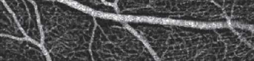
14 Phase Variance OCT OCT Angiography ADVANTAGES No Dye Depth Resolved SLAB LEVEL: Major Retinal Vessels Courtesy of Jeff Fingler, Scott Fraser


15 Phase Variance OCT OCT Angiography ADVANTAGES No Dye Depth Resolved SLAB LEVEL: Superficial Capillary Plexus Courtesy of Jeff Fingler, Scott Fraser


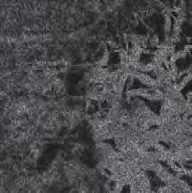
16 Phase Variance OCT OCT Angiography ADVANTAGES No Dye Depth Resolved SLAB LEVEL: Deep Capillary Plexus Courtesy of Jeff Fingler, Scott Fraser


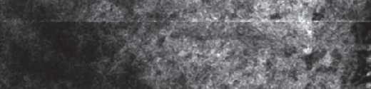
17 Phase Variance OCT OCT Angiography ADVANTAGES No Dye Depth Resolved SLAB LEVEL: Choriocapillaris Courtesy of Jeff Fingler, Scott Fraser


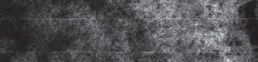
18 Phase Variance OCT OCT Angiography ADVANTAGES No Dye Depth Resolved SLAB LEVEL: Sattler s Layer (medium choroid vessels) Courtesy of Jeff Fingler, Scott Fraser


19 Phase Variance OCT OCT Angiography ADVANTAGES No Dye Depth Resolved SLAB LEVEL: Haller s Layer (large choroid vessels) Courtesy of Jeff Fingler, Scott Fraser
20 PV OCT pitfalls Motion artifact can be a problem for obtaining high-quality images in some patients. Fixation tracking may be a key requirement for optimal imaging


21 PV OCT Eye tracking can yield superb image quality

22 Phase Variance OCT for imaging CNV Neovascular AMD, FVPED s/p >30 ranibizumab injections Old lesion mature vessels within membrane Deep Retinal Capillary Plexus Courtesy of Jeff Fingler, Scott Fraser

23 Phase Variance OCT for imaging CNV Neovascular AMD, FVPED s/p >30 ranibizumab injections Retinal Choroidal Anastomosis Courtesy of Jeff Fingler, Scott Fraser

24 Phase Variance OCT for imaging CNV Neovascular AMD, FVPED s/p >30 ranibizumab injections Retinal Choroidal Anastomosis Courtesy of Jeff Fingler, Scott Fraser

25 Phase Variance OCT for imaging CNV Neovascular AMD, FVPED s/p >30 ranibizumab injections Superficial vessels of CNV Courtesy of Jeff Fingler, Scott Fraser

26 Phase Variance OCT for imaging CNV Neovascular AMD, FVPED s/p >30 ranibizumab injections Larger CNV Vessels Courtesy of Jeff Fingler, Scott Fraser

27 Phase Variance OCT for imaging CNV Neovascular AMD, FVPED s/p >30 ranibizumab injections Larger CNV Vessels Courtesy of Jeff Fingler, Scott Fraser


 Fibrovascular")
28 Increases confidence in our detection of CNV with OCT Spectrum of Pigment Epithelial Detachments Drusenoid PED (medium homogenous) Serous PED (low homogenous) Fibrovascular PED (low heterogenous)


29 Vascular Detail with PV-OCT Zeiss SS-OCT prototype (investigational device, not FDA cleared)
30 Vascular Detail with PV-OCT Zeiss SS-OCT prototype (investigational device, not FDA cleared)


31 Diabetic Retinopathy 2 patients with NPDR --- note microaneurysms and enlarged foveal avascular zone Zeiss SS-OCT prototype (investigational device, not FDA cleared)


32 Depth resolved vascular imaging Superficial Retinal Capillary Plexus Level Zeiss SS-OCT prototype (investigational device, not FDA cleared)


33 Depth resolved vascular imaging Choriocapillaris Level! Zeiss SS-OCT prototype (investigational device, not FDA cleared)
34 Depth resolved vascular imaging Zeiss SS-OCT prototype (investigational device, not FDA cleared)


35 Depth resolved vascular imaging Zeiss SS-OCT prototype (investigational device, not FDA cleared)
36 OCT Angiography Split-Spectrum Amplitude Decorrelation Angiography
37 SSADA Decorrelation refers to fluctuating values of OCT intensities Blood flow results in fluctuation in the amplitude of the OCT fringes ad RBCs enter and exit a particular voxel Greater fluctuation means greater flow Jia et al, Biomed Opt Exp 2012
38 En face retinal and choroidal angiograms at different Z coordinates at macula Yali Jia, PhD; David Huang, MD, PhD.
39 En face retinal and choroidal angiograms at different Z coordinates at ONH Yali Jia, PhD; David Huang, MD, PhD.


 (B)")
 (A) (B) (C) (D) Yali")
40 En face ONH angiograms separately showing the microcirculation within retina, choroid and lamina cribrosa Slab Level Retina Lamina Cribosa Choroid (A) (B) (C) (A) (B) (C) (D) Yali Jia, PhD; David Huang, MD, PhD.


41 Quantitative OCT Angiography Flow and vessel density was reduced in glaucoma patients
42 Summary OCT angiography is an exciting new development in noninvasive imaging The ability to acquire detailed imaging of the retinal and choroidal microvasculature in a depth-resolved fashion, without dye injection, represents a significant advance The prospect of quantitative flow data is an additional major benefit Further refinement of the technology is required to allow ascertainment of leakage The scope/purview of conventional angiography will likely continue to narrow
43 Thank you!
Swept-Source OCT Angiography: SS OCT Angio TM
 Swept-Source OCT Angiography: SS OCT Angio TM Not available in all countries, please check with your distributor. 2015.09 Swept-Source OCT Angiography: SS OCT Angio TM Introduction Optical coherence tomography
ZEISS AngioPlex OCT Angiography Making the revolutionary, routine.
 ZEISS AngioPlex OCT Angiography Making the revolutionary, routine. The moment that revolutionary insight becomes routine. // OCT ANGIOGRAPHY MADE BY ZEISS CIRRUS with AngioPlex creates a new era in both
Visualize. Analyze. Personalize. OCT + OCTA
 Visualize. Analyze. Personalize. OCT + OCTA A New Approach to Protecting Vision AngioVue OCT Angiography brings valuable new information to clinical practice. Non-invasive visualization of retinal vasculature.
OCT Angiography in Primary Eye Care
 OCT Angiography in Primary Eye Care An Image Interpretation Primer Julie Rodman, OD, MS, FAAO and Nadia Waheed, MD, MPH Table of Contents Diabetic Retinopathy 3-6 Choroidal Neovascularization 7-9 Central
OCT Interpretation in Retinal Disease
 OCT Interpretation in Retinal Disease Jay M. Haynie, OD, FAAO Financial Disclosure I have received honoraria or am on the advisory board for the following companies: Carl Zeiss Meditec Advanced Ocular
OCT Angiography. Financial Disclosures: Pre-Test: Which one is Correct?
 OCT Angiography Brandon Lujan, MD Medical Director, Casey Reading Center Assistant Professor of Ophthalmology Financial Disclosures: Genentech (Consultant, Grant support, Educational training) UC Berkeley
Incorporating OCT Angiography Into Patient Care
 Incorporating OCT Angiography Into Patient Care Beth A. Steele, OD, FAAO OCT A: Introduction Isolates microvascular circulation from OCT image data Axial resolution = 5 microns (i.e. fine capillaries visible)
Will OCT-Angiography replace FA?
 ASL Roma A PRESIDIO TERRITORIALE NUOVO REGINA MARGHERITA AMBULATORIO PATOLOGIE RETINICHE Resp. Dott.ssa SUSANNA CATALANO CENTRO ITALIANO MACULA Will OCT-Angiography replace FA? Marco Rispoli, Luca di Antonio,
ZEISS AngioPlex OCT Angiography Overview ZEISS OCT Angiography
 ZEISS AngioPlex OCT Angiography Overview ZEISS OCT Angiography California, ZEISS AngioPlex Ultra-clear visualization of microvascular blood flow using non-invasive OCT angiography 2 AngioPlex OCT Angiography
Is OCT-A Needed As An Investigative Tool During The Management Of Diabetic Macular Edema
 Is OCT-A Needed As An Investigative Tool During The Management Of Diabetic Macular Edema Ayman M Khattab MD, FRCS Professor of Ophthalmology Cairo University Diabetic Macular Edema (DME) Diabetic macular
OCT angiography of ONH blood flow in glaucoma
 Hawaiian Eye Meeting 19-25 January 2013 OCT angiography of ONH blood flow in glaucoma David Huang, MD, Weeks Professor of Ophthalmic Research Professor of Ophthalmology & Biomedical Engineering Casey Eye
Visualize. Analyze. Personalize. OCT + OCTA. with
 Visualize. Analyze. Personalize. OCT + OCTA with Avanti Widefield OCT with AngioVue OCTA Imaging Comprehensive Structural and Functional Imaging in a Single Imaging Platform Comprehensive OCT Imaging The
Clinical Study Optical Coherence Tomography Angiography in Retinal Vascular Diseases and Choroidal Neovascularization
 Hindawi Publishing Corporation Journal of Ophthalmology Volume 2015, Article ID 343515, 8 pages http://dx.doi.org/10.1155/2015/343515 Clinical Study Optical Coherence Tomography Angiography in Retinal
OCT Angiography: The Newest Frontier for the Revolutionary Technology
 Supplement April 2015 OCT Angiography: The Newest Frontier for the Revolutionary Technology OCT Angiography is a new non-invasive, motion contrast micro-vascular imaging modality. Based on two patented
Introducing ANGIOVUE ESSENTIAL. Built on the Avanti Widefield OCT Platform. OCT Angiography for Primary Eye Care
 Introducing ANGIOVUE ESSENTIAL Built on the Avanti Widefield OCT Platform OCT Angiography for Primary Eye Care Transform Your View of the Retina OCT Angiography (OCTA) is a quick non-invasive test that
10/17/2017. FDA Approved. Zeiss AngioPlex TM Optovue AngioVue TM
 Images retinal microvasculature without dye injection Displays structure and function from a single imaging system Standard of Care-2011 DFE, Fundus Photos, VF 10-2, SD-OCT, FAF, or mferg 2016-AAO Baseline
OCT Angiography: The Next Step in Retinal Imaging Jonathan Zelenak D.O.
 OCT Angiography: The Next Step in Retinal Imaging Jonathan Zelenak D.O. Hillsdale Hospital Michigan State University Overview Evolution of OCT How does OCT angiography work? Clinical examples Potential
Deeper visualizations for intervening with confidence.
 CIRRUS OCT with AngioPlex from ZEISS Making the revolutionary routine New vascular quantification Deeper visualizations for intervening with confidence. CIRRUS OCT with AngioPlex from ZEISS can be a much
Go With the Flow: An OCT Angiography Primer Lorne Yudcovitch, OD, MS, FAAO
 Go With the Flow: An OCT Angiography Primer Lorne Yudcovitch, OD, MS, FAAO yudcovil@pacificu.edu OCT Angiography (OCTA) History 2000 - First Doppler flowimetry OCT on human retina 2005 Speckle analysis
OPTICAL COHERENCE TOMOGRAPHY ANGIOGRAPHY OF THE RETINA AND OPTIC NERVE. Lindsay B. Howse, OD
 OPTICAL COHERENCE TOMOGRAPHY ANGIOGRAPHY OF THE RETINA AND OPTIC NERVE Lindsay B. Howse, OD drlindsayhowse@gmail.com None. FINANCIAL DISCLOSURES OUTLINE Introduction/How OCTA works OCTA Analysis Advantages
NIH Public Access Author Manuscript Ophthalmology. Author manuscript; available in PMC 2015 January 01.
 NIH Public Access Author Manuscript Published in final edited form as: Ophthalmology. 2014 January ; 121(1): 180 187. doi:10.1016/j.ophtha.2013.09.002. Phase-Contrast Optical Coherence Tomography: A New
ANSWERING THE WHY? Clinicians discuss the latest imaging technologies for retina practice BY PETER K. KAISER, MD
 Insert to March 2018 Sponsored by MULTI-MODALITY IMAGING: LATEST EVOLUTIONS IN OCTA AND UWF As the array of safe and efficacious medical and surgical options for retinal diseases expands, so does the need
The fovea is the source of highest resolution vision. Its
 Multidisciplinary Ophthalmic Imaging Noninvasive Visualization and Analysis of the Human Parafoveal Capillary Network Using Swept Source OCT Optical Microangiography Laura Kuehlewein, 1,2 Tudor C. Tepelus,
OCT Fundal Angiography Initial Experience The new era in Medical Retina Imaging Based on Cirrus 5000 AngioPlex 2016 Model Sheena George & Nicholas
 OCT Fundal Angiography Initial Experience The new era in Medical Retina Imaging Based on Cirrus 5000 AngioPlex 2016 Model Sheena George & Nicholas Lee Consultants Ophthalmologist at The Hillingdon Hospital
The retinal function imager and clinical applications
 Su and Garg Eye and Vision (2018) 5:20 https://doi.org/10.1186/s40662-018-0114-1 REVIEW Open Access The retinal function imager and clinical applications Daniel Su and Sunir Garg * Abstract Background:
FA vs. OCTA? The status of OCTA, today. Fukuoka, JSOS 2016 Gerd Klose. Korobelnik J Fr Ophthalmol (2015)
") FA vs. OCTA? The status of OCTA, today Korobelnik J Fr Ophthalmol (2015) Fukuoka, JSOS 2016 Gerd Klose 1 2 FA / ICGA a well-founded Gold standard! Benefits Useful for many pathologies High contrast, detailed
FA vs. OCTA? The status of OCTA, today Korobelnik J Fr Ophthalmol (2015) Fukuoka, JSOS 2016 Gerd Klose 1 2 FA / ICGA a well-founded Gold standard! Benefits Useful for many pathologies High contrast, detailed
The leading causes of blindness. ocular circulation: Macular Degeneration
 Taiwan Academy of Ophthalmology Taipei, March 31, 2012 Measurement of Blood Flow in the Retina and Optic Disc with OCT David Huang, MD, Weeks Professor of Ophthalmic Research Professor of Ophthalmology
Functional OCT for Glaucoma Evaluation
 Hawaiian Eye Meeting 18-24 January 2014 Functional OCT for Glaucoma Evaluation David Huang, MD, PhD Weeks Professor of Ophthalmic Research Professor of Ophthalmology & Biomedical Engineering Casey Eye
The role of OCT-A in retinal disease management
 Graefe's Archive for Clinical and Experimental Ophthalmology (2018) 256:2019 2026 https://doi.org/10.1007/s00417-018-4109-3 REVIEW ARTICLE The role of OCT-A in retinal disease management Francisco J. Rodríguez
Disclosures. Definitions. Goals. Imaging and glaucoma 3/22/2016
 Pinakin Davey OD, PhD, FAAO Professor and Director of Research Disclosures Principal investigator for ivue OCT trial Principal investigator Topcon FDA trials for Maestro and OCT 2000 Consultant for Topcon
Angio-OCT. Degenerazione Maculare Legata all Eta. Giuseppe Querques
 Angio-OCT Degenerazione Maculare Legata all Eta Giuseppe Querques Department of Ophthalmology, IRCCS Ospedale San Raffaele, University Vita Salute San Raffaele, Milan, Italy Financial Disclosure ADVISORY
Advances in OCT Murray Fingeret, OD
 Disclosures Advances in OCT Murray Fingeret, OD Consultant Alcon, Allergan, Bausch & Lomb, Carl Zeiss Meditec, Diopsys, Heidelberg Engineering, Reichert, Topcon Currently Approved OCT Devices OCT Devices
Quantitative optical coherence tomography angiography of vascular abnormalities in the living human eye
 Quantitative optical coherence tomography angiography of vascular abnormalities in the living human eye Yali Jia a, Steven T. Bailey a, Thomas S. Hwang a, Scott M. McClintic a, Simon S. Gao a, Mark E.
November Volume 35 - Issue 11
 November 2015 - Volume 35 - Issue 11 pp: 2161-2431,e67-e72 Editorial Optical Coherence Tomography Angiography Spaide, Richard F.; Fujimoto, James G.; Waheed, Nadia K. Original Study IMAGE ARTIFACTS IN
OCT-Angiography Clinical Cases. OCT-Angiography Clinical Cases
 OCT-Angiography Clinical Cases OCT-Angiography Clinical Cases NIDEK RS-3000 Advance AngioScan Daniela Bacherini Andrea Sodi Stanislao Rizzo CONTENTS Page Authors 3 Introduction 4 Case 1 Case 2 Case 3 Case
The Evaluation of Diabetic Macular Ischemia Using Optical Coherence Tomography Angiography
 Retina The Evaluation of Diabetic Macular Ischemia Using Optical Coherence Tomography Angiography Patrick D. Bradley, 1 Dawn A. Sim, 1 Pearse A. Keane, 1 João Cardoso, 1,2 Rupesh Agrawal, 1 Adnan Tufail,
Macular Morphology and Visual Acuity in the Comparison of Age-related Macular Degeneration Treatments Trials
 Macular Morphology and Visual Acuity in the Comparison of Age-related Macular Degeneration Treatments Trials Glenn J. Jaffe, MD, 1 Daniel F. Martin, MD, 2 Cynthia A. Toth, MD, 1 Ebenezer Daniel, MPH, PhD,
Adaptive Optics and OCTA: Update on Retinal Imaging. Judy E. Kim, MD Professor of Ophthalmology Medical College of Wisconsin
 Adaptive Optics and OCTA: Update on Retinal Imaging Judy E. Kim, MD Professor of Ophthalmology Medical College of Wisconsin Financial Disclosure Advisory Board Alimera Science, Allergan, Bayer, Novartis
IMAGE ARTIFACTS IN OPTICAL COHERENCE TOMOGRAPHY ANGIOGRAPHY
 IMAGE ARTIFACTS IN OPTICAL COHERENCE TOMOGRAPHY ANGIOGRAPHY The MIT Faculty has made this article openly available. Please share how this access benefits you. Your story matters. Citation Spaide, Richard
Dr/ Marwa Abdellah EOS /16/2018. Dr/ Marwa Abdellah EOS When do you ask Fluorescein angiography for optic disc diseases???
 When do you ask Fluorescein angiography for optic disc diseases??? 1 NORMAL OPTIC DISC The normal optic disc on fluorescein angiography is fluorescent due to filling of vessels arising from the posterior
OCT Angiography The Next Frontier
 Choroid Retina avascular 5/13/2017 OCT Angiography The Next Frontier Pierce Kenworthy OD, FAAO June 9, 2017 OCT Angiography (OCTA) 2016 Non-invasive, motion contrast imaging Represents erythrocyte movement
Research Article Diabetic Macular Ischemia Diagnosis: Comparison between Optical Coherence Tomography Angiography and Fluorescein Angiography
 Ophthalmology Volume 2016, Article ID 3989310, 6 pages http://dx.doi.org/10.1155/2016/3989310 Research Article Diabetic Macular Ischemia Diagnosis: Comparison between Optical Coherence Tomography Angiography
Instudies of vascular endothelial growth factor
 MONITORING THERAPEUTIC EFFICACY IN THE ANTI-VEGF ERA* SriniVas R. Sadda, MD ABSTRACT One of the most important questions with vascular endothelial growth factor (VEGF) inhibitors for age-related macular
Mark Dunbar: Disclosure
 Important Things to Understand About OCT Mark T. Dunbar, O.D., F.A.A.O. Bascom Palmer Eye Institute University of Miami, School of Medicine Mark Dunbar: Disclosure Optometry Advisory Board for: Allergan
OCT Angiography: An Upcoming Tool for Diagnosis and Treatment of Retinal Vascular Diseases
 E-ISSN 2454-2784 Recent Advances OCT Angiography: An Upcoming Tool for Diagnosis and Treatment of Retinal Vascular Diseases Purnima Sood 1, Nalini Saxena 2, Dinesh Talwar 3 1 Vitreo-Retina Consultant,
Il contributo dell'angio-oct: valutazione integrata della componente nervosa e vascolare della malattia glaucomatosa
 SIMPOSIO G.O.A.L. - LE NUOVE FRONTIERE DIAGNOSTICHE E LE LINEE DI INDIRIZZO AMBULATORIALI DEL GLAUCOMA Coordinatore e moderatore: D. Mazzacane Presidente: L. Rossetti Il contributo dell'angio-oct: valutazione
Quantitative Optical Coherence Tomography Angiography of Choroidal Neovascularization in Age-Related Macular Degeneration
 Quantitative Optical Coherence Tomography Angiography of Choroidal Neovascularization in Age-Related Macular Degeneration The MIT Faculty has made this article openly available. Please share how this access
CAPILLARY NETWORK ANOMALIES IN BRANCH RETINAL VEIN OCCLUSION ON OPTICAL COHERENCE TOMOGRAPHY ANGIOGRAPHY
 CAPILLARY NETWORK ANOMALIES IN BRANCH RETINAL VEIN OCCLUSION ON OPTICAL COHERENCE TOMOGRAPHY ANGIOGRAPHY MARCO RISPOLI, MD, MARIA CRISTINA SAVASTANO, MD, PHD, BRUNO LUMBROSO, MD Purpose: To analyze the
SOUTH-EAST EUROPEAN JOURNAL of OPHTHALMOLOGY 2015; 1 (1) 34 40
 34 40") Review article SOUTH-EAST EUROPEAN JOURNAL of OPHTHALMOLOGY 2015; 1 (1) 34 40 Retinal nerve fiber layer versus peripapillary capillary density assessment A powerful tool for detecting optic nerve head
Review article SOUTH-EAST EUROPEAN JOURNAL of OPHTHALMOLOGY 2015; 1 (1) 34 40 Retinal nerve fiber layer versus peripapillary capillary density assessment A powerful tool for detecting optic nerve head
ZEISS AngioPlex OCT Angiography. Clinical Case Reports
 Clinical Case Reports Proliferative Diabetic Retinopathy (PDR) Case Report 969 PROLIFERATIVE DIABETIC RETINOPATHY 1 1-year-old diabetic female presents for follow-up of proliferative diabetic retinopathy
Clinically Significant Macular Edema (CSME)
") Clinically Significant Macular Edema (CSME) 1 Clinically Significant Macular Edema (CSME) Sadrina T. Shaw OMT I Student July 26, 2014 Advisor: Dr. Uwaydat Clinically Significant Macular Edema (CSME) 2
Clinically Significant Macular Edema (CSME) 1 Clinically Significant Macular Edema (CSME) Sadrina T. Shaw OMT I Student July 26, 2014 Advisor: Dr. Uwaydat Clinically Significant Macular Edema (CSME) 2
Optical Coherence Tomography Angiography in Central Retinal Vein Occlusion: Correlation Between the Foveal Avascular Zone and Visual Acuity METHODS
 Special Issue Optical Coherence Tomography Angiography in Central Retinal Vein Occlusion: Correlation Between the Foveal Avascular Zone and Visual Acuity Manuel Casselholm de Salles, Anders Kvanta, Urban
Clinical Study Optical Coherence Tomography and Optical Coherence Tomography Angiography in Monitoring Coats Disease
 Hindawi Ophthalmology Volume 2017, Article ID 7849243, 8 pages https://doi.org/10.1155/2017/7849243 Clinical Study Optical Coherence Tomography and Optical Coherence Tomography Angiography in Monitoring
Citation. As Published Publisher. Version
 Select Features of Diabetic Retinopathy on Swept-Source Optical Coherence Tomographic Angiography Compared With Fluorescein Angiography and Normal Eyes The MIT Faculty has made this article openly available.
NIH Public Access Author Manuscript JAMA Ophthalmol. Author manuscript; available in PMC 2013 September 10.
 NIH Public Access Author Manuscript Published in final edited form as: JAMA Ophthalmol. 2013 May ; 131(5): 693 694. doi:10.1001/jamaophthalmol.2013.692. Effect of Intravitreous Anti Vascular Endothelial
Simply the best OCT & OCTA image quality.
 Avanti Widefield OCT with AngioVue OCT Angiography Simply the best OCT & OCTA image quality. Dear Friends of Optovue, Since introducing Spectral Domain OCT to the ophthalmology market in 2006, Optovue
Optical Coherence Tomography Angiography In Diagnosis Of Retinal Angiomatous Proliferation
 Optical Coherence Tomography Angiography In Diagnosis Of Retinal Angiomatous Proliferation Stepanov A, (1,4) Jiraskova N, 1,4) Lestak J, 2, 3, 4* 1. Department of Ophthalmology, University Hospital and
THE OPHTHALMOLOGIST S NEEDS FOR THE ANALYSIS OF THE RETINA
 biophotonics end-users needs THE OPHTHALMOLOGIST S NEEDS FOR THE ANALYSIS OF THE RETINA Dr Matonti Frédéric CHU Nord / INT AMU Marseille ANATOMY OF THE RETINA ANATOMY OF THE RETINA ANATOMY OF THE RETINA
PLEX Elite 9000 from ZEISS Swept-Source OCT
 PLEX Elite 9000 from ZEISS Swept-Source OCT Uncovering the undiscovered. ZEISS PLEX Elite 9000 // INNOVATION MADE BY ZEISS 2 Ultra-wide angiography En face montage Image courtesy of Prof. G. Querques,
AMD in 2017: A Review of Treatment Guidelines and the Role of Early Appropriate Therapy
 Supplement to March 2017 CME Activity AMD in 2017: A Review of Treatment Guidelines and the Role of Early Appropriate Therapy Rishi P. Singh, MD, moderator SriniVas Sadda, MD Richard F. Spaide, MD Nadia
OCT Interpretation. Financial Disclosure. Jay M. Haynie, OD, FAAO. OCT Image Layers 7/21/2014
 OCT Interpretation Jay M. Haynie, OD, FAAO Financial Disclosure I have received honoraria or am on the advisory board for the following companies: Olympia Tacoma Renton Kennewick - Washington Carl Zeiss
In vivo human retinal and choroidal vasculature visualization using differential phase contrast swept source optical coherence tomography at 1060 nm
 In vivo human retinal and choroidal vasculature visualization using differential phase contrast swept source optical coherence tomography at 1060 nm Reza Motaghiannezam and Scott Fraser Beckman Institute,
Diurnal variation of choriocapillaris vessel flow density in normal subjects measured using optical coherence tomography angiography
 https://doi.org/10.1186/s40942-018-0140-0 International Journal of Retina and Vitreous ORIGINAL ARTICLE Open Access Diurnal variation of choriocapillaris vessel flow density in normal subjects measured
Fluorescein Angiography
 Last revision: October 2011 by Luis Arias Fluorescein Angiography Authors: Luis Arias, MD Hospital Universitari de Bellvitge - University of Barcelona. Spain Jordi Monés, MD Institut de la Màcula i de
Projection-resolved optical coherence tomographic angiography
 Projection-resolved optical coherence tomographic angiography Miao Zhang, Thomas S. Hwang, J. Peter Campbell, Steven T. Bailey, David J. Wilson, David Huang and Yali Jia * Casey Eye Institute, Oregon Health
Leo Semes, OD, FAAO UAB Optometry
 Leo Semes, OD, FAAO UAB Optometry Safe; inert Has long track record - over 45 years Mixes with plasma and highlights blood vessel compromise Using specific exciting (490 nm)and absorption (510 nm) filters
The diagnostic value of optical coherence tomography angiography in diabetic retinopathy: a systematic review
 https://doi.org/10.1007/s10792-018-1034-8 (0456789().,-volV) (0456789().,-volV) REVIEW The diagnostic value of optical coherence tomography angiography in diabetic retinopathy: a systematic review David
Michael P. Blair, MD Retina Consultants, Ltd Libertyville/Des Plaines, Illinois Clinical Associate University of Chicago 17 October 2015
 Michael P. Blair, MD Retina Consultants, Ltd Libertyville/Des Plaines, Illinois Clinical Associate University of Chicago 17 October 2015 So What Parts of the Eye Retina are Affected by VHL Neural tissue
Citation. As Published Publisher. Version
 Effect of Intravitreous Anti Vascular Endothelial Growth Factor Therapy on Choroidal Thickness in Neovascular Age-Related Macular Degeneration Using Spectral-Domain The MIT Faculty has made this article
The Quick Guide to OCT Mastery 50 Real Cases with Expert Analysis
 OPTICAL COHERENCE TOMOGRAPHY The Quick Guide to OCT Mastery 50 Real Cases with Expert Analysis VOL 1 Sanjay Sharma, MD, FRCS, MSc (Epid), MBA Ophthalmologist, Epidemiologist Queen s University, Canada
TOPCON EURETINA Clinical Advances and Applications With Swept Source OCT and Angiography. JANUARY/FEBRUARY 2019 VOL. 17, NO.
 SUPPLEMENT TO SPONSORED BY JANUARY/FEBRUARY 2019 VOL. 17, NO. 1 TOPCON EURETINA 2018 Clinical Advances and Applications With Swept Source OCT and Angiography. This supplement summarizes highlights from
Comparison of Neovascular Lesion Area Measurements From Different Swept-Source OCT Angiographic Scan Patterns in Age-Related Macular Degeneration
 Retina Comparison of Neovascular Lesion Area Measurements From Different Swept-Source OCT Angiographic Scan Patterns in Age-Related Macular Degeneration Fang Zheng, 1,2 Qinqin Zhang, 3 Elie H. Motulsky,
Diabetic retinopathy (DR), characterized by capillary nonperfusion,
, characterized by capillary nonperfusion,") Retina Automated Quantification of Nonperfusion in Three Retinal Plexuses Using Projection-Resolved Optical Coherence Tomography Angiography in Diabetic Retinopathy Miao Zhang, 1 Thomas S. Hwang, 1 Changlei
Retina Automated Quantification of Nonperfusion in Three Retinal Plexuses Using Projection-Resolved Optical Coherence Tomography Angiography in Diabetic Retinopathy Miao Zhang, 1 Thomas S. Hwang, 1 Changlei
The Human Eye. Cornea Iris. Pupil. Lens. Retina
 The Retina Thin layer of light-sensitive tissue at the back of the eye (the film of the camera). Light rays are focused on the retina then transmitted to the brain. The macula is the very small area in
Diagnosis and treatment of diabetic retinopathy. Blake Cooper MD Ophthalmologist Vitreoretinal Surgeon Retina Associates Kansas City
 Diagnosis and treatment of diabetic retinopathy Blake Cooper MD Ophthalmologist Vitreoretinal Surgeon Retina Associates Kansas City Disclosures Consulted for Novo Nordisk 2017,2018. Will be discussing
Course # Getting to Know Your OCT
 Course # 140 Getting to Know Your OCT Course Title: Lecturer: Getting to Know Your OCT Brad Sutton, OD, FAAO IU School of Optometry Financial Disclosures No financial disclosures Optical Coherence Tomography-OCT
Evaluation of very high- and very low-dose intravitreal aflibercept in patients with neovascular age-related macular degeneration.
 University of Nebraska Medical Center DigitalCommons@UNMC Journal Articles: Ophthalmology Ophthalmology 12-2012 Evaluation of very high- and very low-dose intravitreal aflibercept in patients with neovascular
IQ 532 Micropulse Green Laser treatment for Refractory Chronic Central Serous Retinopathy
 Cronicon OPEN ACCESS EC OPHTHALMOLOGY Case Report IQ 532 Micropulse Green Laser treatment for Refractory Chronic Central Serous Retinopathy Fawwaz Al Mamoori* Medical Retina Department, Eye Specialty Hospital,
Automated segmentation and analysis of layers and structures of human posterior eye
 University of Iowa Iowa Research Online Theses and Dissertations 2015 Automated segmentation and analysis of layers and structures of human posterior eye Li Zhang University of Iowa Copyright 2015 Li Zhang
R&M Solutions
 Mohamed Hosny El-Bradey, MD., Assistant Professor of Ophthalmology, Tanta University. Wael El Haig, MD., Professor of Ophthalmology. Zagazeeg University. 1 Myopic CNV is considered the most common vision
What You Should Know About Acute Macular Neuroretinopathy
 What You Should Know About Acute Macular Neuroretinopathy David J. Browning MD, PhD Chong Lee BS Acute macular neuroretinopathy is a condition characterized by the sudden, painless onset of paracentral
Systemic bevacizumab (Avastin) therapy for exudative neovascular age-related macular degeneration. The BEAT-AMD-Study
 therapy for exudative neovascular age-related macular degeneration. The BEAT-AMD-Study") 1 Department of Ophthalmology, The Ludwig Boltzmann Institute for Retinology and Biomicroscopic Lasersurgery, Rudolf Foundation Clinic, Vienna, Austria; 2 Department of Internal Medicine, Oncology, Rudolf
1 Department of Ophthalmology, The Ludwig Boltzmann Institute for Retinology and Biomicroscopic Lasersurgery, Rudolf Foundation Clinic, Vienna, Austria; 2 Department of Internal Medicine, Oncology, Rudolf
Building The Retina Company
 Building The Retina Company Optos devices produce ultra-widefield (UWF ), high resolution images (optomap ) of approximately 82% (200 ) of the retina. A single optomap can document the retina from the
Clinical Optical Coherence Tomography Angiography Registration and Analysis
 Clinical Optical Coherence Tomography Angiography Registration and Analysis by Morgan Lindsay Heisler B.A.Sc. (Hons.), Simon Fraser University, 2015 Thesis Submitted in Partial Fulfillment of the Requirements
Diabetic retinopathy and OCT angiography: clinical findings and future perspectives
 DOI 10.1186/s40942-017-0062-2 International Journal of Retina and Vitreous REVIEW Open Access Diabetic retinopathy and OCT angiography: clinical findings and future perspectives Jose Mauricio Botto de
APRIL 8th 2016 Therapy
 APRIL 8th 2016 Therapy 09.00-10.00 SESSION 1: Age-related Macular Degeneration Moderators: A. Brucker, E. Souied, M. Stirpe, R. Brancato 09.00-09.15 TBD A. Brucker 09.15-09.30 TBD E. Souied 09.30-09.45
Multifunctional 1050 nm Spectral Domain Oct System at 147 khz for Posterior Eye Imaging
 Multifunctional 1050 nm Spectral Domain Oct System at 147 khz for Posterior Eye Imaging DOI 10.17691/stm2015.7.1.01 Received October 30, 2014 Anqi Zhang, PhD, Postdoctoral Fellow, Department of Bioengineering;
IN NICU OCT UTILIZES A CONCEPT KNOWN AS INTERFEROMETRY APPLICATIONS FOR OCT THE PRIMARY USE IN THE EYE - RETINA
 2016 25 YEARS OF OPTICAL COHERENCE TOMOGRAPHY OPTICAL COHERENCE TOMOGRAPHY IN NICU Marcin Stopa, MD, PhD, FEBO Department of Ophthalmology, Chair of Ophthalmology and Optometry. Poznan University of Medical
Diabetic Retinopathy A Presentation for the Public
 Diabetic Retinopathy A Presentation for the Public Ray M. Balyeat, MD The Eye Institute Tulsa, Oklahoma The Healthy Eye Light rays enter the eye through the cornea, pupil and lens. These light rays are
doi: /s
 doi: 10.1186/s12886-015-0077-0 Miura et al. BMC Ophthalmology (2015) 15:79 DOI 10.1186/s12886-015-0077-0 RESEARCH ARTICLE Open Access Noninvasive vascular imaging of ruptured retinal arterial macroaneurysms
DOME SHAPED MACULOPATHY. Ιωάννης Ν. Βαγγελόπουλος Χειρ. Οφθαλμίατρος - Βόλος
 DOME SHAPED MACULOPATHY Ιωάννης Ν. Βαγγελόπουλος Χειρ. Οφθαλμίατρος - Βόλος DOME SHAPED MACULOPATHY-DEFINITIONS The entity Dome Shaped Macula ( DSM ) was first described by Gaucher and associates in 2008
Disease-Specific Fluorescein Angiography
 Ruth E. Picchiottino, CRA Disease-Specific Fluorescein Angiography 15 Disease-Specific Fluorescein Angiography Recommendations for tailoring retinal fluorescein angiography to diabetic retinopathy, macular
Automated choroidal neovascularization detection algorithm for optical coherence tomography angiography
 Automated choroidal neovascularization detection algorithm for optical coherence tomography angiography Li Liu, 1,2 Simon S. Gao, 1 Steven T. Bailey, 1 David Huang, 1 Dengwang Li, 2,* and Yali Jia 1,*
Retinal Capillary Network and Foveal Avascular Zone in Eyes with Vein Occlusion and Fellow Eyes Analyzed With Optical Coherence Tomography Angiography
 Retinal Capillary Network and Foveal Avascular Zone in Eyes with Vein Occlusion and Fellow Eyes Analyzed With Optical Coherence Tomography Angiography The MIT Faculty has made this article openly available.
Differential intensity contrast swept source optical coherence tomography for human retinal vasculature visualization
 Differential intensity contrast swept source optical coherence tomography for human retinal vasculature visualization Reza Motaghiannezam and Scott Fraser Beckman Institute, Division of Biology, California
The World s fastest OCT. As simple as pressing. the start button
 The World s fastest OCT As simple as pressing the start button lution continues Optopol engineering team, designers of the first commercially available Spectral Domain OCT in the world, are proud to present
Optical Coherence Tomography in Diabetic Retinopathy. Mrs Samantha Mann Consultant Ophthalmologist Clinical Lead of SEL-DESP
 Optical Coherence Tomography in Diabetic Retinopathy Mrs Samantha Mann Consultant Ophthalmologist Clinical Lead of SEL-DESP Content OCT imaging Retinal layers OCT features in Diabetes Some NON DR features
ARVO 2016 Annual Meeting Abstracts
 146 OCT Angiography 1 Sunday, May 01, 2016 3:15 PM 5:00 PM 6B Paper Session Program #/Board # Range: 947 953 Organizing Section: Retina Program Number: 947 Presentation Time: 3:15 PM 3:30 PM Comparisons
Age-related Macular Degeneration Update
 Age-related Macular Degeneration Update AMD: The Burden of Disease Carlo J. Pelino, OD, FAAO cpelino@salus.edu Joseph J. Pizzimenti, OD, FAAO pizzimen@nova.edu Course Goals Statement of the problem Epidemiology