EyePACS Grading System (Part 2): Detecting Presence and Severity of Background (Non-Proliferative) Diabetic Retinopathy Lesion
|
|
|
- Dustin Bell
- 5 years ago
- Views:
Transcription
1 EyePACS Grading System (Part 2): Detecting Presence and Severity of Background (Non-Proliferative) Diabetic Retinopathy Lesion George Bresnick MD MPA Jorge Cuadros OD PhD

2 Anatomy of the eye:

3 3 Normal Retina Retinal Arcades Macula Optic Nerve
4 EyePACS Digital Retinal Image Grading System DIABETIC FUNDUS LESIONS REFLECT: Abnormalities of retinal microvasculature Ischemia (capillary/arteriolar closure) Abnormal permeability (capillary leakage) Lesion type and severity predict risk for future vision loss thereby, determine need and urgency for referral
5 EyePACS Digital Retinal Image Grading System This presentation will emphasize: Appearance and pathophysiology of retinal lesions FUNDUS LESIONS AND THEIR ORIGINS Natural history of diabetic retinopathy WHAT HAPPENS TO UNTREATED EYES? Treatment results (DRS/ETDRS) HOW DO TREATMENT GUIDELINES INFLUENCE GRADING PROGRAM?
6 EyePACS Grading Template
7 Diabetic Retinopathy Pathophysiology Nonproliferative Diabetic Retinopathy (NPDR) background changes within the retina Retinal vessel closure (retinal ischemia) Capillary closure-mild retinal ischemia Arteriolar closure-more severe retinal ischemia Increased retinal vessel permeability (and macular edema)
8 RETINAL CAPILLARY CLOSURE Mild retinal ischemia FLUORESCEIN ANGIOGRAPHY Two processes: Capillary nonperfusion (closure) Compensatory capillary dilation, including microaneurysms

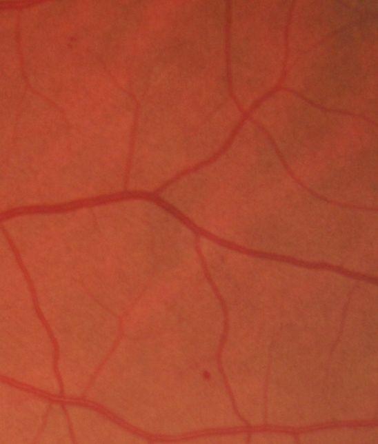
9 Fluorescein angiogram Normal macula Normal capillaries barely visible

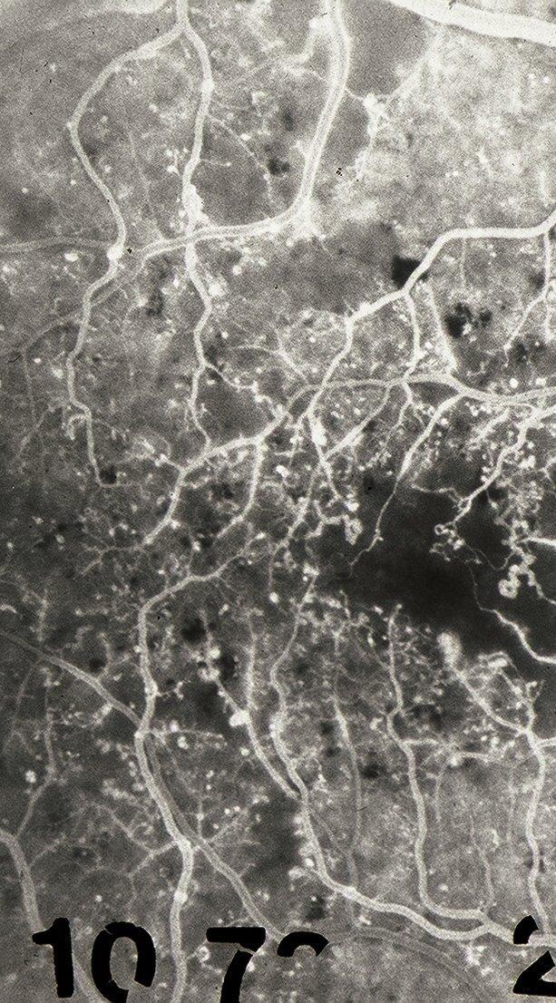
10 Fluorescein angiogram Diabetic macula Dilated capillaries interspersed with Nonperfused dark spaces and hyperfluorescent dots (microaneurysms)

11 Normal Retinal Capillaries Trypsin Digest Regular capillary caliber Round dark pericytes; elongated endothelial nuclei
 capillaries Hypercellular (dilated)")
12 Diabetic Retinal Capillaries Trypsin Digest Acellular (nonperfused) capillaries Hypercellular (dilated) capillaries
13 Fundus Lesions Associated with Capillary Closure Microaneurysms (MA) Appearance: Small red dots microns in diameter Location Clustered adjacent to areas of capillary nonperfusion Significance Earliest manifestation of diabetic retinopathy Numerous MA indicates widespread capillary closure

14 Fundus Signs of Capillary Closure Microaneurysms
15

16 India Ink injection preparation of autopsy eye Microaneurysms face into areas of capillary nonperfusion

17 MICROANEURYSMS ONLY (MA) Mild Nonproliferative retinopathy
18 Mild Nonproliferative Retinopathy (NPDR) = MICROANEURYSMS ONLY (MA) MA defined as small circular red dots with well defined borders Patients with Mild NPDR usually do not require referral to eye care specialist. MA may resolve in about 2 years. MA turnover rate may indicate severity of diabetes.
19 NPDR: RETINAL ARTERIOLAR CLOSURE More severe retinal ischemia FLUORESCEIN ANGIOGRAPHY Nipped arteriolar side branches Non-perfused zone of capillaries in distribution of occluded arterioles
 Nipped arteriole branches")
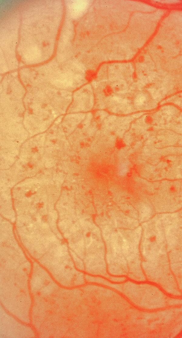
20 Arteriolar nonperfusion Large dark nonperfused areas (Arrows) Nipped arteriole branches (Circled)


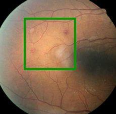
21 Arteriolar nonperfusion Large dark area below major arteriole apparently nonperfused. (Circled)
")

22 Arteriolar Occlusion Nipped arteriolar branch (Arrow) Acellular capillaries in distribution of arteriole (Circled)
23 Fundus Lesions Associated with Arteriolar Closure (ISCHEMIC RETINAL LESIONS) Cotton wool spots (CW) Venous beading (VB) Intraretinal microvascular abnormalities (IRMA) Dark, blot hemorrhages
 Ischemic infarct nerve")
24 Cotton wool spots (CWS) Fluffy white exudates (Arrows) Ischemic infarct nerve fiber layer.
25
26 Cotton Wool Spots Faint CW spots nasal to disc and inf. nasal Case 47492
27 HEMORRHAGES WITH OR WITHOUT MICROANEURYSMS (HMA): Moderate or Severe NPDR Depending on Location and Severity
 H are")

28 Distinguish H(circled) from MA (box) H are larger or more irregular than MA H H MA Case 584 H?H MA
29 GRADING GUIDELINES: Definition of Hemifields EyePACS images can be divided into superior and inferior hemifields by horizontal line through the center of the optic disc. Used for comparison of EyePACS images with ETDRS Standard Photos e.g., HMA>=Standard photo 2A in both superior and inferior hemifields?

30 HEMIFIELDS: SUPERIOR HEMIFELD INFERIOR HEMIFIELD Each imaging field can be divided horizontally into superior and inferior grading hemifields.
31 HEMORRHAGES WITH OR WITHOUT MA (HMA) Consider all intraretinal red spots together (both hemorrhages and microaneurysms) If present, mark the appropriate box: <2A ( ) >=2A ( ) Compare density and areal extent of HMA in the EyePACS images with density and extent of HMA in Standard Photo 2A. (See Photo 2A next slide.) Answer HMA>=2A**, if HMA >=2A in both upper and lower hemifields Answer HMA < 2A, if HMA are <2A in one or both hemifields,

32 Standard Photo 2A Reference Image 2a

33 HMA>=2A Case 46614

34 HMA>=2A Case 45164

35 HMA<2A in both hemifields Case 387
36 HEMORRHAGES WITH OR WITHOUT MA (HMA) If HMA greater than Image 2a in both hemifields: then patient has severe nonproliferative retinopathy and requires referral to eye care specialist within 3 months. associated with 48% chance of developing dangerous new blood vessels (neovascularization) within one year. If HMA is less than Image 2a in either hemifield then patient has Moderate NPDR and should be imaged again in 6 months.
37 Fundus Lesions Associated with Arteriolar Closure (ISCHEMIC RETINAL LESIONS) Cotton wool spots (CW) Venous beading (VB) Intraretinal microvascular abnormalities (IRMA) Dark, blot hemorrhages
38 DEFINITE VENOUS BEADING (VB) = Severe NPDR
39 VENOUS BEADING (VB) = Severe NPDR (depending on severity) Consider definite sausage-like dilation of one or more venous segments anywhere in the EyePACS images. Use Standard Photo 6A for examples of definite VB (See next slide.). Needs to be accompanied by other signs of ischemia

40 Venous Beading (VB) Segmental dilation of vein, often adjacent to cotton wool spots

41 Venous Beading Case 50603
42 VENOUS BEADING (VB) = Severe NPDR (depending on severity) If Venous beading is present and greater than reference image 6a, then patient has Severe NPDR and requires referral within 3 months to eye specialist. Venous beading greater than 6a is associated with a 51% chance of developing dangerous new blood vessels (neovascularization) within one year.
43 Fundus Lesions Associated with Arteriolar Closure (ISCHEMIC RETINAL LESIONS) Cotton wool spots Venous beading (VB) Intraretinal microvascular abnormalities (IRMA) Dark, blot hemorrhages
44 Question 6 INTRARETINAL MICROVASCULAR ABNORMALITIES (IRMA)?
45 INTRARETINAL MICROVASCULAR ABNORMALITIES (IRMA): Moderate or Severe NPDR Intraretinal microvascular abnormalities present? no ( ) yes ( ) Cannot grade ( ) Consider dilated tortuous capillary segments. If (yes), mark the appropriate box: <8A ( ) >=8A ( ) ** Compare the density and severity of IRMA in EyePACS images with extent of IRMA in Standard Photo 8A. (See next slide.) Answer <8A, if IRMA <8A wherever present. Answer IRMA>=8A, if IRMA >=8A anywhere in the EyePACS images.

46 Reference Image 8a Standard Photo 8A

47 Intraretinal Microvascular Abnormalities >= 8a Case 34948
 Dilated tortuous capillary")
48 Intraretinal Microvascular Abnormalities (IRMA) Dilated tortuous capillary segments
 Dilated tortuous capillary")
49 Intraretinal Microvascular Abnormalities (IRMA) Dilated tortuous capillary segments

50 IRMA > 8a Case 47492
51 INTRARETINAL MICROVASCULAR ABNORMALITIES (IRMA) If IRMA is greater than Image 8a in either hemifield: then patient has severe nonproliferative retinopathy and requires referral to eye care specialist within 3 months. associated with 44% chance of developing dangerous new blood vessels (neovascularization) within one year. If IRMA is less than Image 8a then patient has Moderate NPDR and should be imaged again in 6 months.
52 High-Risk Markers for Progression to Proliferative Retinopathy: Early Treatment of Diabetic Retinopathy Study Ischemic Retinal Lesion Progression rate to PDR (1 yr) Extensive retinal hemorrhages (HMA) 48% Venous beading (VB) 51% Intraretinal Microvascular Abnormalities (IRMA) 44% Cotton Wool Spots (CWS) No sig. increase
53 53 Severe Nonproliferative Retinopathy (NPDR) Latin American male, DM II X 12 years, Last eye exam 5 years ago

54 54 Severe Nonproliferative Retinopathy (NPDR) Latin American male, DM II X 12 years, Last eye exam 5 years ago HMA > 2a CW


55 60 yr. old, DM X 9yrs, HbA1C=9.1 1 yr. later, HbA1C=5.5: 55

56 33 yr. old Latin American male Type II DM X 5 years Hyperlipidemia HDL/LDL: 46.2/143.3 Triglycerides: 321 HbA1c = 11.9 Meds: Lantus, Novolog, Pravachol Last Eye Exam: 2-5 years ago
57 33 yr. old Latin American male Microaneurysms Intraretinal hemorrhages

58 48 yr. old Pacific Island female Hypertension DM II Dx s 11/2004 HbA1c 9.3 Cholesterol 223 Triglycerides 216 HDL 29 Meds: Amlodopine, Toprol, Novolog, Lantus, Pravastatin

59 48 yr. old Pacific Island female CW HMA IRMA VB
60 NPDR Summary: Identifying presence and severity of retinal lesions associated with diabetes can help with triage and patient education: No apparent diabetic retinopathy and HbA1c < 7: return in 2 years for imaging Mild NPDR or HBA1c > 7 : return in 1 year for imaging Moderate NPDR: return in 6 months for imaging HMA < 2a, CWS, or IRMA < 8a Severe NPDR: refer to eye specialist within 3 months HMA > 2a, VB, or IRMA > 8a

61 Thank You!
EyePACS Grading System (Part 3): Detecting Proliferative (Neovascular) Diabetic Retinopathy. George Bresnick MD MPA Jorge Cuadros OD PhD
: Detecting Proliferative (Neovascular) Diabetic Retinopathy. George Bresnick MD MPA Jorge Cuadros OD PhD") EyePACS Grading System (Part 3): Detecting Proliferative (Neovascular) Diabetic Retinopathy George Bresnick MD MPA Jorge Cuadros OD PhD Anatomy of the eye: 3 Normal Retina Retinal Arcades Macula Optic
EyePACS Grading System (Part 3): Detecting Proliferative (Neovascular) Diabetic Retinopathy George Bresnick MD MPA Jorge Cuadros OD PhD Anatomy of the eye: 3 Normal Retina Retinal Arcades Macula Optic
The Natural History of Diabetic Retinopathy and How Primary Care Makes A Difference
 The Natural History of Diabetic Retinopathy and How Primary Care Makes A Difference We will discuss - How exactly does blood sugar control affect retinopathy? - What are other factors that we measure in
PART 1: GENERAL RETINAL ANATOMY
 PART 1: GENERAL RETINAL ANATOMY General Anatomy At Ora Serrata At Optic Nerve Head Fundoscopic View Of Normal Retina What Is So Special About Diabetic Retinopathy? The WHO definition of blindness is
Diabetic Retinopathy. Barry Emara MD FRCS(C) Giovanni Caboto Club October 3, 2012
 Giovanni Caboto Club October 3, 2012") Diabetic Retinopathy Barry Emara MD FRCS(C) Giovanni Caboto Club October 3, 2012 Outline Statistics Anatomy Categories Assessment Management Risk factors What do you need to do? Objectives Summarize the
Diabetic Retinopathy Barry Emara MD FRCS(C) Giovanni Caboto Club October 3, 2012 Outline Statistics Anatomy Categories Assessment Management Risk factors What do you need to do? Objectives Summarize the
Diagnosis and treatment of diabetic retinopathy. Blake Cooper MD Ophthalmologist Vitreoretinal Surgeon Retina Associates Kansas City
 Diagnosis and treatment of diabetic retinopathy Blake Cooper MD Ophthalmologist Vitreoretinal Surgeon Retina Associates Kansas City Disclosures Consulted for Novo Nordisk 2017,2018. Will be discussing
DIABETIC RETINOPATHY
 DIABETIC RETINOPATHY C. L. B. Canny, MD FRCSC Diabetic retinopathy is the most serious eye manifestation of diabetes and is responsible for most of the blindness caused by diabetes. Diabetic retinopathy
Diabesity A Public Health Crisis: AOA Evidence Based Translation to Care Series
 Diabesity A Public Health Crisis: AOA Evidence Based Translation to Care Series Joseph J. Pizzimenti, OD, FAAO Associate Professor Nova Southeastern University The Eye Care Institute pizzimen@nova.edu
ZEISS AngioPlex OCT Angiography. Clinical Case Reports
 Clinical Case Reports Proliferative Diabetic Retinopathy (PDR) Case Report 969 PROLIFERATIVE DIABETIC RETINOPATHY 1 1-year-old diabetic female presents for follow-up of proliferative diabetic retinopathy
INTRODUCTION AND SYMPTOMS
 CHAPTER 1 INTRODUCTION AND SYMPTOMS Introduction of Diabetic Retinopathy Diabetic retinopathy (DR) is a potentially blinding complication of diabetes. It is defined as presence of one or more definite
Diabetic Management beyond traditional risk factors and LDL-C control: Can we improve macro and microvascular risks?
 Retinopathy Diabetes has a negative effect on eyes in many ways, increasing the risk of cataracts for example, but the most common and serious ocular complication of diabetes is retinopathy. Diabetic retinopathy
Diabetic Retinopathy
 Diabetic Retinopathy Diabetes can be classified into type 1 diabetes mellitus and type 2 diabetes mellitus, formerly known as insulin-dependent diabetes mellitus, and non-insulin diabetes mellitus, respectively.
OCT Angiography in Primary Eye Care
 OCT Angiography in Primary Eye Care An Image Interpretation Primer Julie Rodman, OD, MS, FAAO and Nadia Waheed, MD, MPH Table of Contents Diabetic Retinopathy 3-6 Choroidal Neovascularization 7-9 Central
CHAPTER 8 EVALUATION OF FUNDUS IMAGE ANALYSIS SYSTEM
 CHAPTER 8 EVALUATION OF FUNDUS IMAGE ANALYSIS SYSTEM Diabetic retinopathy is very common retinal disease associated with diabetes. Efforts to prevent diabetic retinopathy though have yielded some results;
Epidemiology and Pathophysiology of Diabetic Retinopathy
 Epidemiology and Pathophysiology of Diabetic Retinopathy Vincent Reppucci, MD Director, Retina Service Mt. Sinai St. Luke s-roosevelt Hospital Attending Physician, Retina Service New York Eye and Ear Infirmary
Marcus Gonzales, OD, FAAO Cedar Springs Eye Clinic
 Marcus Gonzales, OD, FAAO Cedar Springs Eye Clinic 25.6 million adults 11.3% of the adult population 10.9 million adults 65 years and older 26.9% of this age population 79 million people are Pre-diabetic!!
Guidelines for the Management of Diabetic Retinopathy for the Internist
 Visual Disorder Guidelines for the Management of Diabetic Retinopathy for the Internist JMAJ 45(1): 1 7, 2002 Sadao HORI Professor, Department of Ophthalmology, Tokyo Women s Medical University Abstract:
Jay M. Haynie, O.D.; F.A.A.O. Olympia Tacoma Renton Kennewick Washington
 Jay M. Haynie, O.D.; F.A.A.O. Olympia Tacoma Renton Kennewick Washington I Jay M. Haynie, OD, FAAO have received honoraria from the following companies: Reichert Technologies Notal Vision Carl Zeiss Meditec
Grand Rounds: Interesting and Exemplary Cases From Guanajuato and Djibouti
 Learning Community: January 25, 2015 Grand Rounds: Interesting and Exemplary Cases From Guanajuato and Djibouti JORGE CUADROS, OD, PHD EyePACS In Guanajuato Program started in 2007 Cameras go from clinic
7.1 Grading Diabetic Retinopathy
 Chapter 7 DIABETIC RETINOPATHYGRADING -------------------------------------------------------------------------------------------------------------------------------------- A consistent approach to the
Diabetic and the Eye: An Introduction
 Diabetic and the Eye: An Introduction Lawrence Iu FRCSEd (Ophth), FCOphthHK, FHKAM (Ophthalmology) Department of Ophthalmology, Grantham Hospital & Queen Mary Hospital Background Diabetes mellitus (DM)
Central Mersey Diabetic Retinopathy Screening Programme. Referring patients for Diabetic Retinopathy Screening
 Central Mersey Diabetic Retinopathy Screening Programme Referring patients for Diabetic Retinopathy Screening Information for GPs in Halton & St Helens, Knowsley and Warrington PCT Version: June 2008 Review
Outline. Preventing & Treating Diabetes Related Blindness. Eye Care Center Doctors. Justin Kanoff, MD. Eye Care Center of Northern Colorado
 Outline Preventing & Treating Diabetes Related Blindness Justin Kanoff, MD Eye Care Center of Northern Colorado 303 974 4302 Introduction to Eye Care Center of Northern Colorado How the eye works Eye problems
Clinically Significant Macular Edema (CSME)
") Clinically Significant Macular Edema (CSME) 1 Clinically Significant Macular Edema (CSME) Sadrina T. Shaw OMT I Student July 26, 2014 Advisor: Dr. Uwaydat Clinically Significant Macular Edema (CSME) 2
Clinically Significant Macular Edema (CSME) 1 Clinically Significant Macular Edema (CSME) Sadrina T. Shaw OMT I Student July 26, 2014 Advisor: Dr. Uwaydat Clinically Significant Macular Edema (CSME) 2
The Human Eye. Cornea Iris. Pupil. Lens. Retina
 The Retina Thin layer of light-sensitive tissue at the back of the eye (the film of the camera). Light rays are focused on the retina then transmitted to the brain. The macula is the very small area in
Diabetic Retinopathy A Presentation for the Public
 Diabetic Retinopathy A Presentation for the Public Ray M. Balyeat, MD The Eye Institute Tulsa, Oklahoma The Healthy Eye Light rays enter the eye through the cornea, pupil and lens. These light rays are
PROGRESSION OF DIABETIC RETINOPATHY FOLLOWING CATARACT SURGERY
 PROGRESSION OF DIABETIC RETINOPATHY FOLLOWING CATARACT SURGERY Yayan Heryanto, Iwan Sovani, Arief Kartasasmita, Erwin Iskandar, Djonggi Panggabean. Dept. of Ophthalmology Medical Faculty Unpad, Cicendo
Dr/ Marwa Abdellah EOS /16/2018. Dr/ Marwa Abdellah EOS When do you ask Fluorescein angiography for optic disc diseases???
 When do you ask Fluorescein angiography for optic disc diseases??? 1 NORMAL OPTIC DISC The normal optic disc on fluorescein angiography is fluorescent due to filling of vessels arising from the posterior
Mild NPDR. Moderate NPDR. Severe NPDR
 Diabetic retinopathy Diabetic retinopathy is the most common cause of blindness in adults aged 35-65 years-old. Hyperglycaemia is thought to cause increased retinal blood flow and abnormal metabolism in
Leo Semes, OD, FAAO UAB Optometry
 Leo Semes, OD, FAAO UAB Optometry Safe; inert Has long track record - over 45 years Mixes with plasma and highlights blood vessel compromise Using specific exciting (490 nm)and absorption (510 nm) filters
Eyes on Diabetics: How to Avoid Blindness in Diabetic Patient
 Eyes on Diabetics: How to Avoid Blindness in Diabetic Patient Rova Virgana FK Unpad Pusat Mata Nasional RS Mata Cicendo Bandung Eye Center (Hospital and Clinic) PIT IDI Jabar 2018 Keys Facts from WHO
Diabetic Retinopatathy
 Diabetic Retinopatathy Jay M. Haynie, OD, FAAO Financial Disclosure I have received honoraria or am on the advisory board for the following companies: Carl Zeiss Meditec Arctic DX Macula Risk Advanced
EXUDATES DETECTION FROM DIGITAL FUNDUS IMAGE OF DIABETIC RETINOPATHY
 EXUDATES DETECTION FROM DIGITAL FUNDUS IMAGE OF DIABETIC RETINOPATHY Namrata 1 and Shaveta Arora 2 1 Department of EECE, ITM University, Gurgaon, Haryana, India. 2 Department of EECE, ITM University, Gurgaon,
 measure of your overall performance. An isolated glucose test is helpful to let you know what your sugar level is at one moment, but it doesn t tell you whether or not your diabetes is under adequate control
Vascular Disease Ocular Manifestations of Systemic Hypertension
 Vascular Disease Ocular Manifestations of Systemic Hypertension Maynard L. Pohl, OD, FAAO Pacific Cataract & Laser Institute 10500 NE 8 th Street, Suite 1650 Bellevue, WA 98004 USA 425-462-7664 Cerebrovascular
Diabetes and Eye Health more than meets the eye Vision Initiative - in association with PSA
 Diabetes and Eye Health more than meets the eye Vision Initiative - in association with PSA Vision 2020 Australia Vision Initiative RANZCO & OAA (Vic) Proud members of Vision 2020 Australia Outline Vision
Diabetic Retinopathy Screening in Hong Kong. Dr. Rita Gangwani M.S, FRCS (Ophth), FCOphth(HK), FHKAM Eye Institute, The University of Hong Kong
, FCOphth(HK), FHKAM Eye Institute, The University of Hong Kong") Diabetic Retinopathy Screening in Hong Kong Dr. Rita Gangwani M.S, FRCS (Ophth), FCOphth(HK), FHKAM Eye Institute, The University of Hong Kong Co-Investigators Prof. David Wong Prof. Sarah McGhee Dr. Wico
Diabetic Retinopathy Screening in Hong Kong Dr. Rita Gangwani M.S, FRCS (Ophth), FCOphth(HK), FHKAM Eye Institute, The University of Hong Kong Co-Investigators Prof. David Wong Prof. Sarah McGhee Dr. Wico
Amber Priority. Image Library
 Amber Priority Image Library Amber flag Diabetic Maculopathy (M1) Pre-proliferative Diabetic Retinopathy (R2) Old, treated and now inactive DR (R1/M0/P1or R0/M0/P1) Where only partial or incomplete images
OCCLUSIVE VASCULAR DISORDERS OF THE RETINA
 OCCLUSIVE VASCULAR DISORDERS OF THE RETINA Learning outcomes By the end of this lecture the students would be able to Classify occlusive vascular disorders (OVD) of the retina. Correlate the clinical features
CLINICAL SCIENCES. Computer Classification of Nonproliferative Diabetic Retinopathy. is characterized by structural
 CLINICAL SCIENCES Computer Classification of Nonproliferative Diabetic Retinopathy Samuel C. Lee, PhD; Elisa T. Lee, PhD; Yiming Wang, MS; Ronald Klein, MD; Ronald M. Kingsley, MD; Ann Warn, MD Objective:
RANZCO Screening and Referral Pathway for Diabetic Retinopathy #
 RANZCO Screening and Referral Pathway for Diabetic Retinopathy # Patient Presents a. Screen for Diabetic Retinopathy every 2 years b. Begin screening at diagnosis of Diabetes * Clinical Modifi ers Yearly
FA Conference. Lara Rosenwasser Newman, M.D. 10/2/14 University of Louisville Department of Ophthalmology and Visual Sciences
 FA Conference Lara Rosenwasser Newman, M.D. 10/2/14 University of Louisville Department of Ophthalmology and Visual Sciences Patient Presentation CC: (sent by optometrist) Blurry/foggy vision HPI: 62 yo
OCT Angiography The Next Frontier
 Choroid Retina avascular 5/13/2017 OCT Angiography The Next Frontier Pierce Kenworthy OD, FAAO June 9, 2017 OCT Angiography (OCTA) 2016 Non-invasive, motion contrast imaging Represents erythrocyte movement
Central venous occlusion
 Central venous occlusion Central venous occlusion (right eye) There are dark haemorrhages at the macula and all over the retina. Choroidal haemangioma A choroidal haemangioma has salmon pink colour. There
Documentation, Codebook, and Frequencies
 Documentation, Codebook, and Frequencies Ophthalmology Retinal Imaging Examination Survey Years: 2005 to 2006 SAS Transport File: OPXRET_D.XPT December 2008 NHANES 2005 2006 Data Documentation Exam Component:
Use of the Free Electron Laser for the Noninvasive Determination of Retinal Oxyhemoglobin Saturation by Near Infrared Reflectance Spectrophotometry
 Use of the Free Electron Laser for the Noninvasive Determination of Retinal Oxyhemoglobin Saturation by Near Infrared Reflectance Spectrophotometry Ref: Eye, M.C. Escher, 1946 Ref: Eye, M.C. Escher, 1946
A Systematic Approach to Diabetic Photo Reading
 A Systematic Approach to Diabetic Photo Reading Jacqueline Theis, OD, FAAO Please silence all mobile devices and remove items from chairs so others can sit. Unauthorized recording of this session is prohibited.
FRANZCO, MD, MBBS. Royal Darwin Hospital
 Diabetes and Eye By Dr. Nishantha Wijesinghe FRANZCO, MD, MBBS Consultant Ophthalmologist Royal Darwin Hospital 98% of Diabetics do not need to suffer from severe visual loss Yet Diabetic eye disease is
Case Report: Indocyanine Green Dye Leakage from Retinal Artery in Branch Retinal Vein Occlusion
 Case Report: Indocyanine Green Dye Leakage from Retinal Artery in Branch Retinal Vein Occlusion Hiroki Fujita, Kyoko Ohno-Matsui, Soh Futagami and Takashi Tokoro Department of Visual Science, Tokyo Medical
Disease-Specific Fluorescein Angiography
 Ruth E. Picchiottino, CRA Disease-Specific Fluorescein Angiography 15 Disease-Specific Fluorescein Angiography Recommendations for tailoring retinal fluorescein angiography to diabetic retinopathy, macular
Diabetic Retinopathy
 Diabetic Retinopathy Introduction People with diabetes are more likely to have eye problems that can lead to blindness. Diabetic retinopathy is a disease of the eye s retina that is caused by diabetes.
Clinical Case Presentation. Branch Retinal Vein Occlusion. Sarita M. Registered Nurse Whangarei Base Hospital
 Clinical Case Presentation on Branch Retinal Vein Occlusion Sarita M. Registered Nurse Whangarei Base Hospital Introduction Case Study Pathogenesis Clinical Features Investigations Treatment Follow-up
Brampton Hurontario Street Brampton, ON L6Y 0P6
 Diabetic Retinopathy What is Diabetic Retinopathy Diabetic retinopathy is one of the leading causes of blindness world-wide. Diabetes damages blood vessels in many organs of the body including the eyes.
Is OCT-A Needed As An Investigative Tool During The Management Of Diabetic Macular Edema
 Is OCT-A Needed As An Investigative Tool During The Management Of Diabetic Macular Edema Ayman M Khattab MD, FRCS Professor of Ophthalmology Cairo University Diabetic Macular Edema (DME) Diabetic macular
10/17/2017. FDA Approved. Zeiss AngioPlex TM Optovue AngioVue TM
 Images retinal microvasculature without dye injection Displays structure and function from a single imaging system Standard of Care-2011 DFE, Fundus Photos, VF 10-2, SD-OCT, FAF, or mferg 2016-AAO Baseline
Diabetic retinopathy damage to the blood vessels in the retina. Cataract clouding of the eye s lens. Cataracts develop at an earlier age in people
 Diabetic Retinopathy What is diabetic eye disease? Diabetic eye disease refers to a group of eye problems that people with diabetes may face as a complication of diabetes. All can cause severe vision loss
Diabetic Retinopathy WHAT IS DIABETIC RETINOPATHY? WHAT CAUSES DIABETIC RETINOPATHY? WHAT ARE THE STAGES OF DIABETIC RETINOPATHY?
 Diabetic Retinopathy WHAT IS DIABETIC RETINOPATHY? Diabetic retinopathy affects 8 million Americans with diabetes. A leading cause of blindness in American adults, it is caused by damage to the small blood
Year 2 MBChB Clinical Skills Session Ophthalmoscopy. Reviewed & ratified by: Mr M Batterbury Consultant Ophthalmologist
 Year 2 MBChB Clinical Skills Session Ophthalmoscopy Reviewed & ratified by: o Mr M Batterbury Consultant Ophthalmologist Learning objectives o To understand the anatomy and physiology of the external and
Medical Retina 2011 Nicholas Lee
 Medical Retina 2011 Nicholas Lee 1 Diabetic Retinopathy Epidemiology 1000 registered blind each year 2% diabetics registered as blind (8% of all Blind Registrations) 42% with Mild Background DR will progress
Understanding Diabetic Retinopathy
 Understanding Diabetic Retinopathy What Is Diabetic Retinopathy? Diabetes damages blood vessels in the rear of the eye. This condition is called diabetic retinopathy. It can lead to vision loss or blindness.
Automated Detection of Vascular Abnormalities in Diabetic Retinopathy using Morphological Entropic Thresholding with Preprocessing Median Fitter
 IJSTE - International Journal of Science Technology & Engineering Volume 1 Issue 3 September 2014 ISSN(online) : 2349-784X Automated Detection of Vascular Abnormalities in Diabetic Retinopathy using Morphological
Atypical cotton-wool spots
 Brit. J. Ophthal. (I975) 59, 350 Atypical cotton-wool spots I. EGERER AND H. FREYLER From the First Eye Clinic, Faculty of Medicine, University of Vienna, Austria Atypical cotton-wool spots in diabetic
Posterior Segment Update
 Posterior Segment Update Featured Speaker: Dr. Kyle Cheatham, FAAO, DIP ABO DISCLOSURE STATEMENT We have no direct financial or proprietary interest in any companies, products or services mentioned in
Detection and Classification of Diabetic Retinopathy in Fundus Images using Neural Network
 Detection and Classification of Diabetic Retinopathy in Fundus Images using Neural Network 1 T.P. Udhaya Sankar, 2 R. Vijai, 3 R. M. Balajee 1 Associate Professor, Department of Computer Science and Engineering,
Non-arteritic anterior ischemic optic neuropathy (NAION) with segmental optic disc edema. Jonathan A. Micieli, MD Valérie Biousse, MD
 with segmental optic disc edema. Jonathan A. Micieli, MD Valérie Biousse, MD") Non-arteritic anterior ischemic optic neuropathy (NAION) with segmental optic disc edema Jonathan A. Micieli, MD Valérie Biousse, MD A 75 year old white woman lost vision in the inferior part of her visual
Non-arteritic anterior ischemic optic neuropathy (NAION) with segmental optic disc edema Jonathan A. Micieli, MD Valérie Biousse, MD A 75 year old white woman lost vision in the inferior part of her visual
What is diabetes? Ocolusystemic Disease Essen6als. Statistics, cont. Statistics. Statistics. The Diabetes Epidemic 9/5/12
 What is diabetes? Ocolusystemic Disease Essen6als Steven Ferrucci, OD, FAAO Chief, Optometry Sepulveda VA Associate Professor, SCCO DM is a chronic disorder characterized by a lack of insulin or increased
X-Plain Diabetic Retinopathy Reference Summary
 X-Plain Diabetic Retinopathy Reference Summary Introduction Patients with diabetes are more likely to have eye problems that can lead to blindness. Diabetic retinopathy is a disease of the eye s retina
ROLE OF LASER PHOTOCOAGULATION VERSUS INTRAVITREAL TRIAMCINOLONE ACETONIDE IN ANGIOGRAPHIC MACULAR EDEMA IN DIABETES MELLITUS
 ORIGINAL ARTICLE ROLE OF LASER PHOTOCOAGULATION VERSUS INTRAVITREAL TRIAMCINOLONE ACETONIDE IN ANGIOGRAPHIC MACULAR EDEMA IN DIABETES MELLITUS Aggarwal Somesh VP 1, Shah Sonali N 2, Bharwada Rekha M 3,
MAGNITUDE OF DIABETIC EYE DISEASE IN INDIA
 Dear Doctor This booklet contains information about your role as a physician in preventing blindness in your diabetic patients. You are the first point of contact for your diabetic patients. You see them
Control of Systemic Factors Can Preserve Vision in Diabetic Retinopathy
 dmcjuly05_cme_dr 7/28/05 9:19 AM Page 38 Control of Systemic Factors Can Preserve Vision in Diabetic Retinopathy Jointly sponsored by The Dulaney Foundation and Diabetic Microvascular Complications Today.
Diabetic Retinopathy Classification using SVM Classifier
 Diabetic Retinopathy Classification using SVM Classifier Vishakha Vinod Chaudhari 1, Prof. Pankaj Salunkhe 2 1 PG Student, Dept. Of Electronics and Telecommunication Engineering, Saraswati Education Society
Diabetes mellitus: A risk factor affecting visual outcome in branch retinal vein occlusion
 European Journal of Ophthalmology / Vol. 13 no. 7, 2003 / pp. 648-652 Diabetes mellitus: A risk factor affecting visual outcome in branch retinal vein occlusion J. SWART 1,2, J.W. REICHERT-THOEN 1, M.S.
Step 4: Ask permission to turn off lights or draw the curtains
 STEPS OF EYE EXAMINATION - FUNDUS Step 1: Approach the patient Read the instructions carefully for clues Shake hands, introduce yourself Ask permission to examine him I would like to examine your eyes,
Fundus Fluorescein Angiography in Diabetic Retinopathy: Correlation of Angiographic Findings to the Clinical Maculopathy Abstract: Purpose:
 IOSR Journal of Dental and Medical Sciences (IOSR-JDMS) e-issn: 2279-0853, p-issn: 2279-0861.Volume 15, Issue 2 Ver. XII (Feb. 2016), PP 80-88 www.iosrjournals.org Fundus Fluorescein Angiography in Diabetic
The Era of anti- - - VEGF Kirk L. Halvorson, OD
 The Era of anti- - - VEGF Kirk L. Halvorson, OD Introduction: Anti- - - Vascular Endothelial Growth Factor (Anti- - - VEGF) medication is a relatively a new line of medications used in treating a variety
MANAGING DIABETIC RETINOPATHY. <Your Hospital Name> <Your Logo>
 MANAGING DIABETIC RETINOPATHY It s difficult living with Diabetes Mellitus. Ask any diabetic... Their lives are centered around meal plans, glucose levels, and insulin
Prognosis for rubeosis iridis following central
 British Journal of Ophthalmology, 1979, 63, 735-743 Prognosis for rubeosis iridis following central retinal vein occlusion STEPHEN H. SINCLAIR AND EVANGELOS S. GRAGOUDAS From the Eye Research Institute
Automatic Screening of Fundus Images for Detection of Diabetic Retinopathy
 Volume 02 No.1, Issue: 03 Page 100 International Journal of Communication and Computer Technologies Automatic Screening of Fundus Images for Detection of Diabetic Retinopathy 1 C. Sundhar, 2 D. Archana
Venous Occlusive Diseases
 Venous Occlusive Diseases Bruce R. Saran, MD Adjunct Assistant Clinical Professor of Medicine Scheie Eye Institute University of Pennsylvania School of Medicine Philadelphia, PA -a division of: RVO Demographics
Diabetic Retinopathy in Primary Care
 Diabetic Retinopathy in Primary Care Epidemiology Diabetes is one of the most serious challenges to health care world rld-wide. According to recent projections it will affect 239 million people e by 2010-
A pilot Study of 25-Hydroxy Vitamin D in Egyptian Diabetic Patients with Diabetic Retinopathy
 A pilot Study of 25-Hydroxy Vitamin D in Egyptian Diabetic Patients with Diabetic Retinopathy El-Orabi HA 1, Halawa MR 1, Abd El-Salam MM 1, Eliewa TF 2 and Sherif NSE 1 Internal Medicine and Endocrinology
Diabetes & Your Eyes
 Diabetes & Your Eyes Diabetes is a disease that occurs when the pancreas does not secrete enough insulin or the body is unable to process it properly. Insulin is the hormone that regulates the level of
Spontaneous Regression of Neovascularization at the Disc in Diabetic Retinopathy
 Korean J Ophthalmol Vol. 18:41-46, 2004 Spontaneous Regression of Neovascularization at the Disc in Diabetic Retinopathy Jae Ryong Han, MD, Won Kyung Ju, MD, In Won Park, MD Department of Ophthalmology,
Diabetes Mellitus. Disorder of metabolism (Carb, Prot & Fat) Due to Absolute/relative deficiency of insulin. Characterized by hyperglycemia.
 Due to Absolute/relative deficiency of insulin. Characterized by hyperglycemia.") Diabetes Mellitus Disorder of metabolism (Carb, Prot & Fat) Due to Absolute/relative deficiency of insulin. Characterized by hyperglycemia. Clinically : Polyuria, Polydypsia, Polyphagia. Diabetes Classification
Diabetes Mellitus Disorder of metabolism (Carb, Prot & Fat) Due to Absolute/relative deficiency of insulin. Characterized by hyperglycemia. Clinically : Polyuria, Polydypsia, Polyphagia. Diabetes Classification
OCT Assessment of the Vitreoretinal Relationship in CSME
 December 2007 Sonia Rani John et al. - IFIS 375 ORIGINAL ARTICLE OCT Assessment of the Vitreoretinal Relationship in CSME Dr. Manoj S. DNB FRCS, Dr. Unnikrishnan Nair MS DO FRCS, Dr. Gargi Sathish MS Introduction
Five Things You re Missing with Your Fundus Camera
 ebook Five Things You re Missing with Your Fundus Camera By Donald J. Siegel, OD, Sun City West Eye Care Sponsored by: Before I began incorporating EIDON true-color imaging into my practice, my retinal
Incorporating OCT Angiography Into Patient Care
 Incorporating OCT Angiography Into Patient Care Beth A. Steele, OD, FAAO OCT A: Introduction Isolates microvascular circulation from OCT image data Axial resolution = 5 microns (i.e. fine capillaries visible)
In its initial report, the Early Treatment Diabetic Retinopathy. A Severity Scale for Diabetic Macular Edema Developed from ETDRS Data
 A Severity Scale for Diabetic Macular Edema Developed from ETDRS Data Ronald E. Gangnon, 1,2 Matthew D. Davis, 3 Larry D. Hubbard, 3 Lloyd M. Aiello, 4 Emily Y. Chew, 5 Frederick L. Ferris III, 5 Marian
Facts About Diabetic Eye Disease
 Facts About Diabetic Eye Disease Points to Remember 1. Diabetic eye disease comprises a group of eye conditions that affect people with diabetes. These conditions include diabetic retinopathy, diabetic
Diabetic Retinopathy
 Diabetic Retinopathy Overview This presentation covers the following topics: Definitions Epidemiology of diabetic retinopathy Evidence for public health approaches Screening for diabetic retinopathy Health
Retinal Complications of Obstructive Sleep Apnea A Growing Concern!
 Retinal Complications of Obstructive Sleep Apnea A Growing Concern! Jay M. Haynie, OD, FAAO Financial Disclosure I have received honoraria or am on the advisory board for the following companies: Carl
Building The Retina Company
 Building The Retina Company Optos devices produce ultra-widefield (UWF ), high resolution images (optomap ) of approximately 82% (200 ) of the retina. A single optomap can document the retina from the
DR Screening In Singapore: Achievements & Future Challenges
 DR Screening In Singapore: Achievements & Future Challenges Ecosse Lamoureux Director, Population Research Platform Singapore Eye Research Institute (SERI) Background About 600,000 of Singaporeans aged
Do You See What I See!!! Shane R. Kannarr, OD
 Do You See What I See!!! Shane R. Kannarr, OD skannarr@kannarreyecare.com Define Specialty Testing Additional Test to: Prove/Disprove Diagnosis To monitor progression of a condition To document a condition
Diabetic Retinopathy
 Diabetic Retinopathy Diabetes mellitus is one of the leading causes of irreversible blindness worldwide. In the United States, it is the most common cause of blindness in people younger than 65 years.
10/6/2016. HYPERTENSIVE RETINOPATHY Amiee Ho, O.D.
 1 2 3 4 5 6 HYPERTENSIVE RETINOPATHY Amiee Ho, O.D. COURSE DESCRIPTION This course focuses on the clinical features, diagnosis and management of hypertensive retinopathy. Additionally, some background
A Patient s Guide to Diabetic Retinopathy
 Diabetic Retinopathy A Patient s Guide to Diabetic Retinopathy 840 Walnut Street, Philadelphia PA 19107 www.willseye.org Diabetic Retinopathy 1. Definition Diabetic retinopathy is a complication of diabetes
Neovascular Glaucoma Associated with Cilioretinal Artery Occlusion Combined with Perfused Central Retinal Vein Occlusion
 Neovascular Glaucoma Associated with Cilioretinal Artery Occlusion Combined with Perfused Central Retinal Vein Occlusion Man-Seong Seo,* Jae-Moon Woo* and Jeong-Jin Seo *Department of Ophthalmology, Chonnam
Reappraisal of the retinal cotton-wool spot: a discussion paper
 682 Journal of the Royal Society of Medicine Volume 74 September 1981 Reappraisal of the retinal cotton-wool spot: a discussion paper David McLeod BSC FRCS Moorfields Eye Hospital, London EC] V 2PD In
Slide notes: The major chronic complications of diabetes mellitus are described here. Among these, microvascular complications have an important
 1 2 The major chronic complications of diabetes mellitus are described here. Among these, microvascular complications have an important role. They comprise microangiopathy, diabetic retinopathy, diabetic
New vessel formation in retinal branch vein occlusion
 Brit. 7. Ophthal. (I 976) 6o, 8io New vessel formation in retinal branch vein occlusion JOHN S. SHILLING AND EVA M. KOHNER From the Retinal Diagnostic Unit, Moorfields Eye Hospital, London Neovascularization