Supplementary Information
|
|
|
- Suzan Phillips
- 5 years ago
- Views:
Transcription
1 Nature Immunology doi:1.138/ni.2477 Supplementary Information Capillary and arteriolar pericytes attract innate leukocytes exiting through venules and instruct them with pattern recognition and motility programs Konstantin Stark, Annekathrin Eckart, Selgai Haidari, Anca Tirniceriu, Michael Lorenz, Marie-Luise von Brühl, Florian Gärtner, Alexander Georg Khandoga, Kyle R. Legate, Robert Pless, Ingrid Hepper, Kirsten Lauber, Barbara Walzog, and Steffen Massberg
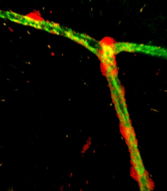
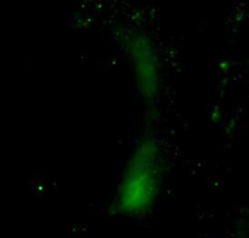
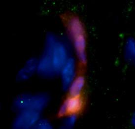
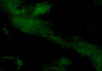
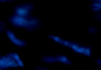
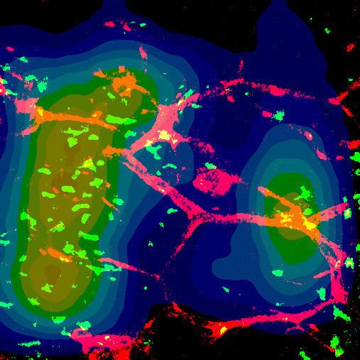
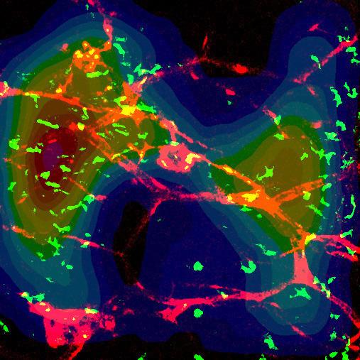
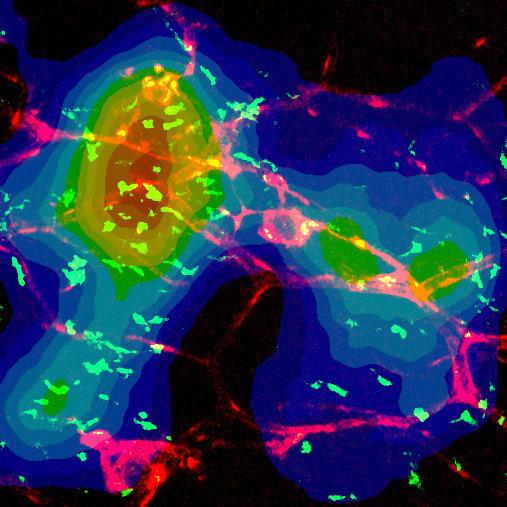
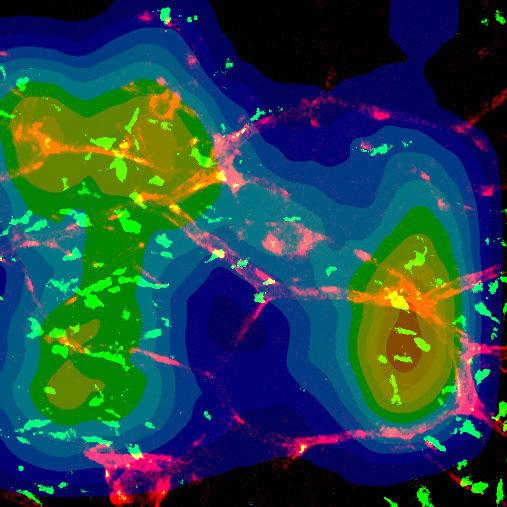
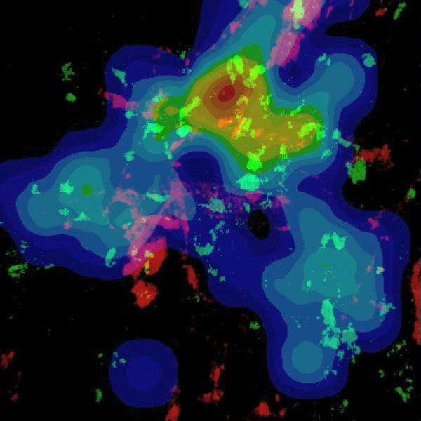
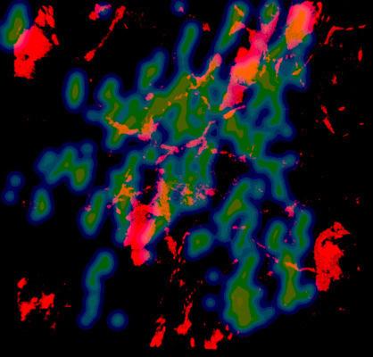
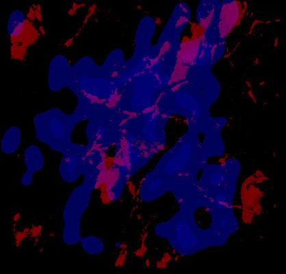
2 Nature Immunology doi:1.138/ni.2477 Supplementary Video Legends Supplementary Video 1. NG2 + pericytes surrounding CD31 + endothelial cells. The video shows a whole mount staining of a microvascular vessel in the ear of an NG2DsRed mouse. NG2 + pericytes (red) in close contact to CD31 + endothelial cells (green), immunostained by a FITC labelled anti- CD31 antibody. Visualized by 2-photon microscopy. Still image is shown in Supplementary Figure 1d. Supplementary Video 2. Interaction of a CX3CR1 + macrophage with a microvascular pericyte during interstitial migration in vivo. The video shows a vessel in the ear skin of an NG2DsRed- CX3CR1eGFP chimera with an NG2 + pericyte (red) around the capillary, stained by i.a. injection of FITC-Dextran (green). The colocalization (yellow, arrow) is zoomed with decreased density in the green and red channel. Inflammation was induced by s.c. injection of TNF and intravital 2-photon microscopy was performed 4 hrs later. A CX3CR1 + macrophage (green, arrowhead) is interacting with a pericyte over a time period of 2 min. Images were acquired at 2 images per minute and the sequence shows a 3 min time period. Still images are shown in Figure 1e. Supplementary Video 3. Interaction of a LysM + neutrophil with a microvascular pericyte during interstitial migration in vivo. The video shows a polarized LysM + neutrophil (green) sequentially interacting with NG2 + cells (red) in the ear skin of an NG2DsRed-LysMeGFP chimera. The track of the cell is shown in blue, colocalization is indicated by yellow. Inflammation was induced by s.c. injection of fmlp 2 hrs before the 2-photon imaging was performed. Images were acquired every 45 sec and the sequence shows a 15 min time period. Still image is shown in Figure 1f. Supplementary Video 4. LysM + cells contacting NG2 + cells during undirected interstitial migration. The video shows LysM + neutrophils (green, cell 1-3) orientating towards NG2 + cells (red) during interstitial migration in the ear skin of an NG2DsRed-LysMeGFP chimera 2 hrs after injection of fmlp. After interaction cell 1 and cell 2 stay in close contact to NG2 + cells and sequentially interact (colocalization in yellow, arrowheads) with them (yellow/cyan track). Cell 3 interacts with a NG2 + cell (green track) and then establishes a long lasting contact (red track). Inflammation was induced by s.c. injection of fmlp 2 hrs before the 2-photon imaging was performed. Images were acquired every 3 sec and the sequence shows a time period of 1 hr. Still image is shown in Figure 2a. Supplementary Video 5. CX3CR1 + cells contacting NG2 + cells during random interstitial migration. The video shows the ear skin of an NG2DsRed-CX3CR1eGFP chimera 4 hrs after s.c. injection of TNF. During undirected migration CX3CR1 + macrophages (green) interact with NG2 + cells (red) and stay in close contact to them. Interaction is visualized by colocalization (yellow). The tracks of cells are shown in different colors. Images were acquired at a rate of 2 images per minute and the sequence shows a time period of 5 min. Still image is shown in Figure 2c. Supplementary Video 6. Heatmap visualization of LysM + cell density over time after fmlp or fmlp and ISO-1 injection. Heatmap visualizations are superimposed on the video, color coding the density of interstitial LysM + cells in the ear skin of NG2DsRed-LysMeGFP chimeras. Black/blue indicating low density, red/white marking areas of high density. Images were acquired at a rate of 2 images per minute by 2-PIVM and the sequence shows a time period of 55 min. Left: The video shows the ear skin 2 hrs after injection of fmlp. Initially, LysM + cells (green) are diffusely distributed in the interstitial space. Over time, there is a concentration around NG2 + (red) cells. Still images are shown in Figure 4c. Right: The video shows the ear skin 2 hrs after injection of fmlp and 3 min after injection of ISO-1. LysM + cells are diffusely distributed over the imaging area and there is no orientation towards NG2 + cells. Still images are shown in Figure 4f. Supplementary Video 7. Heatmap visualization of CX3CR1 + cell density over time after injection of TNF and ISO-1. The video shows the ear skin of an NG2DsRed- CX3CR1eGFP chimera 4 hrs after injection of TNF and 3 min after injection of ISO-1. A heatmap visualization is superimposed on the video, color coding the density of interstitial CX3CR1 + macrophages. Black/blue indicating low
3 Nature Immunology doi:1.138/ni.2477 density, red/white marking areas of high density. The distribution of CX3CR1 + cells over the whole imaging area remains stable over the observation period and there is no concentration around NG2 + cells. Images were acquired at a rate of 2 images per minute and the sequence shows a time period of 45 min. Still images are shown in Supplementary Figure 4f. Supplementary Video 8. LysM + cells contacting NG2 + cells during directed interstitial migration induced by laser injury. The video shows the ear skin of an NG2DsRed-LysMeGFP chimera 45 min after induction of a focal necrosis (yellow) by laser treatment. On their way to the laser injury LysM + neutrophils (green) are contacting NG2 + cells (red) and migrate along them. Interaction is visualized by colocalization (yellow). Images were acquired at a rate of 2 images per minute and the sequence shows a time period of 3 min. Still image is shown in Figure 6a. Supplementary Video 9. CX3CR1 + cells contacting NG2 + cells during interstitial migration induced by laser injury. The video shows the ear skin of an NG2DsRed-CX3CR1eGFP chimera 4 hrs after induction of localized necrosis by laser treatment. During directed migration CX3CR1 + macrophages (green) interact with NG2 + cells (red) and continue their path to the focus of sterile inflammation. Interaction is visualized by colocalization (yellow). The tracks of cells are shown in different colors. Images were acquired at a rate of 2 images per minute and the sequence shows a time period of 75 min. Still image is shown in Supplementary Figure 6g. Supplementary Video 1. LysM + cells contacting NG2 + cells during interstitial migration induced by laser injury after treatment with ISO-1. The video shows the ear skin of an NG2DsRed-LysMeGFP chimera 45 min after induction of a focal necrosis (yellow) by laser treatment. ISO-1 was injected s.c. 3 min before the laser treatment. On their way to the laser injury LysM + neutrophils (green) are contacting NG2 + cells (red), but do not follow them. The tracks of cells are shown in different colors. Images were acquired at a rate of 2 images per minute and the sequence shows a time period of 45 min. Supplementary Video 11. CX3CR1 + cells contacting NG2 + cells during interstitial migration induced by laser injury after treatment with ISO-1. The video shows the ear skin of an NG2DsRed-CX3CR1eGFP chimera 4 hrs after induction of localized necrosis by laser treatment. ISO- 1 was injected 3 min before the laser injury. CX3CR1 + macrophages (green) shortly interact with NG2 + cells (red) on their way to the focus of sterile inflammation. The tracks of cells are shown in different colors. Images were acquired at a rate of 2 images per minute and the sequence shows a time period of 5 min. Still image is shown in Supplementary Figure 7g.
4 Nature Immunology doi:1.138/ni.2477
5 Nature Immunology doi:1.138/ni.2477


 with")


 in")


 Staining for the chemokines CXCL5")

.")
6 Supplementary Figure 3 1 merge b 8 6 MIF ctrl TNF 6h LPS 6h NG2 4 2 CXCR4 CD74 TNF permeabilized TNF x-fold increase in mrna expression of pericytes 12 DAPI ctrl permeabilized a Nature Immunology doi:1.138/ni.2477 NG2 MIF d merge DAPI NG2 CXCL5 merge DAPI NG2 CCL2 merge 5 µm ctrl TNF c ctrl TNF e NG2+CCL2 NG2+CXCL5 NG2+ pericytes express and secrete chemoattractants after stimulation with DAMPs and PAMPs in vivo. (a) rt-pcr for the MIF-receptors CXCR4 and CD74, mrna was obtained from human placental pericytes stimulated with TNF!, LPS or control for 6 hrs. Relative increase in mrna compared to resting pericytes, set as 1. Data from n=3 experiments shown as mean ± s.e.m., P<.5. (b) Immunofluorescence staining of ear tissue sections from NG2DsRed mice after injection of TNF! or normal saline as control (ctrl) with or without permeabilization. Single images of Fig. 3f. Arrowheads indicating MIF expressing NG2+ cells. Bar 25µm. (c) MIF (green) shows a strong signal on NG2+ microvascular pericytes (red) in the ear skin 6 hrs after s.c. injection of TNF!. 3D rendering of images acquired by 2-photon microscopy. Image representative of n=3 experiments. Bar 5 µm. (d) Staining for the chemokines CXCL5 and CCL2 in sections from the ear skin of NG2DsRed mice 6 hrs after s.c. injection of TNF! or normal saline as control (ctrl). CXCL5 is only expressed in NG2+ cells after injection of TNF! (upper panel). CCL2 is localized intracellularly in the control group and exposed on the surface of NG2+ cells after injection of TNF! (lower panel). Bar 25 µm. Epidermal keratinocytes indicated by dashed line. Images representative of n=4 experiments. (e) Whole mount stainings of NG2DsRed ear tissue after stimulation with TNF! visualized by 2-photon microscopy. Stainings for the chemokines CCL2 and CXCL5. NG2+ cells (red) expressed CCL2 and CXCL5 (green) and were surrounded by a strong signal for the chemokines. Orthogonal slices and 3D reconstruction (top, left). Images representative of n=3 experiments. Bar 25 µm.
7 Nature Immunology doi:1.138/ni.2477
8 Nature Immunology doi:1.138/ni.2477
 interaction d e f mean displacement g 2 15 1 5 16 14 12 1 8 6 4 2 before during interacting tracks no interaction after none 1 2 3 4 5 6 time interval (square root of time)")
 + - + - track interaction track interaction + - track interaction fast CX3CR1 + cells slow CX3CR1 + cells 2 15 1 Nature")
9 Supplementary Figure a b c velocity (µm/min) interaction d e f mean displacement g before during interacting tracks no interaction after none time interval (square root of time) Laser injury 3 h meandering index motility coefficient (µm 2 /min) interacting CX3CR1 + cells 15µm injury displacement rate (µm/min) track interaction track interaction + - track interaction fast CX3CR1 + cells slow CX3CR1 + cells Nature Immunology doi:1.138/ni non-interacting CX3CR1 + cells 15µm injury high density of cells low high density of cells low µm 15µm -15µm 15µm -15µm -15µm i 25 j 1. k NS l velocity (µm/min) before during interaction after none meandering index displacement rate (µm/min) track interaction track interaction 5 time to target (relative to non-interacting cells) track interaction NG2 + pericytes support the interstitial migration of CX3CR1 + macrophages. (a) Velocity of individual CX3CR1 + cells before, during, and after interaction with NG2 + pericytes or non-interacting cells (ANOVA/LSD). (b-e) Comparison of several cell motility parameters between interacting and non-interacting tracks of CX3CR1 + cells (Student s t-test): meandering index (b), displacement rate (c), mean displacement plot calculated from tracks <1 min duration (data are shown as mean ± s.e.m.) (d), and motility coefficient (e). (f) Distribution of fast and slow migrating CX3CR1 + cells in the interstitial space. NG2 + cells are outlined by dotted lines, hair follicles are marked by asterisks (). Bar 1 µm. (g) Imaging of macrophage migration 4 hrs after induction of a focal necrosis by laser treatment in an NG2DsRed-CX3CR1eGFP chimera. Tracks of individual macrophages (green) interacting with NG2 + pericytes (red) during interstitial migration on their way to the focus of sterile injury (yellow, arrowheads). Still image of Supplementary Video 1. Bar 6µm. (h) Track plots of CX3CR1 + cells interacting with NG2 + cells (left) and non-interacting cells (right). The relative localization of the injury is depicted in red. (i) Velocity of individual CX3CR1 + cells before, during, and after interaction with NG2 + cells or without interaction during the imaging period (ANOVA/LSD). Comparison of meandering index (j) and displacement rate (k) between interacting and non-interacting tracks of CX3CR1 + cells (Student s t-test) (l) Time to the focus of laser injury for CX3CR1 + cells (n=1) starting at the same distance from the laser injury. Relative migration time of interacting CX3CR1 + cells relative to non-interacting cells (data are shown as mean ± s.e.m.). (a-l) Data from n=4 independent experiments.
10 Nature Immunology doi:1.138/ni.2477


 and extravasation from")


11 Schematic presentation of the role of NG2+ pericytes in sterile inflammation. (a) Sensing of sterile inflammation by pericytes. After cell necrosis, DAMPs are released from dead cells. Microvascular NG2+ pericytes express TLR4, TLR2, FPR2, TNFR1, and NLRP3, which allow them to sense mediators of sterile inflammation. (b) Reaction of pericytes to mediators of sterile inflammation. In response to inflammatory stimuli, pericytes acquire a proinflammatory phenotype, characterized by the up-regulation of chemokines, adhesion molecules, and NLRP3. In addition, pericytes release chemokines and present MIF as well as ICAM-1 on their surface. (c) Pericytes attract and interact with extravasated innate immune cells, thereby supporting their interstitial trafficking to foci of sterile inflammation. After crossing the basement membrane regulated by NG2 pericytes (yellow) and extravasation from postcapillary venules (top right), myeloid leukocytes face a complex field of DAMPs and other chemoattractants in the interstitial space. If they choose to migrate distant from NG2+ pericytes (blue), their path is undirected and slow. However, if innate immune cells interact with NG2+ pericytes (blue) along arterioles and capillaries (bottom), thereby entering the compartment highly enriched in pericyte-derived MIF, they become activated and sensitized to subsequent inflammatory stimuli. This results in a straighter and faster migration through the interstitial space, allowing interacting myeloid leukocytes to scan larger areas and to find a focus of sterile inflammation more efficiently. Pericyte Supplementary Figure 8 a c b DAMPs Directed and fast migration in the pericyte compartment Myeloid leukocyte Indirect and slow migration through the interstitial tissue ICAM-1 MIF Nature Immunology doi:1.138/ni.2477
12 Nature Immunology doi:1.138/ni.2477
13 Nature Immunology doi:1.138/ni.2477
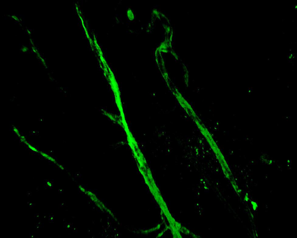
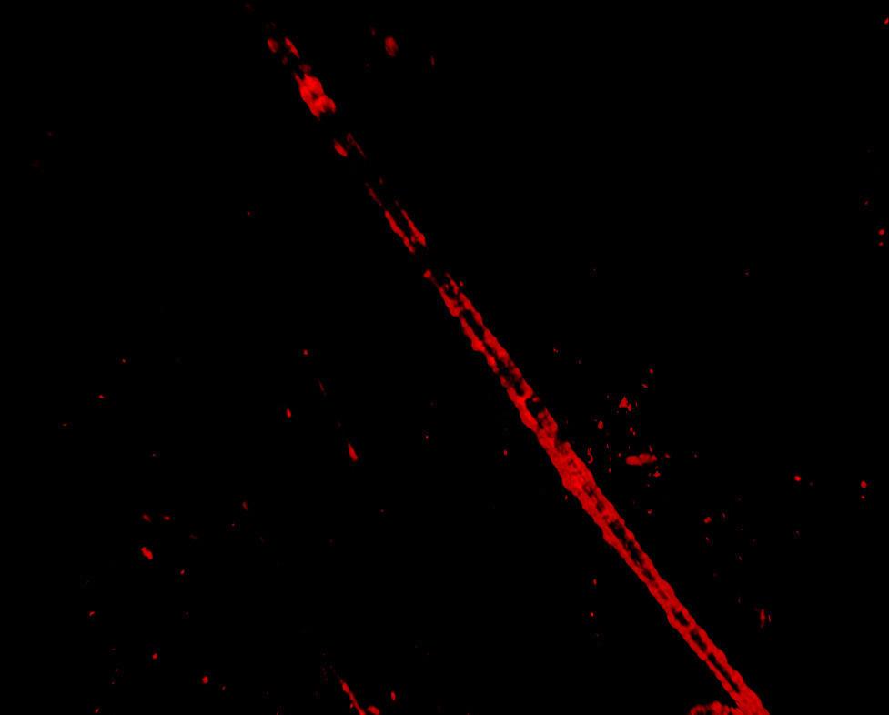
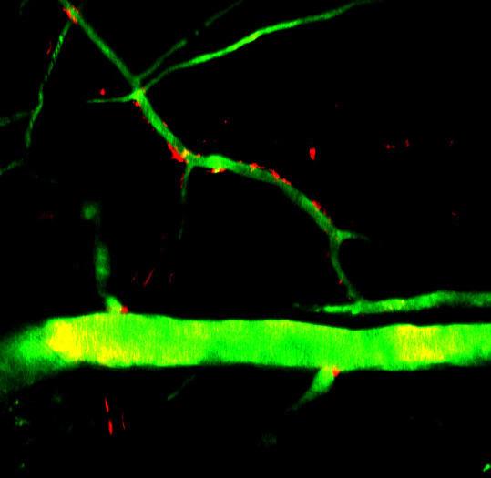
Endogenous TNFα orchestrates the trafficking of neutrophils into and within lymphatic vessels during acute inflammation
 SUPPLEMENTARY INFORMATION Endogenous TNFα orchestrates the trafficking of neutrophils into and within lymphatic vessels during acute inflammation Samantha Arokiasamy 1,2, Christian Zakian 1, Jessica Dilliway
PHENOTYPIC DYNAMICS OF MICROGLIAL AND MONOCYTE-DERIVED CELLS IN GLIOBLASTOMA-BEARING MICE.
 SUPPLEMENTARY FIGURES, TABLES AND VIDEOS PHENOTYPIC DYNAMICS OF MICROGLIAL AND MONOCYTE-DERIVED CELLS IN GLIOBLASTOMA-BEARING MICE. Clément Ricard 1,2,3,4, Aurélie Tchoghandjian 2,4, Hervé Luche 5, Pierre
SUPPLEMENTARY INFORMATION
 DOI:.38/ncb3399 a b c d FSP DAPI 5mm mm 5mm 5mm e Correspond to melanoma in-situ Figure a DCT FSP- f MITF mm mm MlanaA melanoma in-situ DCT 5mm FSP- mm mm mm mm mm g melanoma in-situ MITF MlanaA mm mm
CD4 and CD8 T cells show a similar accumulation in the tumor stroma.
 Fig S1 CD4 Fibronectin EpCM CD8 CD4 and CD8 T cells show a similar accumulation in the tumor stroma. Fluorescently-labeled CD4 (CMFD, green) and CD8 (Hoechst, yellow) T cells were added to a human lung
Supplementary Materials for
 www.sciencetranslationalmedicine.org/cgi/content/full/4/117/117ra8/dc1 Supplementary Materials for Notch4 Normalization Reduces Blood Vessel Size in Arteriovenous Malformations Patrick A. Murphy, Tyson
Type of file: PDF Title of file for HTML: Supplementary Information Description: Supplementary Figures
 Type of file: PDF Title of file for HTML: Supplementary Information Description: Supplementary Figures Type of file: MOV Title of file for HTML: Supplementary Movie 1 Description: NLRP3 is moving along
Santulli G. et al. A microrna-based strategy to suppress restenosis while preserving endothelial function
 ONLINE DATA SUPPLEMENTS Santulli G. et al. A microrna-based strategy to suppress restenosis while preserving endothelial function Supplementary Figures Figure S1 Effect of Ad-p27-126TS on the expression
Nature Immunology: doi: /ni.3866
 Nature Immunology: doi:10.1038/ni.3866 Supplementary Figure 1 The effect of TIPE2 on chemotaxis. a, The expression of TIPE2 in dhl-60c, dhl-60t, TIPE2-expressing and 15/16Q-expressing dhl-60t neutrophils
Suppl Video: Tumor cells (green) and monocytes (white) are seeded on a confluent endothelial
 and monocytes (white) are seeded on a confluent endothelial") Supplementary Information Häuselmann et al. Monocyte induction of E-selectin-mediated endothelial activation releases VE-cadherin junctions to promote tumor cell extravasation in the metastasis cascade
Supplementary Information Häuselmann et al. Monocyte induction of E-selectin-mediated endothelial activation releases VE-cadherin junctions to promote tumor cell extravasation in the metastasis cascade
Supplementary Information. Tissue-wide immunity against Leishmania. through collective production of nitric oxide
 Supplementary Information Tissue-wide immunity against Leishmania through collective production of nitric oxide Romain Olekhnovitch, Bernhard Ryffel, Andreas J. Müller and Philippe Bousso Supplementary
Macrophages form functional vascular mimicry channels in vivo. SI Figures and Legend
 Macrophages form functional vascular mimicry channels in vivo Authors: *Faith H. Barnett, *Mauricio Rosenfeld, Malcolm Wood, William Kiosses, Yoshihiko Usui, Valentina Marchetti, Edith Aguilar, and Martin
Supplementary Figure 1. Nature Neuroscience: doi: /nn.4547
 Supplementary Figure 1 Characterization of the Microfetti mouse model. (a) Gating strategy for 8-color flow analysis of peripheral Ly-6C + monocytes from Microfetti mice 5-7 days after TAM treatment. Living
Serafino et al. Thymosin α1 activates complement receptor-mediated phagocytosis in human monocyte-derived macrophages. SUPPLEMENTARY FIGURES
 Supplementary Fig. S1. Evaluation of the purity and maturation of macrophage cultures tested by flow cytometry. The lymphocytic/monocytic cellular fraction was isolated from buffy coats of healthy donors
Chapter 3, Part A (Pages 37-45): Leukocyte Migration into Tissues
: Leukocyte Migration into Tissues") Allergy and Immunology Review Corner: Chapter 3, Part A (pages 37-45) of Cellular and Molecular Immunology (Seventh Edition), by Abul K. Abbas, Andrew H. Lichtman and Shiv Pillai. Chapter 3, Part A (Pages
Allergy and Immunology Review Corner: Chapter 3, Part A (pages 37-45) of Cellular and Molecular Immunology (Seventh Edition), by Abul K. Abbas, Andrew H. Lichtman and Shiv Pillai. Chapter 3, Part A (Pages
Irf1 fold changes (D) 24h 48h. p-p65. t-p65. p-irf3. t-irf3. β-actin SKO TKO 100% 80% 60% 40% 20%
 24h 48h. p-p65. t-p65. p-irf3. t-irf3. β-actin SKO TKO 100% 80% 60% 40% 20%") Irf7 Fold changes 3 1 Irf1 fold changes 3 1 8h h 8h 8h h 8h p-p6 p-p6 t-p6 p-irf3 β-actin p-irf3 t-irf3 β-actin TKO TKO STKO (E) (F) TKO TKO % of p6 nuclear translocation % % 1% 1% % % p6 TKO % of IRF3
Irf7 Fold changes 3 1 Irf1 fold changes 3 1 8h h 8h 8h h 8h p-p6 p-p6 t-p6 p-irf3 β-actin p-irf3 t-irf3 β-actin TKO TKO STKO (E) (F) TKO TKO % of p6 nuclear translocation % % 1% 1% % % p6 TKO % of IRF3
Supplementary Figure 1: TSLP receptor skin expression in dcssc. A: Healthy control (HC) skin with TSLP receptor expression in brown (10x
 skin with TSLP receptor expression in brown (10x") Supplementary Figure 1: TSLP receptor skin expression in dcssc. A: Healthy control (HC) skin with TSLP receptor expression in brown (10x magnification). B: Second HC skin stained for TSLP receptor in brown
Supplementary Figure 1: TSLP receptor skin expression in dcssc. A: Healthy control (HC) skin with TSLP receptor expression in brown (10x magnification). B: Second HC skin stained for TSLP receptor in brown
Stewart et al. CD36 ligands promote sterile inflammation through assembly of a TLR 4 and 6 heterodimer
 NFκB (fold induction) Stewart et al. ligands promote sterile inflammation through assembly of a TLR 4 and 6 heterodimer a. mrna (fold induction) 5 4 3 2 1 LDL oxldl Gro1a MIP-2 RANTES mrna (fold induction)
The Immune Response in Time and Space
 The Immune Response in Time and Space Chapters 14 & 4 Sharon S. Evans, Ph.D. Department of Immunology 845-3421 sharon.evans@roswellpark.org September 18 & 23, 2014 Inflammation Inflammation Complex response
Influenza virus exploits tunneling nanotubes for cell-to-cell spread
 Supplementary Information Influenza virus exploits tunneling nanotubes for cell-to-cell spread Amrita Kumar 1, Jin Hyang Kim 1, Priya Ranjan 1, Maureen G. Metcalfe 2, Weiping Cao 1, Margarita Mishina 1,
F-actin VWF Vinculin. F-actin. Vinculin VWF
 a F-actin VWF Vinculin b F-actin VWF Vinculin Supplementary Fig. 1. WPBs in HUVECs are located along stress fibers and at focal adhesions. (a) Immunofluorescence images of f-actin (cyan), VWF (yellow),
Afferent lymph-derived T cells and dendritic cells use different CCR7-dependent routes for lymph node entry and intranodal migration
 Braun et al. Supplementary Information 1 Supplementary Information Afferent lymph-derived T cells and dendritic cells use different CCR7-dependent routes for lymph node entry and intranodal migration Asolina
Supplementary Figure 1 IMQ-Induced Mouse Model of Psoriasis. IMQ cream was
 Supplementary Figure 1 IMQ-Induced Mouse Model of Psoriasis. IMQ cream was painted on the shaved back skin of CBL/J and BALB/c mice for consecutive days. (a, b) Phenotypic presentation of mouse back skin
Supplementary table and figures
 3D single molecule tracking with multifocal plane microscopy reveals rapid intercellular transferrin transport at epithelial cell barriers Sripad Ram, Dongyoung Kim, Raimund J. Ober and E. Sally Ward Supplementary
SUPPLEMENTARY INFORMATION
 1. Supplementary Figures and Legends Supplementary Fig. 1. S1P-mediated transcriptional regulation of integrins expressed in OP/monocytoid cells. Real-time quantitative PCR analyses of mrna for two integrins,
Tanimoto et al., http ://www.jcb.org /cgi /content /full /jcb /DC1
 Supplemental material JCB Tanimoto et al., http ://www.jcb.org /cgi /content /full /jcb.201510064 /DC1 THE JOURNAL OF CELL BIOLOGY Figure S1. Method for aster 3D tracking, extended characterization of
SUPPLEMENTARY INFORMATION
 Supplementary Information included with Nature MS 2008-02-01484B by Colantonio et al., entitled The dynein regulatory complex is required for ciliary motility and otolith biogenesis in the inner ear. This
PBS Class #2 Introduction to the Immune System part II Suggested reading: Abbas, pgs , 27-30
 PBS 803 - Class #2 Introduction to the Immune System part II Suggested reading: Abbas, pgs. 15-25, 27-30 Learning Objectives Compare and contrast the maturation of B and T lymphocytes Compare and contrast
Chemical aspects of the cell. Chemicals that control cell signaling: chemotaxis
 Chemical aspects of the cell Chemicals that control cell signaling: chemotaxis Cellular responses Chemotaxis Cellular response to an environmental substance with a directional movement. Chemokinesis Cellular
Dep. Control Time (min)
") aa Control Dep. RP 1s 1 mv 2s 1 mv b % potentiation of IPSP 2 15 1 5 Dep. * 1 2 3 4 Time (min) Supplementary Figure 1. Rebound potentiation of IPSPs in PCs. a, IPSPs recorded with a K + gluconate pipette
aa Control Dep. RP 1s 1 mv 2s 1 mv b % potentiation of IPSP 2 15 1 5 Dep. * 1 2 3 4 Time (min) Supplementary Figure 1. Rebound potentiation of IPSPs in PCs. a, IPSPs recorded with a K + gluconate pipette
Supplementary Materials. for Garmy-Susini, et al, Integrin 4 1 signaling is required for lymphangiogenesis and tumor metastasis
 Supplementary Materials for Garmy-Susini, et al, Integrin 4 1 signaling is required for lymphangiogenesis and tumor metastasis 1 Supplementary Figure Legends Supplementary Figure 1: Integrin expression
Supplementary Material to Manuscript SREP A
 Supplementary Material to Manuscript SREP-15-29162A Monocyte-induced recovery of inflammation-associated hepatocellular dysfunction in a biochip-based human liver model Authors: Marko Gröger a,f,1, Knut
Nature Immunology: doi: /ni Supplementary Figure 1. Production of cytokines and chemokines after vaginal HSV-2 infection.
 Supplementary Figure 1 Production of cytokines and chemokines after vaginal HSV-2 infection. C57BL/6 mice were (a) treated intravaginally with 20 µl of PBS or infected with 6.7x10 4 pfu of HSV-2 in the
SUPPLEMENTARY LEGENDS...
 TABLE OF CONTENTS SUPPLEMENTARY LEGENDS... 2 11 MOVIE S1... 2 FIGURE S1 LEGEND... 3 FIGURE S2 LEGEND... 4 FIGURE S3 LEGEND... 5 FIGURE S4 LEGEND... 6 FIGURE S5 LEGEND... 7 FIGURE S6 LEGEND... 8 FIGURE
Pathologic Stage. Lymph node Stage
 ASC ASC a c Patient ID BMI Age Gleason score Non-obese PBMC 1 22.1 81 6 (3+3) PBMC 2 21.9 6 6 (3+3) PBMC 3 22 84 8 (4+4) PBMC 4 24.6 68 7 (3+4) PBMC 24. 6 (3+3) PBMC 6 24.7 73 7 (3+4) PBMC 7 23. 67 7 (3+4)
Neuronal guidance protein semaphorin 7A influences neutrophil arrest and aggravates inflammatory lung injury. Dr. Tiago Granja
 Neuronal guidance protein semaphorin 7A influences neutrophil arrest and aggravates inflammatory lung injury Dr. Tiago Granja Neuronal guidance proteins (NGPs) force cells to move NPGs give axons and dendrites
(a) Significant biological processes (upper panel) and disease biomarkers (lower panel)
 Significant biological processes (upper panel) and disease biomarkers (lower panel)") Supplementary Figure 1. Functional enrichment analyses of secretomic proteins. (a) Significant biological processes (upper panel) and disease biomarkers (lower panel) 2 involved by hrab37-mediated secretory
Supplementary Figure 1. Functional enrichment analyses of secretomic proteins. (a) Significant biological processes (upper panel) and disease biomarkers (lower panel) 2 involved by hrab37-mediated secretory
Nature Neuroscience: doi: /nn Supplementary Figure 1
 Supplementary Figure 1 Quantification of myelin fragments in the aging brain (a) Electron microscopy on corpus callosum is shown for a 18-month-old wild type mice. Myelin fragments (arrows) were detected
Trim29 gene-targeting strategy. (a) Genotyping of wildtype mice (+/+), Trim29 heterozygous mice (+/ ) and homozygous mice ( / ).
 Genotyping of wildtype mice (+/+), Trim29 heterozygous mice (+/ ) and homozygous mice ( / ).") Supplementary Figure 1 Trim29 gene-targeting strategy. (a) Genotyping of wildtype mice (+/+), Trim29 heterozygous mice (+/ ) and homozygous mice ( / ). (b) Immunoblot analysis of TRIM29 in lung primary
Supplementary Figure 1 Trim29 gene-targeting strategy. (a) Genotyping of wildtype mice (+/+), Trim29 heterozygous mice (+/ ) and homozygous mice ( / ). (b) Immunoblot analysis of TRIM29 in lung primary
Supplementary Figure 1. SA-β-Gal positive senescent cells in various cancer tissues. Representative frozen sections of breast, thyroid, colon and
 Supplementary Figure 1. SA-β-Gal positive senescent cells in various cancer tissues. Representative frozen sections of breast, thyroid, colon and stomach cancer were stained with SA-β-Gal and nuclear fast
Supplementary Figure 1: Fn14 is upregulated in the epidermis and dermis of mice
 Supplementary Figure 1: Fn14 is upregulated in the epidermis and dermis of mice undergoing AD- and psoriasis-like disease. Immunofluorescence staining for Fn14 (green) and DAPI (blue) in skin of naïve
ANGPTL2 increases bone metastasis of breast cancer cells through. Tetsuro Masuda, Motoyoshi Endo, Yutaka Yamamoto, Haruki Odagiri, Tsuyoshi
 Masuda et al. Supplementary information for ANGPTL2 increases bone metastasis of breast cancer cells through enhancing CXCR4 signaling Tetsuro Masuda, Motoyoshi Endo, Yutaka Yamamoto, Haruki Odagiri, Tsuyoshi
well for 2 h at rt. Each dot represents an individual mouse and bar is the mean ±
 Supplementary data: Control DC Blimp-1 ko DC 8 6 4 2-2 IL-1β p=.5 medium 8 6 4 2 IL-2 Medium p=.16 8 6 4 2 IL-6 medium p=.3 5 4 3 2 1-1 medium IL-1 n.s. 25 2 15 1 5 IL-12(p7) p=.15 5 IFNγ p=.65 4 3 2 1
Supplementary Information
 Supplementary Information Title Degeneration and impaired regeneration of gray matter oligodendrocytes in amyotrophic lateral sclerosis Authors Shin H. Kang, Ying Li, Masahiro Fukaya, Ileana Lorenzini,
John Nguyen, Nozomi Nishimura, Robert Fetcho, Costantino Iadecola, Chris B. Schaffer
 Supplemental figures and text for Occlusion of cortical ascending venules causes blood flow decreases, reversals in flow direction, and vessel dilation in upstream capillaries John Nguyen, Nozomi Nishimura,
B220 CD4 CD8. Figure 1. Confocal Image of Sensitized HLN. Representative image of a sensitized HLN
 B220 CD4 CD8 Natarajan et al., unpublished data Figure 1. Confocal Image of Sensitized HLN. Representative image of a sensitized HLN showing B cell follicles and T cell areas. 20 µm thick. Image of magnification
Supplementary Figure 1 Information on transgenic mouse models and their recording and optogenetic equipment. (a) 108 (b-c) (d) (e) (f) (g)
 108 (b-c) (d) (e) (f) (g)") Supplementary Figure 1 Information on transgenic mouse models and their recording and optogenetic equipment. (a) In four mice, cre-dependent expression of the hyperpolarizing opsin Arch in pyramidal cells
Supplementary Figure 1 Information on transgenic mouse models and their recording and optogenetic equipment. (a) In four mice, cre-dependent expression of the hyperpolarizing opsin Arch in pyramidal cells
Nature Biotechnology: doi: /nbt Supplementary Figure 1. Analysis of hair bundle morphology in Ush1c c.216g>a mice at P18 by SEM.
 Supplementary Figure 1 Analysis of hair bundle morphology in Ush1c c.216g>a mice at P18 by SEM. (a-c) Heterozygous c.216ga mice displayed normal hair bundle morphology at P18. (d-i) Disorganized hair bundles
Postn MCM Smad2 fl/fl Postn MCM Smad3 fl/fl Postn MCM Smad2/3 fl/fl. Postn MCM. Tgfbr1/2 fl/fl TAC
 A Smad2 fl/fl Smad3 fl/fl Smad2/3 fl/fl Tgfbr1/2 fl/fl 1. mm B Tcf21 MCM Tcf21 MCM Smad3 fl/fl Tcf21 MCM Smad2/3 fl/fl Tcf21 MCM Tgfbr1/2 fl/fl αmhc MCM C 1. mm 1. mm D Smad2 fl/fl Smad3 fl/fl Smad2/3
Ahtiainen et al., http :// /cgi /content /full /jcb /DC1
 Supplemental material JCB Ahtiainen et al., http ://www.jcb.org /cgi /content /full /jcb.201512074 /DC1 THE JOURNAL OF CELL BIOLOGY Figure S1. Distinct distribution of different cell cycle phases in the
The recruitment of leukocytes and plasma proteins from the blood to sites of infection and tissue injury is called inflammation
 The migration of a particular type of leukocyte into a restricted type of tissue, or a tissue with an ongoing infection or injury, is often called leukocyte homing, and the general process of leukocyte
Supplementary Materials for
 www.sciencesignaling.org/cgi/content/full/2/1/ra81/dc1 Supplementary Materials for Delivery of MicroRNA-126 by Apoptotic Bodies Induces CXCL12- Dependent Vascular Protection Alma Zernecke,* Kiril Bidzhekov,
SUPPLEMENTARY METHODS
 SUPPLEMENTARY METHODS Histological analysis. Colonic tissues were collected from 5 parts of the middle colon on day 7 after the start of DSS treatment, and then were cut into segments, fixed with 4% paraformaldehyde,
SHREE ET AL, SUPPLEMENTAL MATERIALS. (A) Workflow for tumor cell line derivation and orthotopic implantation.
 Workflow for tumor cell line derivation and orthotopic implantation.") SHREE ET AL, SUPPLEMENTAL MATERIALS SUPPLEMENTAL FIGURE AND TABLE LEGENDS Supplemental Figure 1. Derivation and characterization of TS1-TGL and TS2-TGL PyMT cell lines and development of an orthotopic
SHREE ET AL, SUPPLEMENTAL MATERIALS SUPPLEMENTAL FIGURE AND TABLE LEGENDS Supplemental Figure 1. Derivation and characterization of TS1-TGL and TS2-TGL PyMT cell lines and development of an orthotopic
SUPPLEMENTARY INFORMATION
 DOI: 10.1038/ncb2294 Figure S1 Localization and function of cell wall polysaccharides in root hair cells. (a) Spinning-disk confocal sections of seven day-old A. thaliana seedlings stained with 0.1% S4B
Supplementary Figure 1 Chemokine and chemokine receptor expression during muscle regeneration (a) Analysis of CR3CR1 mrna expression by real time-pcr
 Analysis of CR3CR1 mrna expression by real time-pcr") Supplementary Figure 1 Chemokine and chemokine receptor expression during muscle regeneration (a) Analysis of CR3CR1 mrna expression by real time-pcr at day 0, 1, 4, 10 and 21 post- muscle injury. (b)
Supplementary Figure 1 Chemokine and chemokine receptor expression during muscle regeneration (a) Analysis of CR3CR1 mrna expression by real time-pcr at day 0, 1, 4, 10 and 21 post- muscle injury. (b)
Intracellular MHC class II molecules promote TLR-triggered innate. immune responses by maintaining Btk activation
 Intracellular MHC class II molecules promote TLR-triggered innate immune responses by maintaining Btk activation Xingguang Liu, Zhenzhen Zhan, Dong Li, Li Xu, Feng Ma, Peng Zhang, Hangping Yao and Xuetao
Supplementary Figure 1
 Supplementary Figure 1 The average sigmoid parametric curves of capillary dilation time courses and average time to 50% peak capillary diameter dilation computed from individual capillary responses averaged
GFP/Iba1/GFAP. Brain. Liver. Kidney. Lung. Hoechst/Iba1/TLR9!
 Supplementary information a +KA Relative expression d! Tlr9 5!! 5! NSC Neuron Astrocyte Microglia! 5! Tlr7!!!! NSC Neuron Astrocyte! GFP/Sβ/! Iba/Hoechst Microglia e Hoechst/Iba/TLR9! GFP/Iba/GFAP f Brain
Novel model for in vivo imaging of large arteries in steady state and atherosclerosis using 2 photon microscopy
 Fakultät für Medizin Klinikum rechts der Isar Novel model for in vivo imaging of large arteries in steady state and atherosclerosis using 2 photon microscopy A comparison of the role of Mac-1 integrin
activation with anti-cd3/cd28 beads and 3d following transduction. Supplemental Figure 2 shows
 Supplemental Data Supplemental Figure 1 compares CXCR4 expression in untreated CD8 + T cells, following activation with anti-cd3/cd28 beads and 3d following transduction. Supplemental Figure 2 shows the
nature methods Organelle-specific, rapid induction of molecular activities and membrane tethering
 nature methods Organelle-specific, rapid induction of molecular activities and membrane tethering Toru Komatsu, Igor Kukelyansky, J Michael McCaffery, Tasuku Ueno, Lidenys C Varela & Takanari Inoue Supplementary
Title of file for HTML: Supplementary Information Description: Supplementary Figures and Supplementary Table
 Title of file for HTML: Supplementary Information Description: Supplementary Figures and Supplementary Table Title of file for HTML: Peer Review File Description: Innate Scavenger Receptor-A regulates
SUPPLEMENTARY INFORMATION
 DOI: 1.138/ncb222 / b. WB anti- WB anti- ulin Mitotic index (%) 14 1 6 2 T (h) 32 48-1 1 2 3 4 6-1 4 16 22 28 3 33 e. 6 4 2 Time (min) 1-6- 11-1 > 1 % cells Figure S1 depletion leads to mitotic defects
Supplemental Figure 1. Quantification of proliferation in thyroid of WT, Ctns -/- and grafted
 Supplemental Figure 1. Quantification of proliferation in thyroid of WT, Ctns -/- and grafted Ctns -/- mice. Cells immunolabeled for the proliferation marker (Ki-67) were counted in sections (n=3 WT, n=4
label the basement membrane). Different fixation methods of EB-perfused P8 mice to optimize the combination
. Different fixation methods of EB-perfused P8 mice to optimize the combination") Supplementary Figure 1 Optimization of the tissue fixation protocol to combine EB perfusion and IB4 endothelial tip cell staining in the postnatal mouse brain. a-l Labeling of EB-perfused P8 mice with
Supplementary Figure 1 Optimization of the tissue fixation protocol to combine EB perfusion and IB4 endothelial tip cell staining in the postnatal mouse brain. a-l Labeling of EB-perfused P8 mice with
c Ischemia (30 min) Reperfusion (8 w) Supplementary Figure bp 300 bp Ischemia (30 min) Reperfusion (4 h) Dox 20 mg/kg i.p.
 Reperfusion (8 w) Supplementary Figure bp 300 bp Ischemia (30 min) Reperfusion (4 h) Dox 20 mg/kg i.p.") a Marker Ripk3 +/ 5 bp 3 bp b Ischemia (3 min) Reperfusion (4 h) d 2 mg/kg i.p. 1 w 5 w Sacrifice for IF size A subset for echocardiography and morphological analysis c Ischemia (3 min) Reperfusion (8
a Marker Ripk3 +/ 5 bp 3 bp b Ischemia (3 min) Reperfusion (4 h) d 2 mg/kg i.p. 1 w 5 w Sacrifice for IF size A subset for echocardiography and morphological analysis c Ischemia (3 min) Reperfusion (8
3D Tissue Models. Simple, Low Cost Fabrication. Simple, Robust Protocols
 3D Tissue Models SynVivo is a physiological, cell-based microfluidic platform that provides a morphologically and physiologically realistic microenvironment allowing real-time study of cellular behavior,
Nature Immunology: doi: /ni Supplementary Figure 1
 Supplementary Figure 1 NLRP12 is downregulated in biopsy samples from patients with active ulcerative colitis (UC). (a-g) NLRP12 expression in 7 UC mrna profiling studies deposited in NCBI GEO database.
Supplemental information
 Supplemental information PI(3)K p11δ controls the sucellular compartmentalization of TLR4 signaling and protects from endotoxic shock Ezra Aksoy, Salma Taoui, David Torres, Sandrine Delauve, Aderrahman
Nature Neuroscience: doi: /nn Supplementary Figure 1
 Supplementary Figure 1 Relative expression of K IR2.1 transcript to enos was reduced 29-fold in capillaries from knockout animals. Relative expression of K IR2.1 transcript to enos was reduced 29-fold
Supplementary Figure 1: GFAP positive nerves in patients with adenocarcinoma of
 SUPPLEMENTARY FIGURES AND MOVIE LEGENDS Supplementary Figure 1: GFAP positive nerves in patients with adenocarcinoma of the pancreas. (A) Images of nerves stained for GFAP (green), S100 (red) and DAPI
Supplementary Figure 1: Signaling centers contain few proliferating cells, express p21, and
 Supplementary Figure 1: Signaling centers contain few proliferating cells, express p21, and exclude YAP from the nucleus. (a) Schematic diagram of an E10.5 mouse embryo. (b,c) Sections at B and C in (a)
Supplementary Figure 1. Properties of various IZUMO1 monoclonal antibodies and behavior of SPACA6. (a) (b) (c) (d) (e) (f) (g) .
 (b) (c) (d) (e) (f) (g) .") Supplementary Figure 1. Properties of various IZUMO1 monoclonal antibodies and behavior of SPACA6. (a) The inhibitory effects of new antibodies (Mab17 and Mab18). They were investigated in in vitro fertilization
Supplementary Figure 1. Properties of various IZUMO1 monoclonal antibodies and behavior of SPACA6. (a) The inhibitory effects of new antibodies (Mab17 and Mab18). They were investigated in in vitro fertilization
Programmed necrosis, not apoptosis, is a key mediator of cell loss and DAMP-mediated inflammation in dsrna-induced retinal degeneration
 Programmed necrosis, not apoptosis, is a key mediator of cell loss and DAMP-mediated inflammation in dsrna-induced retinal degeneration The Harvard community has made this article openly available. Please
2. Innate immunity 2013
 1 Innate Immune Responses 3 Innate immunity Abul K. Abbas University of California San Francisco The initial responses to: 1. Microbes: essential early mechanisms to prevent, control, or eliminate infection;
Lymphoid architecture & Leukocyte recirculation. Thursday Jan 26th, 2017
 Lymphoid architecture & Leukocyte recirculation Thursday Jan 26th, 2017 Topics The life of immune cells Where are they born? Where are they educated? Where do they function? How do they get there? The
Supplementary fig. 1. Crystals induce necroptosis does not involve caspases, TNF receptor or NLRP3. A. Mouse tubular epithelial cells were pretreated
 Supplementary fig. 1. Crystals induce necroptosis does not involve caspases, TNF receptor or NLRP3. A. Mouse tubular epithelial cells were pretreated with zvad-fmk (10µM) and exposed to calcium oxalate
Aggregated neutrophil extracellular traps limit inflammation by degrading cytokines and chemokines
 CORRECTION NOTICE Nat. Med. doi:10.1038/nm.3547; corrected online 25 August 2014 Aggregated neutrophil extracellular traps limit inflammation by degrading cytokines and chemokines Christine Schauer, Christina
Supplementary Materials for
 advances.sciencemag.org/cgi/content/full/3/8/e1700521/dc1 Supplementary Materials for Functional vascularized lung grafts for lung bioengineering N. Valerio Dorrello, Brandon A. Guenthart, John D. O Neill,
Supplementary Figure 1. Generation of knockin mice expressing L-selectinN138G. (a) Schematics of the Sellg allele (top), the targeting vector, the
 Schematics of the Sellg allele (top), the targeting vector, the") Supplementary Figure 1. Generation of knockin mice expressing L-selectinN138G. (a) Schematics of the Sellg allele (top), the targeting vector, the targeted allele in ES cells, and the mutant allele in
Supplementary Figure 1. Generation of knockin mice expressing L-selectinN138G. (a) Schematics of the Sellg allele (top), the targeting vector, the targeted allele in ES cells, and the mutant allele in
SUPPLEMENTARY FIGURES AND TABLE
 SUPPLEMENTARY FIGURES AND TABLE Supplementary Figure S1: Characterization of IRE1α mutants. A. U87-LUC cells were transduced with the lentiviral vector containing the GFP sequence (U87-LUC Tet-ON GFP).
Dynamic Partitioning of a GPI-Anchored Protein in Glycosphingolipid-Rich Microdomains Imaged by Single-Quantum Dot Tracking
 Additional data for Dynamic Partitioning of a GPI-Anchored Protein in Glycosphingolipid-Rich Microdomains Imaged by Single-Quantum Dot Tracking Fabien Pinaud 1,3, Xavier Michalet 1,3, Gopal Iyer 1, Emmanuel
% of live splenocytes. STAT5 deletion. (open shapes) % ROSA + % floxed
 % ROSA + % floxed") Supp. Figure 1. a 14 1 1 8 6 spleen cells (x1 6 ) 16 % of live splenocytes 5 4 3 1 % of live splenocytes 8 6 4 b 1 1 c % of CD11c + splenocytes (closed shapes) 8 6 4 8 6 4 % ROSA + (open shapes) % floxed
Supp. Figure 1. a 14 1 1 8 6 spleen cells (x1 6 ) 16 % of live splenocytes 5 4 3 1 % of live splenocytes 8 6 4 b 1 1 c % of CD11c + splenocytes (closed shapes) 8 6 4 8 6 4 % ROSA + (open shapes) % floxed
SUPPLEMENTARY INFORMATION
 DOI: 10.1038/ncb2988 Supplementary Figure 1 Kif7 L130P encodes a stable protein that does not localize to cilia tips. (a) Immunoblot with KIF7 antibody in cell lysates of wild-type, Kif7 L130P and Kif7
Distinct Compartmentalization of the Chemokines CXCL1 and CXCL2 and the Atypical Receptor ACKR1 Determine Discrete Stages of Neutrophil Diapedesis
 Article Distinct Compartmentalization of the Chemokines CXCL1 and CXCL2 and the Atypical Receptor ACKR1 Determine Discrete Stages of Neutrophil Diapedesis Graphical Abstract Authors Tamara Girbl, Tchern
Nature Neuroscience: doi: /nn Supplementary Figure 1
 Supplementary Figure 1 Atlas representations of the midcingulate (MCC) region targeted in this study compared against the anterior cingulate (ACC) region commonly reported. Coronal sections are shown on
Fisher et al. Supplemental Figure 1
 Supplemental Figure 1 A TNF IL-1 IL-6 CCL2 CCL5 CXCL10 pg/mg total protein 50 30 10 4,000 3,000 2,000 1,000 n.d. 1 1 14,000 12,000 10,000 8,000 6,000 4,000 2,000 6,000,000 CT26 5,000 16,000 B16 4,000 12,000
hemodynamic stress. A. Echocardiographic quantification of cardiac dimensions and function in
 SUPPLEMENTAL FIGURE LEGENDS Supplemental Figure 1. Fbn1 C1039G/+ hearts display normal cardiac function in the absence of hemodynamic stress. A. Echocardiographic quantification of cardiac dimensions and
Supplementary Figure 1. Deletion of Smad3 prevents B16F10 melanoma invasion and metastasis in a mouse s.c. tumor model.
 A B16F1 s.c. Lung LN Distant lymph nodes Colon B B16F1 s.c. Supplementary Figure 1. Deletion of Smad3 prevents B16F1 melanoma invasion and metastasis in a mouse s.c. tumor model. Highly invasive growth
Supplementary Figure 1 The ability to regenerate an ear hole is discontinuous with wound healing. Ear-hole closure at D85 for each sex within each
 Supplementary Figure 1 The ability to regenerate an ear hole is discontinuous with wound healing. Ear-hole closure at D85 for each sex within each species observed. Data show a binary response to a 4 mm
p = formed with HCI-001 p = Relative # of blood vessels that formed with HCI-002 Control Bevacizumab + 17AAG Bevacizumab 17AAG
 A.. Relative # of ECs associated with HCI-001 1.4 1.2 1.0 0.8 0.6 0.4 0.2 0.0 ol b p < 0.001 Relative # of blood vessels that formed with HCI-001 1.4 1.2 1.0 0.8 0.6 0.4 0.2 0.0 l b p = 0.002 Control IHC:
Thomas HAIDER Journal Club
 Thomas HAIDER Journal Club 20.10.2014 Background Immunology of the CNS - History Ehrlich, 1885 & 1904 dye did not stain brain -> BBB Shirai, Y. (1921) On the transplantation of the rat sarcoma in adult
Supplementary Figures for TSC1 controls macrophage polarization to prevent inflammatory disorder by Linnan Zhu et al
 Supplementary Figures for TSC1 controls macrophage polarization to prevent inflammatory disorder by Linnan Zhu et al Suppl. Fig. 1 Tissue DN C Proteins kd TSC1-17 TSC 1 loxp bp -48-285 ctin PEMs Neutrophils
Supplemental Table 1. Primer sequences for transcript analysis
 Supplemental Table 1. Primer sequences for transcript analysis Primer Sequence (5 3 ) Primer Sequence (5 3 ) Mmp2 Forward CCCGTGTGGCCCTC Mmp15 Forward CGGGGCTGGCT Reverse GCTCTCCCGGTTTC Reverse CCTGGTGTGCCTGCTC
Chronic variable stress activates hematopoietic stem cells
 SUPPLEMENTARY INFORMATION Chronic variable stress activates hematopoietic stem cells Timo Heidt *, Hendrik B. Sager *, Gabriel Courties, Partha Dutta, Yoshiko Iwamoto, Alex Zaltsman, Constantin von zur
Supplementary Figure 1 Expression of Crb3 in mouse sciatic nerve: biochemical analysis (a) Schematic of Crb3 isoforms, ERLI and CLPI, indicating the
 Schematic of Crb3 isoforms, ERLI and CLPI, indicating the") Supplementary Figure 1 Expression of Crb3 in mouse sciatic nerve: biochemical analysis (a) Schematic of Crb3 isoforms, ERLI and CLPI, indicating the location of the transmembrane (TM), FRM binding (FB)
Supplementary Figure 1 Expression of Crb3 in mouse sciatic nerve: biochemical analysis (a) Schematic of Crb3 isoforms, ERLI and CLPI, indicating the location of the transmembrane (TM), FRM binding (FB)
Supplementary Figure 1. Rab27a-KD inhibits speed and persistence of HEp3 cells migrating in the chick CAM. (a) Western blot analysis of Rab27a
 Western blot analysis of Rab27a") Supplementary Figure 1. Rab27a-KD inhibits speed and persistence of HEp3 cells migrating in the chick CAM. (a) Western blot analysis of Rab27a expression in GFP-expressing HEp3 cells. (b) Representative
Supplementary Figure 1. Rab27a-KD inhibits speed and persistence of HEp3 cells migrating in the chick CAM. (a) Western blot analysis of Rab27a expression in GFP-expressing HEp3 cells. (b) Representative
Supplementary Table 1. The primers used for quantitative RT-PCR. Gene name Forward (5 > 3 ) Reverse (5 > 3 )
 Reverse (5 > 3 )") 770 771 Supplementary Table 1. The primers used for quantitative RT-PCR. Gene name Forward (5 > 3 ) Reverse (5 > 3 ) Human CXCL1 GCGCCCAAACCGAAGTCATA ATGGGGGATGCAGGATTGAG PF4 CCCCACTGCCCAACTGATAG TTCTTGTACAGCGGGGCTTG
770 771 Supplementary Table 1. The primers used for quantitative RT-PCR. Gene name Forward (5 > 3 ) Reverse (5 > 3 ) Human CXCL1 GCGCCCAAACCGAAGTCATA ATGGGGGATGCAGGATTGAG PF4 CCCCACTGCCCAACTGATAG TTCTTGTACAGCGGGGCTTG
Nature Neuroscience: doi: /nn Supplementary Figure 1. Large-scale calcium imaging in vivo.
 Supplementary Figure 1 Large-scale calcium imaging in vivo. (a) Schematic illustration of the in vivo camera imaging set-up for large-scale calcium imaging. (b) High-magnification two-photon image from
FOR OPTIMAL GUT HEALTH KEMIN.COM/GUTHEALTH
 FOR OPTIMAL GUT HEALTH KEMIN.COM/GUTHEALTH ALETA A SOURCE OF 1,3-BETA GLUCANS Aleta is highly bioavailable, offering a concentration greater than 5% of 1,3-beta glucans. Aleta provides a consistent response