1. OVERVIEW 2. RADIOPHARMACEUTICAL UTILIZED
|
|
|
- Eileen Warner
- 5 years ago
- Views:
Transcription
1 1. OVERVIEW F-18 Fluorodeoxyglucose Injection is indicated for positron emission tomography (PET) imaging in the following settings: a) Oncology: For assessment of abnormal glucose metabolism to assist in the evaluation of malignancy in patients with known or suspected abnormalities found by other testing modalities, or in patients with an existing diagnosis of cancer. b) Cardiology: For the identification of left ventricular myocardium with residual glucose metabolism and reversible loss of systolic function in patients with coronary artery disease and left ventricular dysfunction, when used together with myocardial perfusion imaging. c) Neurology: For the identification of regions of abnormal glucose metabolism associated with foci of epileptic seizures. Also for postoperative evaluation of patients with head and neck cancer 2. RADIOPHARMACEUTICAL UTILIZED a) F-18 Fluorodeoxyglucose (FDG) is a positron emitting radiopharmaceutical that is used for diagnostic purposes in conjunction with positron emission tomography (PET) imaging. The active ingredient 2-deoxy-2-[18F]-fluoro-D-glucose has the molecular formula of C6H11 18 FO5 with a molecular weight of , and has the following chemical structure:
 FDG is provided as either a unit dose in a syringe or as a multidose vial.")
 F-18 decays by positron decay (97%) and electron capture (3%) and has a half-life of 109.7 minutes.")
2 b) The structure below illustrates the actual spatial positioning of the atoms in the molecule. Color code: red = oxygen; black = carbon; yellow = fluorine c) FDG is provided as either a unit dose in a syringe or as a multidose vial. It is a sterile, pyrogen free, isotonic, clear, colorless citrate buffered solution. Each ml contains between to 7.40 GBq ( mci). The ph of the solution is between 5.5 to 7.5. The solution contains no preservative. 3. CHARACTERISTICS OF THE RADIONUCLIDE a) F-18 decays by positron decay (97%) and electron capture (3%) and has a half-life of minutes. The principal photons useful for diagnostic imaging are the 511 kev gamma photons, resulting from the interaction of the emitted positron with an electron (Table 1). b) The specific gamma ray constant for F-18 is 6.0 R/hr/mCi at 1cm. The half-value layer (HVL) for the 511 kev photons is 4.1 mm lead (Pb). The interposition of an 8.2 mm thickness of Pb, representing 2 half-value layers, will decrease the external radiation by 75%. The tenth-value layer (TVL) for the 511 kev photons is 17 mm of lead
 None required on part of the technologist. The patient dose should be measured in a suitable dose calibration system immediately prior to administration. 6.")
3 c) Decay Scheme of F DRUG AVAILABILITY a) F-18 FDG is readily available all over the US from most Central Radiopharmacies as well as PETNET, IBA, and other PET drug manufacturing facilities. 5. DRUG PREPARATION a) None required on part of the technologist. The patient dose should be measured in a suitable dose calibration system immediately prior to administration. 6. RADIOCHROMATOGRAPHIC QUALITY CONTROL PROCEDURES a) Not required in the Hot Lab- performed by Central Pharmacy. b) May be performed at the production facility by either thin layer chromatography or by HPLC.
4 7. CLINICAL PHARMACOLOGY a) F-18 FDG is a glucose analog that concentrates in cells that rely upon glucose as an energy source, or in cells whose dependence on glucose increases under pathophysiological conditions. It is transported through the cell membrane by facilitative glucose transporter proteins and is phosphorylated within the cell to [18F] FDG-6- phosphate by the enzyme hexokinase. Once phosphorylated it cannot exit until it is dephosphorylated by glucose-6-phosphatase. b) Therefore, within a given tissue or pathophysiological process, the retention and clearance of F-18 FDG reflect a balance involving glucose transporter, hexokinase and glucose-6-phosphatase activities. When allowance is made for the kinetic differences between glucose and F-18 FDG transport and phosphorylation (expressed as the lumped constant ratio), F-18 FDG is used to assess glucose metabolism. c) In comparison to background activity of the specific organ or tissue type, regions of decreased or absent uptake of F-18 FDG reflect the decrease or absence of glucose metabolism. Regions of increased uptake of F-18 FDG reflect greater than normal rates of glucose metabolism. d) F-18 FDG Injection is rapidly distributed to all organs of the body after intravenous administration. After background clearance of F-18 FDG Injection, optimal PET imaging is generally achieved between 30 to 40 minutes after administration. e) F-18 FDG is cleared from most tissues within 24 hours and can be eliminated from the body unchanged in the urine. f) In cancer, the cells are generally characterized by enhanced glucose metabolism partially due to (1) an increase in the activity of glucose transporters, (2) an increased rate of phosphorylation activity, (3) a reduction of phosphatase activity or, (4) a dynamic alteration in the balance among all these processes. However, glucose metabolism of cancer as reflected by F-18 FDG accumulation shows considerable variability. Depending on tumor type, stage, and location, F-18 FDG accumulation may be increased, normal, or decreased. Also, inflammatory cells can have the same variability of uptake of F-18 FDG. g) In the heart, under normal aerobic conditions, the myocardium meets the bulk of its energy requirements by oxidizing free fatty acids. Most of the exogenous glucose taken up by the myocyte is converted into glycogen. However, under ischemic conditions, the oxidation of free fatty acids decreases, exogenous glucose becomes the preferred myocardial substrate, glycolysis is stimulated, and glucose taken up by the myocyte is metabolized immediately instead of being converted into glycogen. Under these conditions, phosphorylated F-18 FDG accumulates in the myocyte and can be detected with PET imaging.
5 h) Normally, the brain relies on anaerobic metabolism. In epilepsy, the glucose metabolism varies. Generally, during a seizure glucose metabolism increases. Interictally, the seizure focus tends to be hypometabolic. 8. MECHANISM OF LOCALIZATION a) Mechanism of localization is referred to as Metabolic trapping. Radiolabeled sugar analog may be trapped by cells for cell metabolism. Tumors, with their higher-thannormal metabolic rate, are able to take up and retain more molecules per gram than normal tissue, resulting in an accumulation of the radioisotope in the tumor. b) Comparison: Structures of FDG and Glucose. Note the similarities.
6 c) Metabolism of Glucose and FDG. both glucose and FDG can penetrate the cell membrane and migrate into the cell interior. In addition, both can be phosphorylated. However, FDG can only do so one time and is trapped in the cell since it can not be metabolized; glucose undergoes multiple phosphorylation steps and is ultimately metabolized into CO2 and H2O. Refer to diagram below. 9. NORMAL DISTRIBUTION OF DRUG Normal Biodistribution (Source: Mettler FA & Guilberteau MJ. Essentials of Nuclear Imaging. 6th ed. Saunders, 2012) a) 20-40% into urine by 2nd hour b) 7% into brain c) 4.5% into liver d) 3.3% into heart e) 1.7% into red bone marrow f) 1.3% into kidneys g) 0.9% into lungs Cerebral cortex, basal ganglia and thalami (i.e., the grey matter) have high FDG activity because these tissues are very metabolically active and use glucose as their primary substrate. Vocal cords at rest are mildly to not FDG-avid. But talking can cause the vocal cords to become FDG-avid. Tonsils (especially palatine tonsils), lymphoid tissue at Waldeyer ring, and parotid and submandibular glands have some FDG activity, sometimes high if they are part of reactive inflammation. As with vocal cords, this FDG activity is symmetric. Thyroid is typically not FDG-avid, but mild diffuse FDG activity is within normal. Thymus is FDG-avid in children and adults up to ~30 years. Thymic FDG activity may be seen due to thymic rebound after chemotherapy.
7 Skeletal muscle at rest (and at basal levels of insulin) has low FDG activity. Increased muscular FDG activity may be seen at the shoulders and upper back due to patient s tension secondary to positioning. Increased FDG activity can also be seen in the diaphragmatic crura, intercostals muscles, psoas muscles, paravertebral muscles, forearms and muscles of mastication. Brown fat (especially in the supraclavicular regions) can be FDG-avid if stimulated in adults, and is more often seen in children. Left ventricular myocardium has variable (i.e., high to low and in-between) FDG activity. Myocardial cells use fatty acids as their primary metabolic substrate at rest, but glucose becomes the primary substrate if blood glucose levels are high (causing increased insulin levels) or oxygen demand is increased beyond supply (ischemia), prompting anaerobic metabolism. Intercollated patches of intense and fainter FDG activity is within normal as different foci of myocardium switch between fatty acids and glucose metabolism. Compared to the LV, the right ventricular myocardium has faint FDG activity. Aortic wall FDG activity is minimal, but can be increased in older adults due to macrophages in atherosclerotic plaques. Lungs have low and diffuse FDG activity (which is more visible on non-ac images versus attenuation corrected images). Breast FDG activity is mild to moderate in young women and postmenopausal women on hormone replacement therapy. Dense breasts have greater FDG activity versus fatty breasts. FDG activity may be intense in lactating breast tissue. Bowel FDG activity varies in intensity and location. Focally high FDG activity in the colonic mucosa can be within normal, though colonic FDG activity tends to be diffuse and highest in the ascending and cecal portions. Rest of GI tract: Esophogeal FDG activity tends to be mild and uniform, and stomach FDG activity can be much greater than liver activity especially if contracted or hiatal hernia. Small bowel activity is less than colon normally. Liver FDG activity is heterogenous or patchy. Benign lesions like focal nodular hyperplasia can be FDG-avid. Urinary FDG activity is intense, so activity in the kidneys, ureters and bladder will be intense. This is because up to 40% of administered FDG is excreted by the kidneys within the first 2 hours. FDG accumulates in the renal calyces, pelvis and ureters. Ureteral activity may be discontinuous due to peristalsis.

8 Pelvic organ FDG activity is mild to minimal (but may be increased in the uterine during menstruation). FDG activity in the testes is mild. Faint FDG activity may be seen in the penis. Bone marrow FDG activity intensity is similar to liver but more homogenous, which is mildly above blood pool FDG activity. FDG activity in the bone marrow may be increased in anemic patients or patients on medications like G-CSF. Splenic FDG activity is low but can be increased by stimulation, similar to bone marrow. Lymph node FDG activity is faint, except when extravasation occurs and infiltrated FDG concentrates in a downstream node. 10. THIS IS AN EXAMPLE OF A NORMAL FDG SCAN. 11. INDICATIONS FOR CLINICAL STUDIES Adapted from Procedure Guideline for Tumor Imaging with 18F-FDG PET/CT 1.0 and reprinted from SNMMI Inc. a) Differentiating benign from malignant lesions b) Searching for an unknown primary tumor when metastatic disease is discovered as the first manifestation of cancer or when the patient presents with a paraneoplastic syndrome c) Staging known malignancies d) Monitoring the effect of therapy on known malignancies e) Determining whether residual abnormalities detected on physical examination or on other imaging studies after treatment represent tumor or posttreatment fibrosis or necrosis f) Detecting tumor recurrence, especially in the presence of elevated levels of tumor markers g) Selecting the region of a tumor most likely to yield diagnostic information for biopsy h) Guiding radiation therapy planning i) Nononcologic applications, such as evaluation of infection and atherosclerosis


9 12. TYPICAL F-18 FDG PET SCANS IN VARIOUS DISEASES Pre- and post- chemotherapy
10

11


12 Images 1 and 2 are the images from the CT scan of the patient. These do not demonstrate any abnormality in the region of the swelling in the neck. Image 3 is the PET scan of the neck region, which shows foci of intense activity in the lower neck in the region of the swelling. Image 4 is the image from the CT scan showing a mass in the abdomen and this area shows intense activity on the PET scan as seen on image 5. Image 6 shows multiple lesions in the spine and image 7 is the whole body projection image. In this patient with known lymphoma in the abdomen, the PET scan identified more lesions in the neck and in the bones.
13 13. PATIENT PREPARATION FOR FDG PET SCAN a) The rationale for performing the procedure and the details of the procedure itself should be explained to the patient in advance. b) Patients should be instructed to fast and not consume beverages, except for water, for at least 4 6 h before the administration of F-18 FDG to decrease physiologic glucose levels and to reduce serum insulin levels to near basal levels. Oral hydration with water is encouraged. Intravenous fluids containing dextrose or parenteral feedings also should be withheld for 4 6 h. c) The blood glucose level should be checked before F-18 FDG administration. Tumor uptake of F-18 FDG is reduced in hyperglycemic states. Most institutions reschedule the patient if the blood glucose level is greater than mg/dl. Reducing the serum glucose level by administering insulin can be considered, but the administration of F-18 FDG should be delayed after insulin administration (with the duration of the delay being dependent on the type and route of administration of insulin). d) For brain imaging, the patient should be in a quiet and dimly lit room for F-18 FDG administration and the subsequent uptake phase. e) For body imaging, the patient should remain seated or recumbent for F-18 FDG administration and the subsequent uptake phase to avoid muscular uptake. 14. TYPICAL ADMINISTERED DOSE FOR ADULTS a) The usual administered activity for adult patients is 5-15 mci, injected intravenously. 15. DRUG ADMINISTRATION PROCEDURE a) The injection is performed intravenously over a period of a few seconds. A small volume of blood is drawn back into the syringe and then re-injected to insure complete delivery of the bone agent. b) Hemostasis is accomplished using a gauze pad and pressure. The gauze pad at the injection site is covered with a Band-Aid or tape.
14 16. IMAGING PROTOCOLS Adapted from Procedure Guideline for Tumor Imaging with 18F-FDG PET/CT 1.0 and reprinted from SNMMI Inc. Field of view, positioning, and pre-acquisition preparation a) Skull base to proximal thigh imaging generally is recommended to survey the body in the search for areas of abnormal F-18 FDG accumulation for most tumor types. Such PET/CT scans typically are acquired from the external auditory meatus to the midthigh region. For tumors with a high likelihood of scalp, skull, or brain involvement or lower-extremity involvement, whole-body tumor imaging is performed. b) Limited-area tumor imaging can be considered when critical abnormalities are likely to be localized in a known region of the body (e.g., solitary pulmonary nodule, probable lung cancer, evaluation of hilar lymph node involvement, diagnosis of head and neck cancer, and monitoring of therapy of locally advanced breast cancer). However, performing whole-body tumor imaging offers the advantage of staging the entire body. c) For optimal imaging of the body, the arms should be elevated over the head if that position can be tolerated by the patient. Arms along the side may produce beamhardening artifacts over the torso. However, for optimal imaging of the head and neck, the arms should be positioned along the side. d) The patient should void the bladder before the acquisition of the images to limit the radiation dose to the renal collecting system and bladder. e) Metallic objects should be removed from the patient whenever possible. Protocol for PET emission imaging a) The radiopharmaceutical should be injected at a site contralateral to the site of concern. Emission images should be obtained at least 45 min after the injection. The optimal F-18 FDG distribution phase is controversial. Many facilities start the acquisition of the images at 60 or 90 min after F-18 FDG administration. Some facilities obtain a second set of images to assess the change in uptake over time. The F-18 FDG uptake time should be constant whenever possible and certainly when 2 studies are compared by use of semiquantitative parameters, especially the SUV. b) The emission image acquisition time varies from 2 to 5 min or longer per bed position for body imaging and is based on the administered activity, patient body weight, and sensitivity of the PET scanner (as determined largely by detector composition and acquisition method). Typically, for imaging skull to midthigh, the total acquisition
15 time ranges from 15 to 45 min. The imaging time typically is prolonged for the acquisition of brain images or for images of a limited region of interest. c) Semiquantitative estimation of tumor glucose metabolism by use of the SUV is based on relative lesion radioactivity measured on images corrected for attenuation and normalized for the injected dose and body weight, lean body mass, or body surface area. This measurement is obtained on a static emission image typically acquired more than 45 min after injection. The accuracy of SUV measurements depends on the accuracy of the calibration of the PET scanner, among other factors. The reproducibility of SUV measurements depends on the reproducibility of clinical protocols, for example, dose infiltration, time of imaging after F-18 FDG administration, type of reconstruction algorithms, type of attenuation maps, size of the region of interest, changes in uptake by organs other than the tumor, and methods of analysis (e.g., maximum and mean). d) Semiquantitative estimation of tumor metabolism can be based on the ratio of F-18 FDG uptake in a lesion to F-18 FDG uptake in internal reference regions, such as the blood pool, mediastinum, liver, and cerebellum. 17. INTERPRETATION CRITERIA a) Normal physiologic uptake of F-18 FDG can be seen to some extent in every viable tissue, including the brain, myocardium (where the uptake is significant in some patients despite prolonged fasting), breast, liver, spleen, stomach, intestines, kidneys and urine, muscle, lymphoid tissue (e.g., tonsils), bone marrow, salivary glands, thymus, uterus, ovaries, testes, and brown adipose tissue (see Section K). b) For whole-body surveys, studies have shown that F-18 FDG PET of the brain is relatively insensitive for the detection of cerebral metastases, because of high physiologic F-18 FDG uptake in the gray matter. c) Increased uptake of F-18 FDG can be seen in neoplasms, granulation tissue (e.g., healing wounds), infections, and other inflammatory processes. d) Although the pattern of F-18 FDG uptake and specific CT findings as well as the correlation with history, physical examination, and other imaging modalities usually are the most helpful features in differentiating benign from malignant lesions, semiquantitative estimates (e.g., SUV) also may be of value, especially for evaluating changes over time or with therapy. i. 18. ADVERSE REACTIONS FOLLOWING IV INJECTION OF F-18 FDG Adverse drug reactions that required medical intervention have not been reported. There have been a few cases of transient hypotension and of hypo- or hyperglycemia. Overall,

16 adverse reactions are very rare. 19. INTERNAL RADIATION DOSIMETRY
FDG-PET/CT for cancer management
 195 REVIEW FDG-PET/CT for cancer management Hideki Otsuka, Naomi Morita, Kyo Yamashita, and Hiromu Nishitani Department of Radiology, Institute of Health Biosciences, The University of Tokushima, Graduate
Imaging Core Laboratory Standard Operating Procedure
 Imaging Core Laboratory Standard Operating Procedure Patient Preparation and FDG Administration Procedure for ACRIN FDG-PET Scans 1.0 PURPOSE To outline the procedure for patient preparation and for 18
Case 1 Normal Distribution of FDG 6
 Chapter 1 Normal Distribution of FDG Case 1 Normal Distribution of FDG 6 Case 2 Myocardial and Liver Uptake 7 Case 3 Cricoarytenoid Muscle 8 Case 4 Ocular Muscles 9 Case 5 Lingual Tonsils 10 Case 6 Salivary
INDICATIONS AND USAGE
 1. INDICATIONS AND USAGE a) Axumin is indicated for positron emission tomography (PET) in men with suspected prostate cancer recurrence based on elevated blood prostate specific antigen (PSA) levels following
PET/CT Frequently Asked Questions
 PET/CT Frequently Asked Questions General Q: Is FDG PET specific for cancer? A: No, it is a marker of metabolism. In general, any disease that causes increased metabolism can result in increased FDG uptake
Page 1 of CONTRAINDICATIONS None (4)
") HIGHLIGHTS OF PRESCRIBING INFORMATION These highlights do not include all the information needed to use AXUMIN safely and effectively. See full prescribing information for AXUMIN. AXUMIN (fluciclovine
HIGHLIGHTS OF PRESCRIBING INFORMATION These highlights do not include all the information needed to use AXUMIN safely and effectively. See full prescribing information for AXUMIN. AXUMIN (fluciclovine
GALLIUM CITRATE Ga 67 INJECTION
 511945-0903 September 2003 USA Bristol-Myers Squibb Medical Imaging 331 Treble Cove Road N. Billerica, MA 01862 USA GALLIUM CITRATE Ga 67 INJECTION FOR DIAGNOSTIC USE DESCRIPTION: Gallium Citrate Ga 67
POSITRON EMISSION TOMOGRAPHY (PET)
") Status Active Medical and Behavioral Health Policy Section: Radiology Policy Number: V-27 Effective Date: 08/27/2014 Blue Cross and Blue Shield of Minnesota medical policies do not imply that members should
Status Active Medical and Behavioral Health Policy Section: Radiology Policy Number: V-27 Effective Date: 08/27/2014 Blue Cross and Blue Shield of Minnesota medical policies do not imply that members should
An Introduction to PET Imaging in Oncology
 January 2002 An Introduction to PET Imaging in Oncology Janet McLaren, Harvard Medical School Year III Basics of PET Principle of Physiologic Imaging: Allows in vivo visualization of structures by their
PET IMAGING (POSITRON EMISSION TOMOGRAPY) FACT SHEET
 FACT SHEET") Positron Emission Tomography (PET) When calling Anthem (1-800-533-1120) or using the Point of Care authorization system for a Health Service Review, the following clinical information may be needed to
Positron Emission Tomography (PET) When calling Anthem (1-800-533-1120) or using the Point of Care authorization system for a Health Service Review, the following clinical information may be needed to
Value of PET/CT in Initial Staging and Subsequent Treatment Strategy for Metastatic Breast Cancer
 Case Study Value of PET/CT in Initial Staging and Subsequent Treatment Strategy for Metastatic Breast Cancer By Dustin Osborne, PhD, and Yong Bradley, MD, University of Tennessee, Knoxville, TN, USA Data
Austin Radiological Association Ga-68 NETSPOT (Ga-68 dotatate)
") Austin Radiological Association Ga-68 NETSPOT (Ga-68 dotatate) Overview Ga-68 dotatate binds to somatostatin receptors, with highest affinity for subtype 2 receptors (sstr2). It binds to cells that express
Austin Radiological Association Ga-68 NETSPOT (Ga-68 dotatate) Overview Ga-68 dotatate binds to somatostatin receptors, with highest affinity for subtype 2 receptors (sstr2). It binds to cells that express
objectives Pitfalls and Pearls in PET/CT imaging Kevin Robinson, DO Assistant Professor Department of Radiology Michigan State University
 objectives Pitfalls and Pearls in PET/CT imaging Kevin Robinson, DO Assistant Professor Department of Radiology Michigan State University To determine the regions of physiologic activity To understand
45 Hr PET Registry Review Course
 45 HR PET/CT REGISTRY REVIEW COURSE Course Control Document Timothy K. Marshel, MBA, R.T. (R), (N)(CT)(MR)(NCT)(PET)(CNMT) The PET/CT Training Institute, Inc. SNMMI-TS 028600-028632 45hr CEH s Voice Credits
Core SPC for Fludeoxyglucose ( 18 F) March 2005
 March 2005") Core SPC for Fludeoxyglucose ( 18 F) March 2005 This FDG Core SPC has been prepared on the basis, and taking into account the available published scientific literature dated from more than 10 years. Then
Core SPC for Fludeoxyglucose ( 18 F) March 2005 This FDG Core SPC has been prepared on the basis, and taking into account the available published scientific literature dated from more than 10 years. Then
Los Angeles Radiological Society 62 nd Annual Midwinter Radiology Conference January 31, 2010
 Los Angeles Radiological Society 62 nd Annual Midwinter Radiology Conference January 31, 2010 Self Assessment Module on Nuclear Medicine and PET/CT Case Review FDG PET/CT IN LYMPHOMA AND MELANOMA Submitted
To report suspected adverse reactions to Axumin, call AXUMIN1 ( ) or contact FDA at FDA-1088 or
 or contact FDA at FDA-1088 or") An Axumin scan can detect and localize recurrent prostate cancer Axumin is an FDA-approved diagnostic imaging agent, also known as a tracer, which may help your physician determine if and where your prostate
An Axumin scan can detect and localize recurrent prostate cancer Axumin is an FDA-approved diagnostic imaging agent, also known as a tracer, which may help your physician determine if and where your prostate
Positron Emission Tomography in Lung Cancer
 May 19, 2003 Positron Emission Tomography in Lung Cancer Andrew Wang, HMS III Patient DD 53 y/o gentleman presented with worsening dyspnea on exertion for the past two months 30 pack-year smoking Hx and
Austin Radiological Association Nuclear Medicine Procedure PET SODIUM FLUORIDE BONE SCAN (F-18 NaF)
") Austin Radiological Association Nuclear Medicine Procedure PET SODIUM FLUORIDE BONE SCAN (F-18 NaF) Overview Indication Sodium Fluoride F18 injection is a radioactive diagnostic agent for positron emission
Austin Radiological Association Nuclear Medicine Procedure PET SODIUM FLUORIDE BONE SCAN (F-18 NaF) Overview Indication Sodium Fluoride F18 injection is a radioactive diagnostic agent for positron emission
Click Here to Continue. Click Here to Return to Table of Contents
 TechneScan Gluceptate Package inserts are current as of January, 1997. Contact Professional Services, 1-888-744-1414, regarding possible revisions. Click Here to Continue Click Here to Return to Table
GLUCEPTATE. Technetium Tc 99m Gluceptate Kit DIAGNOSTIC DESCRIPTION
 27194 0001E m TM GLUCEPTATE Technetium Tc 99m Gluceptate Kit DIAGNOSTIC DESCRIPTION The kit consists of reaction vials which contain the sterile, non-pyrogenic, nonradioactive ingredients necessary to
The Use of PET Scanning in Urologic Oncology
 The Use of PET Scanning in Urologic Oncology Dr Nicholas C. Buchan Uro-oncology Fellow 1 2 Aims To understand the basic concepts underlying PET scanning. Understand the emerging role of PET Scanning for
Breast Cancer PET/CT Imaging Protocol
 Breast Cancer PET/CT Imaging Protocol Scanning Protocol: Patients are scanned from the top of the neck through the pelvis. Arms-up position is used to avoid beam-hardening artifact in the chest and abdomen.
Radiopharmacy. Prof. Dr. Çetin ÖNSEL. CTF Nükleer Tıp Anabilim Dalı
 Prof. Dr. Çetin ÖNSEL CTF Nükleer Tıp Anabilim Dalı What is Nuclear Medicine? Nuclear Medicine is the branch of medicine concerned with the use of radionuclides in the study and the diagnosis of diseases.
Medical imaging X-ray, CT, MRI, scintigraphy, SPECT, PET Györgyi Műzes
 Medical imaging X-ray, CT, MRI, scintigraphy, SPECT, PET Györgyi Műzes Semmelweis University, 2nd Dept. of Medicine Medical imaging: definition technical process of creating visual representations about
Clinical indications for positron emission tomography
 Clinical indications for positron emission tomography Oncology applications Brain and spinal cord Parotid Suspected tumour recurrence when anatomical imaging is difficult or equivocal and management will
GUNDERSEN HEATLH SYSTEM NUCLEAR MEDICINE DEPARTMENT PROTOCOL MANUAL
 GUNDERSEN HEATLH SYSTEM NUCLEAR MEDICINE DEPARTMENT PROTOCOL MANUAL PROCEDURE: POSITRON EMISSION TOMOGRAPHY (PET) SECTION: PET 12.1 ORIGINAL DATE: 2 13 02 DATE REVISED: 6 14-17 REVIEWED: ANNUAL 1 Indications
PHYSICS 2: HSC COURSE 2 nd edition (Andriessen et al) CHAPTER 20 Radioactivity as a diagnostic tool (pages 394-5)
 CHAPTER 20 Radioactivity as a diagnostic tool (pages 394-5)") PHYSICS 2: HSC COURSE 2 nd edition (Andriessen et al) CHAPTER 20 Radioactivity as a diagnostic tool (pages 394-5) 1. (a) A radioisotope is an isotope that is unstable and will emit particles from the nucleus
PHYSICS 2: HSC COURSE 2 nd edition (Andriessen et al) CHAPTER 20 Radioactivity as a diagnostic tool (pages 394-5) 1. (a) A radioisotope is an isotope that is unstable and will emit particles from the nucleus
OTHER NON-CARDIAC USES OF Tc-99m CARDIAC AGENTS Tc-99m Sestamibi for parathyroid imaging, breast tumor imaging, and imaging of other malignant tumors.
 DEFINITION OF CARDIAC RADIOPHARMACEUTICAL: A radioactive drug which, when administered for purpose of diagnosis of heart disease, typically elicits no physiological response from the patient. Even though
Principles of nuclear metabolic imaging. Prof. Dr. Alex Maes AZ Groeninge Kortrijk and KULeuven Belgium
 Principles of nuclear metabolic imaging Prof. Dr. Alex Maes AZ Groeninge Kortrijk and KULeuven Belgium I. Molecular imaging probes A. Introduction - Chemical disturbances will precede anatomical abnormalities
Molecular Imaging and the Brain
 Molecular imaging technologies are playing an important role in neuroimaging, a branch of medical imaging, by providing a window into the living brain. Where CT and conventional MR imaging provide important
Molecular Imaging and Cancer
 Molecular Imaging and Cancer Cancer causes one in every four deaths in the United States, second only to heart disease. According to the U.S. Department of Health and Human Services, more than 512,000
HEALTHFIRST 2011 RADIOLOGY PROGRAM CODE LIST
 HEALTHFIRST 2011 RADIOLOGY PROGRAM CODE LIST Outpatient Radiology utilization call Carecore at 1-877-773-6964 Modality CPT CODE Description CT SCANS 70450 CT HEAD/BRAIN W/O CONTRAST CT SCANS 70460 CT HEAD/BRAIN
Click Here to Continue. Click Here to Return to Table of Contents
 Hippuran I 131 Injection Package inserts are current as of January, 1997. Contact Professional Services, 1-888-744-1414, regarding possible revisions Click Here to Continue Click Here to Return to Table
F NaF PET/CT in the Evaluation of Skeletal Malignancy
 F NaF PET/CT in the Evaluation of Skeletal Malignancy Andrei Iagaru, MD September 26, 2013 School of of Medicine Ø Introduction Ø F NaF PET/CT in Primary Bone Cancers Ø F NaF PET/CT in Bone Metastases
Lymphoma Read with the experts
 Lymphoma Read with the experts Marc Seltzer, MD Associate Professor of Radiology Geisel School of Medicine at Dartmouth Director, PET-CT Course American College of Radiology Learning Objectives Recognize
Sodium Fluoride F 18 Injection* PET/CT Imaging
 Sodium Fluoride F 18 Injection* PET/CT Imaging for Prostate Cancer Sodium Fluoride F 18 Injection* ( 18 F NaF) PET/CT Imaging of Bone Metastases in Prostate Cancer About Prostate Cancer According to the
HIP RADIOLOGY PROGRAM CODE LISTS
 EFFECTIVE OCTOBER 1, 2012 70336 MAGNETIC RESONANCE IMAGING TMJ 70450 COMPUTED TOMOGRAPHY HEAD/BRAIN WITHOUT 70460 COMPUTED TOMOGRAPHY HEAD/BRAIN WITH 70470 COMPUTED TOMOGRAPHY HEAD/BRAIN WITHOUT AND WITH
Ryan Niederkohr, M.D. Slides are not to be reproduced without permission of author
 Ryan Niederkohr, M.D. CMS: PET/CT CPT CODES 78814 Limited Area (e.g., head/neck only; chest only) 78815 78816 Regional (skull base to mid-thighs) True Whole Body (skull vertex to feet) SELECTING FIELD
Subject: PET Scan With or Without CT Attenuation. Original Effective Date: 11/7/2017. Policy Number: MCR: 610. Revision Date(s): Review Date:
: Review Date:") Subject: PET Scan With or Without CT Attenuation Policy Number: MCR: 610 Revision Date(s): MHW Original Effective Date: 11/7/2017 Review Date: DISCLAIMER This Molina Clinical Review (MCR) is intended to
Subject: PET Scan With or Without CT Attenuation Policy Number: MCR: 610 Revision Date(s): MHW Original Effective Date: 11/7/2017 Review Date: DISCLAIMER This Molina Clinical Review (MCR) is intended to
FP/1-61//- p-o37 A mb. Precision Nuclear, LLC. July 7, 2011
 211 JUL Ili A II 5b Precision Nuclear, LLC July 7, 211 Amendment to Citizen Petition docket no. FDA-211-P-337-1/CP filed on 5/6/211 I, Jon L. McReynolds, PharmD, representing Precision Nuclear, LLC submit
New Visions in PET: Surgical Decision Making and PET/CT
 New Visions in PET: Surgical Decision Making and PET/CT Stanley J. Goldsmith, MD Director, Nuclear Medicine Professor, Radiology & Medicine New York Presbyterian Hospital- Weill Cornell Medical Center
Cardiac Imaging Tests
 Cardiac Imaging Tests http://www.medpagetoday.com/upload/2010/11/15/23347.jpg Standard imaging tests include echocardiography, chest x-ray, CT, MRI, and various radionuclide techniques. Standard CT and
General Nuclear Medicine
 General Nuclear Medicine What is General Nuclear Medicine? What are some common uses of the procedure? How should I prepare? What does the equipment look like? How does the procedure work? How is the procedure
Quantitation of Cerebral Glucose Utilization using the Arterial Input Function or the Standardized Uptake Value (SUV)
") Quantitation of Cerebral Glucose Utilization using the Arterial Input Function or the Standardized Uptake Value (SUV) Brian R. Moyer BRMoyer & Associates, LLC, Amherst, NH 03031 http://www.brmassocllc-org.com/
Quantitation of Cerebral Glucose Utilization using the Arterial Input Function or the Standardized Uptake Value (SUV) Brian R. Moyer BRMoyer & Associates, LLC, Amherst, NH 03031 http://www.brmassocllc-org.com/
PET imaging of cancer metabolism is commonly performed with F18
 PCRI Insights, August 2012, Vol. 15: No. 3 Carbon-11-Acetate PET/CT Imaging in Prostate Cancer Fabio Almeida, M.D. Medical Director, Arizona Molecular Imaging Center - Phoenix PET imaging of cancer metabolism
Radiation Dosimetry for CT Protocols
 Radiation Dosimetry for CT Protocols This document contains radiation dosimetry information from CT scans and can be used by investigators to estimate the dosimetry information required by the JRSC or
performed to help sway the clinician in what the appropriate diagnosis is, which can substantially alter the treatment of management.
 Hello, I am Maura Polansky at the University of Texas MD Anderson Cancer Center. I am a Physician Assistant in the Department of Gastrointestinal Medical Oncology and the Program Director for Physician
Palliative treatment of bone metastases with samarium-153
 APPROVED BY: Z. Yang Page 1 of 5 Palliative treatment of bone metastases with samarium-153 Primary Indications: Rationale: To treat bone pain resulting from osteoblastic metastases as defined by bone scan.
Sodium Iodide I 131 Solution. Click Here to Continue. Click Here to Return to Table of Contents
 Sodium Iodide I 131 Solution Package inserts are current as of January, 1997. Contact Professional Services, 1-888-744-1414, regarding possible revisions Click Here to Continue Click Here to Return to
Positron Emission Tomography Computed Tomography (PET/CT)
") Positron Emission Tomography Computed Tomography (PET/CT) What is Positron Emission Tomography Computed Tomography (PET/CT) Scanning? What are some common uses of the procedure? How should I prepare for
Positron Emission Tomography Computed Tomography (PET/CT) What is Positron Emission Tomography Computed Tomography (PET/CT) Scanning? What are some common uses of the procedure? How should I prepare for
DRAXIMAGE SODIUM IODIDE I 131 CAPSULES, USP DIAGNOSTIC. For Oral Use DESCRIPTION
 DRAXIMAGE SODIUM IODIDE I 131 CAPSULES, USP DIAGNOSTIC For Oral Use DESCRIPTION Sodium Iodide I 131 Capsules, USP are color-coded capsules containing sodium iodide I 131 for diagnostic use by oral administration.
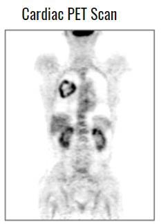
Cardiac FDG Patterns Seen In Oncologic PET Studies: What s Normal and What s Not?
 Cardiac FDG Patterns Seen In Oncologic PET Studies: What s Normal and What s Not? Alan H. Maurer, M.D. Director of Nuclear Medicine Temple University Hospital And School of Medicine Philadelphia, PA Objectives
FDG-PET value in deep endometriosis
 Gynecol Surg (2011) 8:305 309 DOI 10.1007/s10397-010-0652-6 ORIGINAL ARTICLE FDG-PET value in deep endometriosis A. Setubal & S. Maia & C. Lowenthal & Z. Sidiropoulou Received: 3 December 2010 / Accepted:
PSMA PET SCANNING AND THERANOSTICS IN PROSTATE CANCER KEVIN TRACEY, MD, FRCPC PRECISION DIAGNSOTIC IMAGING REGIONAL PET/CT CENTRE
 PSMA PET SCANNING AND THERANOSTICS IN PROSTATE CANCER KEVIN TRACEY, MD, FRCPC PRECISION DIAGNSOTIC IMAGING REGIONAL PET/CT CENTRE DISCLOSURES/CONFLICTS NONE OBJECTIVES Understand current diagnostic role
Imaging in Pediatric Thyroid disorders: US and Radionuclide imaging. Deepa R Biyyam, MD Attending Pediatric Radiologist
 Imaging in Pediatric Thyroid disorders: US and Radionuclide imaging Deepa R Biyyam, MD Attending Pediatric Radiologist Imaging in Pediatric Thyroid disorders: Imaging modalities Outline ACR-SNM-SPR guidelines
Chapter 10. Summary, conclusions and future perspectives
 Chapter 10 Summary, conclusions and future perspectives 10.1 SUMMARY In this thesis, a new tumor imaging tracer in nuclear medicine is studied. This 123 tracer, L-3-[ I]Iodo-alpha-methyl-tyrosine (IMT),
Austin Radiological Association Nuclear Medicine Procedure PROSTATE CANCER STUDY (In-111-Capromab Pendetide [ProstaScint ])
![Austin Radiological Association Nuclear Medicine Procedure PROSTATE CANCER STUDY (In-111-Capromab Pendetide [ProstaScint ])](/thumbs/81/82771892.jpg "Austin Radiological Association Nuclear Medicine Procedure PROSTATE CANCER STUDY (In-111-Capromab Pendetide [ProstaScint ])") Austin Radiological Association Nuclear Medicine Procedure PROSTATE CANCER STUDY (In-111-Capromab Pendetide [ProstaScint ]) Overview Indications The Prostate Cancer Study with an indium-111 labeled murine
Austin Radiological Association Nuclear Medicine Procedure PROSTATE CANCER STUDY (In-111-Capromab Pendetide [ProstaScint ]) Overview Indications The Prostate Cancer Study with an indium-111 labeled murine
Description MRI, TMJ C T Head Without Contrast C T Head With Contrast C T Head Without & With Contrast
 s Requiring Prior Authorization for the Advanced Imaging 70336 MRI, TMJ 70450 C T Head Without Contrast 70460 C T Head With Contrast 70470 C T Head Without & With Contrast 70480 C T Orbit Without Contrast
Core Summary of Product Characteristics for Fludeoxyglucose ( 18 F)
") Core Summary of Product Characteristics for Fludeoxyglucose ( 18 F) This fludeoxyglucose ( 18 F) Core SmPC has been prepared on the basis, and taking into account the available published scientific literature
Core Summary of Product Characteristics for Fludeoxyglucose ( 18 F) This fludeoxyglucose ( 18 F) Core SmPC has been prepared on the basis, and taking into account the available published scientific literature
Indications of PET/CT in oncology
 Monday, August 27, 2012 Session 1, 10:00-10:40 Indications of PET/CT in oncology Helle Westergren Hendel MD, PhD, assistant professor Bacelor in Leadership & Health Ecomomics Head of Clinical PET, Herlev
The Role of PET / CT in Lung Cancer Staging
 July 2004 The Role of PET / CT in Lung Cancer Staging Vlad Vinarsky, Harvard Medical School Year IV Patient AM HPI: 81 yo F p/w hemoptysis x 1 month LLL lesion on CXR, not responsive to Abx 35 pack-year
Austin Radiological Association Nuclear Medicine Procedure THERAPY FOR THYROID CANCER (I-131 as Sodium Iodide)
") Austin Radiological Association Nuclear Medicine Procedure THERAPY FOR THYROID CANCER (I-131 as Sodium Iodide) Overview Indications I-131 therapy for Thyroid Cancer, of the papillo-follicular type, is
Austin Radiological Association Nuclear Medicine Procedure THERAPY FOR THYROID CANCER (I-131 as Sodium Iodide) Overview Indications I-131 therapy for Thyroid Cancer, of the papillo-follicular type, is
Nuclear Medicine and PET. D. J. McMahon rev cewood
 Nuclear Medicine and PET D. J. McMahon 150504 rev cewood 2018-02-15 Key Points Nuclear Medicine and PET: Imaging: Understand how Nuc Med & PET differ from Radiography & CT by the source of radiation. Be
Gastrointestinal tract
 Gastrointestinal tract Colloidal liver-spleen imaging Presented by: Jehad Felemban Introduction: To obtain better anatomic display of liver and spleen architecture, we use (CT Ultrasound). (Radionuclide
Austin Radiological Association BRAIN AMYLOID STUDY (F-18-Florbetapir)
") Austin Radiological Association BRAIN AMYLOID STUDY (F-18-Florbetapir) Overview The Brain Amyloid Study with F-18-florbetapir depicts the extracellular deposition of B- amyloid (Aβ) peptides (or plaques
Austin Radiological Association BRAIN AMYLOID STUDY (F-18-Florbetapir) Overview The Brain Amyloid Study with F-18-florbetapir depicts the extracellular deposition of B- amyloid (Aβ) peptides (or plaques
Index. Surg Oncol Clin N Am 16 (2007) Note: Page numbers of article titles are in boldface type.
 Note: Page numbers of article titles are in boldface type.") Surg Oncol Clin N Am 16 (2007) 465 469 Index Note: Page numbers of article titles are in boldface type. A Adjuvant therapy, preoperative for gastric cancer, staging and, 339 B Breast cancer, metabolic
Surg Oncol Clin N Am 16 (2007) 465 469 Index Note: Page numbers of article titles are in boldface type. A Adjuvant therapy, preoperative for gastric cancer, staging and, 339 B Breast cancer, metabolic
DRAXIMAGE SODIUM IODIDE I 131 SOLUTION USP DIAGNOSTIC. For Oral Use DESCRIPTION
 DRAXIMAGE SODIUM IODIDE I 131 SOLUTION USP DIAGNOSTIC For Oral Use DESCRIPTION Sodium Iodide I 131 Solution is an aqueous solution of sodium iodide I-131 for diagnostic use by oral administration. The
PHYSICAL CHARACTERISTICS
 BRACCO DIAGNOSTICS L/4739/0 1 CHOLETEC Kit for the Preparation of Technetium Tc 99m Mebrofenin For Diagnostic Use DESCRIPTION Each reaction vial contains a nonradioactive, sterile, nonpyrogenic mixture
Society of Nuclear Medicine Procedure Guideline for Tumor Imaging Using F-18 FDG
 Society of Nuclear Medicine Procedure Guideline for Tumor Imaging Using F-18 FDG version 2.0, approved February 7, 1999 Authors: Heinrich R. Schelbert, MD, PhD (UCLA School of Medicine, Los Angeles, CA);
MT09 - Normal Human Tissue Microarray, FDA
 Reveal Biosciences offers Histochemical Staining, Immunohistochemistry (IHC), In Situ Hybridization (ISH), Whole Slide Imaging, and Quantitative Image Analysis on any TMA MT09 - Normal Human Tissue Microarray,
R: March, E D T P A. Kit for the Preparation of Technetium Tc 99m Pentetate Injection. DIAGNOSTIC - For Intravenous Use
 R: March, 1998 27227 0002E m TM DESCRIPTION D T P A Kit for the Preparation of Technetium Tc 99m Pentetate Injection DIAGNOSTIC - For Intravenous Use Each kit consists of reaction vials which contain the
Nuclear Medicine: Manuals. Nuclear Medicine. Nuclear imaging. Emission imaging: study types. Bone scintigraphy - technique
 Nuclear Medicine - Unsealed radioactive preparations the tracer mixes with the patients body fluids on a molecular level (e.g. after intravenous injection) - 3 main fields: - In vitro : measuring concentrations
Anthem Blue Cross and Blue Shield Virginia Advanced Imaging Procedures Requiring Precertification Revised 02/13/2013
 Anthem Blue Cross and Blue Shield Virginia Advanced Imaging Procedures Requiring Precertification Revised 02/13/2013 Modality and CT Head CTA Head: Cerebrovascular MRI Head MRA Head: Cerebrovascular Functional
Pitfalls in Oncologic Diagnosis with PET CT. Nononcologic Hypermetabolic Findings
 Pitfalls in Oncologic Diagnosis with PET CT. Nononcologic Hypermetabolic Findings Poster No.: C-1359 Congress: ECR 2012 Type: Educational Exhibit Authors: J. Rossi 1, C. A. Mariluis 1, E. Delgado 1, R.
Molecular Imaging and Breast Cancer
 Molecular Imaging and Breast Cancer Breast cancer forms in tissues of the breast usually in the ducts, tubes that carry milk to the nipple, and lobules, the glands that make milk. It occurs in both men
RADPrimer Curriculum Breast Topics Covered Basic Intermediate 225
 Breast Anatomy & Normal Variants 11 Breast Imaging Modalities 13 BI RADS Lexicon 3 Mammography: Masses 9 Mammography: Calcifications 17 Mammography: Additional Findings 8 Ultrasound Features 10 Ultrasound
Testicular relapse of non-hodgkin Lymphoma noted on FDG-PET
 Testicular relapse of non-hodgkin Lymphoma noted on FDG-PET Stephen D. Scotti 1*, Jennifer Laudadio 2 1. Department of Radiology, North Carolina Baptist Hospital, Winston-Salem, NC, USA 2. Department of
AN INTRODUCTION TO NUCLEAR MEDICINE
 AN INTRODUCTION TO NUCLEAR MEDICINE WITH RESPECT TO THYROID DISORDERS By: B.Shafiei MD Nuclear Physician Taleghani Medical Center Radioactive: An element with Unstable Nucleus (Excess Energy), can achieve
MRI and CT of the CNS
 MRI and CT of the CNS Dr.Maha ELBeltagy Assistant Professor of Anatomy Faculty of Medicine The University of Jordan 2018 Computed Tomography CT is used for the detection of intracranial lesions. CT relies
Title. CitationJournal of Nuclear Cardiology, 23(3): Issue Date Doc URL. Rights. Type. File Information
: Issue Date Doc URL. Rights. Type. File Information") Title Incidental focal myocardial 18F-FDG uptake indicatin Aikawa, Tadao; Naya, Masanao; Manabe, Osamu; Obara, Author(s) Hiroyuki CitationJournal of Nuclear Cardiology, 23(3): 596-598 Issue Date 2016-06
Title Incidental focal myocardial 18F-FDG uptake indicatin Aikawa, Tadao; Naya, Masanao; Manabe, Osamu; Obara, Author(s) Hiroyuki CitationJournal of Nuclear Cardiology, 23(3): 596-598 Issue Date 2016-06
Whole-body biodistribution and radiation dosimetry estimates for the β-amyloid radioligand [ 11 C]MeS-IMPY in non-human primates
![Whole-body biodistribution and radiation dosimetry estimates for the β-amyloid radioligand [ 11 C]MeS-IMPY in non-human primates](/thumbs/87/95592963.jpg "Whole-body biodistribution and radiation dosimetry estimates for the β-amyloid radioligand [ 11 C]MeS-IMPY in non-human primates") Whole-body biodistribution and radiation dosimetry estimates for the β-amyloid radioligand [ 11 C]MeS-IMPY in non-human primates Molecular Imaging Branch, NIMH Bldg. 1 Rm. B3-10 September 6 th, 2006 The
Whole-body biodistribution and radiation dosimetry estimates for the β-amyloid radioligand [ 11 C]MeS-IMPY in non-human primates Molecular Imaging Branch, NIMH Bldg. 1 Rm. B3-10 September 6 th, 2006 The
CanTrace PRODUCT MONOGRAPH. Parenteral Solution, up to 1.4 GBq/mL (38.2 mci/ml) Diagnostic Radiopharmaceutical
 Diagnostic Radiopharmaceutical") PRODUCT MONOGRAPH CanTrace 18 F-Fluorodeoxyglucose ( 18 F-FDG) Parenteral Solution, up to 1.4 GBq/mL (38.2 mci/ml) Diagnostic Radiopharmaceutical IPET Pharmaceuticals, Inc. Suite 880, 1090 West Georgia,
PRODUCT MONOGRAPH CanTrace 18 F-Fluorodeoxyglucose ( 18 F-FDG) Parenteral Solution, up to 1.4 GBq/mL (38.2 mci/ml) Diagnostic Radiopharmaceutical IPET Pharmaceuticals, Inc. Suite 880, 1090 West Georgia,
Prof. Dr. NAGUI M. ABDELWAHAB,M.D.; MARYSE Y. AWADALLAH, M.D. AYA M. BASSAM, Ms.C.
 Role of Whole-body Diffusion MR in Detection of Metastatic lesions Prof. Dr. NAGUI M. ABDELWAHAB,M.D.; MARYSE Y. AWADALLAH, M.D. AYA M. BASSAM, Ms.C. Cancer is a potentially life-threatening disease,
PET-CT Fusion. imaging in differentiating
 EDUCATION EXHIBIT 1411 PET-CT Fusion Imaging in Differentiating Physiologic from Pathologic FDG Uptake 1 CME FEATURE See accompanying test at http:// www.rsna.org /education /rg_cme.html LEARNING OBJECTIVES
SCINTIGRAPHY OF THE CENTRAL NERVOUS SYSTEM Part 1: Introduction and BBB studies
 SCINTIGRAPHY OF THE CENTRAL NERVOUS SYSTEM Part 1: Introduction and BBB studies George N. Sfakianakis MD Professor of Radiology and Pediatrics Director, Division of Nuclear Medicine October 2009 FIRST
2. RADIOPHARMACEUTICALS UTILIZED
 1. OVERVIEW AND INDICATIONS (adapted from Society of Nuclear Medicine Procedure Guideline for Thyroid Uptake Measurement, Version 3.0; reprinted from http://snmmi.files.cmsplus.com/docs/thyroid%20uptake%20measure%20v3%200.pdf,
PET SCANS: THE WHO, WHEN AND WHY & HOW TO GET REIMBURSED
 PET SCANS: THE WHO, WHEN AND WHY & HOW TO GET REIMBURSED Cecelia E. Schmalbach, MD, FACS Associate Professor of Surgery Head & Neck Surgery Otolaryngology Residency Program Director No Financial Disclosures
PET-CT for radiotherapy planning in lung cancer: current recommendations and future directions
 PET-CT for radiotherapy planning in lung cancer: current recommendations and future directions Gerry Hanna Centre for Cancer Research and Cell Biology Queen s University of Belfast @gerryhanna Talk Outline
Imaging Patient Education What you should know about your PET/CT Bone Scan.
 Imaging Patient Education What you should know about your PET/CT Bone Scan. Purpose: PET/CT uses a small amount of a radioactive sodium fluoride product to detect cancer within the bones. A low dose CT
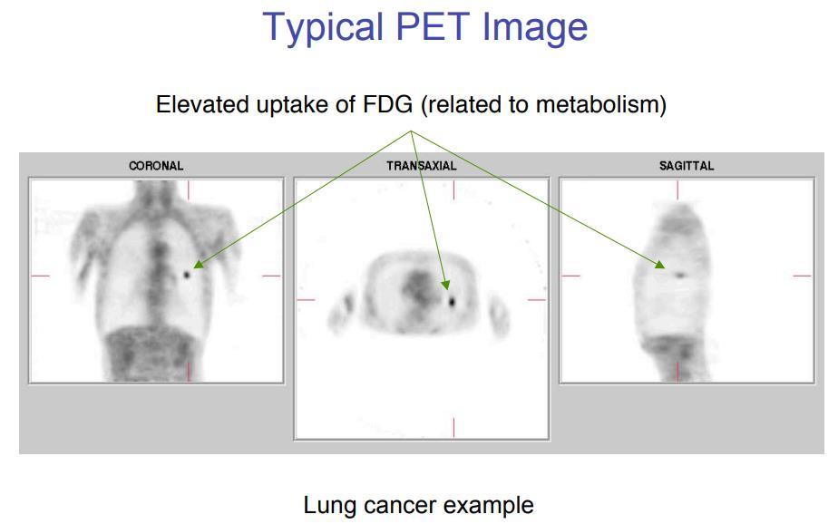
Typical PET Image. Elevated uptake of FDG (related to metabolism) Lung cancer example: But where exactly is it located?
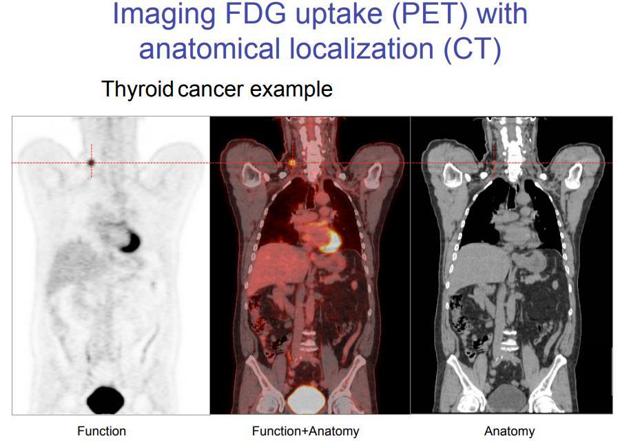
 Lung cancer example: But where exactly is it located?") Typical PET Image Elevated uptake of FDG (related to metabolism) Lung cancer example: But where exactly is it located? PET/CT Oncology Imaging Anatometabolic fusion images are useful in the management
Typical PET Image Elevated uptake of FDG (related to metabolism) Lung cancer example: But where exactly is it located? PET/CT Oncology Imaging Anatometabolic fusion images are useful in the management
Bekir Tasdemir, 1 Zeki Dostbil, 1 Ali Inal, 2 Kemal Unal, 3 Sule Yildirim, 1 andf.selcuksimsek Introduction
 BioMed Research International, Article ID 129683, 5 pages http://dx.doi.org/10.1155/2014/129683 Research Article Evaluation of Clinical Contributions Provided by Addition of the Brain, Calvarium, and Scalp
Radionuclides in Medical Imaging. Danielle Wilson
 Radionuclides in Medical Imaging Danielle Wilson Outline Definitions History and development Radionuclide applications & techniques in imaging Conclusion Definition #1 : Radionuclide An unstable nucleus
42 yr old male with h/o Graves disease and prior I 131 treatment presents with hyperthyroidism and undetectable TSH. 2 hr uptake 20%, 24 hr uptake 50%
 Pinhole images of the neck are acquired in multiple projections, 24hrs after the oral administration of approximately 200 µci of I123. Usually, 24hr uptake value if also calculated (normal 24 hr uptake
Basics of nuclear medicine
 Basics of nuclear medicine Prof. dr. Davor Eterović Prof. dr. Vinko Marković Radioisotopes are used both in diagnostics and in therapy Diagnostics gamma emitters are used since gamma rays can penetrate
PET Guidance of Therapy for BNCT and in vivo B-10 imaging
 INFN LNL Legnaro 17-19 Novembre 2009 Principles of Positron Emission Tomography and Radiopharmaceuticals PET Guidance of Therapy for BNCT and in vivo B-10 imaging Luca Menichetti, Ph.D C.N.R. Institute
Understanding the Diagnostic and Prognostic Role of Imaging in the Evaluation of an Anterior Mediastinal Mass
 Understanding the Diagnostic and Prognostic Role of Imaging in the Evaluation of an Anterior Mediastinal Mass Daniel W. Kim, Harvard Medical School Year III Agenda Mediastinum Menu of tests Anatomy Normal