Role of Imaging Methods in Diagnosis of Acute Pancreatitis. Válek V. Radiologická klinika, FN Brno a LF MU v Brně
|
|
|
- Philippa Douglas
- 5 years ago
- Views:
Transcription
1 Role of Imaging Methods in Diagnosis of Acute Pancreatitis Válek V. Radiologická klinika, FN Brno a LF MU v Brně
2 New Classification: Acute Pancreatitis 2007 revision of Atlanta classification and definitions of collections associated with acute pancreatitis by Acute Pancreatitis Working Group.
 Acute Pancreatitis")
3 Revised Definitions: Acute Pancreatitis Patients who present with two of the following three manifestations are diagnosed as having acute pancreatitis: Abdominal pain suggestive of pancreatic origin Serum amylase, lipase 3 times normal Characteristic findings on CECT Disease Severity First week based on clinical parameters Thereafter on morphologic parameters (CECT) Acute Pancreatitis Classification Working Group May 2007
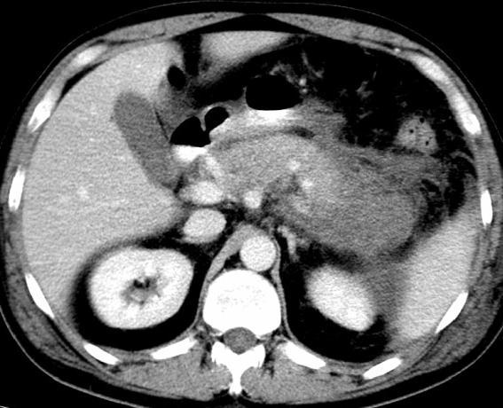
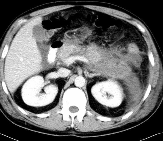
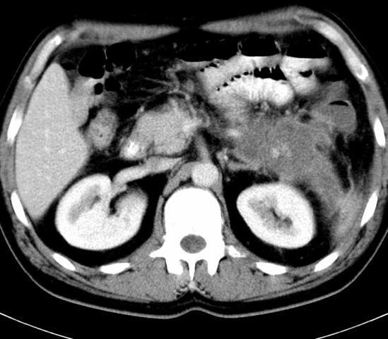
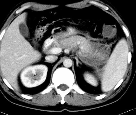
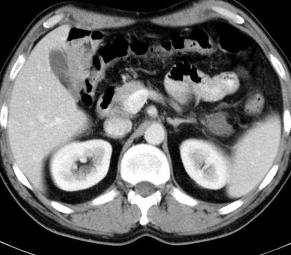
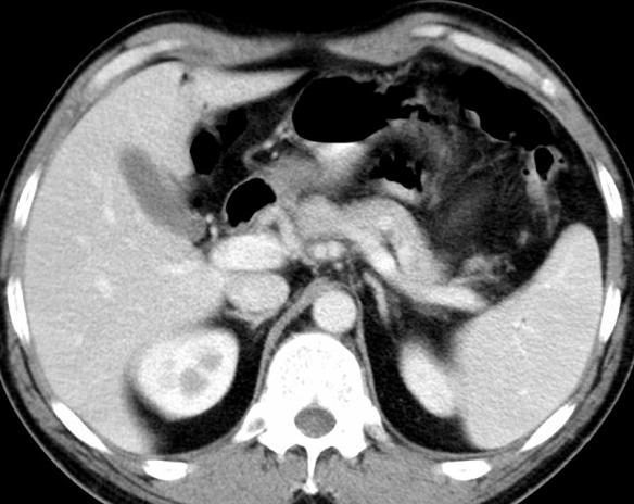
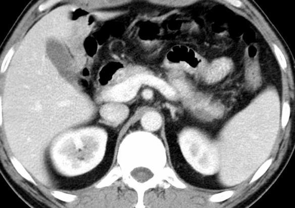
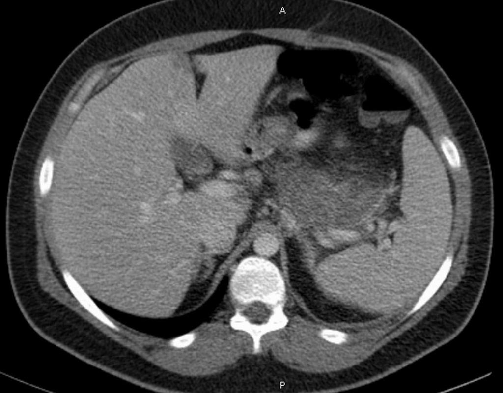
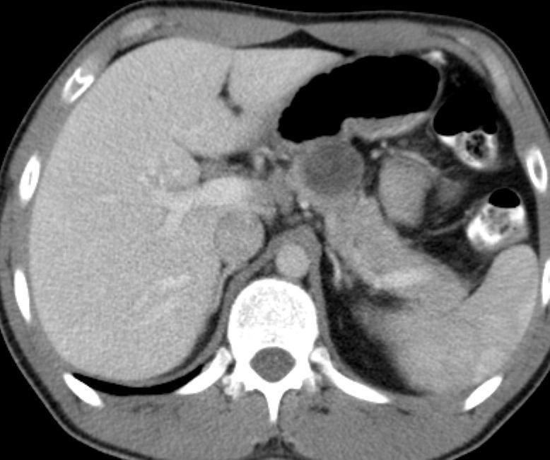
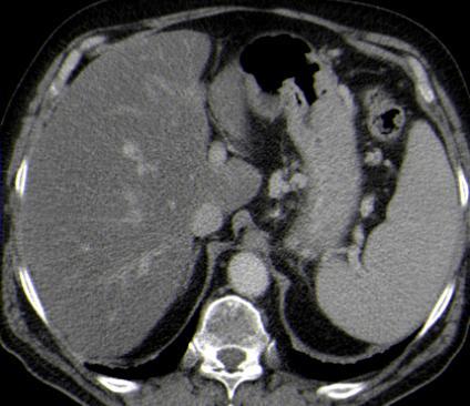
4 CECT
 is basic, because it is strictly correlated to the prognosis of the patient.")
5 Why CECT?? CT study is the most appropriate procedure to confirm image findings of acute pancreatitis. The evaluation of severity (CTSI) is basic, because it is strictly correlated to the prognosis of the patient. Early scanning for the prediction of severity is limited because the full extent of pancreatic necrosis may not develop within the first 48 hour of presentation. Dynamic CECT have % accurate for necrosis detection. Balthazar Radiology 1994; 193: Balthazar Radiology 2002; 223:


6 CTSI Accuracy of CTSI for predicting pancreatitis severity: CTSI > 3 Sensitivity 11/13 Specificity 41/42 PPV 11/12 NPV 52/55 Accuracy 52/55 Gurleyik et al J Pancreas 2005; 6:
7 MRI
8 Acute pancreatitis The MR has actually a secondary role for the diagnosis, but it represents, first instance method in patients with adverse reaction to contrast medium. Standard MRI techniques including T1-weighted and T2- weighted fat-suppressed imaging sequences together with contrast-enhanced imaging.

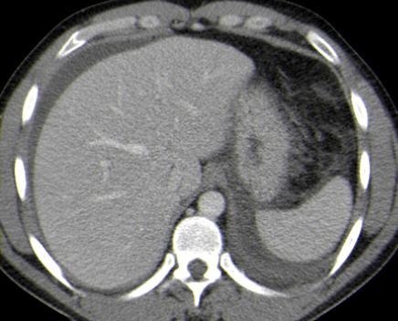
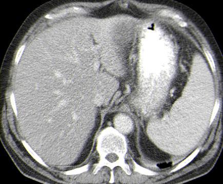
9 CECT vesus MR Non-severe acute pancreatitis
10 US
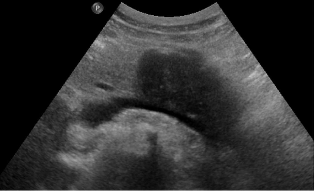
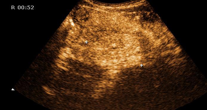
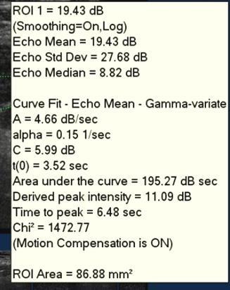
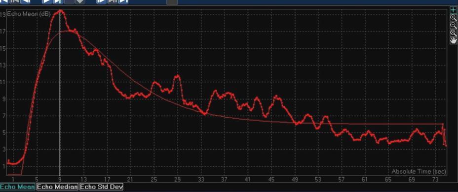
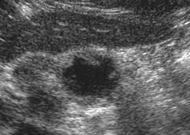
11 Acute pancreatitis The ultrasound can be used as first instance method in patient with clinical suspect of acute pancreatitis. Contrast-enhanced ultrasound is a relatively new technique, currently used for liver tumors diagnosis. Contrast-enhanced ultrasound might represent a valuable additional imaging method to contrast CT for selected cases. Pulse inversion harmonic imaging allows the assessment of the necrotic areas in acute pancreatitis.

12 CECT versus US
13 Non-severe acute pancreatitis

 improve in 48 72 hr Imaging None CECT US to")
14 Old term: Mild acute pancreatitis New Term: Non-severe acute pancreatitis Histology: interstitial edema, micronecrosis Clinical course no MSOF (Multisystem organ failure) improve in hr Imaging None CECT US to evaluate gallstone

15 Old term: Mild acute pancreatitis New Term: Non-severe acute pancreatitis
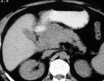
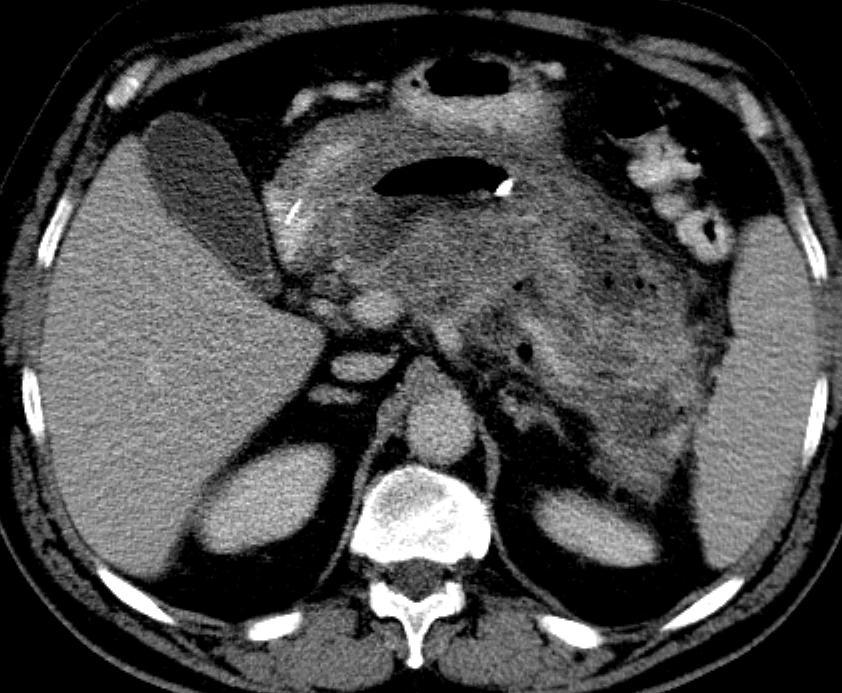
16 Severe acute pancreatitis


17 Old term: Severe acute pancreatitis New Term: Severe Acute Pancreatitis
18 Pancreatic Necrosis

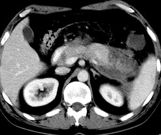
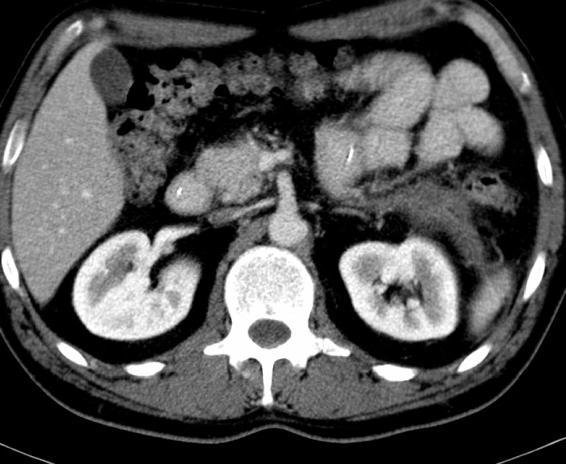
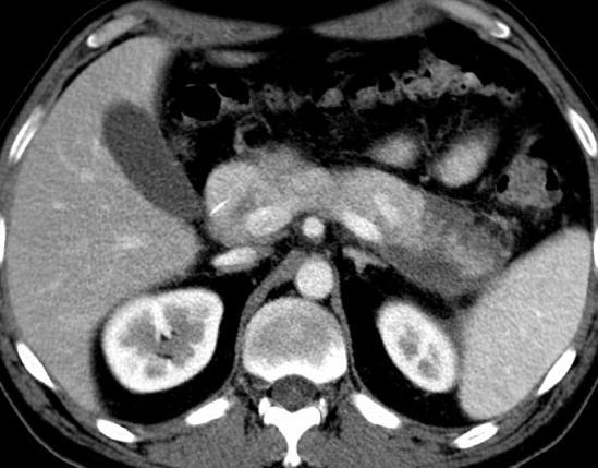
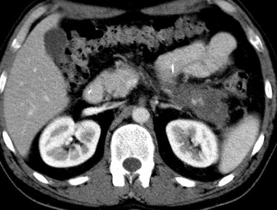

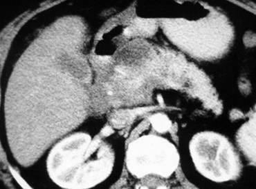
19 26/12 5/1 17/1 6/4
20 New Classification: Necrosis: Based on CECT or MR 1. One or more focal areas of nonenhancing pancreatic parenchyma 2. Focal parenchymal low signal in T1 FS unenhanced sequence 3. Typically accompanied by gross peripancreatic fat necrosis 4. MR more heterogeneous than CT 5. May not be apparent up to 48 hours after onset 6. Potential pitfalls: Apparent diminished enhancement value in patients with normal fatty infiltration Patients with diffuse parenchymal edema in less severe, interstitial pancreatitis Small intrapancreatic focal fluid collections

21 CECT vesus MR: Necrosis
22 Fluid Collections
23 New Classification: Fluid collections: CECT or MRI 1. Enzyme rich pancreatic juice 2. Acute peripancreatic fluid collection (APFC) (occur within 48 hrs in %, majority remain sterile, resolves spontaneously within 2-4 weeks) 3. Post necrotic pancreatic fluid collections (PNPFC) (fluid and necrotic contents fat, initial necrosis liquefactive necrosis 4. Walled off pancreatic necrosis (WOPN) = late stage of PNPFC 5. Pancreatic pseudocyst (4 weeks, contain NO necrosis) 6. Pancreatic abscess = infected pseudocyst (dif.dg. Infected PNPFC and infected WOPN

24 Old term: Acute Fluid Collection New Term: Post Necrotic Pancreatic Colections (PNPC)

")
25 Walled off necrosis (WON)
26 Old term: Pancreatic Pseudocyst New Term: Pancreatic Pseudocyst Well circumscribed, thin walled, homogeneous low attenuation collection of pancreatic juice. Requires 4 weeks, contains no necrosis Resolves spontaneously within 6 weeks 40%, 80% if <6cm Ductal communication important for management Noninfected or infected (suppurative)
27 Pancreatic Pseudocyst - CECT
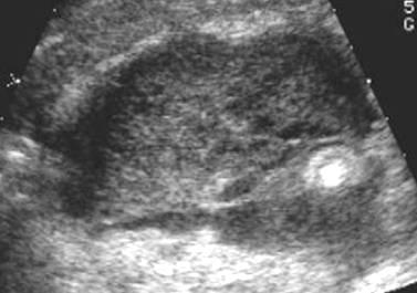
28 Infected Pseudocyst
29 Old term: Pancreatic Absces New Term: Infected Pseudocyst Wall thicker, more irregular than the well delineated thin wall circumscribed collection of pus near pancreas with homogeneously low attenuation center. Requires 4 weeks to form Contains little or no necrosis Should be differentiated from infected PNPC and infected WON
30 Infected Pseudocyst - CECT
31 Conclusion
32 Role of Imaging Methods in Diagnosis of Acute Pancreatitis 1. Early diagnosis 2. Staging (grading) + classification 3. Differential diagnosis 4. Follow-up (treatment)

33 CECT = gold standard 1. Reliable method 2. Widely available 3. Show local extend of inflammation/necrosis and occurrence of complications 4. Balthazar CTSI based on grade of pancreatitis and amount of glandular necrosis 5. CTSI of 7 10 associated with 92% morbidity, 17% mortality 6. Pts with CTSI 3 can safely be discharged from ICU Vriens et all, J.Am.Coll.Surgery, 2005, 2001, Che et al, Eur.J.Radiol., 2006, 57, Leung et al, World J. Gastroenterol., 2005, 11,
34 MRI = second choice 1. Geographic areas of altered T1 signal correspond to areas of pancreatic necrosis depicted on CECT 2. High signal intensity on unenhanced T1 FS SGE images corresponds to hemorrhage and correlates with severity. 3. Sensitivity of MRI for acute pancreatitis may surpass CT (minor peripanc. inflamm) on T2 FS images 4. MRSI =CTSI using dynamic 3DT1 SGE gado enhanced images, with added benefit of PD duct eval. Using secretin MRCP. 5. PD duct evaluation using secretin MRCP Lecesne et al Radiology 1999; 211: Pamuklar Magn Reson Clin NA 2005; 13: Arvanitakis Gastroenterology 2004; 126:
35 Quiz


36 Diagnosis?? 1. Pancreatic atrophy 2. Acute pancreatitis 3. Von Wildelmuth disease 4. Pancreatic agenesis 5. Cystic fibrosis

37 Cystic fibrosis Cystic fibrosis is a condition with autosomal recessive inheritance in which there are defects of serous and mucous secretion involving multiple organs; 85 % of patients have severe exocrine pancreatic insufficiency and steatorrhoea. Obstruction of the main pancreatic duct and side branches by inspissated secretions results in acinar and ductal dilatation and subsequent atrophy of the acinar tissue. Ultrasound (US), computed tomography (CT) and pancreatic duct imaging may show abnormalities, including marked fatty replacement of the normal pancreatic parenchyma, dystrophic calcification and pancreatic cysts resulting from ductal obstruction.


38 Diagnosis?? 1. Pancreastic pseudocysts 2. Pancreatic polycystosis 3. Hippel-Lindau 4. Chronic pancreatitis 5. Multiple cystic tumors

39 Von Hippel-Lindau Von Hippel Lindau disease is inherited as an autosomal dominant condition characterized by renal cell carcinomas, phaeochromocytomas, retinal angiomatosis and haemangioblastomas of the cerebellum. The most common pancreatic lesions in this condition are simple pancreatic cysts, but serous cystic pancreatic neoplasms and pancreatic islet cell tumours may also occur.


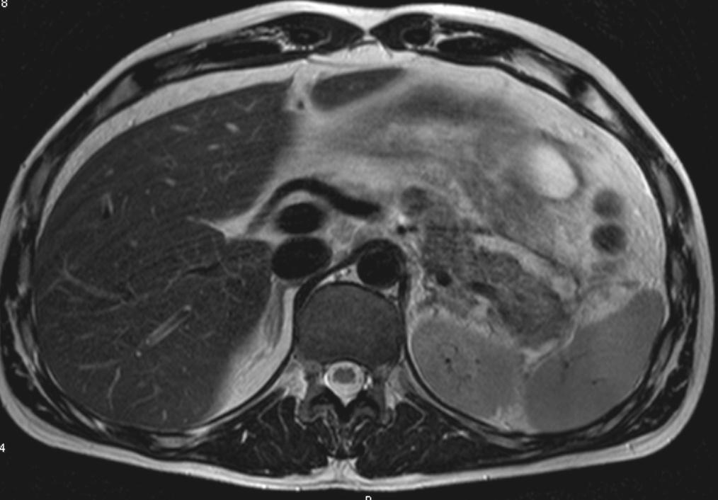
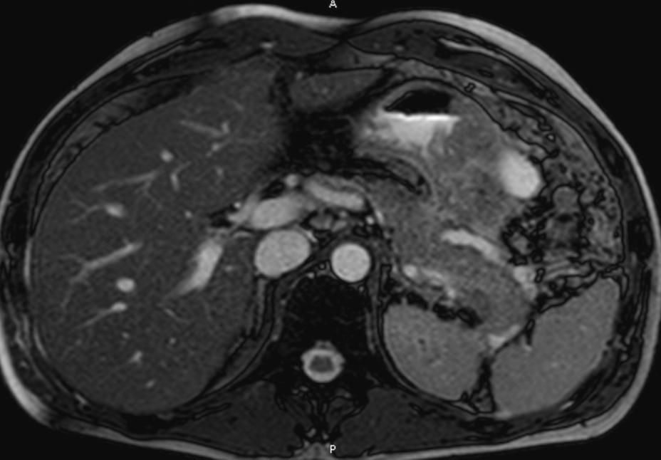
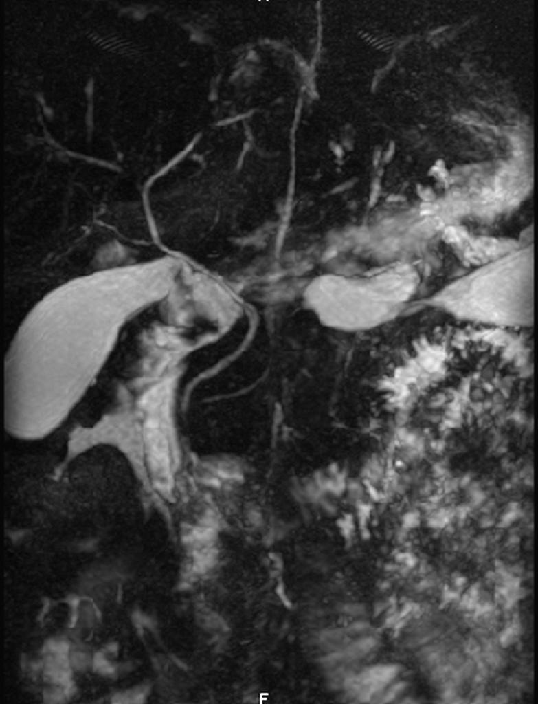
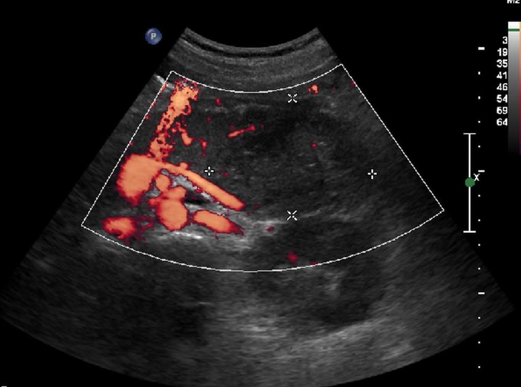
40 Diagnosis?? 1. IPMN 2. Adenocarcinoma 3. Mucinous cystic tumor 4. Serous cystic tumor 5. Pseudocyst
41 Serous cystadenoma A serous cystadenoma consists of inumerable tiny cysts measuring less than 2 cm in diameter. Larger cysts occur in the periphery of the tumor. The cysts are lined with small cuboid epithelial cells containing glycogen but no mucin. The serous (microcystic) cystadenomas are usually large at diagnosis, often measuring more than 8 cm across. The central fibrous area is hypervascular due to a rich subepithelial capillary network, causing enhancement to occur after intravenous contrast injection at computed tomography and magnetic resonance imaging. Calcification occurs in the central fibrous tissue in about half of patients.
U Nordic Forum - Trauma & Emergency Radiology. Lecture Objectives. MDCT in Acute Pancreatitis. Acute Pancreatitis: Etiologies
 Nordic Forum - Trauma & Emergency Radiology Lecture Objectives MDCT in Acute Pancreatitis Borut Marincek Institute of Diagnostic Radiology niversity Hospital Zurich, Switzerland To describe the role of
Anatomy of the biliary tract
 Harvard-MIT Division of Health Sciences and Technology HST.121: Gastroenterology, Fall 2005 Instructors: Dr. Jonathan Glickman Anatomy of the biliary tract Figure removed due to copyright reasons. Biliary
PANCREATIC PSEUDOCYSTS. Madhuri Rao MD PGY-5 Kings County Hospital Center
 PANCREATIC PSEUDOCYSTS Madhuri Rao MD PGY-5 Kings County Hospital Center 34 yo M Case Presentation PMH: Chronic pancreatitis (ETOH related) PSH: Nil Meds: Nil NKDA www.downstatesurgery.org Symptoms o Chronic
Acute pancreatitis is most commonly caused by gallstones
 CLINICAL GASTROENTEROLOGY AND HEPATOLOGY 2008;6:1077 1085 CLINICAL IMAGING Imaging of Acute Pancreatitis and Its Complications DESIREE E. MORGAN Department of Radiology, University of Alabama at Birmingham,
IMAGING OF ACUTE AND CHRONIC PANCREATITIS, INCLUDING EXOCRINE FUNCTION
 IMAGING OF ACUTE AND CHRONIC PANCREATITIS, INCLUDING EXOCRINE FUNCTION Andrew T. Trout, MD @AndrewTroutMD Disclosures Grant support National Pancreas Foundation In-kind support - ChiRhoClin modified from:
Does it matter what we drain?
 Endoscopic Management of Pancreatic Fluid Collections Shyam Varadarajulu, MD Medical Director Center for Interventional Endoscopy Florida Hospital, Orlando Does it matter what we drain? Makes all the difference!
CT 101 :Pancreas and Spleen
 CT 101 :Pancreas and Spleen Shikha Khullar,, MD, MPH Division of Radiology University of South Alabama The Pancreas Normal Pancreas 3 Phase Pancreatic CT Non contrast Arterial phase : 30-35 35 second
Anatomical and Functional MRI of the Pancreas
 Anatomical and Functional MRI of the Pancreas MA Bali, MD, T Metens, PhD Erasme Hospital Free University of Brussels Belgium mbali@ulb.ac.be Introduction The use of MRI to investigate the pancreas has
Evidence based imaging of the pancreas
 Evidence based imaging of the pancreas D.Vanbeckevoort, D.Bielen, K.Op de beeck, R.Vanslembrouck Department of Radiology Chairman Prof. Dr. R.Oyen Non-invasive imaging tests available for the diagnosis
The role of endoscopy in the diagnosis and treatment of cystic pancreatic neoplasms
 The role of endoscopy in the diagnosis and treatment of cystic pancreatic neoplasms CYSTIC LESIONS AND FLUID COLLECTIONS OF THE PANCREAS Their pathology ranges from pseudocysts and pancreatic necrosis
ACG Clinical Guideline: Diagnosis and Management of Pancreatic Cysts
 ACG Clinical Guideline: Diagnosis and Management of Pancreatic Cysts Grace H. Elta, MD, FACG 1, Brintha K. Enestvedt, MD, MBA 2, Bryan G. Sauer, MD, MSc, FACG (GRADE Methodologist) 3 and Anne Marie Lennon,
X-ray Corner. Imaging of The Pancreas. Pantongrag-Brown L
 X-ray Corner 125 Imaging of The Pancreas Modern imaging modalities commonly used in pancreas include ultrasound (US), CT, and MRI. Pancreas is a retroperitoneal organ which makes it difficult to visualize
Dr Claire Smith, Consultant Radiologist St James University Hospital Leeds
 Dr Claire Smith, Consultant Radiologist St James University Hospital Leeds Imaging in jaundice and 2ww pathway Image protocol Staging Limitations Pancreatic cancer 1.2.4 Refer people using a suspected
PATHOLOGY MCQs. The Pancreas
 PATHOLOGY MCQs The Pancreas A patient with cystic fibrosis is characteristically: A. more than 45 years of age B. subject to recurring pulmonary infections C. obese D. subject to spontaneous fractures
Pancreas composed of 2 parts: 1- exocrine gland 2- endocrine gland
 pancreas Pancreas composed of 2 parts: 1- exocrine gland 2- endocrine gland Acute pancreatitis Inflammation of the pancreas associated with acinar cell injury Clinical features: 1-abdominal pain cardinal
40th European Congress of Cytology Liverpool, UK, 2-5 th October 2016
 40th European Congress of Cytology Liverpool, UK, 2-5 th October 2016 EUS FNA of abdominal organs: An approach to reporting and triage for ancillary testing Date and time: Sunday 2 nd October 2016 15.00-16.30
Evaluation and Management of Cystic Lesions of the Pancreas: When to Resect, When to Follow and When to Forget
 Evaluation and Management of Cystic Lesions of the Pancreas: When to Resect, When to Follow and When to Forget Randall Brand, MD Professor of Medicine Division of Gastroenterology, Hepatology and Nutrition
Updated Imaging Nomenclature for Acute Pancreatitis
 Residents Section Structured Review Murphy et al. Imaging Nomenclature for Acute Pancreatitis Residents Section Structured Review Residents inradiology Kevin P. Murphy 1,2 Owen J. O Connor 1,2 Michael
The Pancreas. Basic Anatomy. Endocrine pancreas. Exocrine pancreas. Pancreas vasculature. Islets of Langerhans. Acinar cells Ductal System
 SGNA: Back to Basics Rogelio G. Silva, MD Assistant Clinical Professor of Medicine University of Illinois at Chicago Department of Medicine Division of Gastroenterology Advocate Christ Medical Center GI
Neoplasias Quisticas del Páncreas
 SEAP -Aproximación Práctica a la Patología Gastrointestinal- Madrid, 26 de mayo, 2006 Neoplasias Quisticas del Páncreas Gregory Y. Lauwers, M.D. Director, Service Massachusetts General Hospital Harvard
Chronic pancreatitis mimicking intraductal papillary mucinous neoplasm of the pancreas; Report of tow cases
 Jichi Medical University Journal Chronic pancreatitis mimicking intraductal papillary mucinous neoplasm of the pancreas; Report of tow cases Noritoshi Mizuta, Hiroshi Noda, Nao Kakizawa, Nobuyuki Toyama,
Common and unusual CT and MRI manifestations of pancreatic adenocarcinoma: a pictorial review
 Review Article Common and unusual CT and MRI manifestations of pancreatic adenocarcinoma: a pictorial review Min-Jie Yang, Su Li, Yong-Guang Liu, Na Jiao, Jing-Shan Gong Department of Radiology, Shenzhen
Chief Complaint. Retroperitoneal cystic mass incidentally found at health examination center.
 Personal Information Age: 34 y/o Sex: female Past history: major systemic medical history(-) surgical history(-), family history(-) Denied food or drug allergy Chief Complaint Retroperitoneal cystic mass
Pediatric Retroperitoneal Masses Radiologic-Pathologic Correlation
 Acta Radiológica Portuguesa, Vol.XVIII, nº 70, pág. 61-70, Abr.-Jun., 2006 Pediatric Retroperitoneal Masses Radiologic-Pathologic Correlation Marilyn J. Siegel Mallinckrodt Institute of Radiology, Washington
ACG Clinical Guideline: Management of Acute Pancreatitis
 ACG Clinical Guideline: Management of Acute Pancreatitis Scott Tenner, MD, MPH, FACG 1, John Baillie, MB, ChB, FRCP, FACG 2, John DeWitt, MD, FACG 3 and Santhi Swaroop Vege, MD, FACG 4 1 State University
Matthew McCollough, M.D. April 9, 2009 University of Louisville
 Matthew McCollough, M.D. April 9, 2009 University of Louisville List the differential diagnosis for pancreatic cysts Review the epidemiology Illustrate the types of cysts through case discussions Discuss
Chronic Pancreatitis: When to Scope? Gregory A. Cote, MD, MS Assistant Professor of Medicine Indiana University School of Medicine
 Chronic Pancreatitis: When to Scope? Gregory A. Cote, MD, MS Assistant Professor of Medicine Indiana University School of Medicine Endoscopy & Chronic Pancreatitis Diagnosis EUS ERCP Exocrine Function
X-Ray Corner. Imaging Approach to Cystic Liver Lesions. Pantongrag-Brown L. Solitary cystic liver lesions. Hepatic simple cyst (Figure 1)
") THAI J 136 Imaging Approach to Cystic Liver Lesions GASTROENTEROL 2013 X-Ray Corner Imaging Approach to Cystic Liver Lesions Pantongrag-Brown L Cystic liver lesions are common findings in daily practice
THAI J 136 Imaging Approach to Cystic Liver Lesions GASTROENTEROL 2013 X-Ray Corner Imaging Approach to Cystic Liver Lesions Pantongrag-Brown L Cystic liver lesions are common findings in daily practice
Diagnosis of chronic Pancreatitis. Christoph Beglinger, University Hospital Basel, Switzerland
 Diagnosis of chronic Pancreatitis Christoph Beglinger, University Hospital Basel, Switzerland Pancreatitis Pancreas Pancreas - an organ that makes bicarbonate to neutralize gastric acid, enzymes to digest
HEPATO-BILIARY IMAGING
 HEPATO-BILIARY IMAGING BY MAMDOUH MAHFOUZ MD PROF.OF RADIOLOGY CAIRO UNIVERSITY mamdouh.m5@gmail.com www.ssregypt.com CT ABDOMEN Indications Patient preparation Patient position Scanogram Fasting 4-6 hours
ACUTE PANCREATITIS: NEW CLASSIFICATION OF AN OLD FOE. T Barrow, A Nasrullah, S Liong, V Rudralingam, S A Sukumar
 ACUTE PANCREATITIS: NEW CLASSIFICATION OF AN OLD FOE T Barrow, A Nasrullah, S Liong, V Rudralingam, S A Sukumar LEARNING OBJECTIVES q Through a series of cases illustrate the updated Atlanta symposium
Multidetector CT evaluation of acute pancreatitis and its complications and its correlation with clinical outcome
 INTERNATIONAL JOURNAL OF CURRENT RESEARCH IN BIOLOGY AND MEDICINE ISSN: 2455-944X www.darshanpublishers.com DOI:10.22192/ijcrbm Volume 3, Issue 1-2018 Original Research Article Multidetector CT evaluation
Role of imaging in RCC. Ultrasonography. Solid lesion. Cystic RCC. Solid RCC 31/08/60. From Diagnosis to Treatment: the Radiologist Perspective
 Role of imaging in RCC From Diagnosis to Treatment: the Radiologist Perspective Diagnosis Staging Follow up Imaging modalities Limitations and pitfalls Duangkamon Prapruttam, MD Department of Therapeutic
Autoimmune Pancreatitis: A Great Imitator
 Massachusetts General Hospital Harvard Medical School Autoimmune Pancreatitis: A Great Imitator Dushyant V Sahani MD dsahani@partners.org Autoimmune Pancreatitis: Learning Objectives Clinical manifestations
Diseases of exocrine pancreas
 Diseases of exocrine pancreas The exocrine pancreas constitutes 80% to 85% of the organ and is composed of acinar cells that secrete enzymes needed for digestion. the accessory duct of Santorini, the main
Severe necrotizing pancreatitis. ICU Fellowship Training Radboudumc
 Severe necrotizing pancreatitis ICU Fellowship Training Radboudumc Acute pancreatitis Patients with acute pancreatitis van Dijk SM. Gut 2017;66:2024-2032 Diagnosis Revised Atlanta classification Abdominal
Cystic Lesions of the Pancreas
 Residents Section Pattern of the Month w668 04.29.11 Khan et al. Residents Section Pattern of the Month Residents inradiology tif Khan 1 Faisal Khosa Ronald L. Eisenberg Khan, Khosa F, Eisenberg RL Keywords:
Cystic Pancreatic Lesions: Approach to Diagnosis
 Cystic Pancreatic Lesions: Approach to Diagnosis Poster No.: R-0130 Congress: RANZCR-AOCR 2012 Type: Educational Exhibit Authors: A. AGARWAL, R. M. Mendelson; Perth/AU Keywords: Cysts, Biopsy, Endoscopy,
Radiological Analysis of Cystic lesions of the Pancreas
 September 2002 Radiological Analysis of Cystic lesions of the Pancreas Shruthi Mahalingaiah, Harvard Medical School Year III, Agenda Background Anatomy and histology Radiological workup of a cyst in the
JMSCR Vol 05 Issue 06 Page June 2017
 www.jmscr.igmpublication.org Impact Factor 5.84 Index Copernicus Value: 83.27 ISSN (e)-2347-176x ISSN (p) 2455-0450 DOI: https://dx.doi.org/10.18535/jmscr/v5i6.76 A Comparative Study of Assessment of Different
An Approach to Pancreatic Cysts. Introduction
 An Approach to Pancreatic Cysts Nalini M. Guda, MD Aurora St. Luke s Medical Center, Milwaukee Clinical Adjunct Professor of Medicine, University of Wisconsin School of Medicine and Public Health Introduction
Intraductal papillary mucinous neoplasm of the bile ducts: a rare form of premalignant lesion of invasive cholangiocarcinoma
 Intraductal papillary mucinous neoplasm of the bile ducts: a rare form of premalignant lesion of invasive cholangiocarcinoma Authors: R. Revert Espí, Y. Fernandez Nuñez, I. Carbonell, D. P. Gómez valencia,
Icd 10 pancreatic mass
 Icd 10 pancreatic mass 24-2-2018 islet cell tumor (of pancreas ) ( ICD - 10 -CM Diagnosis Code D13.7.. ICD - 10 - CM Diagnosis Code K90.3. Pancreatic steatorrhea. 2016 2017 2018 Billable. Abdominal wall
Pancreatic Involvement in Von Hippel-Lindau Disease: The Role of Integrated Imaging
 MULTIMEDIA ARTICLE - Clinical Imaging Pancreatic Involvement in Von Hippel-Lindau Disease: The Role of Integrated Imaging Lucia Calculli 1, Marta Fiscaletti 1, Riccardo Casadei 2, Raffaele Pezzilli 3,
Spectrum of Causes of Pancreatic Calcifications
 Pictorial Essay Downloaded from www.ajronline.org by 46.3.200.2 on 12/21/17 from IP address 46.3.200.2. Copyright RRS. For personal use only; all rights reserved Spectrum of Causes of Pancreatic Calcifications
CLASSIFICATION OF CHRONIC PANCREATITIS
 CLASSIFICATION OF CHRONIC PANCREATITIS EAGE, Podstgraduate Course, Prague, April 2010. Tomica Milosavljević School of Medicine, University of Belgrade Clinical Center of Serbia,Belgrade The phrase chronic
Case 1. Intro to Gallbladder & Pancreas Pathology. Case 1 DIAGNOSIS??? Acute Cholecystitis. Acute Cholecystitis. Helen Remotti M.D.
 Cholecystitis acute chronic Gallbladder tumors Adenomyoma (benign) Adenocarcinoma Pancreatitis acute chronic Pancreatic tumors Intro to Gallbladder & Pancreas Pathology Helen Remotti M.D. Case 1 70 year
Imaging of common diseases of hepatobiliary and GI system
 Imaging of common diseases of hepatobiliary and GI system Natthaporn Tanpowpong, M.D. Diagnostic radiology Faculty of Medicine, Chulalongkorn University Normal plain radiograph A = Common bile duct
Newcastle HPB MDM updated radiology imaging protocol recommendations. Author Dr John Scott. Consultant Radiologist Freeman Hospital
 Newcastle HPB MDM updated radiology imaging protocol recommendations Author Dr John Scott. Consultant Radiologist Freeman Hospital This document is intended as a guide to aid radiologists and clinicians
CASE 01 LA Path Slide Seminar 13 March, 08. Deepti Dhall, MD Department of Pathology and Laboratory Medicine Cedars-Sinai Medical Center
 CASE 01 LA Path Slide Seminar 13 March, 08 Deepti Dhall, MD Department of Pathology and Laboratory Medicine Cedars-Sinai Medical Center Clinical History 60 year old male presented with obstructive jaundice
Pancreatic Cysts. Darius C. Desai, MD FACS St. Luke s University Health Network
 Pancreatic Cysts Darius C. Desai, MD FACS St. Luke s University Health Network None Disclosures Incidence Widespread use of cross sectional imaging Seen in over 2% of patients having abdominal imaging
Imaging of liver and pancreas
 Imaging of liver and pancreas.. Disease of the liver Focal liver disease Diffusion liver disease Focal liver disease Benign Cyst Abscess Hemangioma FNH Hepatic adenoma HCC Malignant Fibrolamellar carcinoma
Using T2-Weighted Sequences to More Accurately Characterize Breast Masses Seen on MRI
 Residents Section Pattern of the Month Westra et al. MRI of reast Masses Residents Section Pattern of the Month Downloaded from www.ajronline.org by 46.3.195.58 on 12/28/17 from IP address 46.3.195.58.
Endoscopic Management of Acute Pancreatitis. Theo Doukides, MD Gastroenterology and Therapeutic Endoscopy February 13, 2018
 Endoscopic Management of Acute Pancreatitis Theo Doukides, MD Gastroenterology and Therapeutic Endoscopy February 13, 2018 Objectives Assessment of acute pancreatitis Early management Who needs an ERCP
REVIEW. Rossella Graziani 1, Simona Mautone 2, Mario Vigo 1, Riccardo Manfredi 2, Giuseppe Opocher 4, Massimo Falconi 3
 REVIEW Spectrum of Magnetic Resonance Imaging Findings in Pancreatic and Other Abdominal Manifestations of Von Hippel-Lindau Disease in a Series of 23 patients: A Pictorial Review Rossella Graziani 1,
Acute Pancreatitis: Review of Updated Atlanta Classification and Its Advantages
 Acute Pancreatitis: Review of Updated Atlanta Classification and Its Advantages Poster No.: C-1880 Congress: ECR 2014 Type: Educational Exhibit Authors: U. Koç, B. De#irmenci, A. R. Aktas; Isparta/TR Keywords:
Name : 黃 XX Age : 52 Sex : 女 Occupation : 廚房阿姨 Marital status : 已婚
 Name : 黃 XX Age : 52 Sex : 女 Occupation : 廚房阿姨 Marital status : 已婚 Chief Complaint Mild postprandial fullness for 2 months Present Illness This 52 year-old female suffered from intermittent post-prandial
Primary Pancreatic Lymphoma with Severe Dilatation of Pancreatic Duct: A Case Report 1
 Primary Pancreatic Lymphoma with Severe Dilatation of Pancreatic Duct: A Case Report 1 Tae Wook Heo, M.D., Jin Woong Kim, M.D. 2, Suk Hee Heo, M.D. 2, Sang Soo Shin, M.D., Yong Yeon Jeong, M.D. 2, Heoung
CASE REPORT. Abstract. Introduction. Case Report
 CASE REPORT Branch Duct Intraductal Papillary Mucinous Neoplasms of the Pancreas Involving Type 1 Localized Autoimmune Pancreatitis with Normal Serum IgG4 Levels Successfully Diagnosed by Endoscopic Ultrasound-guided
Chronic Pancreatitis
 Gastro Foundation Fellows Weekend 2017 Chronic Pancreatitis Jose Ramos University of the Witwatersrand Donald Gordon Medical Centre Aetiology in SA Alcohol (up to 80%) Idiopathic Tropical Obstruction Autoimmune
Cystic lesions of the pancreato-duodenal confluence. Who is who?
 Cystic lesions of the pancreato-duodenal confluence. Who is who? Poster No.: C-0183 Congress: ECR 2014 Type: Educational Exhibit Authors: L. Goiburu Gonzalez, M. Paraira Beser, A. Pedrerol Perez, 1 3 1
Outline. Intraductal Papillary Mucinous Neoplasm (IPMN) Guideline Review 4/6/2017. Case Example Background Classification Histology Guidelines
 Guideline Review 4/6/2017. Case Example Background Classification Histology Guidelines") Intraductal Papillary Mucinous Neoplasm (IPMN) Guideline Review The Nurse Practitioner Association New York State Capital Region Teaching Day Matthew Warndorf MD Case Example Background Classification
Intraductal Papillary Mucinous Neoplasm (IPMN) Guideline Review The Nurse Practitioner Association New York State Capital Region Teaching Day Matthew Warndorf MD Case Example Background Classification
The Natural History of Cerebellar Hemangioblastomas in von Hippel-Lindau Disease
 AJNR Am J Neuroradiol 24:1570 1574, September 2003 The Natural History of Cerebellar Hemangioblastomas in von Hippel-Lindau Disease Andrew Slater, Niall R. Moore, and Susan M. Huson BACKGROUND AND PURPOSE:
Neuro-endocrine and pancreatic non-adenocarcinomas. Marc Engelbrecht, AMC, Amsterdam
 Neuro-endocrine and pancreatic non-adenocarcinomas Marc Engelbrecht, AMC, Amsterdam Pancreatic Tumors q Epithelial Exocrine q Mesenchymal Ductal Adenocarcinoma (85-95%) Metastasis Lymfoma Acinar Cell Carcinoma
CT & MRI of Benign Liver Neoplasms Srinivasa R Prasad
 CT & MRI of Benign Liver Neoplasms Srinivasa R Prasad No financial disclosures Acknowledgements Many thanks to Drs. Heiken, Narra & Menias (MIR) Dr. Sahani (MGH) for sharing images Benign Liver Tumors:
Radiologic overview of primary pancreatic neoplasms beyond adenocarcinoma
 Radiologic overview of primary pancreatic neoplasms beyond adenocarcinoma Poster No.: C-0663 Congress: ECR 2014 Type: Educational Exhibit Authors: L. M. Klein, J. A. Pérez Retortillo, L. Gijón de la Santa,
Autosomal Dominant Polycystic Kidney Disease
 Case Studies [1] July 01, 2014 By Amar Udare, MBBS [2] Case History: 45-year-old female with vague pain in the abdomen. Case History: A 45-year-old female presented with vague pain in the abdomen. A USG
Siddharth Gosavi, Vydehi Institute of Medical Sciences & Research Centre, India Under the guidance of Gillian Lieberman, MD
 Under the guidance of Gillian Lieberman, MD March 2016 RADIOLOGICAL HALLMARKS OF NECROTIZING PANCREATITIS Siddharth Gosavi, Vydehi Institute of Medical Sciences & Research Centre, India Under the guidance
Pancreatic hypervascular lesions: Neuroendocrine tumors and beyond
 Pancreatic hypervascular lesions: Neuroendocrine tumors and beyond Poster No.: C-1718 Congress: ECR 2015 Type: Educational Exhibit Authors: P. Pereira, R. Gil, M. E. Abreu, A. B. Bexiga; Lisbon/PT Keywords:
Contrast-Enhanced Ultrasonograpic Findings in Pancreatic Tumors
 Int. J. Med. Sci. 2008, 5 203 Short Research Communication International Journal of Medical Sciences ISSN 1449-1907 www.medsci.org 2008 5(4):203-208 Ivyspring International Publisher. All rights reserved
Select problems in cystic pancreatic lesions
 Disclosure Select problems in cystic pancreatic lesions Five Prime Therapeutics shareholder Adicet Bio shareholder Bristol-Meyer Squibb advisory board grace.kim@ucsf.edu Pancreatic cystic lesions Intraductal
Disclosures. Extra-hepatic Biliary Disease and the Pancreas. Objectives. Pancreatitis 10/3/2018. No relevant financial disclosures to report
 Extra-hepatic Biliary Disease and the Pancreas Disclosures No relevant financial disclosures to report Jeffrey Coughenour MD FACS Clinical Associate Professor of Surgery and Emergency Medicine Division
Report of a case of pancreatic hemangioma: A difficult preoperative diagnosis
 www.edoriumjournals.com CASE REPORT PEER REVIEWED OPEN ACCESS Report of a case of pancreatic hemangioma: A difficult preoperative diagnosis AL Hashmi Al Warith, Lagrange Xavier, Fara Régis, Camerlo Antoine
Video Microscopy Tutorial 19
 Video Microscopy Tutorial 19 EUS FNA of Pancreatic Cysts Martha Pitman, MD There are no disclosures necessary. EUS-FNA of Pancreatic Cysts Martha Bishop Pitman, M.D. Massachusetts General Hospital Harvard
Intro to Gallbladder & Pancreas Pathology
 Cholecystitis acute chronic Gallbladder tumors Adenomyoma (benign) Adenocarcinoma Pancreatitis acute chronic Pancreatic tumors Intro to Gallbladder & Pancreas Pathology Helen Remotti M.D. Gallstones (Cholelithiasis)
Management A Guideline Based Approach to the Incidental Pancreatic Cysts. Common Cystic Pancreatic Neoplasms.
 Management 2016 A Guideline Based Approach to the Incidental Pancreatic Cysts ISMRM 2016 Masoom Haider, MD, FRCP(C) Professor of Radiology, University of Toronto Clinician Scientist, Ontario Institute
16 April 2010 Resident Teaching Conference. Pancreatitis. W. H. Nealon, M.D., F.A.C.S. J.J. Smith, M.D., D.W.D.
 16 April 2010 Resident Teaching Conference Pancreatitis W. H. Nealon, M.D., F.A.C.S. J.J. Smith, M.D., D.W.D. Santorini Wirsung anatomy.med.umich.edu/.../ duodenum_ans.html Bud and ductology Ventral pancreatic
BI-RADS Update. Martha B. Mainiero, MD, FACR, FSBI Brown University Rhode Island Hospital
 BI-RADS Update Martha B. Mainiero, MD, FACR, FSBI Brown University Rhode Island Hospital No Disclosures BI-RADS History 1980s Quality Issues ACR Accreditation BI-RADS 1994 2003 4 th Edition MRI, US January
Imaging in breast cancer. Mammography and Ultrasound Donya Farrokh.MD Radiologist Mashhad University of Medical Since
 Imaging in breast cancer Mammography and Ultrasound Donya Farrokh.MD Radiologist Mashhad University of Medical Since A mammogram report is a key component of the breast cancer diagnostic process. A mammogram
Renal Cystic Disease. Dr H Bierman
 Renal Cystic Disease Dr H Bierman Objectives Be able to diagnose renal cystic disease Genetic / non-genetic Be able to describe patterns of various renal cystic disease on routine imaging studies Be able
Chronic Pancreatitis. Ara Sahakian, M.D. Assistant Professor of Medicine USC core lecture
 Chronic Pancreatitis Ara Sahakian, M.D. Assistant Professor of Medicine USC core lecture What is Chronic Pancreatitis Progressive inflammatory disease Pancreatic parenchyma replaced w/fibrous tissue Destruction
Hepatobiliary and Pancreatic Malignancies
 Hepatobiliary and Pancreatic Malignancies Gareth Eeson MD MSc FRCSC Surgical Oncologist and General Surgeon Kelowna General Hospital Interior Health Consultant, Surgical Oncology BC Cancer Agency Centre
Diseases of pancreas - Chronic pancreatitis
 Corso di laurea in Medicina e Chirurgia Anno accademico 2015-2016 V Anno di corso- Primo Semestre Corso Integrato : Patologia Sistemica C- Gastroenterologia Prof. Stefano Fiorucci Diseases of pancreas
Original Article. Abstract. Introduction
 Original Article Role of Computed Tomography in Acute Pancreatitis and its Complications among Age Groups Ishtiaq Ahmed Chishty, Vaqar Bari, Sajida Pasha, Dawar Burhan, Zishan Haider, Zafar Rafique Radiology
Pancreatic Lesions. Valerie Jefford Pediatric Surgery Rounds June 6, 2003
 Pancreatic Lesions Valerie Jefford Pediatric Surgery Rounds June 6, 2003 Embryology 4 th week 2 buds of endodermal origin from caudal foregut Dorsal and ventral bud Ventral migrates dorsally with CBD (below/behind
Prophylactic Antibiotics in Severe Acute Pancreatitis: Antibiotics are good. Karen Lo R 3 University of Colorado Oct 11, 2010
 Prophylactic Antibiotics in Severe Acute Pancreatitis: Antibiotics are good Karen Lo R 3 University of Colorado Oct 11, 2010 Overview Pancreas: The History Pancreas: The Organ The Disease Pathogenesis
Morphologic features in cystic lesions of pancreas-a retrospective analysis
 International Journal of Advances in Medicine Cicy PJ et al. Int J Adv Med. 2018 Feb;5(1):192-196 http://www.ijmedicine.com pissn 2349-3925 eissn 2349-3933 Original Research Article DOI: http://dx.doi.org/10.18203/2349-3933.ijam20180083
A cute pancreatitis presents with a wide clinical spectrum
 74 PANCREATITIS Echo enhanced ultrasound: a new valid initial imaging approach for severe acute pancreatitis S Rickes, C Uhle, S Kahl, S Kolfenbach, K Monkemuller, O Effenberger, P Malfertheiner... Gut
Case Report Solid Serous Adenoma of the Pancreas: A Case Report and Review of the Literature
 Case Reports in Surgery Volume 2016, Article ID 3730249, 5 pages http://dx.doi.org/10.1155/2016/3730249 Case Report Solid Serous Adenoma of the Pancreas: A Case Report and Review of the Literature Anastasios
'Pan' Modality Imaging of the Pancreas and Pathologies.
 'Pan' Modality Imaging of the Pancreas and Pathologies. Poster No.: C-2650 Congress: ECR 2013 Type: Educational Exhibit Authors: R. P. Patel, S. Barrett, J. Coyle, A. Buckley, W. C. Yee, A. 1 2 4 1 3 2
spect lab Shoot for the Moon. Even if you miss, you ll land among the stars. nuclear medicine services Visit us at :
 Scintillations Shoot for the Moon. Even if you miss, you ll land among the stars. solov - PETCT Volume III / Issue I JAN - MAR 2011 spect lab nuclear medicine services Visit us at : www.spectlab.com Case
Leonard M. Glassman MD
 BI-RADS The New BI-RADS Leonard M. Glassman MD FACR Former Chief of Breast Imaging American Institute for Radiologic Pathology Washington Radiology Associates, PC Breast Imaging Reporting and Data System
Intraductal papillary mucinous neoplasm (IPMN) is a distinct
 is a distinct") CLINICAL GASTROENTEROLOGY AND HEPATOLOGY 2008;6:815 819 Evaluation of the Guidelines for Management of Pancreatic Branch-Duct Intraductal Papillary Mucinous Neoplasm RAYMOND S. TANG,* BENJAMIN WEINBERG,
CLINICAL GASTROENTEROLOGY AND HEPATOLOGY 2008;6:815 819 Evaluation of the Guidelines for Management of Pancreatic Branch-Duct Intraductal Papillary Mucinous Neoplasm RAYMOND S. TANG,* BENJAMIN WEINBERG,
COMPUTED TOMOGRAPHY FINDINGS IN ACUTE PANCREATITIS
 ORIGINAL ARTICLE COMPUTED TOMOGRAPHY FINDINGS IN ACUTE PANCREATITIS Noorul Hadi, Kalsoom Nawab, Ayesha Amin Department Of Radiology, Post Graduate Medical Institute, Hayatabad Medical Complex, Peshawar
Kenneth D. Chi, MD Medical Director GI Lab Advocate Lutheran General Hospital
 Kenneth D. Chi, MD Medical Director GI Lab Advocate Lutheran General Hospital Advances in Digestive Health for the Primary Care Physician Symposium May 2, 2015 None Case Presentation Types of Pancreatic
Evaluation of AGA and Fukuoka Guidelines for EUS and surgical resection of incidental pancreatic cysts
 Evaluation of AGA and Fukuoka Guidelines for EUS and surgical resection of incidental pancreatic cysts Authors Alexander Lee 1, Vivek Kadiyala 2,LindaS.Lee 3 Institutions 1 Texas Digestive Disease Consultants,
International IBD Genetics Consortium
 International IBD Genetics Consortium PRED4 Thiopurine Induced Pancreatitis Case Report Form Please stick study label here On completion, please return to: Claire Bewshea IBD Pharmacogenetics Research
Radiology of hepatobiliary diseases
 GI cycle - Lecture 14 436 Teams Radiology of hepatobiliary diseases Objectives 1. To Interpret plan x-ray radiograph of abdomen with common pathologies. 2. To know the common pathologies presentation.
Pancreatic Benign April 27, 2016
 Department of Surgery Pancreatic Benign April 27, 2016 James Choi Dr. Hernandez Objectives Medical Expert: 1. Anatomy and congenital anomalies of the pancreas and pancreatic duct (divisum, annular pancreas
Interesting Cases from Liver Tumor Board. Jeffrey C. Weinreb, M.D.,FACR Yale University School of Medicine
 Interesting Cases from Liver Tumor Board Jeffrey C. Weinreb, M.D.,FACR Yale University School of Medicine jeffrey.weinreb@yale.edu Common Liver Diseases Hemangioma Cyst FNH Focal Fat/Sparing THID Non-Cirrhotic