CEREBRAL BLOOD FLOW AND METABOLISM
|
|
|
- Jody Arnold
- 5 years ago
- Views:
Transcription

1 Supported by: HURO/0901/069/2.3.1 HU-RO-DOCS CEREBRAL BLOOD FLOW AND METABOLISM Part 3

2 Modern imaging methods SPECT, PET, nmri
3 History of Nuclear Medicine Starts with the invention of the X-ray 1946: radioactive iodine was used to treat a patient s thyroid cancer 1950 s: radioactive nucleotides were being used to treat hyperthyroidism Soon after: snapshot images of form and structure or organs (liver, spleen, brain, gastrointestinal track, ect.) 1980 s: nuclear medicine cameras and imaging computers allowed for the instillation of over 100 different procedures Nuclear medicine became an integral part of patient care, and an important diagnostic and therapeutic specialty.
.")
4 Single Photon-Emitted Computed Tomography (SPECT) Tomographic imaging technique using gamma rays. It is very similar to conventional nuclear imaging using a gamma camera (however, it is able to provide true 3D information). The technique requires injection of a gamma-emitting radioisotope into the bloodstream of the patient, to be carried and bound to a place of interest in the body, which then allows the ligand concentration to be seen by a gamma-camera.

5 Gamma Camera Gamma camera (~scintigraphy) is a device used to image gamma radiation emitting radioisotopes. Information on the position and intensity of incident gamma-ray is recorded by Flat Panel Display Back projection is performed from forward projection data into the space confined by collimators
6 Gamma Camera - Components Collimator: Lead (tungsten or platinum) Narrows a beam of particles or waves Excludes erroneous gamma rays Head Determines spatial resolution to detection efficiency ratio Collimator NaI(Tl) crystals convert the energy deposited by a high energy gamma ray into a large number of lower energy photons Photomultiplier tubes multiply the current produced by incident light by as much as 100 million times NaI Crystal Photomultiplier tubes Computer Transform photons to electrical signals using photocathode Display
7 Image Reconstruction Gamma rays are counted into matrix Filtering Convolution Method (9 point smoothing) Fourier Method Digital vs. Annalog Too few pixels Too few bytes per pixel
8 Radioisotopes Simple soluble dissolved ion Also have chemical properties that allow it to be concentrated in ways of medical interest for disease detection However, most of the time in SPECT, a marker radioisotope (radioactive properties) has been attached to a special radioligand (chemical binding properties to certain types of tissues) Targeted for different tissues Isotope Activity Half-life Energies (KeV) Barium-133 1uCi 10.7 years 81.0, Cadmium-109 1uCi 453 days 88.0 Cobalt 57 1uCi 270 days Cobalt 60 1uCi 5.27 years Europium-152 1uCi 13.5 years , , 344.3, Manganese-54 1uCi 312 days Sodium-22 1uCi 2.6 years Zinc-65 1uCi 244 days 511.0, , Technetium 99m 1uCi 6.01 hours 140
9 History of SPECT Edwards and Kuhl developed the MARK VI, the first Emission Computed Tomography (ECT) device consisted of several sodium iodide photon detectors arranged in the rectangular shape around the head of the patient Tomomatic-32, first SPECT imaging device, was similar to the MARK VI but had 32 photon detectors ECT was discredited due to unusable images Gained acceptance once X-ray CT image reconstruction algorithms were applied to ECT to take into account for attenuation for scatter in the body
10 Technological Advances Problems: Solutions: Long scan times Triple headed cameras drastically reduce scan times Low resolution Improved cameras and computers enhance resolution Visual Tracking Systems to monitor patient movement and correct images accordingly Attenuation Attenuation Correction software
11 SPECT Benefits ECT produces 3-D images that relate an organ s function Allows for better relay of extent of disease and reveals the course of the disease earlier Large amount of data on brain function Simple process with immediate results Much less expensive than MRI or PET Risks Unlike MRI and X-ray, there is an injection Claustrophobia is a cause for concern Quality of image can be lessened by patient movement
12 Clinical Applications In the 70 s & 80 s, SPECT was largely replaced by CAT and MRI scans because they provided superior resolution Recently, SPECT has returned to prominent use, especially in diagnosing cardiac and neurological abnormalities While CAT and MRI scans only provide images of static brain anatomy, SPECT offers clues to brain function by tracing blood allocation


13 SPECT Images of Common Neurological and Psychiatric Disorders Types of brain SPECT images: Surface Image: Active Image: Full symmetrical activity across cortical surface High activity in cerebellum and visual or occipital cortex Examiners look for too much/little activity in a certain area, or asymmetry in areas that should be symmetrical.


14 SPECT Images of Common Neurological and Psychiatric Disorders Right Sided Stroke Alzheimer s Disease pervasive hypoperfusion Head Trauma to left PFC - severe aggression problems/violence Depression increased limbic activity (left) and decreased prefrontal and temporal lobe activity


 and after")


15 SPECT Images of Common Neurological and Psychiatric Disorders Schizophrenia Before (left) and after (right) medication Attention deficit hyperactivity disorder Normally (left) and while performing a concentration task (right) Alcohol Marijuana Cocaine Heroin

16 Positron Emission Tomography PET - nuclear medicine imaging technique that produces a 3D image or picture of functional processes in the body Detects pairs of gamma rays emitted indirectly by a positron-emitting radionuclide (which is introduced into the body on a biologically active molecule)
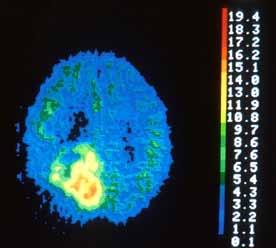
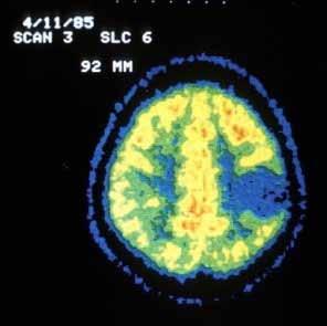
17 PET Scan of the Brain Using radionuclide (FDG Fluodeoxyglucose 18 F) that is like glucose, the PET scan will show how the tissues in the brain are functioning Areas of less function use less energy, and areas with increased metabolic activity use more energy

18 Radiopharmaceuticals Radionuclide 11 C half-life ~20 min 13 N 10 min 15 O 2 min 18 F 110 min The half-life of 18F is long enough that radiotracers can be manufactured commercially at offsite locations and shipped to imaging centers. Localization Biochemical metabolism within the cell Adult Dose Range 5-15 mci ( MBq) + glucose, water, ammonia or other molecule = radiotracers


19 Indications in the CNS Evaluation of cerebrovascular disease Strokes Transient ischemic attacks (TIAs) Evaluation of epilepsy Epilepsy Evaluation of movement disorders Huntington Parkinson Stroke Evaluation of psychiatric disorders Schizophrenia Mood disorders

, bleeding, and/or perfusion (blood and oxygen flow) of the brain")
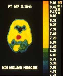
20 Indications in the CNS Evaluation of dementia Alzheimer s disease Evaluation of tumors Evaluation of grade and extend of glioma Evaluation of chemotherapy Alzheimer s disease Tumor To locate the specific surgical site prior to surgical procedures of the brain To evaluate the brain after trauma to detect hematoma (blood clot), bleeding, and/or perfusion (blood and oxygen flow) of the brain tissue
21 Before the PET Examination The patient must fast six hours prior to the appointment time except water! In addition, patient should avoid any carbohydrates from his/her diet (e.g., bread, pasta, potatoes and rice) 24 hours prior to the appointment time, because carbohydrates taken before the test will reduce the effectiveness of it!
22 Brain 3D
23 Functional neuroanatomy of subjective feelings using 3D-PET One of the most exciting methodological advances for brain research has been in functional brain imaging; it enables the localization and characterization of neural activity in the living human brain Recently developed 3D-PET imaging techniques using H 2 15 O and 18 F-FDG make it possible to visualize the brain activity associated with cognitive processes We will visualize functional neuroanatomy of subjective feelings of sleepiness, visceral perception, itching, and emotion using H 2 15 O

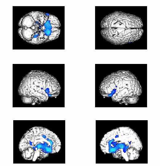
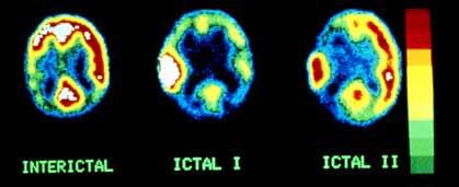
24 3D PET in Diagnostics of Epilepsy During a seizure, the area responsible for the seizure will show up as an area of increased glucose use Between the seizures, PET shows a characteristic pattern of reduced need for glucose
25 Epilepsy

26 Alzheimer s Disease PET scan of a healthy brain compared to a brain at an early stage of Alzheimer's disease Diminished FDG uptake in the temporal lobes which are compatible with Alzheimer s disease
 and a patient with mild Parkinson s disease (right) demonstrating a dramatic reduction in dopaminergic innervation to the")
27 Parkinson s disease Normal Parkinson s disease 18 F-AV-133 images of vesicular monoamine transporters acquired minutes post injection in a normal elderly subject (left) and a patient with mild Parkinson s disease (right) demonstrating a dramatic reduction in dopaminergic innervation to the striatum

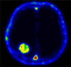
28 Brain Tumor
29 Contraindications Patient too agitated, uncooperative, or claustrophobic to remain still for acquisition Because PET involves cellular metabolism, drugs and food ingestion, or lack thereof, bears specific consideration for each study
30 SPECT vs. PET PET uses beta-plus-emitting radionuclides such as 11 C, 13 N, 15 O, and 18 F which annihilate into two 511-keV photons that travel in opposite directions SPECT involves detection of gamma rays emitted singly from radionuclides such as 99m Tc, 123 I, and 111 In
Molecular Imaging and the Brain
 Molecular imaging technologies are playing an important role in neuroimaging, a branch of medical imaging, by providing a window into the living brain. Where CT and conventional MR imaging provide important
Radionuclides in Medical Imaging. Danielle Wilson
 Radionuclides in Medical Imaging Danielle Wilson Outline Definitions History and development Radionuclide applications & techniques in imaging Conclusion Definition #1 : Radionuclide An unstable nucleus
Basics of nuclear medicine
 Basics of nuclear medicine Prof. dr. Davor Eterović Prof. dr. Vinko Marković Radioisotopes are used both in diagnostics and in therapy Diagnostics gamma emitters are used since gamma rays can penetrate
Medical Use of Radioisotopes
 Medical Use of Radioisotopes Therapy Radioisotopes prove to be useful in the application of brachytherapy, the procedure for using temporary irradiation close to the area of disease (i.e. cancer) 10% Medical
Medical imaging X-ray, CT, MRI, scintigraphy, SPECT, PET Györgyi Műzes
 Medical imaging X-ray, CT, MRI, scintigraphy, SPECT, PET Györgyi Műzes Semmelweis University, 2nd Dept. of Medicine Medical imaging: definition technical process of creating visual representations about
MRI and CT of the CNS
 MRI and CT of the CNS Dr.Maha ELBeltagy Assistant Professor of Anatomy Faculty of Medicine The University of Jordan 2018 Computed Tomography CT is used for the detection of intracranial lesions. CT relies
A Snapshot on Nuclear Cardiac Imaging
 Editorial A Snapshot on Nuclear Cardiac Imaging Khalil, M. Department of Physics, Faculty of Science, Helwan University. There is no doubt that nuclear medicine scanning devices are essential tool in the
PHYSICS 2: HSC COURSE 2 nd edition (Andriessen et al) CHAPTER 20 Radioactivity as a diagnostic tool (pages 394-5)
 CHAPTER 20 Radioactivity as a diagnostic tool (pages 394-5)") PHYSICS 2: HSC COURSE 2 nd edition (Andriessen et al) CHAPTER 20 Radioactivity as a diagnostic tool (pages 394-5) 1. (a) A radioisotope is an isotope that is unstable and will emit particles from the nucleus
PHYSICS 2: HSC COURSE 2 nd edition (Andriessen et al) CHAPTER 20 Radioactivity as a diagnostic tool (pages 394-5) 1. (a) A radioisotope is an isotope that is unstable and will emit particles from the nucleus
SPECT IMAGING AND MAIN MEDICAL APPLICATIONS
 SPECT IMAGING AND MAIN MEDICAL APPLICATIONS Teresa Alonso Ubago Raúl Gijón Villanova María Ramos Ontiveros Medical image and instrumentation. UGR Course 2015-2016 Index 1.Introduction: History 2.What is
Nuclear Medicine and PET. D. J. McMahon rev cewood
 Nuclear Medicine and PET D. J. McMahon 150504 rev cewood 2018-02-15 Key Points Nuclear Medicine and PET: Imaging: Understand how Nuc Med & PET differ from Radiography & CT by the source of radiation. Be
Non-Invasive Techniques
 Non-Invasive Techniques Key: Does not hurt the organism Psychology 372 Physiological Psychology Steven E. Meier, Ph.D. Listen to the audio lecture while viewing these slides or view the video presentation
Non-Invasive Techniques
 Many Procedures Non-Invasive Techniques Key: Does not hurt the organism Psychology 372 Physiological Psychology Steven E. Meier, Ph.D. Listen to the audio lecture while viewing these slides or view the
45 Hr PET Registry Review Course
 45 HR PET/CT REGISTRY REVIEW COURSE Course Control Document Timothy K. Marshel, MBA, R.T. (R), (N)(CT)(MR)(NCT)(PET)(CNMT) The PET/CT Training Institute, Inc. SNMMI-TS 028600-028632 45hr CEH s Voice Credits
Nuclear neurology. Zámbó Katalin Department of Nuclear Medicine
 Nuclear neurology Zámbó Katalin Department of Nuclear Medicine To refresh your memory Brain has a high rate of oxidative metabolism. It has no reserves either of oxygen or of glucose and has a very limited
Option D: Medicinal Chemistry
 Option D: Medicinal Chemistry Basics - unstable radioactive nuclei emit radiation in the form of smaller particles alpha, beta, positron, proton, neutron, & gamma are all used in nuclear medicine unstable
Positron Emission Tomography Computed Tomography (PET/CT)
") Positron Emission Tomography Computed Tomography (PET/CT) What is Positron Emission Tomography Computed Tomography (PET/CT) Scanning? What are some common uses of the procedure? How should I prepare for
Positron Emission Tomography Computed Tomography (PET/CT) What is Positron Emission Tomography Computed Tomography (PET/CT) Scanning? What are some common uses of the procedure? How should I prepare for
COMENIUS-Project: SM&CLIL Radiation & Medicine
 Medical imaging refers to the techniques and processes used to create images of the human body (or parts thereof) for clinical purposes. Thanks to modern mathematics and computer technology, medical imaging
COGNITIVE SCIENCE 17. Peeking Inside The Head. Part 1. Jaime A. Pineda, Ph.D.
 COGNITIVE SCIENCE 17 Peeking Inside The Head Part 1 Jaime A. Pineda, Ph.D. Imaging The Living Brain! Computed Tomography (CT)! Magnetic Resonance Imaging (MRI)! Positron Emission Tomography (PET)! Functional
USAAA 2007 International Conference. Autism- you'll never see it the same way again
 J. Michael Uszler, MD USAAA 2007 International Conference www.drspectscan.com Autism- you'll never see it the same way again Do you have a concern that your brain is not working right? Are you having trouble
General Nuclear Medicine
 General Nuclear Medicine What is General Nuclear Medicine? What are some common uses of the procedure? How should I prepare? What does the equipment look like? How does the procedure work? How is the procedure
Yin-Hui Siow MD, FRCPC Director of Nuclear Medicine Southlake Regional Health Centre
 Yin-Hui Siow MD, FRCPC Director of Nuclear Medicine Southlake Regional Health Centre Today Introduction to CT Introduction to MRI Introduction to nuclear medicine Imaging the dementias The Brain ~ 1.5
María del Pilar Garrido Ruiz Teresa Mendoza Dobaño Cristian Jesús Lucena Morales
 María del Pilar Garrido Ruiz Teresa Mendoza Dobaño Cristian Jesús Lucena Morales Index 1. Introduction. 2. Physical principles: annihilation reaction. 3. PET image creation. 4. Advantages of PET use. 5.
Laura Tormoehlen, M.D. Neurology and EM-Toxicology Indiana University
 Laura Tormoehlen, M.D. Neurology and EM-Toxicology Indiana University Disclosures! No conflicts of interest to disclose Neuroimaging 101! Plain films! Computed tomography " Angiography " Perfusion! Magnetic
Fundamentals of Nuclear Cardiology. Terrence Ruddy, MD, FRCPC, FACC
 Fundamentals of Nuclear Cardiology Terrence Ruddy, MD, FRCPC, FACC Objectives To understand the Principles of Nuclear Cardiac Imaging Radiotracers Image acquisition and processing Stress protocols To appreciate
OTHER NON-CARDIAC USES OF Tc-99m CARDIAC AGENTS Tc-99m Sestamibi for parathyroid imaging, breast tumor imaging, and imaging of other malignant tumors.
 DEFINITION OF CARDIAC RADIOPHARMACEUTICAL: A radioactive drug which, when administered for purpose of diagnosis of heart disease, typically elicits no physiological response from the patient. Even though
KEYWORDS: nuclear medicine; gamma camera; radiopharmaceutical activities.
 Radiopharmaceutical Activities Administered for Diagnostic Procedures in Nuclear Medicine in the First Six Months of the Gamma Camera Use in the Clinical Center of Montenegro - Podgorica Nevenka Antovic
Medical imaging. Medical imaging uses electromagnetic radiation, sound or ingestion of radioactive substances. 10/6/2011 Medical imaging 1
 Medical imaging Medical imaging uses electromagnetic radiation, sound or ingestion of radioactive substances 10/6/2011 Medical imaging 1 0 Ultrasound Imaging Transducer Reflector Use high-frequency sound
Physics of MBI (~10 slides)
") Physics of MBI (~10 slides) Molecular Breast Imaging (MBI) physics and performance testing JW Hugg, BR Simrak, PD Smith, BE Patt Gamma Medica, Inc., Northridge, CA Molecular Breast Imaging (MBI) is an
Physics of MBI (~10 slides) Molecular Breast Imaging (MBI) physics and performance testing JW Hugg, BR Simrak, PD Smith, BE Patt Gamma Medica, Inc., Northridge, CA Molecular Breast Imaging (MBI) is an
ADVANCES IN RADIATION TECHNOLOGIES IN THE TREATMENT OF CANCER
 ADVANCES IN RADIATION TECHNOLOGIES IN THE TREATMENT OF CANCER Bro. Dr. Collie Miller IARC/WHO Based on trends in the incidence of cancer, the International Agency for Research on Cancer (IARC) and WHO
Ingenuity TF PET/CT. Comfort and quality care in one. Your PET/CT scan with Philips Ingenuity TF PET/CT
 Ingenuity TF PET/CT Comfort and quality care in one Your PET/CT scan with Philips Ingenuity TF PET/CT What s a PET/CT scan, an A PET/CT scan provides your doctor and medical specialists with valuable information
Brain Perfusion SPECT
 APPROVED BY: Director of Radiology Page 1 of 5 Brain Perfusion SPECT Primary Indications: Brain perfusion SPECT is most commonly performed (1) to aid in identification of the epileptogenic focus in patients
Introduction, use of imaging and current guidelines. John O Brien Professor of Old Age Psychiatry University of Cambridge
 Introduction, use of imaging and current guidelines John O Brien Professor of Old Age Psychiatry University of Cambridge Why do we undertake brain imaging in AD and other dementias? Exclude other causes
Molecular Imaging and Cancer
 Molecular Imaging and Cancer Cancer causes one in every four deaths in the United States, second only to heart disease. According to the U.S. Department of Health and Human Services, more than 512,000
Outline. Biological Psychology: Research Methods. Dr. Katherine Mickley Steinmetz
 Biological Psychology: Research Methods Dr. Katherine Mickley Steinmetz Outline Neuroscience Methods Histology Electrophysiological Recordings Lesion Neuroimaging Neuroanatomy Histology: Brain structure
Molecular Imaging and Breast Cancer
 Molecular Imaging and Breast Cancer Breast cancer forms in tissues of the breast usually in the ducts, tubes that carry milk to the nipple, and lobules, the glands that make milk. It occurs in both men
Radiation Detection and Measurement
 Radiation Detection and Measurement Range of charged particles (e.g.,!: µm; ": mm) Range of high energy photons (cm) Two main types of interactions of high energy photons Compton scatter Photoelectric
Physical Bases : Which Isotopes?
 Physical Bases : Which Isotopes? S. Gnesin Institute of Radiation Physics, Lausanne University Hospital, Lausanne, Switzerland 1/53 Theranostic Bruxelles, 2 Octobrer 2017 Theranostic : use of diagnostic
Ways to Study Brain Structures and Functioning. Can physically trace connections. Ablation. Is the most primitive Can be done with any structures
 Ways to Study Brain Structures and Functioning Can physically trace connections Is the most primitive Can be done with any structures Ablation Can remove a piece of the brain and see what happens If the
Nuclear Medicine: Manuals. Nuclear Medicine. Nuclear imaging. Emission imaging: study types. Bone scintigraphy - technique
 Nuclear Medicine - Unsealed radioactive preparations the tracer mixes with the patients body fluids on a molecular level (e.g. after intravenous injection) - 3 main fields: - In vitro : measuring concentrations
Exam 1. Mean 78.0% Median 80% Mode 86% Min 26% Max 98% Std Dev 12.6%
 Exam 1 Mean 78.0% Median 80% Mode 86% Min 26% Max 98% Std Dev 12.6% None at the moment! Announcements VII. Imaging techniques of the brain A. CT: anatomical B. MRI: anatomical C. fmri: functional D. SPECT
Positron Emission Tomography: Tool to Facilitate Drug Development and to Study Pharmacokinetics
 Positron Emission Tomography: Tool to Facilitate Drug Development and to Study Pharmacokinetics Robert B. Innis, MD, PhD Molecular Imaging Branch National Institute Mental Health 1 Outline of Talk 1. PET
Methods of Visualizing the Living Human Brain
 Methods of Visualizing the Living Human Brain! Contrast X-rays! Computerized Tomography (CT)! Magnetic Resonance Imaging (MRI)! Positron Emission Tomography (PET)! Functional MRI! Magnetoencephalography
Nuclear Medicine in the Diabetic Foot
 26.11.2015, Uniklinik Balgrist Nuclear Medicine in the Diabetic Foot Martin Hüllner Nuklearmedizin und Neuroradiologie, USZ / UZH Outline A. Imaging modalities brief technical overview B. Nuclear medicine
The Physics of Medical Imaging
 VEA Bringing Learning to Life Program Support Notes Senior Secondary The Physics of Medical Imaging 27mins Program Support Notes by Ian Walter, Dip.App.Chem.; G.Dip.Ed.Admin.; TTTC Produced by VEA Pty
Positron Emission Tomography: Tool to Facilitate Drug Development and to Study Pharmacokinetics
 Positron Emission Tomography: Tool to Facilitate Drug Development and to Study Pharmacokinetics Robert B. Innis, MD, PhD Molecular Imaging Branch National Institute Mental Health 1 Outline of Talk 1. PET
Gastrointestinal tract
 Gastrointestinal tract Colloidal liver-spleen imaging Presented by: Jehad Felemban Introduction: To obtain better anatomic display of liver and spleen architecture, we use (CT Ultrasound). (Radionuclide
Austin Radiological Association BRAIN AMYLOID STUDY (F-18-Florbetapir)
") Austin Radiological Association BRAIN AMYLOID STUDY (F-18-Florbetapir) Overview The Brain Amyloid Study with F-18-florbetapir depicts the extracellular deposition of B- amyloid (Aβ) peptides (or plaques
Austin Radiological Association BRAIN AMYLOID STUDY (F-18-Florbetapir) Overview The Brain Amyloid Study with F-18-florbetapir depicts the extracellular deposition of B- amyloid (Aβ) peptides (or plaques
Announcements. Exam 1. VII. Imaging techniques of the brain. Anatomical/Structural Scans. Structural Scans: CT. Structural Scans: CT 2/17/2014
 Exam 1 None at the moment! Announcements Mean 78.0% Median 80% Mode 86% Min 26% Max 98% Std Dev 12.6% VII. Imaging techniques of the brain A. CT: anatomical B. MRI: anatomical C. fmri: functional D. SPECT
Itroduction to the Nuclear Medicine: biophysics and basic principles. Zámbó Katalin Department of Nuclear Medicine
 Itroduction to the Nuclear Medicine: biophysics and basic principles Zámbó Katalin Department of Nuclear Medicine NUCLEAR MEDICINE Application of the radioactive isotopes in the diagnostics and in the
Children's (Pediatric) Nuclear Medicine
 Nuclear Medicine") Scan for mobile link. Children's (Pediatric) Nuclear Medicine Children s (pediatric) nuclear medicine imaging uses small amounts of radioactive materials called radiotracers, a special camera and a computer
Scan for mobile link. Children's (Pediatric) Nuclear Medicine Children s (pediatric) nuclear medicine imaging uses small amounts of radioactive materials called radiotracers, a special camera and a computer
Typical PET Image. Elevated uptake of FDG (related to metabolism) Lung cancer example: But where exactly is it located?
 Lung cancer example: But where exactly is it located?") Typical PET Image Elevated uptake of FDG (related to metabolism) Lung cancer example: But where exactly is it located? PET/CT Oncology Imaging Anatometabolic fusion images are useful in the management
Typical PET Image Elevated uptake of FDG (related to metabolism) Lung cancer example: But where exactly is it located? PET/CT Oncology Imaging Anatometabolic fusion images are useful in the management
Austin Radiological Association Nuclear Medicine Procedure WHITE BLOOD CELL MIGRATION STUDY (In-111-WBCs, Tc-99m-HMPAO-WBCs)
") Austin Radiological Association Nuclear Medicine Procedure WHITE BLOOD CELL MIGRATION STUDY (In-111-WBCs, Tc-99m-HMPAO-WBCs) Overview Indications The White Blood Cell Migration Study demonstrates the distribution
Austin Radiological Association Nuclear Medicine Procedure WHITE BLOOD CELL MIGRATION STUDY (In-111-WBCs, Tc-99m-HMPAO-WBCs) Overview Indications The White Blood Cell Migration Study demonstrates the distribution
Atoms for Health. Atoms for Health The. Atoms for Health - Division of Nuclear Health - Dept of Nuclear Aplications P Andreo DIR-NAHU 1
 International Atomic Energy Agency Atoms for Health The human side of nuclear applications NUCLEAR APPLICATIONS IN HEALTH A UNIQUE MANDATE OF THE UN SYSTEM The Agency shall seek to accelerate and enlarge
Appendix A: Introduction to Imaging Modalities for Which Data Were Collected in the 2017 Imaging Inventory
 Appendix A: Introduction to Imaging Modalities for Which Data Were Collected in the 207 Imaging Inventory Computed Tomography Computed tomography (CT) employs X-rays as a source of ionizing radiation,
STRUCTURAL ORGANIZATION OF THE NERVOUS SYSTEM
 STRUCTURAL ORGANIZATION OF THE NERVOUS SYSTEM STRUCTURAL ORGANIZATION OF THE BRAIN The central nervous system (CNS), consisting of the brain and spinal cord, receives input from sensory neurons and directs
Radionuclide & Radiopharmaceuticals
 Radionuclide & Radiopharmaceuticals 1. Generator & Reactors 2. Cyclotrons & PET tracer 3. Quality control 4. Renal 5. GIT 6. CNS & Psychiatrics 7. Tumor Diagnosis & Treatment 8. Bones & joints 9. Thyroid
The Biological Level of Analysis: Studying the Brain
 The Biological Level of Analysis: Studying the Brain In the past the study of the brain was limited to people suffering from head injuries and the effects of accidental damage. It was only possible to
Nuclear Medicine - Hepatobiliary
 Scan for mobile link. Nuclear Medicine - Hepatobiliary Hepatobiliary nuclear medicine imaging helps evaluate the parts of the biliary system, including the liver, gallbladder and bile ducts, using small
2. RADIOPHARMACEUTICALS UTILIZED
 1. OVERVIEW AND INDICATIONS (adapted from Society of Nuclear Medicine Procedure Guideline for Thyroid Uptake Measurement, Version 3.0; reprinted from http://snmmi.files.cmsplus.com/docs/thyroid%20uptake%20measure%20v3%200.pdf,
Introduction to the Course and the Techniques. Jeffry R. Alger, PhD Ahmanson-Lovelace Brain Mapping Center Department of Neurology
 Introduction to the Course and the Techniques Jeffry R. Alger, PhD Ahmanson-Lovelace Brain Mapping Center Department of Neurology (jralger@ucla.edu) CTSI Neuroimaging April 2014 Rationale for the Course
What Is Nuclear Medicine?
 What Is Nuclear Medicine? An Introductory Guide For Patients And Their Families What is nuclear medicine? Nuclear medicine is a type of medical imaging that uses small amounts of radioactive material (called
Austin Radiological Association Nuclear Medicine Procedure THYROID UPTAKE MEASUREMENT (I-123 or I-131 as Sodium Iodide)
") Austin Radiological Association Nuclear Medicine Procedure THYROID UPTAKE MEASUREMENT (I-123 or I-131 as Sodium Iodide) Overview Indications The Thyroid Uptake Measurement measures the metabolic activity
Austin Radiological Association Nuclear Medicine Procedure THYROID UPTAKE MEASUREMENT (I-123 or I-131 as Sodium Iodide) Overview Indications The Thyroid Uptake Measurement measures the metabolic activity
III. Studying The Brain and Other Structures
 III. Studying The Brain and Other Structures 1. Accidents (case study) In 1848, a railroad worker named Phineas Gage was involved in an accident that damaged the front part of his brain. Gage s doctor
Department of Nuclear Medicine with Positron Emission Tomography
 (PET) Unit [1] Contact information: Registration: +48 41 367 4850 Main office: +48 41 367 4860 Fax: +48 41 367 4887 e-mail: zmnsco@onkol.kielce.pl [2] Head of the Department: Professor Janusz Braziewicz
Cardiac Nuclear Medicine
 Cardiac Nuclear Medicine What is Cardiac Nuclear Medicine? What are some common uses of the procedure? How should I prepare? What does the equipment look like? How does the procedure work? How is the procedure
Unit 3: The Biological Bases of Behaviour
 Unit 3: The Biological Bases of Behaviour Section 1: Communication in the Nervous System Section 2: Organization in the Nervous System Section 3: Researching the Brain Section 4: The Brain Section 5: Cerebral
Positron Emission Tomography - Computed Tomography (PET/CT)
") Scan for mobile link. Positron Emission Tomography - Computed Tomography (PET/CT) Positron emission tomography (PET) uses small amounts of radioactive materials called radiotracers, a special camera and
Scan for mobile link. Positron Emission Tomography - Computed Tomography (PET/CT) Positron emission tomography (PET) uses small amounts of radioactive materials called radiotracers, a special camera and
Neuroimaging for dementia diagnosis. Guidance from the London Dementia Clinical Network
 Neuroimaging for dementia diagnosis Guidance from the London Dementia Clinical Network Authors Dr Stephen Orleans-Foli Consultant Psychiatrist, West London Mental Health NHS Trust Dr Jeremy Isaacs Consultant
Cardiac Imaging. Kimberly Delcour, DO, FACC. Mahi Ashwath, MD, FACC, FASE. Director, Cardiac CT. Director, Cardiac MRI
 Cardiac Imaging Kimberly Delcour, DO, FACC Director, Cardiac CT Mahi Ashwath, MD, FACC, FASE Director, Cardiac MRI Cardiac Imaging Discuss the clinical applications of and indications for: Cardiac CT Nuclear
Austin Radiological Association Nuclear Medicine Procedure PET SODIUM FLUORIDE BONE SCAN (F-18 NaF)
") Austin Radiological Association Nuclear Medicine Procedure PET SODIUM FLUORIDE BONE SCAN (F-18 NaF) Overview Indication Sodium Fluoride F18 injection is a radioactive diagnostic agent for positron emission
Austin Radiological Association Nuclear Medicine Procedure PET SODIUM FLUORIDE BONE SCAN (F-18 NaF) Overview Indication Sodium Fluoride F18 injection is a radioactive diagnostic agent for positron emission
Applicable Neuroradiology
 For the Clinical Neurology Clerkship LSU Medical School New Orleans Amy W Voigt, MD Clerkship Director Introduction The field of Radiology first developed following the discovery of X-Rays by Wilhelm Roentgen
AN INTRODUCTION TO NUCLEAR MEDICINE
 AN INTRODUCTION TO NUCLEAR MEDICINE WITH RESPECT TO THYROID DISORDERS By: B.Shafiei MD Nuclear Physician Taleghani Medical Center Radioactive: An element with Unstable Nucleus (Excess Energy), can achieve
SCINTIGRAPHY OF THE CENTRAL NERVOUS SYSTEM Part 1: Introduction and BBB studies
 SCINTIGRAPHY OF THE CENTRAL NERVOUS SYSTEM Part 1: Introduction and BBB studies George N. Sfakianakis MD Professor of Radiology and Pediatrics Director, Division of Nuclear Medicine October 2009 FIRST
Cardiac Imaging Tests
 Cardiac Imaging Tests http://www.medpagetoday.com/upload/2010/11/15/23347.jpg Standard imaging tests include echocardiography, chest x-ray, CT, MRI, and various radionuclide techniques. Standard CT and
Arteriogram An X-ray of an artery after the injection of dye.
 A Abscess A localized collection of pus in any part of the body, usually surrounded by inflamed tissue. Anesthetic An agent that causes loss of sensation with or without the loss of consciousness. Angiography,
PET IMAGING (POSITRON EMISSION TOMOGRAPY) FACT SHEET
 FACT SHEET") Positron Emission Tomography (PET) When calling Anthem (1-800-533-1120) or using the Point of Care authorization system for a Health Service Review, the following clinical information may be needed to
Positron Emission Tomography (PET) When calling Anthem (1-800-533-1120) or using the Point of Care authorization system for a Health Service Review, the following clinical information may be needed to
General Nuclear Medicine
 Scan for mobile link. General Nuclear Medicine Nuclear medicine imaging uses small amounts of radioactive materials called radiotracers that are typically injected into the bloodstream, inhaled or swallowed.
An investigation of the effect of ionising radiation on nurses and their patients during dialysis
 International Scholars Journals African Journal of Nursing and Midwifery ISSN 2198-4638 Vol. 2 (7), pp. 548-552, September, 2015. Available online at www.internationalscholarsjournals.org International
Fellows on this rotation are expected to attend nuclear conferences and multimodality imaging conference.
 Rotation: Imaging 1 Imaging 1 provides COCATS Level 1 experience for nuclear cardiology (including SPECT and PET) and cardiac CT. Fellows will administer, process, and read cardiac nuclear studies with
Cardiac PET. John Buscombe
 Cardiac PET John Buscombe Why PET? Improved resolution-not really required in cardiology Improved sensitivity this may be important-financially as reduced acquisition time Improved attenuation correction-good
Facility IBA 18/9 Cyclotron Accelerates p and d. Facility GMP Grade Clean Room Automated Dispenser Synthera Rig FDG 07/11/2014
 Dr Chris Marshall Director Introduction to PET Positron Emission Tomography Positron is anti matter equivalent of the electron Isotopes that are proton rich can decay by emission of a positron Positron
The Effect of Administered Activity on Patient Radiation dose and Image Quality in SPECT at Korle-Bu Teaching Hospital
 2017 IJSRST Volume 3 Issue 1 Print ISSN: 2395-6011 Online ISSN: 2395-602X Themed Section: Science and Technology The Effect of Administered Activity on Patient Radiation dose and Image Quality in SPECT
Myocardial viability testing. What we knew and what is new
 Myocardial viability testing. What we knew and what is new Dr B K S Sastry, MD, DM. CARE Hospitals, Hyderabad What is Viability Viability Dysfunctional myocardium subtended by diseased coronary arteries
Nuclear Medicine Head and Neck Region. Bán Zsuzsanna, MD University of Pécs, Department of Nuclear Medicine
 Nuclear Medicine Head and Neck Region Bán Zsuzsanna, MD University of Pécs, Department of Nuclear Medicine Thyroid scintigraphy Parathyroid scintigraphy F18-FDG PET examinations in head and neck cancer
Austin Radiological Association Nuclear Medicine Procedure PROSTATE CANCER STUDY (In-111-Capromab Pendetide [ProstaScint ])
![Austin Radiological Association Nuclear Medicine Procedure PROSTATE CANCER STUDY (In-111-Capromab Pendetide [ProstaScint ])](/thumbs/81/82771892.jpg "Austin Radiological Association Nuclear Medicine Procedure PROSTATE CANCER STUDY (In-111-Capromab Pendetide [ProstaScint ])") Austin Radiological Association Nuclear Medicine Procedure PROSTATE CANCER STUDY (In-111-Capromab Pendetide [ProstaScint ]) Overview Indications The Prostate Cancer Study with an indium-111 labeled murine
Austin Radiological Association Nuclear Medicine Procedure PROSTATE CANCER STUDY (In-111-Capromab Pendetide [ProstaScint ]) Overview Indications The Prostate Cancer Study with an indium-111 labeled murine
I. Cancer staging problem
 Instrumentation Lab (Craig Levin) Angela Craig Peter Jin Frezghi Guillem Billie Garry Research interests (by imaging modality) I. High resolution radionuclide imaging : positron emission tomography (PET)
Radiation Exposure to Staff Using PET/CT Facility
 Egyptian J. Nucl. Med., Vol. 8, No. 2, December 2013 1 Editorial Radiation Exposure to Staff Using PET/CT Facility Taalab, Kh; and Mohsen, Z Department of Nuclear Medicine, International Medical Center;
Radiopharmacy. Prof. Dr. Çetin ÖNSEL. CTF Nükleer Tıp Anabilim Dalı
 Prof. Dr. Çetin ÖNSEL CTF Nükleer Tıp Anabilim Dalı What is Nuclear Medicine? Nuclear Medicine is the branch of medicine concerned with the use of radionuclides in the study and the diagnosis of diseases.
The Nervous System. Biological School. Neuroanatomy. How does a Neuron fire? Acetylcholine (ACH) TYPES OF NEUROTRANSMITTERS
 TYPES OF NEUROTRANSMITTERS") Biological School The Nervous System It is all about the body!!!! It starts with an individual nerve cell called a NEURON. Synapse Neuroanatomy Neurotransmitters (chemicals held in terminal buttons that
Biological School The Nervous System It is all about the body!!!! It starts with an individual nerve cell called a NEURON. Synapse Neuroanatomy Neurotransmitters (chemicals held in terminal buttons that
Study of the utility of PET image in refractory epilepsy
 Author:. Facultat de Física, Universitat de Barcelona, Diagonal 645, 08028 Barcelona, Spain. Advisor: Domènec Ros. Abstract: The surgical removal of the epileptogenic region is considered as a possible
Nuclear pulmonology. Katalin Zámbó Department of Nuclear Medicine
 Nuclear pulmonology Katalin Zámbó Department of Nuclear Medicine Imaging techniques Morphology Physiology Metabolism Molecules X-ray / CT MRI NM - SPECT/ PET MR spectroscopy fmri Ultrasound Hybrid imaging:
biological psychology, p. 40 The study of the nervous system, especially the brain. neuroscience, p. 40
 biological psychology, p. 40 The specialized branch of psychology that studies the relationship between behavior and bodily processes and system; also called biopsychology or psychobiology. neuroscience,
Description of the consecutive stages (which took place from December 2002 to July 2003)
") Radiation Protection Issues in a PET/CT Installation M. Coronado 1, R. Plaza 2, R. Couto 1, MD. Marin 1, C. Huerga 2, J. Coya 1, LM Martin Curto 1, M. Téllez de Cepeda 2 1 Servicio de Medicina Nuclear,
Radiation Protection Issues in a PET/CT Installation M. Coronado 1, R. Plaza 2, R. Couto 1, MD. Marin 1, C. Huerga 2, J. Coya 1, LM Martin Curto 1, M. Téllez de Cepeda 2 1 Servicio de Medicina Nuclear,
Brain and Cognition. Cognitive Neuroscience. If the brain were simple enough to understand, we would be too stupid to understand it
 Brain and Cognition Cognitive Neuroscience If the brain were simple enough to understand, we would be too stupid to understand it 1 The Chemical Synapse 2 Chemical Neurotransmission At rest, the synapse
Round table: Moderator; Fereshteh Sedaghat, MD, PhD Brain Mapping in Dementias and Non-invasive Neurostimulation
 Round table: Moderator; Fereshteh Sedaghat, MD, PhD Brain Mapping in Dementias and Non-invasive Neurostimulation 1. Reflection of Mild Cognitive Impairment (MCI) and Dementias by Molecular Imaging, PET
Acetylcholine (ACh) Action potential. Agonists. Drugs that enhance the actions of neurotransmitters.
 Action potential. Agonists. Drugs that enhance the actions of neurotransmitters.") Acetylcholine (ACh) The neurotransmitter responsible for motor control at the junction between nerves and muscles; also involved in mental processes such as learning, memory, sleeping, and dreaming. (See
Acetylcholine (ACh) The neurotransmitter responsible for motor control at the junction between nerves and muscles; also involved in mental processes such as learning, memory, sleeping, and dreaming. (See
LESSON 1.3 WORKBOOK. How can we study the behaving brain?
 LESSON 1.3 WORKBOOK How can we study the behaving brain? We are in the middle of a technological revolution when it comes to how closely we can look at the behaving brain. Scientists and doctors now have
Austin Radiological Association Ga-68 NETSPOT (Ga-68 dotatate)
") Austin Radiological Association Ga-68 NETSPOT (Ga-68 dotatate) Overview Ga-68 dotatate binds to somatostatin receptors, with highest affinity for subtype 2 receptors (sstr2). It binds to cells that express
Austin Radiological Association Ga-68 NETSPOT (Ga-68 dotatate) Overview Ga-68 dotatate binds to somatostatin receptors, with highest affinity for subtype 2 receptors (sstr2). It binds to cells that express
Calculation methods in Hermes Medical Solutions dosimetry software
 Calculation methods in Hermes Medical Solutions dosimetry software Helena McMeekin MSc. Clinical Applications Scientist, Hermes Medical Solutions MRTDosimetry Scientific Workshop The Principals and Clinical
Medical Physics 4 I3 Radiation in Medicine
 Name: Date: 1. This question is about radiation dosimetry. Medical Physics 4 I3 Radiation in Medicine Define exposure. A patient is injected with a gamma ray emitter. The radiation from the source creates