controls. <Conclusions> These data support the hypothesis that JME and FLE involve neuronal dysfunction within the temporal lobe as well as the
|
|
|
- Homer Cannon
- 5 years ago
- Views:
Transcription
1 A single-voxel spectroscopy study of hippocampal metabolic dysfunction in patients with juvenile myoclonic epilepsy, frontal lobe epilepsy, and psychogenic nonepileptic seizures Epilepsy Center, National Hospital Organization, Nara Medical Center Tohru HOSHIDA, Nobuyuki Maruyama, Yasuko Sawai, Kazuhiro Kawata, Hidehiro Hirabayashi <Purpose> Proton magnetic resonance spectroscopy (MRS) studies have shown neuronal dysfunction with differing patterns of abnormality in various types of epilepsy pathogenesis. Our aim was to identify metabolic differences in the hippocampi of patients with juvenile myoclonic epilepsy (JME), frontal lobe epilepsy (FLE), and psychogenic nonepileptic seizure (PNES) compared to normal healthy subjects by using single-voxel MRS. <Methods> The study included 18 patients with JME, 38 with FLE, and 15 with PNES who had no true epileptic seizures. The control group consisted of 24 age-matched healthy volunteers (mean age: JME, 22.3; FLE, 23.7; PNES, 25.0; controls, 25.8). All patients and controls underwent normal neurological examinations and magnetic resonance imaging. Quantitative single-voxel MRS was conducted at 1.5 Tesla with a sequence of TR/TE = 1,323/136 ms with a voxel size of mm in both hippocampi. LC-Model was used to estimate the absolute concentrations of N-acetyl-aspartate (NAA), choline (Cho), creatine (Cr), and the ratio of NAA to Cho + Cr (NAA ratio). <Results> Significant reductions in NAA and the NAA ratio were observed in the left hippocampus in the JME group compared to controls (NAA: 8.22 vs. 8.89, p < 0.05; NAA ratio: 0.92 vs. 1.03, p < 0.01). Furthermore, significant reductions in NAA were found in both hippocampi in the FLE group compared to controls (right: 7.79 vs. 8.28, p < 0.05; left: 8.14 vs. 8.89, p < 0.01). The bilateral hippocampal NAA ratios were not reduced significantly in the FLE patients. In PNES patients, NAA and the NAA ratio in both hippocampi were not significantly lower than in the
2 controls. <Conclusions> These data support the hypothesis that JME and FLE involve neuronal dysfunction within the temporal lobe as well as the frontal lobe. However, neuronal dysfunction in PNES might demonstrate normal hippocampal metabolism and differ from epileptic pathogenesis.


3 A single-voxel spectroscopy study of hippocampal metabolic dysfunction in patients with juvenile myoclonic epilepsy, frontal lobe epilepsy, and psychogenic nonepileptic seizures Epilepsy Center, National Hospital Organization, Nara Medical Center Tohru HOSHIDA, Nobuyuki Maruyama, Kazuhiro Kawata, Hidehiro Hirabayashi
4 <Purpose> Proton magnetic resonance spectroscopy (MRS) studies have shown neuronal dysfunction with differing patterns of abnormality in various types of epilepsy pathogenesis. Our aim was to identify metabolic differences in the hippocampi of patients with juvenile myoclonic epilepsy (JME), frontal lobe epilepsy (FLE), and psychogenic nonepileptic seizure (PNES) compared to normal healthy subjects by using single-voxel MRS.
5 Choline peak at 3.24ppm metabolism of phospholipid cell membrane and synaptic terminal of cholinergic nerve fiber neuron<glia increased in tumor tissue and plaque of multiple sclerosis Creatine peak at 3.04ppm sum of creatine and phosphocreatine relatively stable in brain tissue neuron<glia decreased in metastatic brain tumor and infectious disease N-acethyl aspartate(naa) peak at 2.02ppm only in central nervous system white matter<grey matter possibility of regulate action of neuronal protein synthesis indicator of neuronal function decreased in neuronal cell loss or dysfunction
6 <Methods> The study included 18 patients with JME, 38 with FLE, and 15 with PNES who had no true epileptic seizures. The control group consisted of 24 age-matched healthy volunteers (mean age: JME, 22.3; FLE, 23.7; PNES, 25.0; controls, 25.8). All patients and controls underwent normal neurological examinations and magnetic resonance imaging. Quantitative single-voxel MRS was conducted at 1.5 Tesla with a sequence of TR/TE = 1,323/136 ms with a voxel size of 30X15X15 mm in both hippocampi. LC-Model was used to estimate the absolute concentrations of N-acetyl-aspartate (NAA), choline (Cho), creatine (Cr), and the ratio of NAA to Cho + Cr (NAA ratio).

7 right left

8 right 18.9 y.o. male JME NAA/Cr Choline Creatinine NAA NAA/Cho+Cr left
9 <Results> 1. In normal control, there is no difference between male and female in hippocampal NAA value and ratio. 2. Left hippocampal NAA value is higher than right in normal control, and no difference in NAA ratio. 3. Significant reductions in NAA and the NAA ratio were observed in the left hippocampus in the JME group compared to controls (NAA: 8.22 vs. 8.89, p < 0.05; NAA ratio: 0.92 vs. 1.03, p < 0.01). 4. Significant reductions in NAA were found in both hippocampi in the FLE group compared to controls (right: 7.79 vs. 8.28, p < 0.05; left: 8.14 vs. 8.89, p < 0.01). The bilateral hippocampal NAA ratios were not reduced significantly in the FLE patients. 5. In PNES patients, NAA and the NAA ratio in both hippocampi were not significantly lower than in the controls.
10 Normal control (n=24) Result 1 No neurological deficit, negative MRI findings, MMSE:27>/30, WMS-R:85> mean+/-sd range significance Male (n=13) /- 4.9y.o. 17~35y.o. Female (n=11) / ~33 p=0.99 Male r-naa / ~9.95 l-naa / ~10.46 p=0.07 r-naa/cho+cr / ~1.12 l-naa/cho+cr / ~1.14 p=0.94 Female r-naa / ~9.38 l-naa / ~10.29 p=0.07 r-naa/cho+cr / ~1.16 l-naa/cho+cr / ~1.28 p=0.96 Gender difference r-naa p=0.93 l-naa p=0.66 r-naa/cho+cr p=0.79 l-naa/cho+cr p=0.70
11 Result 2 age No male/female mean+/-sd range JME 18 6/ / ~ FLE 38 18/ / ~ PNES 15 7/ / ~ Control 24 13/ / ~35 mean+/-sd range right-left difference control (n=24) r-naa / ~9.95 l-naa / ~10.46 p=0.008 r-naa/cho+cr / ~1.16 l-naa/cho+cr / ~1.28 p=0.99
12 Result 3,4,5 right left NAA NAA/Cho+Cr NAA NAA/Cho+Cr JME * 0.92** FLE 7.79* ** 0.99 PNES Control * p<0.05 ** p<0.01 mean+/-sd range right-left difference JME (n=18) r-naa / ~9.60 l-naa / ~9.75 p=0.20 r-naa/cho+cr / ~1.26 l-naa/cho+cr / ~1.04 p=0.014 FLE (n=38) r-naa / ~9.99 l-naa / ~10.41 p=0.026 r-naa/cho+cr / ~1.39 l-naa/cho+cr / ~1.18 p=0.71 PNES (n=15) r-naa / ~9.90 l-naa / ~10.48 p=0.07 r-naa/cho+cr / ~1.25 l-naa/cho+cr / ~1.24 p=0.67
13 Discussion Duncan JS: Imaging Idiopathic Generalized Epilepsy. Clinical EEG and Neuroscience 2004;35: MRS indicates neuronal dysfunction with differing patterns of abnormality in the IGE sub-syndrome. Haki C et al: Proton magnetic resonance spectroscopy study of bilateral thalamus in juvenile myoclonic epilepsy. Seizure 2007;16: Thalami NAA/Cr ratios were significantly decreased in JME patients as compared with controls. Savic I et al: MR spectroscopy shows reduced frontal lobe concentrations of N-acetyl aspartate in patients with JME. Epilepsia 2000;41: JME had significantly reduced prefrontal concentrations of NAA in relation to controls The other regions showed normal NAA values, as did the other metabolites. The observed reduction in NAA levels suggests a prefrontal neuronal lesion in patients with JME. Ristić AJ et al: Hippocampal metabolic dysfunction in JME: 3D multivoxel spectroscopy study. Journal of the Neurological Sciences 2011;305: Significant differences of NAA/Cr in the head, body and tail, NAA/Cho+Cr in the body and tail of The left hippocampus, and NAA/Cho+Cr in the body and tail of the right hippocampus. The hippocampus may have a certain role in the pathogenesis of JME.
14 <Conclusions> These data support the hypothesis that JME and FLE involve neuronal dysfunction within the temporal lobe as well as the frontal lobe. However, neuronal dysfunction in PNES might demonstrate normal hippocampal metabolism and differ from epileptic pathogenesis.
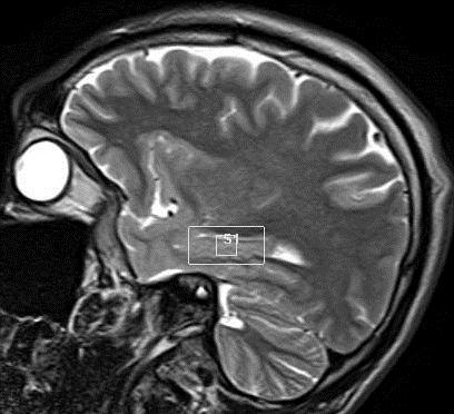
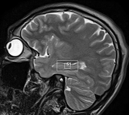
Fig. 1. Localized single voxel proton MR spectroscopy was performed along the long axis of right hippocampus after extension of patient s head to
 125 A B C Fig. 1. Localized single voxel proton MR spectroscopy was performed along the long axis of right hippocampus after extension of patient s head to obtain entire dimension of the hippocampal body.
Proton MR Spectroscopy in Patients with Acute Temporal Lobe Seizures
 AJN Am J Neuroradiol 22:152 157, January 2001 Proton M Spectroscopy in Patients with Acute Temporal obe Seizures Mauricio Castillo, J. Keith Smith, and ester Kwock BACKGOUND AND PUPOSE: Decreases in N-acetyl
Usefulness of Single Voxel Proton MR Spectroscopy in the Evaluation of Hippocampal Sclerosis
 Usefulness of Single Voxel Proton MR Spectroscopy in the Evaluation of Hippocampal Sclerosis 1, 2, 3 Kee-Hyun Chang, MD Hong Dae Kim, MD 1 Sun-Won Park, MD 1 In Chan Song, PhD 2 In Kyu Yu, MD 1 1, 2, 3
José A Mendes-Ribeiro, Raquel Soares, Fernanda Simões-Ribeiro, M Luiza Guimarães
 58 Neurophysiology Unit J A Mendes-Ribeiro M L Guimarães Department of Neurology and Neurosurgery, Hospital S João, Porto, Portugal F Simões-Ribeiro Magnetic Resonance Unit, IPO, Porto, Portugal R Soares
RADIOLOGY NEURORADIOLOGY. Iranian Journal of
 NEURORADIOLOGY Iranian Journal of RADIOLOGY RADIOLOGYwww.iranjradiol.com Value of Proton-MR-Spectroscopy in the Diagnosis of Temporal Lobe Epilepsy; Correlation of Metabolite Alterations With Electroencephalography
A study of the relationship between metabolism using 1 H-MRS and function using several neuropsychological
 Seizure 2001; 10: 188 193 doi:10.1053/seiz.2000.0498, available online at http://www.idealibrary.com on A study of the relationship between metabolism using 1 H-MRS and function using several neuropsychological
Laura Tormoehlen, M.D. Neurology and EM-Toxicology Indiana University
 Laura Tormoehlen, M.D. Neurology and EM-Toxicology Indiana University Disclosures! No conflicts of interest to disclose Neuroimaging 101! Plain films! Computed tomography " Angiography " Perfusion! Magnetic
Case reports functional imaging in epilepsy
 Seizure 2001; 10: 157 161 doi:10.1053/seiz.2001.0552, available online at http://www.idealibrary.com on Case reports functional imaging in epilepsy MARK P. RICHARDSON Medical Research Council Fellow, Institute
Correlation between 1 H MRS and Memory before and after Surgery in Mesial Temporal Lobe Epilepsy with Hippocampal Sclerosis
 Epilepsia, 45(6):632 640, 2004 Blackwell Publishing, Inc. C 2004 International League Against Epilepsy Correlation between 1 H MRS and Memory before and after Surgery in Mesial Temporal Lobe Epilepsy with
Focal epilepsy recruiting a generalised network of juvenile myoclonic epilepsy: a case report
 Clinical commentary Epileptic Disord 2014; 16 (3): 370-4 Focal epilepsy recruiting a generalised network of juvenile myoclonic epilepsy: a case report Myo Khaing 1,2, Kheng-Seang Lim 1, Chong-Tin Tan 1
Proton magnetic resonance spectroscopy study of bilateral thalamus in juvenile myoclonic epilepsy
 Seizure (2007) 16, 287 295 www.elsevier.com/locate/yseiz REVIEW Proton magnetic resonance spectroscopy study of bilateral thalamus in juvenile myoclonic epilepsy Cemile Haki a,oğuzhan G. Gümüştaş b, *,İbrahim
Theroleofclinicalin vivo 1H-MR spectroscopy in the evaluation of epilepsies
 Spectroscopy 16 (2002) 297 306 297 IOS Press Theroleofclinicalin vivo 1H-MR spectroscopy in the evaluation of epilepsies T. Hammen a,,h.stefan a and B. Tomandl b a Clinics of Neurology Center Epilepsy,
Epilepsy: diagnosis and treatment. Sergiusz Jóźwiak Klinika Neurologii Dziecięcej WUM
 Epilepsy: diagnosis and treatment Sergiusz Jóźwiak Klinika Neurologii Dziecięcej WUM Definition: the clinical manifestation of an excessive excitation of a population of cortical neurons Neurotransmitters:
Value of Single-Voxel Proton MR Spectroscopy in Temporal Lobe Epilepsy
 Value of Single-Voxel Proton MR Spectroscopy in Temporal Lobe Epilepsy Eric Achten, Paul Boon, Tom Van De Kerckhove, Jacques Caemaert, Jacques De Reuck, and Marc Kunnen PURPOSE: To study the value of different
Proton Magnetic Resonance Spectroscopy
 1432/Cap.10/2b 12-11-2001 16:55 Pagina 3 Chapter 10 Proton Magnetic Resonance Spectroscopy Z. CARAMANOS, A.C. SANTOS, S.J. FRANCIS, S. NARAYANAN, D. PELLETIER, D.L. ARNOLD Introduction Primary Progressive
Is DTI Increasing the Connectivity Between the Magnet Suite and the Clinic?
 Current Literature In Clinical Science Is DTI Increasing the Connectivity Between the Magnet Suite and the Clinic? Spatial Patterns of Water Diffusion Along White Matter Tracts in Temporal Lobe Epilepsy.
University of Groningen. Biomarkers in premanifest Huntington's disease van Oostrom, Joost Cornelis Hendricus
 University of Groningen Biomarkers in premanifest Huntington's disease van Oostrom, Joost Cornelis Hendricus IMPORTANT NOTE: You are advised to consult the publisher's version (publisher's PDF) if you
Magnetic Resonance Imaging. Alex MacKay University of British Columbia
 Magnetic Resonance Imaging Alex MacKay University of British Columbia Magnetic Resonance Imaging A) What is MRI? B) Why do MRI? C) What can we do with an MRI scanner? What is MRI? Magnetic Resonance Imaging
Comparison of 1.5T and 3T 1 H MR Spectroscopy for Human Brain Tumors
 Comparison of 1.5T and 3T 1 H MR Spectroscopy for Human Brain Tumors Ji-hoon Kim, MD 1 Kee-Hyun Chang, MD 2-4 Dong Gyu Na, MD 2 In Chan Song, PhD 2,3 Seung Ja Kim, MD 2 Bae Ju Kwon, MD 2 Moon Hee Han,
Multisection Proton MR Spectroscopy for Mesial Temporal Lobe Epilepsy
 AJNR Am J Neuroradiol 23:1359 1368, September 2002 Multisection Proton MR Spectroscopy for Mesial Temporal Lobe Epilepsy Arístides A. Capizzano, Peter Vermathen, Kenneth D. Laxer, Gerald B. Matson, Andrew
Proton magnetic resonance spectroscopy and electroencephalographic activity in attention deficit disorder
 1 Proton magnetic resonance spectroscopy and electroencephalographic activity in attention deficit disorder Arturo Alvarado, Germán Zapata, Larry Díaz, Zhilma Sucre, Gladys Veracochea, Raiza Pérez, Maritza
Lateralizing Ability of Single-voxel Proton MR Spectroscopy in Hippocampal Sclerosis: Comparison with MR Imaging and Positron Emission Tomography
 AJNR Am J Neuroradiol 22:625 631, April 2001 Lateralizing Ability of Single-voxel Proton MR Spectroscopy in Hippocampal Sclerosis: Comparison with MR Imaging and Positron Emission Tomography Sun-Won Park,
Magnetic resonance spectroscopy of the thalamus in patients with typical absence epilepsy
 Seizure (2006) 15, 533 540 www.elsevier.com/locate/yseiz Magnetic resonance spectroscopy of the thalamus in patients with typical absence epilepsy Dagmar Fojtiková a, *, Milan Brázdil a, Jaroslav Horký
MR Spectroscopic Evaluation of Psychomotor Delay of Unknown Cause in Children
 Pediatric Imaging Original Research Koşucu et al. MR Spectroscopy of Psychomotor Delay Pediatric Imaging Original Research Polat Koşucu 1 Sema Erdemli 2 Müjgan Sönmez 3 Sibel Kul 1 Ayşe Aksoy 3 Koşucu
Epilepsy Surgery: A Pediatric Neurologist s Perspective
 Epilepsy Surgery: A Pediatric Neurologist s Perspective Juliann M. Paolicchi, MD, MA Associate Professor of Neurology and Pediatrics Director, Pediatric Neurology Director, Pediatric Epilepsy and EEG Vanderbilt
Epilepsy DOJ Lecture Masud Seyal, M.D., Ph.D. Department of Neurology University of California, Davis
 Epilepsy DOJ Lecture - 2005 Masud Seyal, M.D., Ph.D. Department of Neurology University of California, Davis Epilepsy SEIZURE: A temporary dysfunction of the brain resulting from a self-limited abnormal
Cerebral structural lesions are found in approximately. Surgery of Intractable Temporal Lobe Epilepsy Presented with Structural Lesions
 Original Article J Chin Med Assoc 2003;66:565-571 Surgery of Intractable Temporal Lobe Epilepsy Presented with Structural Lesions Yang-Hsin Shih 1 Jiang-Fong Lirng 2 Der-Jen Yen 3 Donald M. Ho 4 Chun-Hing
Department of Radiology, Faculty of Paramedicine, Shahrekord University of Medical Sciences, Shahrekord, Iran. 2
 ISSN: 0974-2115 Idiopathic generalized epilepsies and efficiency of advanced magnetic resonance imaging techniques in present era; perspectives in future Hossien Masoumi 1, Farhad Naleini 2, Mohammad Gharib
Study on the clinical application of the MRS in the cognitive assessment after stroke
 European Review for Medical and Pharmacological Sciences 2017; 21: 2437-2442 Study on the clinical application of the MRS in the cognitive assessment after stroke S.-Y. WANG 1,2,3, M. WANG 1,2,3, X.-X.
True Epileptiform Patterns (and some others)
") True Epileptiform Patterns (and some others) a) What is epileptiform b) Some possible surprises c) Classification of generalized epileptiform patterns An epileptiform pattern Interpretative term based
True Epileptiform Patterns (and some others) a) What is epileptiform b) Some possible surprises c) Classification of generalized epileptiform patterns An epileptiform pattern Interpretative term based
Proton MR Spectroscopy of Polymicrogyria and Heterotopia
 AJNR Am J Neuroradiol 24:2077 2081, November/December 2003 Proton MR Spectroscopy of Polymicrogyria and Heterotopia Elysa Widjaja, Paul D. Griffiths, and Iain D. Wilkinson BACKGROUND AND PURPOSE: Proton
Intracranial Studies Of Human Epilepsy In A Surgical Setting
 Intracranial Studies Of Human Epilepsy In A Surgical Setting Department of Neurology David Geffen School of Medicine at UCLA Presentation Goals Epilepsy and seizures Basics of the electroencephalogram
Effects of Brain Region and Gender on Proton Magnetic Resonance Spectroscopy in Normal Subjects
 Yale University EliScholar A Digital Platform for Scholarly Publishing at Yale Yale Medicine Thesis Digital Library School of Medicine 12-13-2002 Effects of Brain Region and Gender on Proton Magnetic Resonance
Medical Policy Original Effective Date: Revised Date: Page 1 of 5
 Disclaimer Description Coverage Determination Refer to the member s specific benefit plan and Schedule of Benefits to determine coverage. This may not be a benefit on all plans or the plan may have broader
MR spectroscopy in diagnosing intracranial lesions: comparison of diagnostic accuracy at different TE
 MR spectroscopy in diagnosing intracranial lesions: comparison of diagnostic accuracy at different TE Poster No.: C-1359 Congress: ECR 2013 Type: Authors: Keywords: DOI: Scientific Exhibit A. S. DUNGDUNG;
Attending: a medical doctor (MD or OD) who has completed medical school, residency, and often a specialized fellowship
 who has completed medical school, residency, and often a specialized fellowship") Descriptions for members of the Epilepsy Team Attending: a medical doctor (MD or OD) who has completed medical school, residency, and often a specialized fellowship Fellow: a medical doctor (MD or OD)
Descriptions for members of the Epilepsy Team Attending: a medical doctor (MD or OD) who has completed medical school, residency, and often a specialized fellowship Fellow: a medical doctor (MD or OD)
Source analysis of polyspike and wave complexes in juvenile myoclonic epilepsy
 Seizure 2002; 11: 320 324 doi:10.1053/seiz.2002.0676, available online at http://www.idealibrary.com on Source analysis of polyspike and wave complexes in juvenile myoclonic epilepsy EFRAÍN SANTIAGO-RODRÍGUEZ,
Assessing Temporal Brain Metabolite Changes in Preterm Infants Using Multivoxel Magnetic Resonance Spectroscopy
 Magn Reson Med Sci, Vol. XX, No. X, pp. XXX XXX, 205 205 Japanese Society for Magnetic Resonance in Medicine MAJOR PAPER by J-STAGE doi:0.2463/mrms.mp.205-004 Assessing Temporal Brain Metabolite Changes
Imaging for Epilepsy Diagnosis December 2, 2011
 Imaging for Epilepsy Diagnosis December 2, 2011 Samuel Wiebe, MD University of Calgary Canada American Epilepsy Society Annual Meeting Disclosure University of Calgary Hopewell Professorship of Clinical
Advanced MR Imaging of Cortical Dysplasia with or without Neoplasm: A Report of Two Cases
 AJNR Am J Neuroradiol 23:1686 1691, November/December 2002 Case Report Advanced MR Imaging of Cortical Dysplasia with or without Neoplasm: A Report of Two Cases Jay J. Pillai, Richard B. Hessler, Jerry
Proton spectroscopy of the thalamus in a homogeneous sample of patients with easy-to-control juvenile myoclonic epilepsy
 Original Article Leite CC et al. / MRS in patients with juvenile myoclonic epilepsy Proton spectroscopy of the thalamus in a homogeneous sample of patients with easy-to-control juvenile myoclonic epilepsy
Multimodal Imaging in Extratemporal Epilepsy Surgery
 Open Access Case Report DOI: 10.7759/cureus.2338 Multimodal Imaging in Extratemporal Epilepsy Surgery Christian Vollmar 1, Aurelia Peraud 2, Soheyl Noachtar 1 1. Epilepsy Center, Dept. of Neurology, University
Key Words: Parkinson's disease, Magnetic resonance spectroscopy (MRS) 대한신경과학회지 21 권 6 호
 대한신경과학회지 21 권 6 호") Jong-Ki Kim,.D., Byeong-Chae Kim,.D., Kee-Ra Lee,.D., in-kyung Song,.D., an-seok Park,.D., yeong-kyu Kim,.D., Ki-Hyun Cho,.D., Jeong-Jin Seo,.D. Background: Parkinson's disease is a progressive, common
Jong-Ki Kim,.D., Byeong-Chae Kim,.D., Kee-Ra Lee,.D., in-kyung Song,.D., an-seok Park,.D., yeong-kyu Kim,.D., Ki-Hyun Cho,.D., Jeong-Jin Seo,.D. Background: Parkinson's disease is a progressive, common
Method. NeuRA MRS March 2017
 Introduction (MRS) is a specialised nuclear imaging technique that utilises magnetic resonance imaging (MRI) technology to investigate biochemical alterations within tissues of interest. Different biochemicals
Magnetic resonance spectroscopic imaging of brain injury after nasopharyngeal cancer radiation in early delayed reaction
 Magnetic resonance spectroscopic imaging of brain injury after nasopharyngeal cancer radiation in early delayed reaction W.-S. Chen 1, J.-J. Li 1, J.-H. Zhang 2, L. Hong 1, Z.-B. Xing 1, F. Wang 1 and
MR Detection of Hippocampal Disease in Epilepsy: Factors Influencing T2 Relaxation Time
 MR Detection of Hippocampal Disease in Epilepsy: Factors Influencing T2 Relaxation Time R. A. Grunewald, G. D. Jackson, A. Connelly, and J. S. Duncan PURPOSE: To assess the reproducibility and stability
Epilepsy in the Primary School Aged Child
 Epilepsy in Primary School Aged Child Deepak Gill Department of Neurology and Neurosurgery The Children s Hospital at Westmead CHERI Research Forum 15 July 2005 Overview The School Age Child and Epilepsy
Late-onset temporal lobe epilepsy in a patient with juvenile myoclonic epilepsy
 Clinical commentary Epileptic Disord 2012; 14 (2): 190-4 Late-onset temporal lobe epilepsy in a patient with juvenile myoclonic epilepsy Octavian V Lie 1,2, Mark D Holmes 1 1 Department of Neurology, University
MRS and Perfusion of Brain Tumors
 Department of Radiology University of California San Diego MRS and Perfusion of Brain Tumors John R. Hesselink, M.D. MRS & Perfusion of Brain Tumors Tumor histology Degree of malignancy Delineate tumor
Magnetic Resonance Spectroscopy as applied to epilepsy
 P a g e 1 Magnetic Resonance Spectroscopy as applied to epilepsy Robert Simister, MRCP Department of Clinical and Experimental Epilepsy, Institute of Neurology, University College London (UCL), Queen Square,
Frontal Contributions to Memory Encoding Before and After Unilateral Medial Temporal Lobectomy
 Frontal Contributions to Memory Encoding Before and After Unilateral Medial Temporal Lobectomy Jeff Ojemann, MD Department of Neurological Surgery University of Washington Children s Hospital & Regional
Child Neurology. The Plural. of anecdote. is not evidence. University of Texas Health Science Center at San Antonio
 Child Neurology Management of Seizure Disorders The stated goal of advocacy groups for patients with seizures, is to have the patient seizure free. S W Atkinson, MD Management of When to pharmacologically
Single-Voxel Proton MR Spectroscopy and Positron Emission Tomography for Lateralization of Refractory Temporal Lobe Epilepsy
 AJNR Am J Neuroradiol 19:1 8, January 1998 Single-Voxel Proton MR Spectroscopy and Positron Emission Tomography for Lateralization of Refractory Temporal Lobe Epilepsy Eric Achten, Patrick Santens, Paul
J Neurol Neurosurg Psychiatry 2005; 76(Suppl III):iii2 iii10. doi: /jnnp RAY COMPUTED TOMOGRAPHY
:iii2 iii10. doi: /jnnp RAY COMPUTED TOMOGRAPHY") iii2 See end of article for authors affiliations Correspondence to: Professor John S Duncan, Department of Clinical and Experimental Epilepsy, Institute of Neurology, Queen Square, University College London,
iii2 See end of article for authors affiliations Correspondence to: Professor John S Duncan, Department of Clinical and Experimental Epilepsy, Institute of Neurology, Queen Square, University College London,
Epilepsy T.I.A. Cataplexy. Nonepileptic seizure. syncope. Dystonia. Epilepsy & other attack disorders Overview
 : Clinical presentation and management Markus Reuber Professor of Clinical Neurology Academic Neurology Unit University of Sheffield, Royal Hallamshire Hospital. Is it epilepsy? Overview Common attack
CASE 49. What type of memory is available for conscious retrieval? Which part of the brain stores semantic (factual) memories?
 memories?") CASE 49 A 43-year-old woman is brought to her primary care physician by her family because of concerns about her forgetfulness. The patient has a history of Down syndrome but no other medical problems.
CASE 49 A 43-year-old woman is brought to her primary care physician by her family because of concerns about her forgetfulness. The patient has a history of Down syndrome but no other medical problems.
INVESTIGATION OF BRAIN METABOLITES AND CORTICAL GLUCOSE METABOLISM IN HUMAN PARTIAL EPILEPSY
 INVESTIGATION OF BRAIN METABOLITES AND CORTICAL GLUCOSE METABOLISM IN HUMAN PARTIAL EPILEPSY PhD theses Zoltán Pfund, MD Pécs University, Faculty of Medicine, Department of Neurology Pécs, Hungary 2002
Research Article Altered Neurochemical Ingredient of Hippocampus in Patients with Bipolar Depression
 Depression Research and Treatment Volume 2012, Article ID 485249, 6 pages doi:10.1155/2012/485249 Research Article Altered Neurochemical Ingredient of Hippocampus in Patients with Bipolar Depression Murad
Fernando Cendes, MD, PhD,* Zografos Caramanos, MA, Frederick Andermann, MD, FranGois Dubeau, MD, and Douglas L. Arnold, MD
 P Proton Magnetic Resonance Spectroscopic Imaging and Magnetic Resonance Imaging Volumetry in the Lateralization of Temporal Lobe Epilepsy: A Series of 100 Patiekts Fernando Cendes, MD, PhD,* Zografos
Magnetic resonance spectroscopy of the thalamus in patients with mesial temporal lobe epilepsy and hippocampal sclerosis
 Epileptology in Czech Republic Epileptic Disord 2007; 9 (Suppl. 1): S59-67 Magnetic resonance spectroscopy of the thalamus in patients with mesial temporal lobe epilepsy and hippocampal sclerosis Dagmar
Magnetic Resonance Imaging of Mesial Temporal Sclerosis (MTS): What radiologists ought to know?
: What radiologists ought to know?") Magnetic Resonance Imaging of Mesial Temporal Sclerosis (MTS): What radiologists ought to know? Poster No.: C-0856 Congress: ECR 2012 Type: Educational Exhibit Authors: P. Singh, G. Mittal, R. Kaur, K.
Magnetic Resonance Imaging of Mesial Temporal Sclerosis (MTS): What radiologists ought to know? Poster No.: C-0856 Congress: ECR 2012 Type: Educational Exhibit Authors: P. Singh, G. Mittal, R. Kaur, K.
A U. Methods of Cognitive Neuroscience 2/8/2016. Neat Stuff! Cognitive Psychology
 Methods of Cognitive Neuroscience Neat Stuff! Optogenetics http://spie.org/newsroom/technical-articles-archive/videos/0411-boyden Stimulating the brain with light Cognitive Psychology Mental Representation
Introduction to Brain Imaging
 Introduction to Brain Imaging Human Brain Imaging NEUR 570 & BIC lecture series September 9, 2013 Petra Schweinhardt, MD PhD Montreal Neurological Institute McGill University Montreal, Canada Various techniques
Primary Central Nervous System Lymphoma with Lateral Ventricle Involvement
 The Open Medical Imaging Journal, 2012, 6, 103-107 103 Open Access Primary Central Nervous System Lymphoma with Lateral Ventricle Involvement Yumi Oie 1,*, Kazuhiro Murayama 1, Shinya Nagahisa 2, Masato
Ictal pain: occurrence, clinical features, and underlying etiologies.
 Thomas Jefferson University Jefferson Digital Commons Department of Neurology Faculty Papers Department of Neurology 8-1-2016 Ictal pain: occurrence, clinical features, and underlying etiologies. Ali Akbar
Reference Values for Long Echo Time MR Spectroscopy in Healthy Adults
 AJNR Am J Neuroradiol 26:1439 1445, June/July 2005 Reference Values for Long Echo Time MR Spectroscopy in Healthy Adults Yair Safriel, MarlyAnne Pol-Rodriguez, Edward J. Novotny, Douglas L. Rothman, and
What Can the Brain Teach Us About Treating PTSD? Thomas C. Neylan, MD Norbert Schuff, PhD Charles R. Marmar, MD Michael W.
 What Can the Brain Teach Us About Treating PTSD? Thomas C. Neylan, MD Norbert Schuff, PhD Charles R. Marmar, MD Michael W. Weiner, MD Stress and Biological Sciences Abram Kardiner (1891-1981) Described
Journal of Neurology, Neurological Science and Disorders
 v Clinical Group Journal of Neurology, Neurological Science and Disorders DOI CC By Ossama Y Mansour 1 *, Doaa Hanfy 2, Sameh Fathy 3 and Rania E Mohammed 4 1 Alexandria university neurology and endovascular
A Method of Connectivity Analysis of Epileptiform Discharges during Epileptic Seizure
 International Journal of Bioelectromagnetism Vol. 19, No.1, pp. 6-10, 2017 www.ijbem.org A Method of Connectivity Analysis of Epileptiform Discharges during Epileptic Seizure Hisashi Yoshida a, Yasuto
4/12/2016. Seizure description Basic EEG ICU monitoring Inpatient Monitoring Elective admission for continuous EEG monitoring Nursing s Role
 Kathleen Rieke, MD Chari Ahrenholz Curt Devos Understand why continuous EEG is being requested in certain patient populations Understand what the EEG can tell us about our patient. Understand nursing role
Methods of Sample Preparation for Analysis and Quality Assurance of Prostate MR Spectroscopy
 Methods of Sample Preparation for Analysis and Quality Assurance of Prostate MR Spectroscopy Kristina KRISTINAITYTĖ 1,2*, Jonas RAŽANSKAS 3, Vaida PAKETURYTĖ 3, Nomeda R. VALEVIČIENĖ 2,3, Vytautas BALEVIČIUS
DISTINCTION BETWEEN RECURRENT GLIOMA AND RADIATION INJURY USING MAGNETIC RESONANCE SPECTROSCOPY IN COMBINATION WITH DIFFUSION-WEIGHTED IMAGING
 doi:10.1016/j.ijrobp.2006.12.001 Int. J. Radiation Oncology Biol. Phys., Vol. 68, No. 1, pp. 151 158, 2007 Copyright 2007 Elsevier Inc. Printed in the USA. All rights reserved 0360-3016/07/$ see front
Radiation-Induced Brain Metabolic Changes in the Acute and Early Delayed Phase Detected With Quantitative Proton Magnetic Resonance Spectroscopy
 ORIGINAL ARTICLE Radiation-Induced Brain Metabolic Changes in the Acute and Early Delayed Phase Detected With Quantitative Proton Magnetic Resonance Spectroscopy Tatsuro Kaminaga, MD and Katsuo Shirai,
Epilepsy, a common chronic neurological disorder, is a
 10 SUPPLEMENT TO Journal of the association of physicians of india august 2013 VOL. 61 Epilepsy: Diagnostic Evaluation JMK Murthy* Epilepsy, a common chronic neurological disorder, is a potentially treatable
1) Diffusion weighted imaging DWI is a term used to describe moving molecules due to random thermal motion. This motion is restricted by boundaries
 Diffusion weighted imaging DWI is a term used to describe moving molecules due to random thermal motion. This motion is restricted by boundaries") 1) Diffusion weighted imaging DWI is a term used to describe moving molecules due to random thermal motion. This motion is restricted by boundaries such as ligaments, membranes and macro molecules. Diffusion
1) Diffusion weighted imaging DWI is a term used to describe moving molecules due to random thermal motion. This motion is restricted by boundaries such as ligaments, membranes and macro molecules. Diffusion
3.02 Understand the functions and disorders of the nervous system Understand the functions and disorders of the nervous system
 3.02 Understand the functions and disorders of the nervous system 1 3.02 Essential Questions What are the functions of the nervous system? What are some disorders of the nervous system? How are nervous
TEMPORAL LOBE EPILEPSY AND SLEEP: FOCUS ON INTERICTAL SPIKES AND MEMORY CONSOLIDATION
 Ph.D thesis TEMPORAL LOBE EPILEPSY AND SLEEP: FOCUS ON INTERICTAL SPIKES AND MEMORY CONSOLIDATION Zsófia Clemens National Institute of Psychiatry and Neurology Semmelweis University Budapest János Szentágothai
Name: Period: Chapter 2 Reading Guide The Biology of Mind
 Name: Period: Chapter 2 Reading Guide The Biology of Mind The Nervous System (pp. 55-58) 1. What are nerves? 2. Complete the diagram below with definitions of each part of the nervous system. Nervous System
Functional diagnostic imaging in epilepsy
 Alberta Heritage Foundation for Medical Research Functional diagnostic imaging in epilepsy Paula Corabian, David Hailey August 1998 HTA 10 Functional diagnostic imaging in epilepsy Paula Corabian, David
Terce Liana Menezes, Luciana Patrízia A. Andrade-Valença, Marcelo Moraes Valença
 DOI:.59/4-8X ARTICLE Magnetic resonance imaging study cannot individually distinguish individuals with mild cognitive impairment, mild Alzheimer s disease, and normal aging Estudo por ressonância magnética
Table 9: Vascularity and Hemorrhage
 Table 9: Vascularity and Hemorrhage Di Ieva (2007) 120 Fractal dimension as a quantitator the microvasculat ure normal and adenomatous tissue. Clinical experience characterizing vascular surface fractal
University of Groningen. Fluoxetine as disease modifying treatment in multiple sclerosis Mostert, Jop Pieter
 University of Groningen Fluoxetine as disease modifying treatment in multiple sclerosis Mostert, Jop Pieter IMPORTANT NOTE: You are advised to consult the publisher's version (publisher's PDF) if you wish
PSYC& 100: Biological Psychology (Lilienfeld Chap 3) 1
 1") PSYC& 100: Biological Psychology (Lilienfeld Chap 3) 1 1 What is a neuron? 2 Name and describe the functions of the three main parts of the neuron. 3 What do glial cells do? 4 Describe the three basic
PSYC& 100: Biological Psychology (Lilienfeld Chap 3) 1 1 What is a neuron? 2 Name and describe the functions of the three main parts of the neuron. 3 What do glial cells do? 4 Describe the three basic
Challenges for multivariate and multimodality analyses in "real life" projects: Epilepsy
 Challenges for multivariate and multimodality analyses in "real life" projects: Epilepsy Susanne Mueller M.D. Center for Imaging of Neurodegenerative Diseases Background: Epilepsy What is epilepsy? Recurrent
Correlation of Myo-inositol Levels and Grading of Cerebral Astrocytomas
 AJNR Am J Neuroradiol 21:1645 1649, October 2000 Correlation of Myo-inositol Levels and Grading of Cerebral Astrocytomas Mauricio Castillo, J. Keith Smith, and Lester Kwock BACKGROUND AND PURPOSE: In a
Neuropsychological Outcomes of Pediatric Epilepsy. John B. Fulton, Ph.D. Barrow Neurological Institute at Phoenix Children s Hospital
 Neuropsychological Outcomes of Pediatric Epilepsy John B. Fulton, Ph.D. Barrow Neurological Institute at Phoenix Children s Hospital Objectives Cognitive Behavioral Risk factors Outline 1. Definitions,
Imaging is routinely used for the
 Magnetic resonance spectroscopy of the brain Ajay Kumar Singh, MD; Ay-Ming Wang, MD; William Sanders, MD In vivo magnetic resonance (MR) spectroscopy is a noninvasive imaging modality useful for obtaining
Idiopathic Epileptic Syndromes
 Idiopathic Epileptic Syndromes Greek words idios = self, own and personal pathic = suffer Kamornwan Katanuwong MD Chiangmai University Hospital 1 st Epilepsy Camp, Hua Hin 20 th August 2010 Is a syndrome
Chapter 6 Section 1. The Nervous System: The Basic Structure
 Chapter 6 Section 1 The Nervous System: The Basic Structure Essential Question: How does studying the biology of the brain give us an understanding of our behavior? Draw or type 2 things you already know
Spike voltage topography in temporal lobe epilepsy
 Thomas Jefferson University Jefferson Digital Commons Department of Neurology Faculty Papers Department of Neurology 5-17-2016 Spike voltage topography in temporal lobe epilepsy Ali Akbar Asadi-Pooya Thomas
Diagnosis, Assessment and Evaluation for Seizures
 Lehigh Valley Health Network LVHN Scholarly Works Neurology Update for the Non-Neurologist 2013 Neurology Update for the Non-Neurologist Feb 20th, 7:40 PM - 8:10 PM Diagnosis, Assessment and Evaluation
Sincerely, Ms. Paoloni and Mrs. Whitney
 Dear Students, Welcome to AP Psychology! We will begin our course of study focusing on the nervous system with a particular emphasis on how the brain and neurotransmitters influence our behaviors. In preparation
Fire-Related Post-Traumatic Stress Disorder: Brain 1 H-MR Spetroscopic Findings
 Fire-Related Post-Traumatic Stress Disorder: Brain 1 H-MR Spetroscopic Findings Myung Kwan Lim, MD 1 Chang Hae Suh, MD 1 Hyung Jin Kim, MD 1 Sung Tae Kim, MD 1 Jeong Seop Lee, MD 2 Min Hee Kang, MD 2 Ji
Objectives. Amanda Diamond, MD
 Amanda Diamond, MD Objectives Recognize symptoms suggestive of seizure and what those clinical symptoms represent Understand classification of epilepsy and why this is important Identify the appropriate
Evaluation and management of drug-resistant epilepsy
 Evaluation and management of drug-resistant epilepsy Fateme Jahanshahifar Supervised by: Professor Najafi INTRODUCTION 20 to 40 % of patients with epilepsy are likely to have refractory epilepsy. a substantive
ORIGINAL CONTRIBUTION. Ictal Fear in Temporal Lobe Epilepsy
 ORIGINAL CONTRIBUTION Ictal Fear in Temporal Lobe Epilepsy Surgical Outcome and Focal Hippocampal Changes Revealed by Proton Magnetic Resonance Spectroscopy Imaging Michael Feichtinger, MD; Elisabeth Pauli,
Review of Anticonvulsant Medications: Traditional and Alternative Uses. Andrea Michel, PharmD, CACP
 Review of Anticonvulsant Medications: Traditional and Alternative Uses Andrea Michel, PharmD, CACP Objectives Review epidemiology of epilepsy Classify types of seizures Discuss non-pharmacologic and pharmacologic
Analysis of metabolic changes of brain in HIV-1 seropositive patients with proton magnetic resonance spectroscopy
 Signature: Pol J Radiol, 2010; 75(2): 27-32 ORIGINAL ARTICLE Received: 2010.03.25 Accepted: 2010.04.06 Analysis of metabolic changes of brain in HIV-1 seropositive patients with proton magnetic resonance
doi: /brain/awq006 Brain 2010: 133; Imaging memory in temporal lobe epilepsy: predicting the effects of temporal lobe resection
 doi:10.1093/brain/awq006 Brain 2010: 133; 1186 1199 1186 BRAIN A JOURNAL OF NEUROLOGY Imaging memory in temporal lobe epilepsy: predicting the effects of temporal lobe resection Silvia B. Bonelli, 1,2
High Resolution Ictal SPECT: Enhanced Epileptic Source Targeting?
 High Resolution Ictal SPECT: Enhanced Epileptic Source Targeting? Marvin A Rossi MD, PhD RUSH Epilepsy Center Research Lab http://www.synapticom.net Chicago, IL USA Medically-Refractory Epilepsy 500,000-800,000